Cardiogenic shock and acute kidney injury: the rule rather than
the exception
N Ghionzoli1 &C Sciaccaluga1&GE Mandoli1&G Vergaro2,3&F Gentile3&F D’Ascenzi1
&S Mondillo1&M Emdin2,3&
S Valente1&M Cameli1
Accepted: 23 September 2020 # The Author(s) 2020 Abstract
Cardiogenic shock (CS) is a life-threatening condition of poor end-organ perfusion, caused by any cardiovascular disease resulting in a severe depression of cardiac output. Despite recent advances in replacement therapies, the outcome of CS is still poor, and its management depends more on empirical decisions rather than on evidence-based strategies. By its side, acute kidney injury (AKI) is a frequent complication of CS, resulting in the onset of a cardiorenal syndrome. The combination of CS with AKI depicts a worse clinical scenario and holds a worse prognosis. Many factors can lead to acute renal impairment in the setting of CS, either for natural disease progression or for iatrogenic causes. This review aims at collecting the current evidence-based acknowledgments in epidemiology, pathophysiology, clinical features, diagnosis, and management of CS with AKI. We also attempted to highlight the major gaps in evidence as well as to point out possible strategies to improve the outcome.
Keywords Cardiogenic shock . Acute kidney injury . Heart failure . Replacement therapy . Outcome
Abbreviations
AKI Acute kidney injury AMI Acute myocardial infarction CRS Cardiorenal syndrome
CS Cardiogenic shock
CVP Central venous pressure
CVVH Continuous veno-venous hemofiltration ECMO Extracorporeal membrane oxygenation IABP Intra-aortic balloon pump
LV Left ventricular
MCS Mechanical support devices RV Right ventricular
PCWP Pulmonary capillary wedge pressure PEEP Positive end-expiration pressure RRT Renal replacement therapy
Introduction
Cardiogenic shock (CS) is a critical condition of end-organ hypoperfusion, consequent to a severe decrease in cardiac output, in spite of adequate intravascular volume. Therefore, hypotension requiring volume resuscitation and signs of end-organ hypoperfusion represent the clinical landmarks of CS, urgently demanding for pharmacological and/or mechanical intervention [1–3].
Acute kidney injury (AKI) represents a sudden insult to renal function that encompasses several clinical scenarios, ranging from a mild increase in serum creatinine to end-stage renal disease, as stated by Risk-Injury-Failure-Loss-End-stage (RIFLE) criteria. In order to make a diagnosis of AKI, at least one of the following criteria has to be met: in-crease in serum creatinine≥ 0.3 mg/dL within 48 h; increase in basal serum creatinine by≥ 1.5 times within the previous 7 days; urine volume < 0.5 mL/kg/h for 6 h [4]. For further details about RIFLE criteria, please see Table1.
Management of both CS and AKI is strictly time-depen-dent: the longer they persist, the higher is the likelihood of developing irreversible organ damages. They can occur alone as well as clustered in the same patients. Evidences of inter-dependency between the heart and the kidney are described as cardiorenal syndromes (CRSs) [5]. With concerns to the spe-cific setting of critical care, the most prevalent is type-1 CRS, * N Ghionzoli
1
Department of Medical Biotechnologies, Division of Cardiology, University of Siena, Policlinico Le Scotte, Viale Bracci 16, Siena, Italy
2
Institute of Life Sciences, Scuola Superiore Sant’Anna, Pisa, Italy
3 Division of Cardiology and Cardiovascular Medicine, Fondazione
Toscana Gabriele Monasterio, Pisa, Italy https://doi.org/10.1007/s10741-020-10034-0
whose pivotal pathogenetic mechanism lays on an abrupt de-crease in renal perfusion, typical of patients with CS. Since kidneys are considered end-organs, it follows that a setting of CS can be the cause itself of AKI [1,6]. Type-2 CRS refers to patients with a chronic heart disease that progressively affects renal function, e.g., chronic heart failure; type-3 CRS refers to patients with an abrupt reduction in renal function, thus lead-ing to an acute cardiac disorder, i.e., glomerulonephritis caus-ing fluid retention, hypertension, and then heart failure; type-4 CRS regards the consequences of chronic kidney disease on the cardiovascular system, i.e., vascular calcification; type 5 is for all the systemic disorders that affect both cardiac and renal functions, i.e., diabetes mellitus [6].
Nevertheless, whether many steps have been taken towards the comprehension of the mechanisms underlying both the conditions, few improvements have been made regarding ef-fective therapies for this rare but often fatal disease. Still, a prompt intervention represents the leading modifier of the outcome for these patients.
Epidemiology
Data regarding epidemiological features of CS combined to AKI are still poor. Most of them derive from patients with acute myocardial infarction (AMI), as it represents the leading cause of CS [7]. Hence, the applicability of the available data may be nebulous in clinical settings other than AMI. Thus, further studies investigating CS-AKI when other etiologies are responsible for CS should be promoted.
The incidence of AKI complicating CS (type-1 CRS) is considerably high, since it ranges from 20 to 35% according to studies [8]. This cohort was also burdened by a higher rate of complications, in-hospital mortality, and healthcare sources utilization as compared with patients suffering from only CS. Furthermore, the more impaired renal function is, the higher is the mortality, so that patients requiring hemodialysis had worse outcome than those who did not need it [9]. In a Danish population of 5079 patients with CS, 13% developed AKI requiring renal replacement therapy (RRT). Among them, the in-hospital mortality was 62% for those who
required RRT and 36% for those who did not; this trend was further confirmed on a 5-year follow-up analysis, with a mor-tality of 43% for the first group and 29% for the second one [10]. In a US population of 440,257 patients admitted for CS complicating AMI, 35.3% developed AKI, and 3.4% AKI requiring hemodialysis. All-cause in-hospital mortality was higher in CS-AKI patients than in those with only CS, with a poorer trend in those who needed hemodialysis. Additionally, length of stay was proportionally higher in pa-tients with CS without AKI, with AKI, and with AKI requir-ing RRT (9 ± 10, 12 ± 13, and 18 ± 19 days, respectively, p < 0.001) [11]. As a confirmation, AKI was often found to be an independent predictor of mortality in CS [12,13].
Of note, gender analyses have shown uneven results. Males suffered from CS-AKI significantly more than females, as well as from other end-organs failure, despite women were older and with more comorbidities at presentation [14,15]. On the other hand, in-hospital mortality was found to be sur-prisingly higher in women. The lower rate of coronary angi-ography performed in women as compared with men may at least partially explain this divergence, since prompt revascu-larization was not always guaranteed [6]. In contrast, gender did not influence in-hospital mortality in a setting of post-cardiotomy CS complicated by AKI [16].
Pathophysiology
All cardiorenal syndromes are usually described as the mixing of either hemodynamic or non-hemodynamic factors, with type 1 making no exception [6]. The abrupt reduction in renal perfusion due to pump failure reduces the ability of the neph-ron to filter, with consequent reduction in urine output. This setting is described as prerenal AKI, since the leading cause is upstream to the kidney itself.
Moreover, especially when RV failure occurs, an increase in central venous pressure (CVP) may be observed, with con-sequent renal venous congestion and so loss of function [17]. The clinical relevance of such mechanism was further con-firmed by a recent study by van den Akker et al., as they found that CVP was the only independent predictor for AKI in the Table 1 Staging of acute kidney
injury according to Kidney Disease: Improving Global Outcomes (KDIGO). Five stages of renal impairment have been described and ranked in ascending order according to the severity
Stage Serum creatinine Glomerular filtration rate
Urine output (mL/kg)
R: risk 1.5-fold increase 25% decrease < 0.5 in 6 h I: injury 2-fold increase 50% decrease < 0.5 in 12 h F: failure 3-fold increase or value
≥ 4 mg/dL 75% decrease < 0.3 in 24 h (oliguria) or anuriafor 12 h L: loss (of
function)
Complete loss of renal function for≥ 4 weeks, requiring dialysis
context of CS, and by the role of RV performance in this particular clinical scenario [18,19].
Beyond these hemodynamic factors, the acute reduction in heart function is theprimum movens for the activation of sev-eral neurohormonal systems, aiming at restoring hemodynam-ic stability. The balance of these molecular determinants is responsible for both the activation of life-saving pathways and detrimental effects. In this regard, the massive activation of the adrenergic system in CS has been well established, with proven positive inotropic effect and peripheral vasoconstric-tion [20,21]. Although this response is partially adaptive in supporting vital functions, vasoconstriction involves also the kidneys. This leads to possible ischemic effects when autoreg-ulation is exceeded—especially at the renal medulla—and is overall responsible for an increase in cardiac afterload [22,
23]. Furthermore, beta-1 stimulation of the kidney induces renin release, thus activating the renin-angiotensin-aldosterone cascade. The consequent sodium-water retention further increases cardiac afterload (Fig.1). Whether the stim-ulation of mineralocorticoid receptors in the acute setting may be harmful still has to be clarified, especially with regard to non-epithelial tissues such as the heart.
The double-edged nature of neurohormonal drivers is further confirmed by the current clinical use of inotropes in the setting of CS. Norepinephrine and dobutamine are the most used agents, but their long-standing use is asso-ciated with an increased risk of cardiac arrhythmias, as well as severe ischemia in multiple organs [24]. Safer approaches may derive from replacement therapies, in-cluding both ultrafiltration and cardiac devices for tran-sient mechanical support.
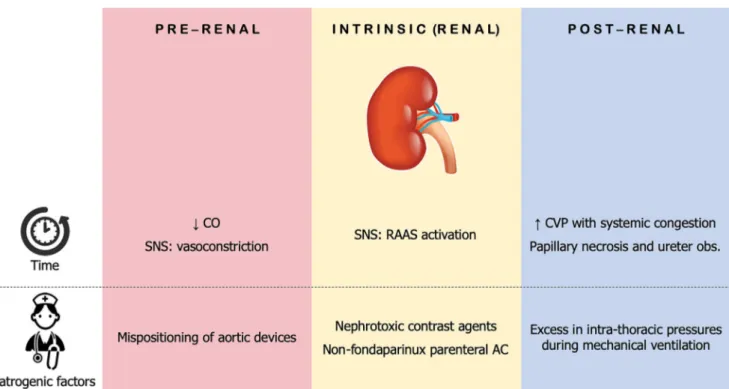
Given this background, the interdependence between the heart and the kidney comes clear, with mutual detrimental amplification. The timing of occurrence of CS-AKI may vary among patients, with two main scenarios: those presenting with both CS and AKI at admission and those who develop AKI during hospitalization (Fig.2). The latter cases may rep-resent the natural evolution of the disease, as well as the com-plication of medical intervention. Iatrogenic factors indeed are often addressed as responsible or, at least, precipitating mech-anism. For example, the use of nephrotoxic contrast agents as part of the investigation of CS complicating AMI may precip-itate a preexistent borderline renal function alongside with ischemia-induced damages. Nevertheless, data from the Bremen STEMI registry did not find any correlation between the amount of the administered contrast agent and the onset of AKI, whilst the only driver was actually the impairment of the left ventricular (LV) function [25]. It is not clear whether the quantity of contrast could predict AKI in the specific subcat-egory of CS. Eventually, trans-femoral positioning of cardiac support devices (such as intra-aortic balloon pump) may com-plicate with renal artery occlusion, especially when delivered distally in the aorta [26].
Clinical phenotypes
CS is described as a status of hypoperfusion due to a signifi-cant reduction in cardiac index, leading to peripheral vasocon-striction and increased pulmonary capillary wedge pressure (PCWP) [27]. Although a reduced cardiac index is a necessary prerequisite for the diagnosis of CS, both peripheral vascular resistance and PCWP may vary among patients, as demon-strated by the SHould we emergently revascularize Occluded Coronaries for Cardiogenic shock (SHOCK) trial [2]. New phenotypes were then categorized, since a minority of patients could present as“wet and warm” or “dry and cold” over the traditional“wet and cold” (Fig.3). With regard to the former cases, systemic inflammatory response syndrome was shown to occur also in the setting of cardiogenic shock, thus activat-ing the inflammasome and leadactivat-ing to inappropriate systemic vasodilation. This can be due to two orders of factors: the release of cytokines—in particular TNF alpha—from the in-jured myocardium, and the release of endotoxins and bacteria translocating from the hypoxic intestine, thus leading to a catecholamine-unresponsive profile of shock [28].
The“dry and cold” phenotype includes patients with sig-nificant vasoconstriction but low or normal PCWP (6– 12 mmHg). This is considered a diuretic-responsive class of patients who usually suffer from chronic heart failure and are less likely to have chronic kidney disease as a comorbidity [29]. Therefore, as clinical presentation may be heteroge-neous, studies addressing potential differences in renal func-tion and the likelihood of developing AKI are required for each class. As renal function can be affected in the settings of both peripheral and pulmonary congestion, it is reasonable to think that each phenotype may present with a various de-gree of renal impairment.
Management
Whether therapy for CS is a tough challenge for clinicians when presenting alone, the combination of both CS and AKI is a further complication in the decision-making process. Indeed, most of the interventions are still made on an empir-ical basis rather than on solid results from clinempir-ical trials. A thorough invasive assessment and prompt interventions of both revascularization and replacement therapies are strongly recommended when needed.
Diagnostic assessment
An in-depth assessment of hemodynamic parameters is key in the management of CS as well as in CS-AKI. CVP monitoring (normal values 3–8 mmHg) can predict the onset of AKI com-plicating CS, as it can reflect systemic venous congestion, with the kidneys that are no exception. CVP can be further influenced
by the regulation of ventilatory parameters when mechanical ventilation is required (see“Ventilatory support”) [18].
Invasive arterial pressure measurement should also be en-couraged. In particular, pulse pressure and stroke volume var-iations derived from arterial waveform demonstrated to pre-vent the onset of AKI in a setting of CS from post-resuscitated cardiac arrest [30]. More comprehensive studies are needed in order to assess whether this strategy may be successfully ap-plied to CS from other causes.
A close monitoring of renal function and urine output is strongly recommended. The estimation of renal function can be achieved by the measurement of specific plasma proteins (such as creatinine and cystatin C) and dedicated biomarkers of renal injury, as well as by the monitoring of urine output. To our knowledge, only the study by Tarvasmäki et al. inves-tigated renal function in the context of CS and performed a
between-methods comparison using KDIGO criteria for AKI [31]. AKI according to cystatin C was defined on a par with creatinine. The elevation of both molecules did not show a significant difference in the prediction of outcomes; thus, their use for AKI stratification is comparable and valuable. On the contrary, KDIGO cut-off of 0.5 mL/kg/min was not that ad-vantageous, albeit a stricter cut-off of 0.3 mL/kg/min proved effective and independently associated with 90-day mortality. The introduction of renal injury biomarkers (i.e., neutrophil gelatinase–associated lipocalin [NGAL]) in clinical practice may play a useful role, as their increase in plasma occurs earlier than the changes in parameters of function and is less dependent on hemodynamic modifications [32] (see Table2). Although they did not provide any additional prognostic in-formation, predicting AKI before it actually occurs may influ-ence therapeutic decisions, as a prompter intervention with Fig. 1 Hemodynamic and non-hemodynamic factors in CS, their
inter-play with the kidneys, and associated therapeutic strategies. Injuries to the heart can determine both a reduction in cardiac output and an increase in central venous pressure. Alongside, the activation of the sympathetic nervous system induces renal vasoconstriction and RAAS activation, thus reducing urine output. Replacement and pharmacological strategies are
displayed for each organ.CO, cardiac output; CVP, central venous pres-sure;CVVH, continuous veno-venous hemofiltration; IABP, intra-aortic balloon pump;MCS, mechanical circulatory support; PEEP, positive end-expiration pressure;RAAS, renin-angiotensin-aldosterone system; RR, respiratory rate; RRT, renal replacement therapy; SNS, sympathetic nervous system;TV, tidal volume; UO, urinary output
mechanical circulatory support devices may avoid its onset and therefore a worsening in outcome [33].
Cardiogenic shock related to ischemic heart disease
As CS represents the ultimate step for multiple severe inju-ries to the myocardium, defining and readily solving the underlying cause should be considered the gold standard of treatment. Since AMI is still the leading etiology for CS, coronary angiography with a view to revascularization is often the treatment of choice. By its side, percutaneous revascularization includes the utilization of contrast agents that are nephrotoxic, so possibly triggering AKI, even if the correlation between AKI and the amount of contrast has been questioned [25]. Furthermore, when prompt and inva-sive evaluation of the coronary anatomy is required, unfractionated heparin should be considered the anticoagu-lant treatment of choice, given that both low molecular weight heparin and fondaparinux may precipitate AKI [1,
34]. A shared guideline is missing with regard to the strat-egy of revascularization in the context of multivessel dis-ease, since current studies came to different conclusions. Results from the CULPRIT-SHOCK trial demonstrated an increase in mortality or need for RRT for patients receiving an“all-in-one” revascularization, thus preferring a culprit-dedicated approach [35]. Accordingly, the treatment of non-culprit artery disease should be postponed to a time when clinical stabilization is reached. A recent subanalysis from the same trial also showed that no different approaches should be undertaken between men and women, even if the latter presented with a different profile of risk [36]. Fig. 2 Prerenal, renal, and post-renal main causes of acute kidney injury
complicating cardiogenic shock. Causes are distinguished as part of the natural history of the disease and as iatrogenic factors during in-hospital
management.AC, anticoagulation; CO, cardiac output; CVP, central ve-nous pressure;obs., obstruction; RAAS, renin-angiotensin-aldosterone system;SNS, sympathetic nervous system
Fig. 3 Clinical phenotypes of cardiogenic shock. Two additional types of cardiogenic shock have been described over the classic“wet and cold” phenotype. These are“dry and cold,” with cold extremities and no pulmonary congestion, and“wet and warm,” where a peripheral vasodilation is observed mainly as the consequence of a systemic inflammatory syndrome response.CS, cardiogenic shock; SIRS, systemic inflammatory response syndrome
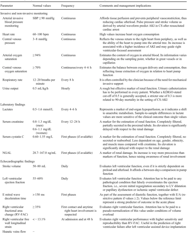
Table 2 Diagnostic work-up and management in patients with CS-AKI, according to invasive and non-invasive monitoring strategies and laboratoristic and echocardiographic findings. Name of the parameters, normal values, frequency of measurements, and further comments are
here reported.AKI, acute kidney injury; CS, cardiogenic shock; KDIGO, Kidney Disease: Improving Global Outcome; NGAL, neutrophil gelatinase–associated lipocalin and kidney injury molecule; SBP, systolic blood pressure
Parameter Normal values Frequency Comments and management implications Invasive and non-invasive monitoring
Arterial invasive blood pressure monitoring
SBP≥ 90 mmHg Continuous Affords tissue perfusion and prevents peripheral vasoconstriction, thus reducing cardiac afterload. Pulse pressure and stroke volume as derived by arterial waveform predict AKI in CS after resuscitated cardiac arrest
Heart rate 60–100 bpm Continuous High values increase heart oxygen consumption Central venous
pressure
3–8 mmHg Continuous Reflects the venous return to the right heart from periphery, as well as the ability of the heart to pump into the arterial tree. Its increase is associated with a higher incidence of AKI and may guide right ventricular-focused assessment
Arterial oxygen saturation
≥ 94% Continuous Estimates the content of oxygen in arterial blood. Its information varies depending on the sampling point, whether in great vessels or in capillaries
Central venous oxygen saturation
≥ 70% Continuous/every 4–6 h Estimates the balance between oxygen delivery and consumption, thus reflecting tissue extraction of oxygen in relation to heart pump function
Respiratory rate 12–20 breaths per minute
Every 8 h It is often controlled by the clinician because of the need for mechanical invasive support
Urine output 0.5 mL/kg/h Hourly A rough but effective marker of renal function. Urinary catheterization has to be performed in every patient. Whether a KDIGO-stated cut-off of 0.5 is generally accepted, a stricter one of 0.3 is more related to 90-day mortality in the setting of CS-AKI
Laboratory findings
Lactates 0.5–1.6 mmol/L Every 4–6 h Represents a marker of end-organ hypoperfusion, as it indicates a shift to anaerobic metabolism. Sample-to-sample differences in lactate values are more sensitive of the clinical outcome than single values Serum creatinine 0.8–1.3 mg/dL
(men) 0.6–1.1 mg/dL
(women)
Every 12–24 h A marker for the estimation of renal function. Completely filtered, partially secreted in the proximal tubule. Its elevation is significantly delayed with respect to the renal damage
Serum cystatin C 0.60–1.55 mg/L First phases (if available) A marker for the estimation of renal function. Completely filtered, no secreted or reabsorbed. Less dependent on age, gender, ethnicity, and muscle mass compared with creatinine. Its elevation is significantly delayed with respect to the renal damage
NGAL 28.7–167.0 ng/mL First phases (if available) A marker of renal damage. Its increase is way more precocious than markers of function, hence raising awareness of renal involvement Echocardiographic findings
Stroke volume 50–80 mL Daily Evaluates left ventricular function, even if it is strictly dependent on preload and afterload. It affords a between-days comparison in pump function
Left ventricular ejection fraction
55–60% Daily Evaluates left ventricular function. Attention has to be paid to any pathological condition that falsely overestimates the ejection fraction, i.e., severe mitral regurgitation secondary to LV dilatation or papillary dysfunction or ischemic septal ventricular defect E mitral wave
deceleration time
> 150 ms First phases As part of the assessment of diastolic function, together with E/A (re-strictive pattern if values≥ 2). Values below the reference limit represent a strong predictor of outcome in the acute phase Right ventricular
fractional area change (RV-FAC)
≥ 35% First contact and anytime right heart involvement is suspected
Evaluates right ventricular function. Attention has to be paid to a pseudo-normalization of this value under conditions of volume overload
Right ventricular free wall longitudinal strain
<− 13.1% At admission and at 48 h Evaluates right ventricular performance with higher sensitivity and reproducibility than RV-FAC. Useful in the prediction of right ventricular failure after left ventricular assisted device implantation Hepatic veins flow −
Ventilatory support
When a ventilatory support is required, a setting with low tidal volumes is strongly recommended in order to reduce the inci-dence of RV failure and to achieve an appropriate venous return, thus avoiding venous renal stasis and edema. [37]. Additionally, despite initial concerns regarding a worsening in cardiac output, moderate values of positive end-expiration pressure (PEEP, 5 cmH2O) were able to reduce LV oxygen demand as well as to improve myocardial oxygen delivery, perhaps due to reduced afterload and preload, with consequent LV unloading [38–41]. Indeed, patients experiencing CS are more prone to be afterload-dependent rather than preload-de-pendent, with the exception of RV failure and/or hypovole-mia. In these two scenarios, clinicians should initiate a low PEEP regimen (3–5 cmH2O) only when euvolemic state is achieved, with a view to up-titration. Close evaluations of blood gas analysis can help to adjust the ventilatory strategy, as well as to evaluate lactates, whose high levels were found to predict persistent AKI [42].
Pump failure and mechanical circulatory support
LV pump failure, as assessed by several hemodynamic (i.e., cardiac index), laboratoristic (lactates) and non-invasive pa-rameters (LV ejection fraction, diastolic function, RV perfor-mance), requires adequate support to antagonize peripheral hypoperfusion. A long-standing experience with inotropes has been collected, reporting controversial findings. Norepinephrine and dobutamine are the first-line agents for patients with evidence of CS, but their use should be as limited as possible given the increase in arrhythmic risk. Moreover, the higher the dose of catecholamines required, the higher is the likelihood of developing persistent AKI [42]. Novel agents have been tested in order to overcome the high burden of adverse effects from first-generation drugs. Among them, the infusion of levosimendan on top of ineffective treatment with catecholamines was well tolerated and responsible for improved cardiovascular hemodynamics, as estimated by car-diac power and carcar-diac power index. Results were further confirmed also for patients requiring RRT. Hypotension was not significantly observed even if levosimendan holds an “inodilator” role [43]. Despite these promising observations, no benefits were observed regarding mortality [44].
In the era of replacement therapies, a breakthrough in CS and CS-AKI management was expected. Unfortunately, most of the studies demonstrated a poor contribution of first-generation devices supporting cardiac function. Routine use of intra-aortic balloon pump (IABP) is contraindicated in the management of CS [45,46]. Data from new-generation de-vices are quite scarce, but Impella showed no superiority to IABP itself in mortality, at the cost of a higher bleeding risk [47]. Only a very early use of Impella—also before PCI was
performed—showed improvements in patient outcome, ac-cording to analyses of the USpella registry [48]. This affords a proper LV unloading, perhaps accelerating muscle recovery when combined with revascularization of the culprit artery. Although data regarding benefits in the prevention or amelio-ration of AKI are incomplete, it is known by previous findings that CS-AKI patients are more likely to require temporary mechanical support devices (MCS) than those without AKI [9]. Given their increased need for MCS, early implantation may be crucial in the management of AKI before it occurs, with a likely improvement in outcome.
The use of the extracorporeal membrane oxygenation (ECMO) has increased in the setting of CS, with the veno-arterial being the most indicated technique, especial-ly when both cardiac and respiratory failures are docu-mented. The rationale of its use includes both a supply in the oxygenation of blood and a supplemental pump function that is complementary to the one from the failing heart, thus improving hemodynamics. Concerns were raised about a possible increase in afterload, requiring a further unloading device or intervention in combination (central ECMO or LV apical vent) [49]. The use of veno-arterial ECMO showed transient benefits in the con-text of post-cardiotomy CS, even if AKI may be a com-plication itself of this technique, ranging from 70 to 85% depending on centers and requiring RRT in about half of patients [50]. The onset of AKI during ECMO may be caused by infections, hypotension, an inflammatory-like reaction to the extracorporeal support and fluid overload [51, 52]. Moreover, the longer is the time of support, the higher is the risk for major complications, including se-vere bleedings (especially intracranial hemorrhages), he-molysis, infections, and multiorgan failure [53]. Of note, complications may be at least partially attributed to the anticoagulant regimen more than to the ECMO itself, Table 2 (continued)
Parameter Normal values Frequency Comments and management implications First contact and anytime
right heart involvement is suspected
Evaluates right ventricular function. A bi- or tri-phasic waveform with D-wave greater than S-wave may suggest right heart failure, as well as tricuspid regurgitation. An irregular pattern of the waveform may suggest arrhythmias
but are still unacceptably high [54]. Trials assessing the actual utility of ECMO in CS and CS-AKI are needed, especially in the widespread context of AMI.
Renal replacement therapy
The use of continuous veno-venous hemofiltration (CVVH) as a technique of RRT was tested in the setting of post-cardiotomy CS-AKI. When promptly used (especially in the early perioperative period), CVVH at high rates was associat-ed with better in-hospital and long-term survival [54]. This finding is in agreement with the superiority of CVVH to in-termittent hemodialysis, whose fluid shifts are scarcely toler-ated in patients with CS. However, the impact of RRT on outcome decreased when the number of comorbidities in-creased; hence, a multifocused, optimized treatment for each clinical condition should be perceived [10]. The timing of RRT is widely debated, but it should be considered at stage 2 kidney injury or whenever life-threatening changes in fluid, electrolyte, and acid-base balance precipitate the need for di-alysis [4].
Prognosis
Despite recent advances, especially with regard to re-placement therapies, CS is still a life-threatening condi-tion with a high mortality rate. Renal involvement should be considered more the rule than the exception, as kidneys receive 20–25% of the whole blood supply. The superim-position of AKI is a deleterious clinical complication that further affects prognosis in a direct proportional fashion to the grade of renal damage. Higher mortality rates, length of stay, and costs were observed in patients requir-ing replacement therapy as compared with those who did not [9]. Albeit veno-venous hemofiltration actually im-proved outcomes, RRT following AMI-related CS was also found to predict long-term risk of chronic dialysis and mortality [11].
Since the onset of AKI heavily affects the outcome, a switch in the focus from renal function to renal damage should be strongly promoted. This may afford an earlier detection of renal impairment, guiding the clinician to more precocious invasive strategies that prevent AKI to occur or to worsen. In these terms, the introduction in clinical practice of biomarkers of renal injury may be a step forward a better comprehension and treatment of CS-AKI, as their increase in plasma is more rapid than markers of function. Further studies should assess wheth-er device implantation guided by these biomarkwheth-ers may actually improve outcome.
Conclusions
Despite the large number of scenarios in which CS and AKI can coexist, just the one regarding AKI complicating AMI-related CS is well described, hence representing an important gap in evidence. CS-AKI holds high complexity and high fatality rate, and growing evidences should discourage clini-cians to consider it as a unique syndrome. Whether the abrupt depression in cardiac function is the common thread through the entire disease spectrum, variability in peripheral and pul-monary vascular tone has been observed. This complicates the understanding of the disease, but on the other hand opens the possibility to tailored clinical trials and therapies that rely on the combination of hemodynamic parameters. A thorough neurohormonal investigation may further integrate this ap-proach and should be collected from each patient.
As only a very early use of MCS showed beneficial, it may follow that a precocious LV unloading affords the muscle to recover, even before proper revascularization is restored. The optimal window in order to improve progno-sis appears to be really short in time, thus requiring a well-organized connection between the territory, spoke, and hub centers. Patient’s and emergency medical system’s delays should be as reduced as possible, with no hesitation in recurring to MCS.
Clinical trials for CS-AKI are still poor, despite its relatively high frequency, the high lethality rate, and the amount of unanswered questions. Although the emer-gency setting of care often complicates the design and the feasibility of clinical trials, many efforts should be made to obtain a better comprehension of the disease, especially with concerns to the correct timing of inter-ventions other than revascularization.
Acknowledgements Open access funding provided by Università degli Studi di Siena within the CRUI-CARE Agreement.
Compliance with ethical standards
Conflict of interest The authors declare that they have no conflicts of interest.
Open Access This article is licensed under a Creative Commons Attribution 4.0 International License, which permits use, sharing, adap-tation, distribution and reproduction in any medium or format, as long as you give appropriate credit to the original author(s) and the source, pro-vide a link to the Creative Commons licence, and indicate if changes were made. The images or other third party material in this article are included in the article's Creative Commons licence, unless indicated otherwise in a credit line to the material. If material is not included in the article's Creative Commons licence and your intended use is not permitted by statutory regulation or exceeds the permitted use, you will need to obtain permission directly from the copyright holder. To view a copy of this licence, visithttp://creativecommons.org/licenses/by/4.0/.
References
1. van Diepen S, Katz JN, Albert NM, Henry TD, Jacobs AK, Kapur NK, Kilic A, Menon V, Ohman EM, Sweitzer NK, Thiele H, Washam JB, Cohen MG, American Heart Association Council on Clinical Cardiology; Council on Cardiovascular and Stroke Nursing; Council on Quality of Care and Outcomes Research; and Mission: Lifeline (2017) Contemporary management of cardio-genic shock: a scientific statement from the American Heart Association. Circulation. 136:e232–e268
2. Hochman JS, Sleeper LA, Webb JG, Sanborn TA, White HD, Talley JD, Buller CE, Jacobs AK, Slater JN, Col J, McKinlay SM, Picard MH, Menegus MA, Boland J, Dzavik V, Thompson CR, Wong SC, Steingart R, Forman R, Aylward PE, Godfrey E, Desvigne-Nickens P, LeJemtel TH (1999) Early revascularization in acute myocardial infarction complicated by cardiogenic shock. SHOCK Investigators. Should we emergently revascularize occlud-ed coronaries for cardiogenic shock. N Engl J Mocclud-ed 341:625–634 3. Ponikowski P, Voors AA, Anker SD, Bueno H, Cleland JGF, Coats
AJS, Falk V, González-Juanatey JR, Harjola VP, Jankowska EA, Jessup M, Linde C, Nihoyannopoulos P, Parissis JT, Pieske B, Riley JP, Rosano GMC, Ruilope LM, Ruschitzka F, Rutten FH, van der Meer P, ESC Scientific Document Group (2016) 2016 ESC guidelines for the diagnosis and treatment of acute and chronic heart failure: the Task Force for the diagnosis and treatment of acute and chronic heart failure of the European Society of Cardiology (ESC). Developed with the special contribution of the Heart Failure Association (HFA) of the ESC. Eur Heart J 37:2129–2200 4. Kidney Disease: Improving Global Outcomes (KDIGO) (2012)
Acute Kidney Injury Work Group. KDIGO clinical practice guide-line for acute kidney injury. Kidney Int Suppl 2:1–138
5. Chua HR, Glassford N, Bellomo R (2012) Acute kidney injury after cardiac arrest. Resuscitation. 83:721–727
6. Ronco C, Haapio M, House AA, Anavekar N, Bellomo R (2008) Cardiorenal syndrome. J Am Coll Cardiol 52:1527–1539 7. Reynolds HR, Hochman JS (2008) Cardiogenic shock: current
con-cepts and improving outcomes. Circulation. 117:686–697 8. Khan I, Dar MH, Khan A, Iltaf K, Khan S, Falah SF (2017)
Frequency of acute kidney injury and its short-term effects after acute myocardial infarction. J Pak Med Assoc 67:1693–1697 9. Adegbala O, Inampudi C, Adejumo A et al (2019) Characteristics
and outcomes of patients with cardiogenic shock utilizing hemodi-alysis for acute kidney injury. Am J Cardiol 123:1816–1821 10. Lauridsen MD, Gammelager H, Schmidt M (2015) Acute kidney
injury treated with renal replacement therapy and 5-year mortality after myocardial infarction-related cardiogenic shock: a nationwide population-based cohort study. Crit Care 19:452
11. Vallabhajosyula S, Dunlay SM, Barsness GW, Vallabhajosyula S, Vallabhajosyula S, Sundaragiri PR, Gersh BJ, Jaffe AS, Kashani K (2019) Temporal trends, predictors, and outcomes of acute kidney injury and hemodialysis use in acute myocardial infarction-related cardiogenic shock. PLoS One 14:e0222894
12. Ho HH, Ong HA, Arasaratnam P, Ooi YW, Tan J, Loh KK, Foo D, Jafary FH, Ong PJL (2014) Predictors of in-hospital mortality in patients with acute myocardial infarction complicated by cardio-genic shock in the contemporary era of primary percutaneous cor-onary intervention. Int J Cardiol Heart Vessel 3:88–89
13. Koreny M, Karth GD, Geppert A, Neunteufl T, Priglinger U, Heinz G, Siostrzonek P (2002) Prognosis of patients who develop acute renal failure during the first 24 hours of cardiogenic shock after myocardial infarction. Am J Med 112:115–119
14. Vallabhajosyula S, Ya'Qoub L, Dunlay SM (2019) Sex disparities in acute kidney injury complicating acute myocardial infarction with cardiogenic shock. ESC Heart Fail 6:874–877
15. Aimo A, Vergaro G, Barison A, Maffei S, Borrelli C, Morrone D, Cameli M, Palazzuoli A, Ambrosio G, Coiro S, Savino K, Cerbai E, Marcucci R, Pedrinelli R, Padeletti L, Passino C, Emdin M (2018) Sex-related differences in chronic heart failure. Int J Cardiol 255: 145–151
16. Li SY, Yang WC, Chuang CL (2014) Effect of early and intensive continuous venovenous hemofiltration on patients with cardiogenic shock and acute kidney injury after cardiac surgery. J Thorac Cardiovasc Surg 148:1628–1633
17. Viswanathan G, Scott G (2011) The cardiorenal syndrome: making the connection. Int J Nephrol 2011:1–10
18. van den Akker JPC, Bakker J, Groeneveld ABJ, den Uil CA (2019) Risk indicators for acute kidney injury in cardiogenic shock. J Crit Care 50:11–16
19. Cameli M, Pastore MC, Henein MY, Mondillo S (2019) The left atrium and the right ventricle: two supporting chambers to the fail-ing left ventricle. Heart Fail Rev 24:661–669
20. Alho A, Jäättelä A, Lahdensuu M, Rokkanen P, Avikainen V, Karaharju E, Tervo T, Lepistö P (1977) Catecholamines in shock. Ann Clin Res 9:157–163
21. Leont'eva GV, Frolova TM, Bobkov II, Kazak LE, Sukhova ZI (1981) State of the cardiovascular and sympathetic-adrenal system in experimental cardiogenic shock. Patol Fiziol Eksp Ter 6:13–18 22. Karth GD (2010) Heart and kidney: the cardiorenal syndrome in
cardiogenic shock. Crit Care Med 38:699–700
23. Metra M, Nodari S, Parrinello G et al (2008) Worsening renal function in patients hospitalized for acute heart failure: clinical im-plications and prognostic significance. Eur J Heart Fail 10:188e95 24. Fozzard HA (1992) Afterdepolarizations and triggered activity.
Basic Res Cardiol 87:105–113
25. Schmucker J, Fach A, Becker M, Seide S, Bünger S, Zabrocki R, Fiehn E, Würmann-Busch B, Pohlabeln H, Günther K, Ahrens W, Hambrecht R, Wienbergen H (2018) Predictors of acute kidney injury in patients admitted with ST-elevation myocardial infarction – results from the Bremen STEMI Registry. Eur Heart J Acute Cardiovasc Care 7:710–722
26. Sukhodolya T, Damjanovic D, Beyersdorf F et al (2013) Standard intra-aortic counterpulsation balloon may cause temporary occlu-sion of mesenterial and renal arteries. ASAIO J 59:593–599 27. Freis ED, Schnaper HW, Johnson RL, Schreiner GE (1952)
Hemodynamic alterations in acute myocardial infarction, I: cardiac output, mean arterial pressure, total peripheral resistance, central and total blood volumes, venous pressure and average circulation time. J Clin Invest 31:131–140
28. Peschel T, Schönauer M, Thiele H, Anker S, Schuler G, Niebauer J (2003) Invasive assessment of bacterial endotoxin and inflammato-ry cytokines in patients with acute heart failure. Eur J Heart Fail 5: 609–614
29. Kohsaka S, Menon V, Lowe AM, Lange M, Dzavik V, Sleeper LA, Hochman JS, SHOCK Investigators (2005) SHOCK Investigators. Systemic inflammatory response syndrome after acute myocardial infarction complicated by cardiogenic shock. Arch Intern Med 165: 1643–1650
30. Adler C, Reuter H, Seck C, Hellmich M, Zobel C (2013) Fluid therapy and acute kidney injury in cardiogenic shock after cardiac arrest. Resuscitation 84:194–199
31. Tarvasmäki T, Haapio M, Mebazaa A, Sionis A, Silva-Cardoso J, Tolppanen H, Lindholm MG, Pulkki K, Parissis J, Harjola VP, Lassus J, on behalf of the CardShock Study Investigators (2018) Acute kidney injury in cardiogenic shock: definitions, incidence, haemodynamic alterations, and mortality. Eur J Heart Fail 20: 572–581
32. McCullough PA, Bouchard J, Waikar SS et al (2013) Implementation of novel biomarkers in the diagnosis, prognosis, and management of acute kidney injury: executive summary from
the tenth consensus conference of the Acute Dialysis Quality Initiative. Contrib Nephrol 182:5–12
33. Fuernau G, Poenisch C, Eitel I, Denks D, de Waha S, Pöss J, Heine GH, Desch S, Schuler G, Adams V, Werdan K, Zeymer U, Thiele H (2015) Prognostic impact of established and novel renal function biomarkers in myocardial infarction with cardiogenic shock: a bio-marker substudy of the IABP-SHOCK II-trial. Int J Cardiol 191: 159–166
34. Fabbian F, De Giorgi A, Pala M et al (2011) Low molecular weight heparins and glomerular filtration rate: a report to be considered. Curr Vasc Pharmacol 9:693–697
35. Thiele H, Akin I, Sandri M (2017) PCI strategies in patients with acute myocardial infarction and cardiogenic shock. N Engl J Med 377:2419–2432
36. Rubini Gimenez M, Zeymer U, Desch S et al (2020) Sex-specific management in patients with acute myocardial infarction and car-diogenic shock: a substudy of the CULPRIT-SHOCK trial. Circ Cardiovasc Interv 13:e008537
37. Price LC, Wort SJ, Finney SJ, Marino PS, Brett SJ (2010) Pulmonary vascular and right ventricular dysfunction in adult crit-ical care: current and emerging options for management: a system-atic literature review. Crit Care 14:R169
38. Hevroy O, Reikeras O, Grundnes O et al (1988) Cardiovascular effects of positive end-expiratory pressure during acute left ventric-ular failure in dogs. Clin Physiol 8:287–301
39. Fessler HE, Brower RG, Wise RA, Permutt S (1988) Mechanism of reduced LV afterload by systolic and diastolic positive pleural pres-sure. J Appl Physiol 65:1244–1250
40. Peters J, Kindred MK, Robotham JL (1988) Transient analysis of cardiopulmonary interactions. II. Systolic events. J Appl Physiol 64:1518–1526
41. Grace MP, Greenbaum DM (1982) Cardiac performance in re-sponse to PEEP in patients with cardiac dysfunction. Crit Care Med 10:358–360
42. Roman-Pognuz E, Elmer J, Rittenberger JC, Guyette FX, Berlot G, de Rosa S, Peratoner A, de Arroyabe BML, Lucangelo U, Callaway CW (2019) Markers of cardiogenic shock predict persistent acute kidney injury after out of hospital cardiac arrest. Heart Lung 48: 126–130
43. Russ MA, Prondzinsky R, Christoph A, Schlitt A, Buerke U, Söffker G, Lemm H, Swyter M, Wegener N, Winkler M, Carter JM, Reith S, Werdan K, Buerke M (2007) Hemodynamic improve-ment following levosimendan treatimprove-ment in patients with acute myo-cardial infarction and cardiogenic shock. Crit Care Med 35:2732– 2739
44. Fang M, Cao H, Wang Z (2018) Levosimendan in patients with cardiogenic shock complicating myocardial infarction: a meta-anal-ysis. Med Int 42:409–415
45. Neumann FJ, Sousa-Uva M, Ahlsson A et al (2018) 2018 ESC/ EACTS Guidelines on myocardial revascularization. Eur Heart J 40:87–165
46. Christoph A, Prondzinsky R, Russ M (2008) Early and sustained haemodynamic improvement with levosimendan compared to intraaortic balloon counterpulsation (IABP) in cardiogenic shock complicating acute myocardial infarction. Acute Card Care 10: 49–57
47. Cheng JM, den Uil CA, Hoeks SE, van der Ent M, Jewbali LSD, van Domburg RT, Serruys PW (2009) Percutaneous left ventricular assist devices vs. intra-aortic balloon pump counterpulsation for treatment of cardiogenic shock: a meta-analysis of controlled trials. Eur Heart J 30:2102–2108
48. O’Neill WW, Schreiber T, Wohns DH et al (2014) The current use of Impella 2.5 in acute myocardial infarction complicated by car-diogenic shock: results from the USpella Registry. J Interv Cardiol 27:1–11
49. Koeckert MS, Jorde UP, Naka Y, Moses JW, Takayama H (2011) Impella LP 2.5 for left ventricular unloading during venoarterial extracorporeal membrane oxygenation support. J Card Surg 26: 666–668
50. Chen YC, Tsai FC, Fang JT, Yang CW (2014) Acute kidney injury in adults receiving extracorporeal membrane oxygenation. J Formos Med Assoc 113:778–785
51. Chang WW, Tsai FC, Tsai TY, Chang CH, Jenq CC, Chang MY, Tian YC, Hung CC, Fang JT, Yang CW, Chen YC (2012) Predictors of mortality in patients successfully weaned from extra-corporeal membrane oxygenation. PLoS One 7:e42687
52. Askenazi DJ, Selewski DT, Paden ML et al (2012) Renal replace-ment therapy in critically ill patients receiving extracorporeal mem-brane oxygenation. Clin J Am Soc Nephrol 7:1328e36
53. Peek GJ, Mugford M, Tiruvoipati R, Wilson A, Allen E, Thalanany MM, Hibbert CL, Truesdale A, Clemens F, Cooper N, Firmin RK, Elbourne D, CESAR trial collaboration (2009) Efficacy and eco-nomic assessment of conventional ventilatory support versus extra-corporeal membrane oxygenation for severe adult respiratory fail-ure (CESAR): a multicentre randomised controlled trial. Lancet 374:1351–1363
54. Bartlett RH, Gattinoni L (2010) Current status of extracorporeal life support (ECMO) for cardiopulmonary failure. Minerva Anestesiol 76:534–540
Publisher’s note Springer Nature remains neutral with regard to jurisdic-tional claims in published maps and institujurisdic-tional affiliations.