PSICOART n. 1 – 2010
Cinzia Di Dio
Emiliano Macaluso Giacomo Rizzolatti
The Golden Beauty: Brain Response to Classical and Renaissance Sculptures*
Introduction
One of the most debated issues in aesthetics is whether beauty may be defined by some objective parameters or whether it merely depends on subjective factors. The first perspective goes back to Plato's objectivist view of aes-thetic perception, in which beauty is regarded as a prop-erty of an object that produces a pleasurable experience in any suitable viewer. This stance may be rephrased in
bio-logical terms by stating that human beings are endowed with species-specific mechanisms that resonate in re-sponse to certain parameters present in works of art. The alternative stance is that the viewers' evaluation of art is fully subjective. It is determined by experience and per-sonal values.1
Although it is commonly accepted that subjective criteria play a major role in one's aesthetic experience,2 it is also
biologically-Cinzia Di Dio Emiliano Macaluso Giacomo Rizzolatti The Golden Beauty
http://psicoart.cib.unibo.it
2
PSICOART n. 1 - 2010
based principles which may facilitate the perception of beauty in the beholder. After all, new artists typically first master the ability to represent standard principles of beauty, such as symmetry and proportion, and only then eventually bend these rules to represent their overall vi-sion of the world.3
In the present study we investigated the aesthetic effect of objective parameters in the works of art by studying brain activations (fMRI) in viewers naïve to art criticism who observed images of sculptures selected from masterpieces of Classical and Renaissance art that are commonly ac-cepted as normative Western representations of beauty. An important feature that characterized the present study distinguishing it from others that also have attempted to clarify the neural correlates of aesthetic perception4 was
the use of two sets of stimuli that were identical in every aspects but one: proportion. More specifically, a parame-ter that is considered to represent the ideal beauty,
namely the golden ratio (1:0.618),5 was modified to create
a degraded aesthetic value of the same stimuli in a con-trolled fashion (Figure 1). Stimulus manipulation was very contained and in no cases were the modified sculp-tures judged as deformed representations of the human body, as assessed in post-scanning debriefing. Another important feature of the present study was that the same stimuli were presented in experimental conditions that varied in the instructions given to the participants. In one condition-observation (O) – viewers were asked to ob-serve the sculptures as if they were in a museum, without any explicit request to judge them. By inducing a “simply enjoy” contextual frame and without having the volun-teers perform any specific cognitive task, we meant to elicit a most spontaneous/unbiased brain response to the artworks. In a second-aesthetic judgment (AJ)- and third -proportion judgment (PJ)- condition, on the other hand, the viewers had to judge the stimuli on the basis of their
Cinzia Di Dio Emiliano Macaluso Giacomo Rizzolatti The Golden Beauty
http://psicoart.cib.unibo.it
3
PSICOART n. 1 - 2010
aesthetic or proportion quality, respectively. Therefore, in both these conditions the participants were involved in an additional cognitive evaluation of the stimuli. Whereas the aesthetic judgment condition allowed us to determine brain activations in response to the volunteer's subjective evaluation of the stimuli, the PJ condition was used to ob-serve brain response during a task of overt proportion evaluation.
In order to assess both “objective” and “subjective” aes-thetic values, two types of analysis were carried out. In the first one, aimed at establishing the neural responses to objective beauty parameters, we contrasted brain acti-vations during the presentation of the canonical sculp-tures vs. their modified counterparts. The underlying ra-tionale was that the canonical proportions intrinsic to the original works of art would elicit enhanced activity in ar-eas mediating plar-easure and, in particular, in the insula, the cortical region known to be involved in the feeling of
emotion.6 We also expected signal increase to be
particu-larly strong during the observation condition, where brain response to the artworks was not interfered with by addi-tional cognitive requests (i.e. aesthetic or proportion judgment). The second type of analysis, on the other hand, was aimed at the evaluation of brain responses re-lated to the overt subjective appreciation of the stimuli by contrasting the brain activations obtained during the presentation of the judged-as-beautiful against the judged-as-ugly images. In this analysis, we expected the judged-as-beautiful images to produce a stronger activa-tion, than the judged-as-ugly images, in areas involved in the subjective emotional appraisal of the stimuli. In this case, however, we did not bring forward any specific pre-diction due to the divergent existing evidence in the field.
Cinzia Di Dio Emiliano Macaluso Giacomo Rizzolatti The Golden Beauty
http://psicoart.cib.unibo.it
4
PSICOART n. 1 - 2010
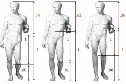
Figure 1. Example of canonical and modified stimuli.
The original image (Doryphoros by Polykleitos) is shown at the centre of the figure. This sculpture obeys to canonical propor-tion (golden ratio = 1:1.618). Two modified versions of the same sculpture are presented on its left and right sides. The left image was modified by creating a short legs: long trunk rela-tion (ratio = 1:0.74); the right image by creating the opposite relation pattern (ratio = 1:0.36). All images were used in be-havioural testing. The central image (judged-as-beautiful on 100%) and left one (judged-as-ugly on 64%) were employed in the fMRI study.
2. Materials and Methods 2.1 Participants
Fourteen healthy right-handed volunteers (8 males, 6 fe-males; mean age 24.5, range 12 years) participated in this study. They were educated undergraduate or graduate students, with no experience in art theory. After receiving an explanation of the experimental procedure, partici-pants gave their written informed consent. The study was approved by the independent Ethics Committee of the Santa Lucia Foundation (Scientific Institute for Research Hospitalization and Health Care).
2.2 Stimuli
Fifteen 2-dimentional images of Classical and Renais-sance sculptures were chosen following a specific
selec-Cinzia Di Dio Emiliano Macaluso Giacomo Rizzolatti The Golden Beauty
http://psicoart.cib.unibo.it
5
PSICOART n. 1 - 2010
tion method.7 All the original pictures met the criteria of
canonical proportions defined by the ratio 1:1.618 be-tween body parts; among the 15 modified image-versions, 7 presented a “long-trunk, short-legs” modification (range = 1:1.47-1:1.59), whereas the remaining 8 images pre-sented the opposite pattern of modification (range = 1:1.64-1:1.82). Twenty sculptures represented male bodies and 10 female bodies.
2.3 Paradigm
The stimuli were presented in three experimental condi-tions: observation (O), aesthetic judgment (AJ), and pro-portion judgment (PJ). Each participant underwent 6 separate fMRI runs, repeating each condition twice. The condition order was maintained fixed across all partici-pants, with observation condition first, explicit aesthetic judgment second, and explicit proportion judgment, last.
By keeping the observation runs first, we aimed at meas-uring unbiased (spontaneous) brain responses to the type of the stimuli (canonical and modified). To make sure that volunteers were not biased in their aesthetic judg-ment by explicit proportion evaluation, the aesthetic judgment condition always preceded the proportion judgment runs.
Within each run we presented 30 stimuli (15 canonical and 15 modified) in a randomized order, but never repeat-ing the same image within a run. A question mark in-structed the participants to respond to the images after a 4s-fix interval following each stimulus presentation by using a response box placed inside the scanner.
2.4 Task
Participants lay in the scanner in a dimly lit environment. The stimuli were presented on a black background and
Cinzia Di Dio Emiliano Macaluso Giacomo Rizzolatti The Golden Beauty
http://psicoart.cib.unibo.it
6
PSICOART n. 1 - 2010
were displayed on a screen visible through a mirror mounted on the interior of the head coil. At the beginning of each session, a 5 s visual instruction informed the vol-unteers about the upcoming condition/task. On each trial, a 400ms central fixation point plus 1000 ms blank-screen interval preceded the presentation of the sculpture stimu-lus. The stimulus then appeared at the centre of the screen for 2 s and it was followed by another 4 s blank-screen interval.8 After this, a question mark instructed the
observer to respond to the stimulus (see below). The question mark remained on screen for 400 ms and was followed by a jittered interval ranging 2–5 s, with a uni-form distribution.
During observation condition (O), the volunteers were required to observe the images as if they were in a mu-seum and, when the question mark appeared, they had to indicate whether they paid attention to the picture or not. During the aesthetic and proportion judgment conditions,
the volunteers were required to decide whether they liked the image (AJ) or whether they found it proportional (PJ), respectively. Thus, all 3 conditions required a re-sponse from the participants. Using the index or middle finger of the right hand, the participants answered yes or no, according to the instruction presented at the start of each run. Specifically, before the observation sessions, the participants were instructed to answer “yes” if they paid attention to the stimulus just presented, whereas to press “no” to indicate that they did not pay attention to the stimulus.
The question “did you pay attention to the image?” was introduced to make sure that participants were actually looking at the stimuli during fMRI scanning. During AJ condition, participants were required to indicate “yes” if they aesthetically liked the image and “no” if they did not. Finally, PJ condition required the observers to explicitly indicate whether they thought that the image was
propor-Cinzia Di Dio Emiliano Macaluso Giacomo Rizzolatti The Golden Beauty
http://psicoart.cib.unibo.it
7
PSICOART n. 1 - 2010
tional by pressing “yes” or if they thought that the image was disproportionate by pressing “no”.
The volunteers underwent six subsequent scanning runs, each lasting approximately 5.6 min. Each fMRI runs con-sisted of 30 trials with each sculpture images presented once.
2.5 Image acquisition
Functional images were acquired with a Magnetom Vision MRI scanner (Siemens, Erlangen, Germany) operating at 3T. Blood oxygenation level dependent (BOLD) contrast was obtained using echo-planar T2* weighted imaging (EPI). The acquisition of 32 transverse slices with an ef-fective repetition time of 2.08 s, provided coverage of the whole cerebral cortex. The in-plane resolution was 3×3 mm.
2.6 Data analysis
Two types of analyses of fMRI data were performed. A stimulus-based analysis (“objective beauty”) considered only the type of image that was presented to the partici-pants: i.e. with canonical (C) or modified (M) propor-tions. The second analysis (“subjective beauty”) catego-rized each sculpture image according to the behavioural responses measured during AJ runs. For this analysis, we included only images that were consistently judged either beautiful (B) or ugly (U) in both runs requiring aesthetic judgment.
Event-related fMRI data were processed with SPM2.9 The
first four image volumes of each run were discarded to allow for stabilization of longitudinal magnetization. For each participant, the remaining 162 volumes were re-aligned with the first volume, and the acquisition timing was corrected using the middle slice as reference.10 To
al-Cinzia Di Dio Emiliano Macaluso Giacomo Rizzolatti The Golden Beauty
http://psicoart.cib.unibo.it
8
PSICOART n. 1 - 2010
low inter-subject analysis, images were normalised to the Montreal Neurological Institute (MNI) standard space,11
using the mean of the 162 functional images. All images were smoothed using an isotropic Gaussian kernel (full width at half maximum = 10 mm).
Statistical inference was based on a random effects ap-proach.12 This comprised two steps. First, for each
sub-ject, the data were best-fitted (least-square fit) at every voxel using a linear combination of the effects of interest. The effects of interest were the timing of the fixation point onsets, the presentation times of the sculptures (C & M; or B & U), and the presentation times of the question mark that cued overt responses. All event-types were con-volved with the SPM2 standard haemodynamic response function (HRF). Linear compounds (contrasts) were used to determine common effect (C+M vs. rest) and differen-tial effects associated with the presentation of the sculp-tures (C-M and M-C; or B-U and U-B), separately for each
of the three conditions (O, AJ and PJ). For each subject, this led to the creation of six contrast-images, that is three contrasts C+M vs. rest–one for each condition, and three contrasts C-M vs. rest, again one for each condition. Ad-ditionally, three contrast-images were also created, which contrasted judged-as-beautiful vs. judged-as-ugly images for each condition.
These contrast-images then underwent the second step that comprised three separate ANOVAs. One considering overall pattern of activation C+M vs. rest modeled for each condition; one considering “objective beauty” (C vs. M) modeled for each condition; and one considering “subjective beauty” (B vs. U) for each condition. Finally, for each of the three separate ANOVAs, linear compounds were used to compare these effects, now using between-subjects variance. Correction for non-sphericity13 was
used to account for possible differences in error variance across conditions and any non-independent error terms
Cinzia Di Dio Emiliano Macaluso Giacomo Rizzolatti The Golden Beauty
http://psicoart.cib.unibo.it
9
PSICOART n. 1 - 2010
for the repeated measures.
The following contrasts were tested. First, within the “common effects”, ANOVA (C+M vs. rest) averaging across all experimental conditions (O, AJ, PJ). For this, the SPM-maps were thresholded at P-corrected = 0.05 (voxel-level). The other two ANOVAs assessed any stimu-lus -specific effect (“objective”: C-M, M-C; or “subjective”: B-U, U-B). We tested for main effects of stimulus across the three experimental conditions (O, AJ, PJ); and for in-teractions between stimulus and condition. Additional contrasts explored simple effects separately for the differ-ent conditions (e.g. B-U, during AJ only). For all these stimulus-specific effects, we used P-corrected = 0.05 at the cluster-level (cluster size estimated with a voxel-level threshold of P-uncorrected = 0.001, extent threshold = 10 voxels).
In addition, because of our prior hypothesis concerning the possible involvement of the insula in aesthetic
appre-ciation, we used a small volume correction procedure14 to
test for the effect of “objective beauty” (C-M; within and across O/AJ/PJ conditions) specifically in this region. The search volume was derived from Mario Livio15
center-ing a sphere at MNI x, y, z = 30, 18, 18; with a radius of 10 mm.
3. Results and Discussion 3.1 fMRI behavioural data
The viewers' evaluation of the stimuli, as expressed in the aesthetic judgment condition, showed that the canonical images were mostly evaluated positively (76%, sd = 0.18), whereas the modified images were generally scored with a negative rating (63%, sd = 0.25). This finding was in ac-cord with a preliminary behavioural testing used for
im-Cinzia Di Dio Emiliano Macaluso Giacomo Rizzolatti The Golden Beauty
http://psicoart.cib.unibo.it
10
PSICOART n. 1 - 2010
ages selection that also showed the relevance of propor-tion in aesthetic evaluapropor-tion. In this test violapropor-tion of ca-nonical proportions accounted for 77% of the variance in aesthetic rating (partial Eta2).16
3.2 Overall effect of viewing the sculptures
MRI analysis was carried out by first assessing the overall effect of viewing the sculptures contrasting canonical (C) and modified (M) images (pooled together, C+M) with rest, across all three conditions (O, AJ, PJ; P-corrected<0.05).
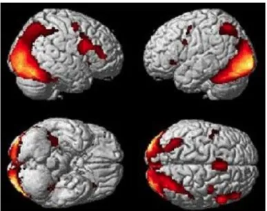
As shown in Figure 2, activations were found in occipital and temporal visual areas, including lingual and fusiform gyri. Additionally, activations were observed in the infe-rior parietal lobule (IPL) bilaterally, in the SMA/pre-SMA complex, ventral premotor areas, and in the posterior part of right inferior frontal gyrus (IFG). Signal increase
was also found in the insula and hippocampus. Most of the activations were bilateral, although stronger in the right hemisphere. These results are summarized in Table 1.
Among the visual activations, besides the primary visual cortex, signal increase was found in the lateral occipital cortex and the inferior temporal lobe (shape sensitive ar-eas), as well as in the MT/MST complex. This last finding, although surprising at first considering that the MT/MST complex is involved in the analysis of motion,17 is
consis-tent with previous data showing that activation of these areas may be elicited by static images that imply motion.18
Most noteworthy was the activation of the inferior parie-tal lobule and especially of the premotor cortex. These ar-eas are known to become active during the observation of actions done by others.19 It is likely that their activation
was dependent on the intrinsic dynamic properties of the sculptures used in this study and the sense of action that
Cinzia Di Dio Emiliano Macaluso Giacomo Rizzolatti The Golden Beauty
http://psicoart.cib.unibo.it
11
PSICOART n. 1 - 2010
they evoked in the observer.20
Figure 2. Brain activation of canonical and modified sculp-tures vs. rest.
The analysis was carried out by averaging activity across the three experimental conditions (observation, aesthetic judgment, proportion judgment). Group-averaged statisti-cal parametric maps are rendered onto the MNI brain tem-plate (P-corrected<0.05)
3.3 Canonical vs. Modified Sculptures: “Objective Beauty”
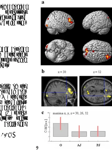
The direct contrast of canonical vs. modified images across the three experimental conditions revealed signal increase for the canonical stimuli in the right occipital cortex extending into lingual gyrus; in the precuneus bi-laterally; in the right posterior cingulate gyrus; and in the depth of right inferior frontal sulcus extending to the ad-jacent convexity of the middle frontal gyrus (P-corrected<0.05; Figure 3a; see also Table 2a).
The lateral occipital cortex (LOC)21 and the temporal
vis-ual areas are known to be responsive to the presentation of body parts or even the whole human body.22 Signal
in-crease within these areas may be therefore due to a greater representation of canonical body structures rela-tive to the disproportionate ones. The activation of the medial parietal areas and of the prefrontal lobe, on the
Cinzia Di Dio Emiliano Macaluso Giacomo Rizzolatti The Golden Beauty
http://psicoart.cib.unibo.it
12
PSICOART n. 1 - 2010
other hand, might be related to mnemonic functions,23
possibly elicited by the retrieval of plausible motor con-figurations, better represented by the proportional mate-rial.
The central hypothesis underlying the present study was that the contrast of canonical vs. modified stimuli would produce signal enhancement within the insula. Accord-ingly, we carried out a small volume correction within the main effects analysis (C-M) using the anatomical coordi-nates reported in A. Damasio, T.J. Grabowski et alii24 on
the feeling of emotion.25 The results revealed a significant
signal increase in the anterior sector of the right insular cortex extending to the operculum region (maxima x, y, z = 30, 26, 12; Figure 3b, P<0.05, corrected for small vol-ume).
This effect was particularly strong during observation condition (P<0.02, corrected for the whole brain volume, Table 2b; P = 0.005, corrected for small volume), which is
.
Figure 3. Brain activation in the contrast canonical vs. modi-fied stimuli.
a. Main effect of canonical
vs. modified sculptures
across conditions rendered onto the MNI brain tem-plate.
b, Parasagittal and coronal view showing activations of the right insular region in the main effect.
c. Activity profile of the right insula. For each con-dition (O, AJ, PJ) the sig-nal plots show the differ-ence between canonical (C) minus modified (M) sculp-tures in arbitrary units (a.u), +/− 10% confidence intervals
Cinzia Di Dio Emiliano Macaluso Giacomo Rizzolatti The Golden Beauty
http://psicoart.cib.unibo.it
13
PSICOART n. 1 - 2010
in the condition in which the volunteers were in a merely observational (museum-like) context (see Figure 3c). Sig-nal increase in AJ and PJ conditions, on the other hand, was virtually the same. The most likely interpretation for this result stands in the different cognitive demands be-tween the first (O) and the last two (AJ, PJ) conditions. In the latter, in fact, the explicit request of overtly judging the stimuli diverted the volunteers' attention resources towards a specific cognitive demand, thus lessening the natural neural response within the insula.
These data are in apparent contrast with some previous findings where symmetry was employed as an objective parameter of aesthetic evaluation.26 In this study, the
au-thors did find significant activation in the anterior insula in the comparison of aesthetic judgment vs. control con-dition as well as in symmetry judgment vs. control condi-tion. However, they considered those areas that were ac-tivated by both aesthetic and symmetry judgment to be
not involved in pure aesthetic judgment and hence omit-ted them from the analysis that directly contrasomit-ted brain activity for the judged-as-beautiful vs. the judged-as-ugly stimuli. In this way, therefore, they also disregarded the insular activation elicited by objective parameters (i.e. symmetry) intrinsic to the stimuli and involved in mediat-ing the sense of beauty.
The question now arises of what possible mechanisms are responsible for the insula activation during the observa-tion of canonical sculptures. The anterior sector of the in-sula has an agranular/disgranular cytoarchitectonic or-ganization and is characterized by extensive connections with limbic structures and with centers involved in auto-nomic functions.27 Functionally, anterior insula is thought
to mediate feelings associated with specific emotional states.28 Now, considering the pattern of activity
de-scribed in the main effect (C+M vs. rest), there are two concurrent possibilities that may explain insula
activa-Cinzia Di Dio Emiliano Macaluso Giacomo Rizzolatti The Golden Beauty
http://psicoart.cib.unibo.it
14
PSICOART n. 1 - 2010
tion. One is that in LOC and in the parietal cortex there are neurons specifically sensitive to the canonic body im-ages and that have privileged access to the insula. Alter-natively, one may suppose that the canonical sculptures simply determined a stronger activation of cortical neu-rons sending their output to the insula.
Another possible explanation, based on both main and simple effect analyses (C-M), is that the insula was acti-vated, not by simplest aspects of the visual stimuli (e.g. shape or motion), but rather by higher order information coming from prefrontal areas 45 and 46. Studies in pri-mates29 showed that area 45 integrates information about
object shape with that about actions. While human left area 45 subserves language functions, it is plausible that human right area 45, selectively activated in the present experiment, could be involved in action/shape integration as well. In this light, canonical stimuli could be more effi-ciently coded in this area and determined, therefore, a
stronger activation of the insula relative to the modified one. In this context, also the functional role of prefrontal area 46 could be noteworthy in confronting information from memory (e.g. standard body configuration) with online incoming information (observation of canonical and modified stimuli).
To summarize, we propose that the positive emotional feeling elicited in the viewer by the canonical images was determined by a preferential coding of these images, rela-tive to the modified ones, by various cortical areas and by a concurrent, joint activation of the anterior insula.
3.4 Judged-as-Beautiful vs. Judged-as-Ugly Sculptures: “Subjective Beauty”
With this further analysis, we investigated the neuronal substrate associated with subjective appreciation of the sculptures as expressed by each participant in the AJ
Cinzia Di Dio Emiliano Macaluso Giacomo Rizzolatti The Golden Beauty
http://psicoart.cib.unibo.it
15
PSICOART n. 1 - 2010
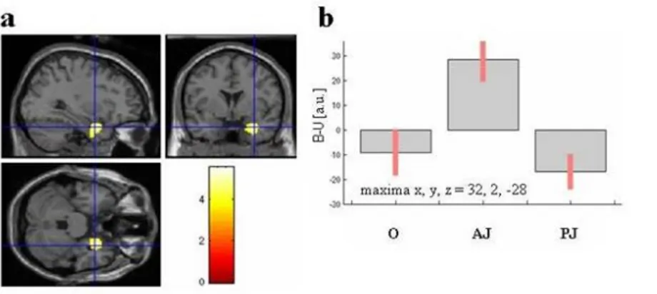
condition (2 runs). Behavioural data showed that 49% and 38% of stimuli were consistently judged, respectively, beautiful (B) and ugly (U) over both AJ runs, whereas 13% was rated inconsistently. Only the stimuli that were rated in a consistent way were employed for analysis. The judged-as-beautiful images selectively activated the right amygdala. This effect was observed for the aesthetic judgment condition, as demonstrated by the stimu-lus×condition interaction analysis (maxima: x, y, z = 32, 2, −28; P-corrected<0.03; Figure 4 a,b).
The amygdala is a complex nuclear structure. It is inter-connected with several cortical areas and subcortical brain centers and subserves a variety of functional roles. However, a fundamental amygdalar function is to provide neutral stimuli with positive or negative values through association learning.30
For a long time, studies involving the amygdala have mainly focused on negative stimulus conditioning.
How-ever, more recent studies support a role of the amygdala also for positive emotions, both in animals31 and
hu-mans.32 This property puts the amygdala as a prime
can-didate in the storing of implicit emotional memories that can be subsequently accessed and used. In this light, the judged-as-beautiful stimuli could have been judged as such, not on the basis of their objective parameters, but because they were associated with memories charged with positive emotional values. The distinctiveness of each own experience would then partly explain the variance observed in the subjective rating of the observed images. Finally, we compared ugly versus judged-as-beautiful stimuli. As shown in Figure 4c, the only acti-vated area was a region straddling the central sulcus (somatomotor cortices; P-corrected<0.05; see also Table 3a). Figure 4d shows signal change in this region, reveal-ing a particularly strong effect of “ugly” versus “beautiful” images during the explicit aesthetic judgment condition.
Cinzia Di Dio Emiliano Macaluso Giacomo Rizzolatti The Golden Beauty
http://psicoart.cib.unibo.it
16
PSICOART n. 1 - 2010
This selectivity was confirmed by the significant stimulus-by-condition interaction, as reported in Table 3.
These data are in accord with previous findings by Kawa-bata and Zeki showing that a negative evaluation of paint-ings (landscapes, abstract paintpaint-ings, portraits, still life) determined the activation of the somatomotor region.33
There is also evidence from other studies that negative emotional stimuli may determine unilateral or bilateral activation in this region.34
The activation of the somatomotor region during aes-thetic judgment seems rather surprising in the absence of actual movements. However, this activation may find an explanation if one also considers the activity pattern (de-activation) of the orbito-frontal cortex reported in Kawa-bata and Zeki35 and also found in our work in a post-hoc
analysis.36 Although much attention has been drawn in
recent years on the role of the orbito-frontal cortex in re-lation to positive rewards,37 there is also evidence coming
from lesion studies that damage to orbito-frontal cortex causes a liberation of a variety of behaviours, ranging from extreme irritability, hot temper, antisocial behav-iour, to euphoria, locomotor hyperactivity and sexual dis-inhibition.38 If one admits that a decrease of activity in
orbito-frontal cortex mimics, although to a different ex-tent, the effect of a lesion one may account for the motor activation in response to ugly stimuli as a covert release of an appropriate motor behaviour.
Cinzia Di Dio Emiliano Macaluso Giacomo Rizzolatti The Golden Beauty
http://psicoart.cib.unibo.it
17
PSICOART n. 1 - 2010
Figure 4. Brain activations in the contrasts “judged-as-beautiful vs.
a. Parasagittal, coronal and transaxial sections showing activation of the right amygdala in the interaction stimulus (beautiful vs. ugly)×condition (observation; aesthetic judgment; proportion judg-ment).
b. Activity profile of the right amygdala. For each condition (O = ob-servation, AJ = aesthetic judgment, PJ = proportion judgment) the signal plots show the difference between beautiful (B) minus ugly (U)-as judged sculptures in arbitrary units (a.u), +/− 10% confidence intervals.
judged-as-ugly” and “judged-as-ugly vs. judged-as-beautiful” stimuli
c. Statistical parametric maps rendered onto the MNI brain template showing activity within left somatomotor cortex in the contrast of ugly vs. beautiful stimuli averaged across the three conditions.
d. Activity profile (ugly-beautiful) of the left motor cortex. For each condition (O, AJ, PJ) the signal plots show the difference between ugly (U) minus beautiful (B)-as judged sculptures in arbitrary units (a.u), +/− 10% confidence intervals (P-corrected<0.05).
Cinzia Di Dio Emiliano Macaluso Giacomo Rizzolatti The Golden Beauty
http://psicoart.cib.unibo.it
18
PSICOART n. 1 - 2010
3.5 Final considerations
The main question we addressed in the present study was whether there is an objective beauty, i.e., if objective pa-rameters intrinsic to works of art are able to elicit a spe-cific neural pattern underlying the sense of beauty in the observer. Our results gave a positive answer to this ques-tion. The presence of a specific parameter (the golden ra-tio) in the stimuli we presented determined brain activa-tions different to those where this parameter was vio-lated. The spark that changed the perception of a sculp-ture from “ugly” to beautiful appears to be the joint acti-vation of specific populations of cortical neurons respond-ing to the physical properties of the stimuli and of neu-rons located in the anterior insula.
Insula mediates emotion feelings. It would be too reduc-tive, however, to think that the sense of beauty occurs be-cause of the activation of this structure alone. Insula is
also activated by non-artistic stimuli; however, the feeling that these stimuli produce in the observer differs qualita-tively from that determined by artworks. Our view is that this specific quality–the sense of beauty-derives from a joint activity of neural cortical populations responsive to specific elementary or high order features present in works of art and neurons located in emotion controlling centers.
It has often been claimed that beauty, objectively deter-mined, does not exist because of profound subjective dif-ferences in the evaluation of what is beautiful and what is not. Although individual biases are undeniable, it is also rather implausible to maintain that beauty has no biologi-cal substrate and is merely a conventional, experientially determined concept. As Gombrich39 wrote, elements in a
picture which determine aesthetical experience are “deeply involved in our biological heritage”, although we are unable to give a conscious explanation to them.40
Cinzia Di Dio Emiliano Macaluso Giacomo Rizzolatti The Golden Beauty
http://psicoart.cib.unibo.it
19
PSICOART n. 1 - 2010
The results of our experiment concerning what we called subjective beauty are also relevant here. In the condition in which the viewers were asked to indicate explicitly which sculptures they liked, there was a strong increase in the activity of the amygdala, a structure that responds to incoming information laden with emotional value. Thus, instead of allowing their nervous centers to “resonate” in response to the observed stimuli (observation condition), when the viewers judged the stimuli according to their individual idiosyncratic criteria (explicit aesthetic judg-ment), that structure was activated that signals which stimuli had produced pleasant experiences in the past. In conclusion, both objective and subjective factors inter-vene in determining our appreciation of an artwork. The history of art is replete with the constant tension between objective values and subjective judgments. This tension is deepened when artists discover new aesthetic parameters that may appeal for various reasons, be they related to our
biological heritage, or simply to fashion or novelty. Still, the central question remains: when the fashion and nov-elty expire, could their work ever become a permanent patrimony of humankind without a resonance induced by some biologically inherent parameters?
Cinzia Di Dio Emiliano Macaluso Giacomo Rizzolatti The Golden Beauty
http://psicoart.cib.unibo.it
20
PSICOART n. 1 - 2010
TABLE 1. Brain activity reflecting the common effects of Canonical and Modified images vs. baseline across conditions (observation; aesthetic judgment; proportion judgment).
BRAIN STRUCTURE SPHERE MAXIMA
x y z
Z p. corr
(vx) Occipital Lobe
Inferior occipital gyrus (LO)
L -45 -84 -6 Inf 0.000 R 38 -88 -10 Inf 0.000 Middle occipital gyrus L -32 -96 -6 Inf 0.000 R 28 -92 0 Inf 0.000 R 30 -92 -2 Inf 0.000
Parietal Lobe
Supramarginal gyrus R 64 -20 38 5.08 0.006
Frontal Lobe
Middle frontal gyrus R 38 0 54 5.49 0.001 R 38 -2 54 4.89 0.015 R 38 0 52 4.65 0.041 Inferior frontal gyrus R 50 14 24 7.32 0.000 R 52 14 24 5.36 0.002 R 45 40 8 5.33 0.002 R 50 34 18 5.25 0.003 R 48 35 14 5.24 0.003
BRAIN STRUCTURE SPHERE MAXIMA Z p. corr
(vx) Precentral gyrus R 50 10 12 6.21 0.005 R 54 8 42 4.84 0.019 Precentral gyrus L -56242 4.58 0.036 L -50 8 30 4.62 0.047 L -52 6 26 4.56 0.05 Supplementary motor area – 0 10 52 6.36 0.000 Supplementary motor area R 4 16 48 5.16 0.005 R 2 16 50 6.01 0.000 R 14 8 58 5.21 0.004 Subcortical/insula Ippocampus R 24 -32 -6 5.49 0.001 R 22 -32 -6 5.35 0.002 Ippocampus L -22 -32 -6 6.14 0.000 4.92 0.013 Insula R 36 20 -6 5.05 0.008 Insula L -34 24 -4 5.58 0.001 Cerebellum Cerebellum 4-5 R 32 -34 -28 4.81 0.021
Cinzia Di Dio Emiliano Macaluso Giacomo Rizzolatti The Golden Beauty
http://psicoart.cib.unibo.it
21
PSICOART n. 1 - 2010
TABLE 2. Brain activity reflecting the main effect (a) and the
sim-ple effect (b) of Canonical vs. Modified images
BRAIN STRUCTURE SPHERE MAXIMA Z p. corr
x y z cluster
level
a Main Effect (C-M)
Medial parietal lo-be/Precuneus
R 12, -52, 46 3.79 0.04 L -2, -42, 58 3.21
Posterior cingulum R 8, -52, 30 3.33
Inferior occipital gyrus R 30, -94, -8 3.75 0.0001 Lingual gyrus R 16, -66, -6 3.56
Cuneus L -4, -78, 30 3.55
Inferior frontal gyrus R 44, 42, 20 3.65 0.03 Middle frontal gyrus R 30, 40, 30 3.65
b Simple Effect Observation (C-M)
Anterior insula/frontal operculum
R 36, 22, 16 3.86 0.016 Middle frontal gyrus R 38, 36, 20 3.62
Superior frontal gyrus R 18, 44, 26 3.31
TABLE 3. Brain activity reflecting main effect (a) and interaction (b) of judged-as-ugly vs. judged-as-beautiful images
BRAIN STRUCTURE SPHERE MAXIMA Z p. corr
x y z cluster
level
a. Main Effect
Precentral gyrus L -36, -14, 60 4.68 0.0001 Postcentral gyrus L -38, -28, 52 4.34
b Interaction (Stimulus by Condition)
Precentral gyrus L -36, -12, 58 4.35 0.003 Postcentral gyrus L -40, -34, 56 3.88
Inferior parietal lobule L -50, -26, 40 3.82 L -52, -32, 52 3.34
Cinzia Di Dio Emiliano Macaluso Giacomo Rizzolatti The Golden Beauty http://psicoart.cib.unibo.it 22 PSICOART n. 1 - 2010 AC K N O W L E D G M E N T S
We thank G. Berlucchi, V. Gallese and D. Freedberg for comments on the manuscript, G. Buccino for providing methodological support, K.D. Albano for helping in stimulus modification, S. Gazzitano for assistance in conducting MRI imaging acquisition.
* The text The Golden Beauty. Brain Response to Classical and
Ren-aissance Sculptures has been published in “PLoS ONE”, 2, 11, 2007.
NOTE
1 W. Tatarkiewicz, History of Aesthetics, Mouton, The Hague 1970
and R. Reber, N. Schwarz, P. Winkielman, Processing Fluency and
Aesthetic Pleasure: Is Beauty in the Perceiver’s Processing Experi-ence?, “Personality and Social Psychology Review”, n. 8, November
2004, pp. 364-382.
2 C.W. Valentine, The experimental psychology of beauty, Methuen,
London 1962.
3 D. Bayles, T. Orland, Art and Fear. Observations on the perils (and
rewards) of artmaking, Image Continuum Press Edition, Santa Cruz
2001.
4 C.J. Cela-Conde, G. Marty, F. Maestú, T. Ortiz, E. Munar, et al.,
Ac-tivation of the prefrontal cortex in the human visual aesthetic percep-tion, “Psychology”, n. 101, April 2004, pp. 6321-6325; H. Kawabata, S. Zeki, Neural Correlates of Beauty, “Journal of Neurophysiology”, n. 91, April 2004, pp. 1699-1705; O. Vartanian, V. Goel,
Neuro-anatomical correlates of aesthetic preference for paintings,
“Neu-roreport”, n. 15, April 2004, pp. 893-897; T. Jacobsen, R.I. Schubots, L. Hofel, D.V. v. Cramon, Brain Correlates of aesthetic judgment of
Cinzia Di Dio Emiliano Macaluso Giacomo Rizzolatti The Golden Beauty
http://psicoart.cib.unibo.it
23
PSICOART n. 1 - 2010
5 For reviews, see: H.E. Huntley, The divine proportion. A study in
mathematical beauty, Dover Publications, New York 1970 and M.
Livio, The Golden Ratio. The story of Phi, the extraordinary number
of nature, art and beauty, Headline Book Publishing, London 2002.
6 A. Damasio, The Feeling of What Happens: Body and Emotion in
the Making of Consciousness, Harcourt Brace, New York 1999; A.
Damasio, T.J. Grabowski, A. Bechara, H. Damasio, L.L. Ponto, et al.,
Subcortical and cortical brain activity during the feeling of self-generated emotions, “Nature Neuroscience”, n. 3, October 2000, pp.
1049–1056; A.D. Craig, Interoception: the sense of the physiological
condition of the body, “Current opinion in Neurobiology”, n. 13,
Au-gust 2003, pp. 500-505; H.D. Critchley, S. Wiens, P. Rotshtein, A. Ohman, R.J. Dolan, Neural systems supporting interoceptive
awareness, “Nature Neuroscience”, 7, February 2004, pp. 189-195;
H.D. Critchley, P. Rotshtein, Y. Nagai, J. O’Doherty, C.J. Mathias, et al., Activity in the human brain predicting differential heart rate
responses to emotional facial expressions, “NeuroImage”, n. 24,
Feb-ruary 2005, pp. 751-762.
7 See Supporting Information in the previous version of this paper
published in:
<http://www.plosone.org/article/info:doi/10.1371/journal.pone.000 1201>.
8 Also see: C.J. Cela-Conde, G. Marty, F. Maestú, T. Ortiz, E. Munar,
et al., Activation of the prefrontal cortex in the human visual
aes-thetic perception, cit. and P. Winkielman, J.T. Cacioppo, Mind at ease puts a smile on the face: Psychophysiological evidence that processing facilitation leads to positive affect, “Journal of
Personal-ity and Social Psychology”, n. 81, December 2001, pp. 989–1000.
9 For further information about the topic, see
<http://www.fil.ion.ucl.ac.uk>.
10 R.N.A. Henson, C. Buechel, O. Josephs, K. Friston, The slice-timing
problem in event-related fMRI, “NeuroImage”, n. 9, 1999, p. 125.
11 D.L. Collins, P. Neelin, T.M. Peters, A.C. Evans, Automatic 3D
in-tersubject registration of MR volumetric data in standardized Ta-lairach space, “Journal of Computer Assisted Tomography”, n. 18,
March-April 1994, pp. 192–205.
12 A.P. Holmes, K.J. Friston, Generalisability, random effects and
population inference, “NeuroImage”, n. 7, May 1998, p. S754.
13 K.J. Friston, Bayesian estimation of dynamical systems: an
Cinzia Di Dio Emiliano Macaluso Giacomo Rizzolatti The Golden Beauty
http://psicoart.cib.unibo.it
24
PSICOART n. 1 - 2010
14 K.J. Worsley, S. Marrett, P. Neelin, A.C. Vandal, K.J. Friston, et al.,
A unified statistical approach for determining significant signals in images of cerebral activation, “Human Brain Mapping”, 4, January
1996, pp. 58-83.
15 M. Livio, The Golden Ratio. The story of Phi, the extraordinary
number of nature, art and beauty, cit. See also: H.D. Critchley, S.
Wiens, P. Rotshtein, A. Ohman, R.J. Dolan, Neural systems
support-ing interoceptive awareness, cit. and H.D. Critchley, P. Rotshtein, Y.
Nagai, J. O’Doherty, C.J. Mathias, et al., Activity in the human brain
predicting differential heart rate responses to emotional facial ex-pressions, cit.
16 For details on the preliminary behavioral experiment, see
Support-ing Information in the previous version of this paper published in: <http://www.plosone.org/article/info:doi/10.1371/journal.pone.000 1201>.
17 J.D. Watson, R. Myers, R.S.J. Frackowiak, J.V. Hajnal, R.P. Woods,
et al., Area V5 of the human brain: evidence from a combined study
using positron emission tomography and magnetic resonance imag-ing, “Cerebral Cortex”, n. 3, March-April 1993, pp. 79-94; P. Dupont,
G.A. Orban, B. De Bruyn, A. Verbruggen, L. Mortelmans, Many areas
in the human brain respond to visual motion, “Journal of
physiology”, n. 72, September 1994, pp. 1420–1424; G.A. Orban, P. Dupont, B. De Bruyn, R. Vogels, R. Vandenberghe, et al., A motion
area in human visual cortex, “Proceedings of the National Academy
of Science”, n. 92, February 1995, pp. 993 –997.
18 Z. Kourtzi, N. Kanwisher, Activation in Human MT/MST by Static
Images with Implied Motion, “Journal of Cognitive Neuroscience”, n.
12, January 2000, pp. 48-55.
19 G. Rizzolatti, L. Craighero, The Mirror Neuron System, “Annual
Review of Neuroscience”, n. 27, 2004, pp. 169–192.
20 D. Freedberg, V. Gallese, Motion, emotion and empathy in esthetic
experience, “Trends in Cognitive Sciences”, n. 11, May 2007, pp.
197-203.
21 R. Malach, J.B. Reppas, R.R. Benson, K.K. Kwong, H. Jiang, et al.,
Object-related activity revealed by functional magnetic resonance imaging in human occipital cortex, “Neurobiology”, n. 92, August
1995, pp. 8135-8139; K. Grill-Spector, Z. Kourtzi, N. Kanwisher, The
lateral occipital cortex and its role in object recognition, “Vision
re-search”, n. 41, May 2001, pp. 1409-1422.
22 P.E. Downing, Y. Jiang, M. Shuman, N. Kanwisher, A cortical area
selective for visual processing of the human body, “Science”, n. 293,
Cinzia Di Dio Emiliano Macaluso Giacomo Rizzolatti The Golden Beauty
http://psicoart.cib.unibo.it
25
PSICOART n. 1 - 2010
Shulman, M. Corbetta, Extrastriate body area in human occipital
cortex responds to the performance of motor actions, “Nature
Neu-roscience”, n. 7, May 2005, pp. 542-548.
23 T. Shallice, P. Fletcher, C.D. Frith, P. Grasby, R.S.J. Frackowiak, et
al., Brain regions associated with acquisition and retrieval of verbal
episodic memory, “Nature”, n. 368, April 1994, pp. 633-635; E.
Tulv-ing, S. Kapur, H.J. Varkovitsch, F.I.M. Craik, R. Harbib, et al.,
Neu-roanatomical correlates of retrieval in episodic memory: auditory sentence recognition, “Proceedings of the National Academy of
Sci-ences of the United States of America”, n. 91, March 1994, pp. 2012-5. For a review, see A.E. Cavanna, M.R. Trimble, The precuneus: a
re-view of its functional anatomy and behavioural correlates, “Brain”,
n. 129, March 2006, pp. 1-20.
24 A. Damasio, T.J. Grabowski, A. Bechara, H. Damasio, L.L. Ponto, et
al., Subcortical and cortical brain activity during the feeling of
self-generated emotions, cit.
25 See also: H.D. Critchley, S. Wiens, P. Rotshtein, A. Ohman, R.J.
Dolan, Neural systems supporting interoceptive awareness, cit.; H.D. Critchley, P. Rotshtein, Y. Nagai, J. O’Doherty, C.J. Mathias, et al., Activity in the human brain predicting differential heart rate
responses to emotional facial expressions, cit.
26 T. Jacobsen, R.I. Schubots, L. Hofel, D.V. v. Cramon, Brain
Corre-lates of aesthetic judgment of beauty, cit.
27 M.M. Mesulam, E.J. Mufson, Insula of the Old World monkey
(III): Efferent cortical output and comments on function, “The
Jour-nal of Comparative Neurology”, n. 212, November 1982, pp. 38 –52; M.M. Mesulam, E.J. Mufson, The insula of Reil in man and monkey,
Architectonics, Connectivity and Function, “Cerebral Cortex”, vol. 4,
1985, pp. 179-226; S. Dupont, V. Bouilleret, D. Hasboun, F. Semah, M. Baulac, Functional anatomy of the insula: new insights from
im-aging, “Surgical and Radiologic Anatomy”, n. 25, May 2003, pp.
113-119.
28 J.R. Augustine, Circuitry and functional aspect of the insular lobe
in primates including humans, “Brain Research Review”, n. 22,
Oc-tober 1996, pp. 229-244; A. Damasio, The Feeling of What Happens:
Body and Emotion in the Making of Consciousness, cit.; A.D. Craig, Interoception: the sense of the physiological condition of the body,
cit.; H.D. Critchley, S. Wiens, P. Rotshtein, A. Ohman, R.J. Dolan,
Neural systems supporting interoceptive awareness, cit.; H.D.
Critchley, P. Rotshtein, Y. Nagai, J. O’Doherty, C.J. Mathias, et al.,
Activity in the human brain predicting differential heart rate re-sponses to emotional facial expressions, cit.
Cinzia Di Dio Emiliano Macaluso Giacomo Rizzolatti The Golden Beauty
http://psicoart.cib.unibo.it
26
PSICOART n. 1 - 2010
29 K. Nelissen, G. Luppino, W. Vanduffel, G. Rizzolatti, G. Orban,
Ob-serving others: Multiple action representation in the frontal lobe,
“Science”, n. 310, October 2005, pp. 332-336.
30 E.g. J.E. LeDoux, The Emotional Brain: The Mysterious
Under-pinnings of Emotional Life Simon and Schuster, New York 1996; P.
Rotshtein, R. Malach, U. Hadar, M. Graif, T. Hendler, Feeling or
fea-tures: different sensitivity to emotion in high-order visual cortex and amygdala, “Neuron”, n. 32, November 2001, pp. 747–757; E.A.
Phelps, J.E. LeDoux, Contribution of the Amygdala to Emotion
Processing: From Animal Models to Human Behaviour, “Neuron”, n.
48, October 2005, pp. 175-187; J.J. Paton, M.A. Belova, S.E. Morri-son, C.D. Salzman, The primates amygdala represents the positive
and negative value of visual stimuli during learning, “Nature”, n.
439, February 2006, pp. 865-870.
31 Ibid.
32 E.A. Phelps, J.E. LeDoux, Contribution of the Amygdala to
Emo-tion Processing, cit.
33 H. Kawabata, S. Zeki, Neural Correlates of Beauty, cit.
34 About fear, see J.L. Armony, R.J. Dolan, Modulation of spatial
at-tention by fear conditioned stimuli: an event-related fMRI study,
“Neuropsychologia”, n. 40, July 2002, pp. 817–826; about anger, see:
H. Zald, The human amygdala and the emotional evaluation of
sen-sory stimuli, “Brain Research. Brain Research Reviews”, n. 41,
Janu-ary 2003, pp. 88-123 and D.D. Dougherty, S.L. Rauch, T. Deckers-bach, C. Marci, R. Loh, et al., Ventromedial prefrontal cortex and
amygdala dysfunction during an anger induction PET study in pa-tients with depression with anger attacks, “Archives of General
Psy-chiatry”, n. 61, August 2004, pp. 795–804.
35 H. Kawabata, S. Zeki, Neural Correlates of Beauty, cit.
36 See Supporting Information Text S1 and Figure S1 in the previous
version of this paper published in: <http://www.plosone.org/article/info:doi/10.1371/journal.pone.000
1201>.
37 For a review, see E.T. Rolls, The orbitofrontal cortex and reward,
“Cerebral Cortex”, n. 10, January 2000, pp. 284–294 and M.L. Kringelbach, The human orbitofrontal cortex: linking reward to
he-donic experience, “Nature Reviews Neuroscience”, n. 6, September
2005, pp. 691-702.
38 K K. Kleist, Bericht über die Gehirnpatologie und ihrer Bedeutung
für Neurologie und Psychiatrie, “Zeitschrift für die gesamte
re-Cinzia Di Dio Emiliano Macaluso Giacomo Rizzolatti The Golden Beauty
http://psicoart.cib.unibo.it
27
PSICOART n. 1 - 2010
view, see F. Boller, J.Grafman, Handbook of Neuropsychology, El-sevier, Amsterdam 2001.
39 E.H. Gombrich, Tributes. Interpreters of our cultural tradition,
Phaidon Press, Oxford 1984.
40 See also V.S. Ramachandran, A brief tour of human consciousness,