Concerted action of cellular JNK and
Pin-1 restricts HIV-1 genome integration
to activated CD4
+T lymphocytes
Lara Manganaro
Ph.D. Thesis in Molecular Biology
Supervisor: Prof Mauro Giacca
Scuola Normale Superiore
!
!
a mamma, papà
e Paolo
!
!
!
!
!
!
!
!
!
!
!
!
The only means of strengthening one's intellect is to make up one's mind about nothing, to let the mind be a thoroughfare for all thoughts. L'unico modo per rafforzare l'intelletto è quello di non decidere niente riguardo a nulla, di lasciare che la mente sia una strada percorribile da tutti i pensieri.
Introduction
!
!
!
!
!
!
!
!
1!
1. The Human Immunodeficiency Virus 1 (HIV-1) 1
1.1 HIV-1 replication cycle and host factors functions 1 1.1.1 Cellular proteins regulating HIV-1 infection 5 1.1.2 Regulation of HIV-1 infection early phase by cellular
proteins: a balance between restriction and permissivity
factors 7
1.1.3 Interactions between cellular and viral proteins during HIV integration and transcription 14 1.1.4 Cellular proteins involved in the late phase of HIV-1 24 1.1.5 Cellular microRNAs: novel partners for HIV-1 27 1.2 Lymphocyte activation status and HIV-1 infection 29
2. The IN protein 34
2.1 IN structure and domains 34
2.2 IN enzimatic activity 37
2.3 IN interactors 40
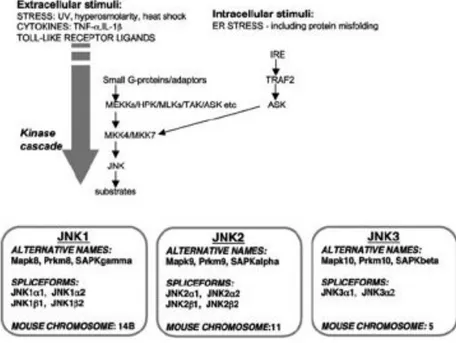
3. The Jun N-Terminal Kinase 44
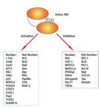
3.1 JNK structure and function 44
3.2 JNK substrates 47
3.3 JNK function in the immune system 49
4. The Prolyl Isomerase Pin1 52
4.2 Pin1 in the cell cycle 55 4.3 Pin1 in the immune system 58
Materials and Methods
611. In vitro binding assays and immunoprecipitation 61
2. Subtilisin Proteolysis 62
3. Kinase Assay 62
4. Cell culture and treatments 62 5. Plasmids, siRNA and Antibodies 64
6. HIV-1 RT Assay 65
7. Dot Blot 65
8. Strand Transfer 65
9. Virus Production, Infection and Alu-PCR 66
Results
69
1. Cellular JNK activity is required for efficient HIV-1 infection and integration in primary human T-lymphocytes 69 2. HIV-1 IN is phosphorylated on Ser 57 by cellular JNK 78 3. The cellular prolyl-isomerase Pin1 interacts with HIV-1 IN depending on JNK mediated phosphorylation 91 4. Pin1 regulates IN stability 96 5. Inhibition of Pin1 activity and mutation of IN Ser 57 impair HIV-1
Discussion
105
1. A role for JNK in determining permissivity to HIV-1 infection 106
2. HIV-1 IN is a substrate of JNK activity 110
3. A role for the prolyl-isomerase Pin1 in HIV-1 infection 113
4. A model to explain the poor susceptibility of resting CD4" T cells to HIV-1 infection 119
Introduction
1. The Human Immunodeficiency Virus 1 (HIV-1)
1.1 HIV-1 Life Cycle and host factors functions
Human Immunodeficiency virus (HIV) is a retrovirus, classified into the Lentivirus genus, which is the causative agent of the Acquired Immunodeficiency Syndrome (AIDS). A hallmark of all members of the Retroviridae family is to produce DNA from an RNA genome via the enzyme reverse transcriptase. The DNA is incorporated into the host genome by the integrase enzyme (IN), then it replicates as part of the host cell DNA. In 1983 Dr. Montagnier isolated from lymphonodes of an asymptomatic individual, who presented a generalized lymphadenopatia, an agent containing a reverse transcriptase activity that was highly cytopathic in human peripheral blood mononuclear cells (Barré-Sinoussi et al., 1983). In the same period, Dr. Gallo and Dr. Levy isolated a retrovirus from both immunodeficiency patients and healthy individuals from the various risk groups
!
(Gallo et al., 1984; Popovic et al., 1984). This new retrovirus was originally named LAV by the French group and HTLV-III by the US group, and later called Human Immunodeficiency virus (HIV); the new disease was named Acquired Immunodeficiency Syndrome (AIDS). In 1986 a related virus was isolated from African individuals and was named HIV-2, which is less pathogenic than HIV-1 (Clavel et al., 1986). In the last 25 years therapeutic strategies have been successful to control viral spread, however AIDS is still considered a devastating and incurable disease. There are several reasons for the impossibility to cure HIV-1 infection, that relate to both the HIV-1 cellular targets and its life cycle. First HIV-1 mainly infects CD4" T cells and macrophages causing catastrophic effects on the immune system such as CD4" T cell depletion and destruction of lymphoid organs. Moreover the emergence of viral strains that are resistant tocurrently available drugs is a crucial feature of the infection. One of the major reasons for the emergence of resistant strains is that both RNA Polymearse II and in particular Reverse Transcriptase, the two enzymes that synthesize viral genomes during the infection cycle are an error prone enzymes, lacking exonucleolytic proof-reading activity. In addition, during reverse transcription template switching occurs, causing deletions, insertions and mutations; these variations allow the virus to evade the immune response. Besides immune escape, a major reason why HIV-1 infection cannot be eradicated by the current therapies is due to the remarkable property of the virus to establish latent infection in resting CD4" T cells: since these cells do not express viral proteins, they remain completely unseen by the immune system and untouched by current antiretroviral therapies (Chun et al., 1995; Lassen et al., 2004; Williams and Greene 2007).
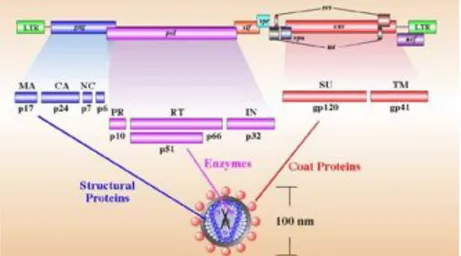
The HIV-1 genome is composed of two copies of positive single-stranded RNA that codes for the viral genes (Alizon et al., 1984). These two copies of the viral genome are enclosed by a conical capsid composed of 2,000 copies of the viral protein p24; single-stranded RNA is bound to nucleocapsid proteins. The virion contains two enzymes needed for the early steps of infection: Reverse Transcriptase (RT) and Integrase (IN), and Protease (PR), which ia important for virion maturation. A matrix composed of the viral protein p17 surrounds the capsid ensuring the integrity of the virion particle. This is, in turn, surrounded by the viral envelope, which is composed by phospholipid bilayer derived from the cell membrane when a newly formed viral particle buds from it. Two viral proteins that derive from the Env gene are embedded in the viral envelope: the glycoprotein 120 (gp120) and the glycoprotein 41 (gp41). These molecules are responsible for the recognition, attachment and fusion with the target cell.
The RNA genome consists of at least nine genes (gag, pol, and env, tat, rev, nef, vif, vpr, vpu) encoding 19 proteins. Two of these genes, gag and env, contain informations needed to make the structural proteins for new viral particles. Env codes for a polyprotein called gp160 that is cleaved by a viral enzyme to form gp120 and gp41 and gag is translated into a p55 polyprotein that is cleaved to form p17 matrix (MA), p24 capsid (CA), p7 nucleocapsid (NC) and p6. Pol codifies for RT, IN and PR, while the six remaining genes, tat, rev, nef, vif, vpr, and vpu are regulatory genes (Figure 1) (Martin et al., 2000). The Tat protein (p16 and p14) is a transcriptional transactivator for the LTR promoter acting by binding the TAR RNA element (Berkhout et al., 1989). The Rev protein (p19) is involved in shuttling RNAs from the nucleus to the cytoplasm by binding to the RRE RNA element (Rev Nekhai and Jeang 2006). A role for Vif (p23) has only recently been uncovered. This protein is necessary for an efficient infection of certain cells types, while other cells support infection in the absence of it (Sakai et al., 1991). Now it has been demonstrated that Vif prevents the action of the restriction factor APOBEC3G (Stopak et al., 2003; Sheehy et al., 2003). The Vpr protein (p14) seems to play a role in the translocation of the PIC from cytoplasm to nucleus and has been shown to arrest cell division at G2/M phase when transcription from LTR promoter is more efficient (He et al., 1995). The Nef protein (p27) downregulates both CD4 and MHC molecules probably through the recruitment of lck (leukocyte-specific protein tyrosine kinase) and PACS-1 (Phosphofurin acidic cluster sorting protein 1) (Piguet et al., 2000; Salghetti et al., 1995). A number of studies suggests that Nef may control the activation status of infected cells and their survival responses as it interacts with p21-activated kinase, Src-family kinases (PAK), Phosphoinositide 3-kinases (PI3-Kinase) and apoptosis signal-regulating kinase (ASK) (Nunn et al., 1996; Wolf et al., 2001; Geleziunas et al., 2001; Graziani et al 1996; Blagoveshchenskaya et al., 2002). However its role in signal transduction pathways still re mains
controversial (Marsh, 1999). The Vpu protein (p16) is an integral membrane phosphoprotein. Early studies have demonstrated that it is necessary for an efficient release of the viral particles (Klimkait et al., 1990). The mechanisms by which Vpu influences particle release was disclosed only recently by two different groups. Biesniasz and colleagues discovered a new cellular factor (Tetherin) that blocks viral particle release and is antagonized by Vpu (Neil et al., 2008) and Freed laboratory found that Calcium-modulating cyclophilin ligand (CAML), which restricts HIV-1 release, is also a Vpu-interacting factor (Varthakavi et al., 2008).
Figure 1. Schematic representation of the HIV-1 genome. (Standford
1.1.1 Cellular proteins regulating HIV-1 infection
HIV-1 is a very sophisticated virus despite its simple genomic structure. Virtually all steps of its viral replication cycle involve cellular factors, suggesting a very complex dynamic relationship between the virus and the infected cell. Cellular proteins seem to have either pro-viral or anti-viral functions. Over the past several years, many cellular proteins involved in HIV-1 infection have been discovered, nevertheless the complexity of HIV-1 viral cycle suggests that presence of several other cellular partners. In 2008 three different large scale RNA interference screenings were performed to identify these partners (Brass et al., 2008; König et al., 2008; Zhou et al., 2008). Hundreds of proteins have been found to be involved in HIV-1 infection unlocking a broad range of possible future investigations.
Cellular
Factor Viral Phase Activity Reference
CD4 Entry Binding with Env Dalgleish et al., 1984
CXCR4 Entry Binding with Env Endres et al 1996 CCR5 Entry Binding with Env Choe et al., 1996 DC-SIGN Entry Virion
Internalization in DC cells
Geijtenbeek et al., 2000
Dynamins Entry Necessary for
virions endocytosis Miyauchi et al., 2009 Trim5 alpha Early Phase Interferes with the
uncoating process RESTRICTION FACTOR Hatziioannou et al., 2004 Keckesova et al., 2004
Cyclophilin A Early Phase Protects HIV-1 from an unknown
antiviral factor
Luban et al., 1993 Braaten and Luban 1996
APOBEC3 Reverse
Transcription Induces mutations in viral cDNA
RESTRICTION FACTOR
Sheeny et al 2002 Harris et al., 2003 Uracil-DNA Reverse Controls dUTP Bouhamdan et al.,
glycosylase Transcription misincorporation in
viral cDNA 1996 Priet et al., 2005 Cofilin Post-Entry
Migration Actin depolymerizing factor Yoder et al., 2008 Transportin-SR2 (TRN-SR2) Nuclear
Translocation PIC entry Chirst et al., 2008 Importin7 Nuclear
Translocation PIC entry Fassati et al., 2003 BAF Integration Prevents
autointegration Lee and Craigie, 1994 INI1/hSNF5 Integration IN interactor
involved in virion production
Kalpana et al., 1994 Sorin et al., 2006 LEDGF Integration Tethering factor Cherepanov et al.,
2003
Maartens et al., 2003
Emerin Integration Bridges together chromatin and viral cDNA Jacque and Stevenson 2006 Shun et al., 2007 p300/CBP Integration/ Transcription Positively modulates integration and transcription Cereseto et al., 2005 Marzio et al., 1998 Benkirane et al., 1998
Cyclin T1 Transcription Regulation of HIV-1
transcription Fujinaga et al., 1998 Bieniasz et al., 1999 NAP-1 Transcription Regulation of HIV-1
transcription Vardabasso et al., 2007 CRM-1 Nuclear
export Nuclear export receptor for unspliced viral RNA
Askjaer et al., 1998 TSG 101 Assembly and
Budding Interaction with ESCRT machinery Garrus et al., 2001 Tetherin Assembly and
Budding Inhibits Budding RESTRICTION FACTOR Neil et al., 2008 Calcium-modulating cyclophilin ligand Assembly and
Budding Inhibits Budding RESTRICTION FACTOR
Varthakavi et al., 2008
(CAML)
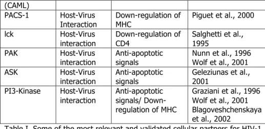
PACS-1 Host-Virus
Interaction Down-regulation of MHC Piguet et al., 2000 lck Host-Virus interaction Down-regulation of CD4 Salghetti et al., 1995 PAK Host-Virus
interaction Anti-apoptotic signals Nunn et al., 1996 Wolf et al., 2001 ASK Host-Virus
interaction Anti-apoptotic signals Geleziunas et al., 2001 PI3-Kinase Host-Virus interaction Anti-apoptotic signals/ Down-regulation of MHC Graziani et al., 1996 Wolf et al., 2001 Blagoveshchenskaya et al., 2002
Table I. Some of the most relevant and validated cellular partners for HIV-1 infection; restriction factors are indicated in red.
1.1.2 Regulation of HIV-1 infection early phase by cellular proteins: a balance between restriction and permissivity factors
ADSORPTION AND INTERNALIZATION
HIV-1 is an enveloped virus. The envelope, formed during budding, is a lipid bilayer carrying phospholipids and both viral and cellular proteins. Cellular components, which constitute a significant fraction of the envelope, represent particular areas of the plasma membrane from which budding occurred. The HIV-1 envelope is rich in cholesterol, indicating its preferential derivation from lipid rafts, as well as in MHC molecules. Two viral proteins are present in the envelope: gp120 or the surface env subunit (SU) and gp41, the transmembrane subunit (TM); these proteins are products of proteolytic cleavage of the gp160 precursor .
The sequence of gp120 is highly variable and heavily glicosylated: the most variable domains, termed hypervariable loops, are exposed at the surface of the virion while the conserved domains are folded to form the core of the protein (Fennie et al., 1989). gp120 interacts with specific cellular
receptors, the CD4 molecule and two different chemokine receptors (Dalgleish et al., 1984; Klatzmann et al., 1984; Alkhatib et al., 1996; Choe et al., 1996; Endres et al., 1996; Deng et al., 1996, Feng et al., 1996).
!
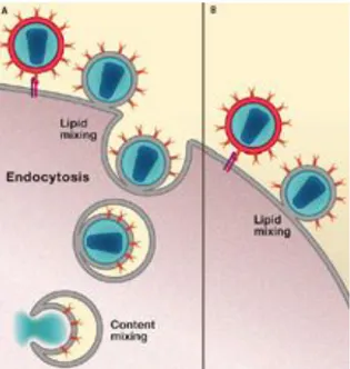
The CCR5 protein functions as a receptor for chemokines belonging to the CC group, including RANTES, MIP-1! and MIP-1". This receptor is predominantly expressed on T cells, macrophages, dendritic cells and microglia. The CXCR4 molecule, also called fusin, is an alpha-chemokine receptor specific for stromal-derived-factor-1 (SDF-1), a molecule possessing a potent chemotactic activity for lymphocytes. Both CCR5 and CXCR4 belong to the family of G-protein-coupled chemokine receptors and share a seven transmembrane-spanning alpha-helix structure that mediates signal propagation from biological membranes (rev: Lodowski et al., 2009). Based on its ability to bind these chemokine receptors, HIV-1 can be divided into different strains: CCR5 (R5 strain) or CXCR4R (X4 strains) or both (R5X4 strains). R5 strains can be isolated throughout the natural course of human infection and reflects the capability of HIV-1 to infect macrophages and monocytes. X4 and R5X4 strains appear in the late stages of infection, when the immune system is impaired; appearance of strains possessing this receptor specificity is an aggravating factor because it reflects the ability of HIV-1 to infect a larger spectrum of cells including resting T lymphocytes. The adhesion of HIV-1 to cells is mediated by the aspecific interaction between gp120 and charged groups on the cell surface. These interactions are important for the subsequent binding between gp120 and its specific receptors (Brelot and Alizon, 2001). HIV-1 has long been assumed to fuse directly with the plasma membrane (Stein et al., 1987; McClure et al 1988; Melikyan 2008). This fusion occurs after the induction of a conformational change in TM (Chan and Kim, 1998). Recent work entailing live cell imaging demonstrates that HIV-1 entry also occurs after virion endocytosis and shows that the cellular protein dynamin plays a pivotal role in this process (Figure 2) (Miyauchi 2009).Adhesion of HIV-1 to the cell surface is particularly important for the mucosal transmission of the infection. It has been shown that dendritic cells present in the skin and mucosae can uptake virions thorough the mannose-binding C-type lectin domain of a type II membrane protein named DC-SIGN (Geijtenbeek et al., 2000). The virions are retained in dendritic cells for extended periods of time in an infectious state and are presented to permissive T cells. The sites of contact and HIV-1 presentation between DC and CD4+ T cells, called immunological synapses
or virological synapses, were visualized by high resolution microspcopy. The authors showed that, in these contact sites, the viral receptors cluster, thus allowing highly efficient infection (Stoll at al., 2002; McDonald et al., 2003).
Figure 2. The new model of HIV-1 entry. Fusion events that occur at the
plasma membrane and proceed at least to the stage of hemifusion and subsequent endocytosis (A). Fusion events at the plasma membrane that do not result in any subsequent content mixing (B) (Uchil and Mothes 2009).
UNCOATING
Following entry, the uncoating steps take place; removal of the CA and release of the viral genome into the cytoplasm are necessary for the initiation of reverse transcription.
The early steps of viral replication seems to be the main targets for host proteins that elicit an innate antiviral response. These factors are generally termed as “Restriction Factors”. The existence of retroviral restriction factors was discovered by Lilly and colleagues in 1967. In particular these authors found a protein that confers resistance to the Friend’s murine leukemia virus in murine cells. This factor was named Fv1. This molecule probably exerts its restriction activity after the initiation of reverse transcription but before viral integration (DesGroseillers and Jolicoeur 1983). Interestingly it was found that restriction can be blocked by saturation of target cells with non infectious viral particles and that resistance was abrogated by a single mutation in the CA protein. In human cells, the factor that conferred resistance to N-MLV at the early post-entry stages of viral infection was termed restriction factor 1 (Ref1) while, in primate cells, a group of factors restricting lentiviral infection was termed lentivirus susceptibility factor 1 (Lv1) (Towers et al., 2002; Hatziioannou et al., 2003). Lv1 was found to induce resistance to HIV-1 and numerous other lentiviruses in Old World and New World monkeys and to act in a dominant manner (Münk et al., 2002). A few years later, Tripartite motif protein 5 alpha (Trim5 !) was identified through a screening of a rhesus cDNA library, and was shown to confer resistance to HIV-1 infection in HeLa cells (Stremlau et al., 2004). Both Ref1 and Lv1 were shown to belong to the Trim5! family (Hatziioannou et al., 2004; Keckesova et al., 2004). The antiviral mechanism of Trim5! action has not been completely elucidated, and seems to involve blockade of HIV-1 infection at several steps. The protein possesses three well defined domains, a RING finger, a B-box and a coiled coil domain, common for all the TRIM family members,
as well as an additional SPRY domain which is necessary for its binding with viral CA. Several proteins having a RING finger domain possess a E3 ubiquitin ligase activity, thus it was initially hypothesized that TRIM5! might acta as an antiviral factor by inducing the degradation of the core proteins of the virions; despite that TRIM5! mutants in the RING finger domain still possess an antiviral activity, albeit lower than the wild type (see reviews Luban, 2007; Towers, 2007). Moreover, proteosomal inhibition induces accumulation of viral DNA in the cytoplasm, but the nuclear import remained impaired (Perez-Caballero et al., 2005; Javanbakht et al., 2005). Therefore it was proposed that TRIM5! acts through blocking the nuclear import of PICs (Wu et al., 2006). Interestingly, in New World owl monkeys, which are the only New World primates resistant to HIV-1 infection, Trim5! action was found to be dependent on Cyclophilin A (CypA), a cytoplasmatic peptidyl-prolyl isomerase that belongs to the family of Cyclosporin A (CsA)-binding cyclophilins. In particular the authors found that, in owl monkeys, three different short hairpin RNAs (shRNAs) against CypA were able to eliminate resistance; however reintroduction of CypA did not restore the antiviral activity. Only after a screen of owl monkey cDNAs, the existence of a mRNA coding a fusion protein between CypA and Trim5! was revealed. Hence, in these primates, the antiviral activity is determined by the presence of a fusion protein composed by 299 N terminal aminoacids of Trim5! and CypA (Sayah et al., 2004). The fact that CypA was required for Trim5! restiction against HIV-1 seems paradoxical, since it has been previously demonstrated that human CypA is required for the early steps of HIV-1 infection exerting its function on CA, the same protein targeted by Trim5! (Luban eal., 1993). CypA binds to CA protein in the producer cell and it is incorporated into the virions. Therefore, it was then proposed that CypA might play a role in viral assembly (Franke et al., 1994; Thali et al., 1994). However, knockdown of CypA does not affect viral particle
formation and release. Additionally in the same period it was demonstrated that CypA was necessary for the early steps of viral life cycle (Braaten et al 1996; Braaten and Luban 2001). In paricular, isomerization of CA by CypA seemed to protect the virion from TRIM5! antiviral activity. In closing, it should be emphasized that other studies have demonstrated that CypA protects HIV-1 from an unknown antiviral factor that is independent from TRIM5! (Keckesova et al., 2006; Sokolskaja et al., 2006).
REVERSE TRANSCRIPTION
Anotother mechanism that can suppress HIV-1 replication is mediated by the apolipoprotein B mRNA editing enzyme family (APOBEC3), also known as CEM-15, which most probably exerts its antiviral function during the step of reverse transcription. The first member of APOBEC to be isolated as a restriction factor was APOBEC3G (Sheehy et al., 2002). It was known that certain cells are not permissive to HIV-1 strains harbouring mutation in the vif gene, while other cell types remain permissive to HIV-1 lacking a functional Vif protein (Gabuzda et al., 1992). Through subtractive cloning between non-permissive and permissive cells it was discovered that the inhibitory factor for HIV-1 replication in non permissive cells was APOBEC3G. Later, its close relatives APOBEC3F, and to a lesser degree APOBEC3B, were found to posses similar antiviral activities (Zheng et al., 2004). APOBEC3G from a producer cell is incorporated into new virions. Both virion-associated and cellular APOBEC3G play an antiviral function in the newly infected cell. Vif counteracts this antiviral activity recruiting a E3 ubiquitin ligase complex that target APOBEC3G to proteosomal degradation (Stopak et al., 2003; Sheehy et al., 2003). APOBEC3G belongs to the family of polynucleotide cytosine deaminase (CDAs), that catalyzes the deamination of cytosine to uracil in DNAand/or RNA strands (Teng et al
1993). Hypermutation of viral genomes clearly is deleterious for the spread of HIV-1 infection by causing replication defects at multiple steps. Nonetheless there is still an ongoing discussion whether editing of the viral genome can explain completely the antiviral effect of APOBEC. In particular, it was shown that a catalytic inactive mutant of APOBEC still possessed antiviral effect. These authors observed that overexpression of inactive APOBEC3G reduced the accumulation of reverse transcripts similar to the wild type protein, and proposed that APOBEC3G interferes with the removal of tRNA primer and thus exerted antiviral effects independent from its enzymatic activity (Guo et al., 2006). Recent work however demonstrate that an enxymatically inactive APOBEC3G display less efficient resctriction activity, thus calling again into question the relevance of deamination (Miyagi et al., 2007; Browne et al., 2008; Aguiar and Peterlin, 2008). One possible mechanism explaining the antiviral activity of APOBEC is the degradation of uracilated viral cDNA through the activity of cellular DNA glycosylases, e.g. uracil-N-glycosidase (UNG) and Single-strand-selective monofunctional uracil-DNA glycosylase 1 (SMUG1). During reverse transcription, APOBEC proteins introduce C to U mutations in the newly synthetized minus strand viral cDNA. Since uracils are not tolerated in the
!
DNA, they are removed by a cellular enzyme named (UNG) and the nicked DNA is further degraded in the cytoplasm or, once integrated, might lead to the production of aberrant mRNA and protein products (Lecossier et al., 2003; Mangeat et al., 2003; Harris et al., 2003). Again, the interplay between APOBEC and UNG enzymes is still not clear. One report descirbed that the nuclear form of UNG (UNG2) is packaged into HIV-1 virions through an interaction with Vpr to modulate viral mutation rate (Mansky et al., 2000). In contrast, another study concluded that Vpr induces UNG and SMUG proteasomal degradation thus reducing their packaging into virions (Schrofelbauer et al., 2005). A third study did not observe any effect of Vpron UNG packaging (Kaiser et al., 2006). Consistent with this latter study, a fourth investigation reported that UNG packaging was, indeed, Vpr-independent and instead involved an interaction with the HIV-1 IN (Willets et al., 1999). Thus, the mode of UNG packaging remains under discussion; however, most studies agree on the presence of UNG2 in HIV-1 virions (Rev Goila-Gaur and Strebel 2008). An additional interesting role of APOBEC3G is the regulation of the permissivity of CD4+ resting T cells to
HIV-1 infection. Resting CD4+ T cells are highly resistant to HIV infection
and, as demonstrated recently, in these cells APOBEC3G is associated with a low molecular weight complex in which it is enzymatically active and thus restricts HIV-1 infection. In activated T cells, APOBEC3G becomes associated with a high molecular mass complex (HMM) and its enzymatic activity is inhibited (Chiu et al., 2005).
1.1.3 Interactions between cellular and viral proteins during HIV-1 integration and transcription
NUCLEAR TRANSPORT OF VIRAL cDNA
A hallmark of all retroviruses is the integration of the their genomes into the host DNA, however only lentiviruses have the capability to integrate into the genome of non-dividing cells. In the case of HIV-1, this feature is very important since non-dividing macrophages are fundamental reservoirs of the virus in infected individuals (Yamashita et al., 2006). Products of reverse transcription are transported through the cytoplasm and into the nucleus as a structure termed Pre-integration complex (PIC), which contains cDNA and viral proteins as well as some cellular proteins. The
mechanism by which PICs are transported through the nuclear pores into the nucleus is still poorly understood, and depends on both host and viral factors. Once in the nucleus, full length linear copies of reverse transcripts are integrated in to the host genome by the viral enzyme IN. Although each virion contains two RNA molecules, only one copy ends up integrated into the host cell genome (Suzuki et al., 2007). PICs consist of double stranded viral cDNA and both viral (IN, NC, MA, RT and Vpr) and cellular proteins (BAF, HMGs, LAP2a, Ku, LEDGF/p75). Nuclear pores (NP) allow the active transport of complexes and macromolecules, they have diameter of 25 nm while the diameter of the PIC is about 56 nm, thus suggesting that HIV-1 PICs use active transport to reach the nucleus (Bukrinsky et al., 2004). Studies using fluorescently labelled PICs indicate that they co-localize with microtubules organizing centres (MTOC), which are located in cytoplasm close to the nuclear membrane (McDonald et al., 2002). The molecular processes of PIC entry into the nucleus are still poorly understood but it is possible that the virus uses a redundancy of mechanisms. In general, all the PIC components play a role in nuclear translocation.
MA was the first viral protein to be implicated in nuclear translocation of the PIC (Bukrinsky et al., 1993). Early studies discovered that MA, although itself unable to localize in the nucleus, contains a nuclear localization signal (NLS). This peptide induced nuclear translocation of other proteins when fused to them (Depienne et al., 2000). Phosphorylation of Tyr 132 in the MA protein was also proposed to be important for nuclear entry, however in the absence of functional MA HIV-1 can still efficiently infect non dividing cells such as macrophages (Gallay et al., 1995 ; Reil et al., 1998). The accessory protein Vpr was also suggested to be involved in the nuclear import of the PIC since it possesses karyophilic sequences and localizes to the nucleus. The role of Vpr also remained unclear since some groups
reported that it is important for HIV-1 replication in resting macrophages (but not in cycling T cells), while others showed that it is not necessary for nuclear import of the PIC in growth arrested cell lines (Sherman and Greene 2002). An intriguing hypothesis regarding the function of Vpr was made by Greene and colleagues, who demonstrated that Vpr alters nuclear structure by causing the formation of herniations. These ruptures in the nuclear envelope provide an access for the PICs (de Noronha et al., 2001). The particular structure present in the viral DNA, named DNA-Flap, seems to participate in PICs nuclear import. DNA-Flap is a triple stranded intermediate created during reverse transcription and mutations in this sequence greatly impairs nuclear import (Zennou et al., 2000). However, the mechanism by which this DNA sequence functions as a nuclear localization signals remains unclear.
Although PIC entry into the nucleus remains still poorly understood, it is clear that IN plays a leading role in the process. IN accumulates in the nucleus and contains several putative NLS, the most important of which seems to be the sequence present in the catalytic domain. When this sequence is mutated, the nuclear import is greatly impaired albeit IN catalytic activity remains intact (Bouyac-Bertoia et al., 2001). Some more recent studies propose that, even in the absence of a transferable NLS, IN still localizes in the nucleus due to its interaction with LEDGF/p75 (Maertens et al., 2003; Vanegas et al., 2005).
In general, the import of PICs into the nucleus also involves cellular proteins. Several components of the PIC interact with the family of importin alpha while Vpr also interacts with nucleoporins (Suzuki and Craigie 2007). A yeast two-hybrid screen recently revealed a new IN partner, named transportin-SR2 (TRN-SR2) involved in regulation of the nuclear import. By using fluorescently labeled HIV-1 particles, the authors demonstrated that,
under conditions of TRN-SR2 knockdown, nuclear import was impaired and that HIV-1 replication in macrophages was blocked (Christ et al., 2008).
INTEGRATION
Once in the nucleus, viral DNA stably interacts with chromosomal DNA. LEDGF/p75 seems to be the key factor that contributes to stable tethering of IN to chromatin (Maertens et al., 2003; Emiliani et al., 2005; Hombrouck et al., 2007). A role in tethering HIV-1 to chromatin has also been attributed to emerin, a component of the nuclear envelope. In particular, this protein was demonstrated to regulate HIV-1 integration in macrophages (Jacque and Stevenson 2006). However, subsequent studies failed to confirm the involvement of this protein in tethering HIV-1 to chromatin (Shun et al., 2007).
HIV-1 integration is not site-specific and in vitro studies revealed that different primary DNA sequences can function as acceptor sites for viral integration (Bor et al., 1996). Early in vitro studies demonstrated that the presence of proteins bound to the acceptor DNA inhibits the integration reaction by steric hindrance (Bor et al., 1995). However, incorporation of histones does not inhibit integration but rather distorts the target DNA, thus creating hot spots that often favour integration (Pryciak and Varmus 1992).
Retroviral integration does not occur at random in the host DNA and a number of different studies suggest a role for chromatin in the site selection of HIV-1. It has been shown that centromeric heterochromatin is an unfavourable site for HIV-1 integration probably due to the poor accessibility of these regions (Carteau et al., 1998). Nevertheless, integrations into heterochromatic regions do occur and are proposed to be
tightly connected with a phenomenon of post-integration latency (Jordan et al., 2001 ; Jordan et al., 2003 ; Lewinski et al., 2005). Sequencing of more that 500 sites of HIV-1 integration in SupT1 T cell line revealed that gene regions were preferentially chosen by HIV-1 for its integration (Schroder et al., 2002). Moreover, comparative analysis of integration sites of HIV-1, MLV and ASLV showed that these retroviruses have different preferences for integration. In particular, MLV preferentially integrates into the so-called DNase I hypersensitivity regions, CpG island and promoters, whereas HIV-1 preferentially integrates into the reading frames of active genes (often intronic regions). At the same time, high rate of transcription seems to inhibit ASLV integration (Vijaya et al., 1986; Bushman et al., 2005; Lewinski et al., 2006).
In achieving efficient integration into the genome of infected cell, viral IN is assisted by cellular proteins, or co-factors. The barrier-to-autointegration factor (BAF) has been identified as a part of the PIC that binds IN and was suggested to have a role in preventing autointegration. Its ability to bridge DNA and the finding that the nuclear lamina-associated polypeptide-2alpha interacts with BAF suggest a role in nuclear structure organization (Lin et al., 2003). IN interactor 1 (INI1) was found to directly interact with HIV-1 IN and to activate its DNA-joining activity, and the high mobility group chromosomal protein A1 (HMGA1) might approximate both long terminal repeat (LTR) ends and facilitate IN binding by unwinding the LTR termini (rev Van Maele et al., 2006).
Recently, we shown that HIV-1 IN interacts and is acetylated by p300, a transcriptional co-activator and histone acetyl-transferase (HAT) that facilitates the access of the transcription machinery to DNA by acetylating histones. p300mediated acetylation increased the affinity of IN for its DNA template and suggested that this protein may also function as a tethering factor for an open chromatin structure (Cereseto et al., 2005).
TRANSCRIPTION
Upon integration into the cellular DNA, the HIV-1 provirus adopts a chromatinized conformation with two nucleosomes that are precisely positioned in the 5# long terminal repeat (LTR). These two nucleosomes overlap binding sites for several transcription factors in the transcriptional initiation site; the modification status of these nucleosomes is a key modulator of HIV-1 transcriptional activity (Williams and Greene 2007). Transcription of the integrated viral DNA is critical for the establishment of efficient infection. This process is essentially regulated by a variety of host cell factors that act in concert with the viral protein Tat. Tat is a small nuclear protein of 86 to 101 amino acids (depending on the viral strain) and is encoded from two separate exons. Tat binds to an RNA sequence named transactivation-responsive region (TAR) that is located downstream of the initiation site for transcription. TAR RNA sequence forms a highly stable stem – loop structure (Berkhout et al., 1989). Mutations that destabilize the TAR stem - loop structure impair Tat – stimulated transcription. The interaction of Tat with TAR permits activation of HIV-1 transcription by promoting the assembly of transcriptionally active complexes at the LTR by multiple protein–protein interactions (Marcello et al., 2001; Marcello et al., 2004).
HIV-1 transcription is highly dependent on host proteins and consequently is influenced by the cellular activation status. The expression of viral genes is a complex balance that involves both trancriptional activators and repressors and it is also influenced by external stimuli. HIV transcription can be induced by a wide range of stimuli, including T-cell receptor ligation by anti-CD3 antibodies, cytokines, including IL-1$, IL-2 and TNF-%, and mitogens. Such signalling ultimately drives HIV-1 transcription through the
induction of activating cellular transcription factors, including NF-&B, NFAT and AP-1. The LTR contains also several additional transcription factors binding domains that recruit various cellular transcription factors, including Sp1, LEF-1, COUP-TF, YY1, Ets-1 and USF (Rohr et al., 2003). In addition to binding of different factors to the promoter/enhancer region in the LTR, different histone acetyltransferases were shown to be recruited to the viral promoter. These HATs (p300/CBP, P/CAF and GCN5) are recruited by the viral transactivator Tat protein and were shown to acetylate Tat and to induce changes in the histone hyperacetylation and remodeling of nuc-1 at the LTR (Marzio et al., 1998; Benkirane et al., 1998; Lusic et al., 2003). Recently in our lab, through a proteomic screening, we found that the human Nucleosome Assembly Protein-1 (hNAP-1), an histone chaperone that shuttles histones H2A/H2B into the nucleus, assembles nucleosomes and promotes chromatin fluidity, interacts and cooperates with Tat during trancription of the viral genes (Vardabasso et al., 2008).
After initiation of transcription, expression of full-length HIV-1 transcripts requires the concerted action of several cellular proteins. Tat is necessary for an efficient elongation of the transcripts by RNA pol II. In particular, Tat is known to interact with the cyclin component of the cellular transcriptional kinase P-TEFb. This kinase is a heterodimer composed of CDK9 and CylinT1 (CycT1). P-TEFb phosphorylates two serines in the eptad repeats present in the C-terminal domain (CTD) of the largest subunit of RNA polymerase II thus increasing its elongation activity. Chemical inhibition of CDK9 with DRB of flavopiridol or genetic inhibition with CDK9 dominant negative mutants strongly impairs Tat-mediated activation of HIV gene expression (Wei et al., 1998; Fujinaga et al., 1998; Bieniasz et al., 1998). It has been proposed, in some models of latency, that the limited expression of CycT1 in resting CD4" T cells might be the responsible for the block in HIV-1 expression in these cells (Liou et al., 2002).
It has been hypothesized that chromosomal location of integrated provirus may affect the transcription of HIV-1. In particular it has been proposed that latently infected cells harbour proviruses in disfavoured regions. Taking advantage of T cell line models of latency it has been found that, in latently infected cells, integration occurs in three different chromatin regions: centromeric heterochromatin, gene deserts and surprisingly highly transcribed genes. It is possible that a high rate of transcription of host genes might inhibit viral transcription (Lewinski et al., 2005). These findings suggest that chromatin status and environment can influence both HIV-1 integration and transcription.
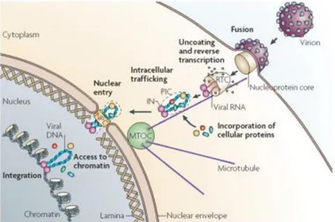
Figure 3. From cell entry to DNA integration. The virus enters the target
cell by fusion between the cellular and viral membranes. The nucleoprotein core containing the genomic RNA is delivered into the cytoplasm where reverse transcription takes place. The viral cDNA with viral and cellular proteins form the pre-integration complex (PIC), that reaches the nuclear envelope by active transport along microtubules and then cross the intact nuclear envelope, presumably through the nuclear pore complex (NPC). The PIC gains access to chromatin and the viral protein IN catalyzes the integration reaction of the viral DNA into the host genome (Suzuki et al., 2007).
HIV-1 encodes at least nine genes which must be expressed during viral life cycle in the correct temporal order. Early in the viral infection, small, multiply spliced transcripts (2 kb) encoding Tat, Rev and Nef predominate in the cytoplasm. During the late phase of HIV infection, genomic (unspliced, 9kb) RNAs and singly spliced RNAs (4 kb) become leading species in the infected cell. A key factor controlling late phase transition is the viral protein Rev. Rev is a small, positively charged RNA-binding protein that is approximately 116 amino acids in size. It is encoded from two exons which are joined by splicing to produce a monocystronic transcript early in the viral replication cycle. Rev contains both nuclear localization sequence (NLS) and nuclear export signal (NES). Hence, Rev is a shuttling protein. Rev permits export of unspliced/partially spliced transcripts from the nucleus to the cytoplasm, where they serve as templates for translation of the gag-pol open reading frame or as the full-length genomic RNA (Zapp and Green, 1989). Rev specifically recognizes an RNA element located within the coding sequence for Env. This sequence, called RRE (Rev-responsive element), is present within the env-coding region and is about 200 nucleotides in size and forms secondary RNA structure. Rev exports the intron-containing HIV-1 mRNA via a CRM1 shuttling system (Askjaer et al., 1998). CRM1 is a member of the karyopherin family of nucleocytoplasmic-transport factors and, like others karyopherins involved in nuclear export, binds its cargo in the nucleus in the presence of the GTP-bound form of the Ran GTPase. After nuclear export, hydrolysis of the bound GTP to GDP causes a conformational shift that induces cargo release in the cytoplasm, thus providing the directionality of this export pathway. CRM1 also interacts with components of the nuclear pore complex (NPC) and this interaction is essential for nuclear RNA export. In the nucleus, Ran-GTP bound CRM1 binds the NES domain of Rev, which in turn is bound to RRE-containing HIV-1 transcripts. This interaction enables CRM1 to export the resulting RNA/protein complex into the cytoplasm. In the
cytoplasm, conversion from Ran-GTP to Ran-GDP releases the Rev/RNA cargo. Rev returns to the nucleus by binding to importin-$ and Ran-GDP for subsequent rounds of export (Suhasini and Reddy 2009).
Although it is now clear that the primarily effect of Rev is in promoting the nuclear export of RRE- containing HIV RNAs the mechanism of Rev function is not fully understood. This protein functions through several cellular posttranscriptional mechanisms, such as mRNA splicing, RNA stability, nucleocytoplasmic transport, and translation. Nevertheless, numerous studies have provided insights on several posttranscriptional steps in viral gene expression regulated by Rev. Studies of Cochrane and co-workers indicate that Rev acts early in HIV biogenesis (Iacampo and Cochrane 1996). In addition to influencing the fate of viral mRNA within the nucleus, Rev has been proposed to affect utilization of viral mRNA for translation. Rev efficiently loads RRE-containing mRNAs onto polysomes leading to high levels of structural proteins translation (D'Agostino et al., 1992). There are many lines of evidence which suggest the involvement of Rev in stabilization of unspliced HIV transcripts either by disassembly of spliceosomes or by overcoming the destabilization effect of cis-acting elements present in HIV RNAs.
1.1.4 Cellular proteins involved in the late phases of HIV-1 infection
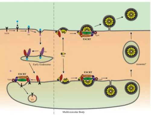
The late phases of HIV-1 life cycle are characterized by the assembly of new viral particles, their release from the plasma membrane and their maturation. These steps are also highly dependent on cellular proteins. ASSEMBLY, BUDDING AND MATURATION
Several groups have shown that HIV-1 recruits the cellular endocytosis machinery for assembly and budding (Martin-Serrano et al., 2003; von Schwedler et al., 2003; Pornillos et al., 2002). The HIV-1 envelope contains a high percentage of cholesterol suggesting that HIV may bud from specific membrane microdomains named lipid rafts (Campbell et al., 2001). The viral particles assemble at the plasma membrane, then are wrapped with the host membrane and bud out from the cell surface. The gag polyprotein, which is composed by the matrix domain (MA), the capsid domain (CA) and the nucleocapsid domain (NC), plays a pivotal role in driving all these processes. The MA domain targets gag to the site of assembly, where CA plays a role in gag multimerization and NC domain packages viral genomic RNA during assembly. In addition to these domains, the p6 domain positioned at the carboxyl terminus of gag is necessary for particle release from the host cell.
The mature virion is generated during viral release upon cleavage of the gag precursor by the viral protease (PR). Different studies have suggested that HIV-1 viral particles recruit the high molecular weight endosomal sorting complexes (ESCRTI, ESCRTII, ESCRTIII) (Rev Demirov and Freed, 2004). The ESCRT machineries are usually involved in the sorting of cargo proteins, such as activated receptors, to Multi Vesicular Bodies (MVB) before their degradation in the lysosomes (Fujii et al., 2007).
Independent studies have shown that p6 interacts with Tsg101, a component of ESCRTI machinery, and this interaction is necessary for viral particle release. Depletion of endogenous Tsg101 or disruption of Tsg101-p6 interaction inhibits virus release (Garrus et al., 2001; VerPlank et al., 2001). Another minor player in HIV-1 budding is the apoptosis-linked-gene 2 interacting protein (Alix) protein. Strack and colleagues demonstrated that Alix was involved in EIAV (Equine Infectious Anemia Virus) release (Strack et al., 2003). However many observation suggest that Alix can also be used by HIV-1 as an alternative route when Tsg101 is not available (Fisher et al., 2007; Usami et al., 2007). All these findings suggest that, under particular circumstances, retrovirus assembly and budding can take place in the MVB. Additional electromicroscopy experiments describe virus positive intracellular compartments that also display MVB specific markers. After budding of the virus into the MVB lumen, particle release occurs via the endosomal pathway (Figure 4) (Sherer et al., 2003; Ono and Freed, 2004).
In certains types of human cells the absence of Vpu leads to inefficient HIV particle release due to the failure to detach from the plasma membrane and accumulation in large numbers at the cell surface (Klimkait et al., 1990). Interestingly, in simian cells, Vpu is dispensable for efficient HIV release (Varthakavi et al 2003). Neil and colleagues found a new protein (Tetherin) that, in the absence of Vpu, inhibits viral particle release; this factor is induced by IFN-alpha. The mechanism by which Tetherin inhibits viral release is still unknown, but it has been shown that this factor also antagonizes the release of MLV and that its overexpression, induced by IFN alpha, blocks infection, thus suggesting that Tetherin could be a generic defence against enveloped viruses (Neil et al., 2008).
A second release restriction factor is represented by calcium-modulating cyclophilin ligand (CAML-1), an integral membrane protein involved in T cell
development and regulation (Tran et al., 2004). In 2008 it was shown that CAML is a Vpu-sensitive human host restriction factor to HIV-1 release. Indeed, expression of human CAML rendered simian cells restrictive for viral release in the absence of Vpu, suggesting that CAML is a human host restriction factor blocking viral paricle release or budding, the action of which is counteracted by Vpu (Varthakavi et al 2008).
Figure 4. Model for retrovirus release. On the left is a schematic
representation of endocytosis and MVB sorting of an activated growth factor receptor. An Hrs- containing complex (dark purple) recognizes and sequesters ubiquitylated cargo (Ub, light purple) at clathrin-rich regions of the early endosomal membrane. On the right is depicted the hijacking of MVB sorting machinery for virus release. Virus particles are shown to assemble and bud at the plasma or to be released (e.g., from macrophages) through the exosome pathway following assembly in the MVB (Demirov and Freed, 2004).
1.1.5 Cellular MicroRNAs: novel partner for HIV-1
In addition to cellular proteins that regulate HIV-1 infection, there is mounting interest in the regulatory roles served by non-coding RNAs (NcRNA) and by the RNAi cellular machinery. The best known subtype of NcRNAs are the small, 21–23 nucleotides long, single-stranded microRNA (miRNA) molecules, which are key modulators of eukaryotic gene expression and are involved in many cellular processes such as development oncogenesis, cell cycle control and immunity (Calin and Croce 2006; Scaria and Jadhav 2007). Pri-miRNAs are encoded in introns, intergenic regions, and specific transcription units in both sense and antisense orientations and transcribed by RNA polymerase II. The pri-miRNA is processed in the nucleus by the Drosha-DGCR8 complex (RNase III endonuclease complex), into a shorter 70 nucleotide RNA (pre-miRNA) containing a stem-loop structure (Han et al., 2006). The pre-miRNA is then transported into the cytoplasm where it is further processed by a second RNase III endonuclease complex, Dicer-TRBP, to generate a 21–23 nucleotide imperfectly duplexed mature miRNA (Chendrimada et al., 2005; Haase et al., 2005; rev Bushati and Cohen 2007). A mature miRNA is assembled into an RNA-induced silencing complex (RISC). The miRNA-RISC (miRISC) complex recognizes the target mRNA via imperfect base pairing at the 3' -UTR region (Grimson et al., 2007). Since the base pairing is ‘‘imperfect’’ a single miRNA could potentially recognize and downregulate up to 100 mRNAs (Brennecke et al., 2005).
It has been shown that plants and lower eukaryotes use de novo synthesized virus-derived small interfering RNAs to regulate infecting viruses (Ding and Voinnet 2007). Although it remains unclear if mammals conserve an RNAi-based antiviral strategy, recent evidence indicate that this might be the case (Berkhout and Jeang 2007; Yeung et al., 2005). The
first finding of a cellular miRNA involvement in antiviral immunity describes a cellular miRNA (miR-32) as a inhibitor of primate foamy virus type 1 (PFV-1) infection (Lecellier et al., 2005).
Recently, it has been shown that cellular miRNAs are involved in the regulation of HIV-1 latency. In particular, Huang and colleagues found that the 3’ end of HIV-1 mRNA is targeted by 28, 125b, 150, miR-223, miR-382, which are more abundant in resting primary CD4" T cells, and that inhibition of these miRNA induces HIV-1 expression in latently infected cells (Huang et al., 2007). Moreover, it has been recently reported that another miRNA (miR-198) functions to restrict HIV-1 replication in monocytes, by repressing Cyclin T1 expression (Sung et al., 2009).
This restrictive role of microRNAs on HIV infection in part explains the reason why HIV-1 has evolved ways to affect the cellular RNAi machinery. First of all, it seems that HIV-1 can reshape the infected cell miRNA expression profile (Yeung et al., 2005; Houzet et al., 2008). Moreover it was demonstrated that HIV-1 actively suppressed the expression of the polycistronic miRNA cluster miR-17/92 and that this suppression is required for efficient viral replication (Triboulet et al., 2007).
Since HIV-1 produces a large number of small viral RNA hairpins, it is probable that it evolved a strategy to avoid viral RNA processing by the RNAi machinery (Bennasser et al., 2006; Bennasser et al., 2006; Klase et al., 2007; Ouellet et al., 2008). However, miRNAs and RNAi are conserved factors/processes whose complete suppression is incompatible with cellular viability (Muljo et al., 2005). Most likely HIV-1 avoids RNAi antiviral mechanisms by mutating viral RNA-sequences to alter base-complementarity with cellular miRNAs. Indeed, there is evidence that selective and evasive nucleotide changes in HIV-1 sequences can be
elicited rapidly by siRNA/shRNA induced RNAi (Dash et al., 2004; Westerhout et al., 2005).
Finally, a large number of viral miRNAs (vmiRNAs) have been described to be encoded by viral genomes, in particular herpes and polyoma viruses (SV40) (Pfeffer et al., 2005; Sullivan et al., 2005). In the specific case of HIV-1, a vmiRNA encoded by the nef region of HIV-1 termed miRN367 was physically identified and isolated by Omoto and colleagues (Omoto et al., 2005). The role of this vmiRNA in HIV-1 infection remains unclear.
!
1.2 Lymphocytes activation status and HIV-1 infection
HIV-1 replication is greatly influenced by the activation status of the target cell. Activated CD4" T lymphocytes are permissive to HIV-1 infection, whereas in resting T cells, despite the efficient entry of HIV-1, no viral progeny is produced. Since the early 1990s, different hypothesis have been made to explain this block. Initially, Zack and colleagues demonstrated that quiescent T cells can be infected by HIV-1 and that viral cDNA synthesis initiates at levels comparable with those of activated T cells. However, the viral genome remains incompletely reverse transcribed and may persist in an inactive state until subsequent mitogenic stimulation (Zack et al., 1990). In the same year, Stevenson and colleagues suggested that the block in resting T cells occurs at the level of integration. They found that, in resting T cells, viral cDNA was unable to integrate into the host cell genome and was maintained extrachromosomally for several weeks. Subsequent T cell activation allowed integration of extrachromosomal DNA that seemed to be transcriptionally active (Stevenson et al., 1990). However studies from different groups performed later on, suggested that, in resting T cells, a block to HIV-1 infection occurs at the level of reverse transcription and that a progression through the G1b phase was needed to achieve efficient
reverse transcription (Korin and Zack, 1998). Latest findings from Greene and colleagues better elucidated this mechanism of resting T cell resistance to HIV-1 infection: they demonstrated that APOBEC3G, in a form of a low molecular mass ribonucleoprotein complex present exclusively in resting CD4" T lymphocytes, blocks formation of late products of reverse transcription via the RNA binding ability of APOBEC3G. In activated T cells, the APOBEC-containing low molecular mass complex forms a larger complex that looses the capacity to restrict the viral infection thus allowing fully efficient reverse transcription (Chiu et al., 2005). Understanding the fate of HIV-1 in resting T cells is particularly important since the majority of T lymphocytes are in resting state (Tang et al., 1995). Siliciano and colleagues monitored the kinetics of HIV-1 decay in resting CD4" T cells and found that slow kinetics of reverse transcription and blocks at subsequent steps limit HIV-1 infection in these cells. They also showed that the reservoir of unintegrated HIV-1 in recently infected resting CD4" T cells is highly labile. By examining the decay of integration-competent HIV-1 DNA, they found that this form of fully retrotranscribed HIV-1 has a half-life of 1 day in resting T cells. They proposed that degradation of either viral DNA and viral proteins that constitute the preintegration complex would lead to a functional decay of the virus (Zhou et al., 2005). Moreover, several studies suggest that inhibition of the proteasome increases the production of proviral DNA by blocking the degradation of the preintegration complexes (Butler et al., 2002; Schwartz et al., 1998). In 2007, a work performed using immunofluorescence experiments on CA viral protein and FISH analysis on viral DNA in resting infected cells demonstrated that full length reverse transcribed HIV-1 cDNA together with CA localizes at the centrosomes 4 days post infection (Zamborlini et al., 2007). Collectively, these data suggest that, in resting CD4" T cells, different blocks that impair viral replication may exist at multiple steps of
the early infection. It seems that reverse transcription is the most affected step as it resulted to be both impaired and delayed. However, when the full length transcripts accumulate in the infected cells, they are anyhow unable to reach the nucleus and stably integrate into the host genome.
In addition to this pre-integration latency mechanisms, another typical characteristic of HIV-1 infection, that correlates with T cell activation status, is the establishment of a latent reservoir of infected cells (post-integration latency). The major obstacle to HIV-1 eradication is the establishment of a latent infection. The formation of this latent reservoir is a natural consequence of the fact that the virus replicates in activated CD4" T cells (Persaud et al., 2003), while the vast majority of CD4"! T lymphocytes is in a resting G0 state. In adults, about half of the resting cells are naïve, having yet to encounter an appropriate antigen (Ag) and the other half is represented by memory cells, that have previously responded to an Ag. Ag-driven responses involve a burst of cellular proliferation and differentiation, giving rise to effector cells, most of which die quickly; a surviving subset reverts to the resting G0 state as memory. These cells are characterized by long-term survival and rapid responses to the Ag in the future (review ref. Kaech et al., 2002). The virus replicates preferentially in activated CD4" T cells with cytopathic effects (Ho et al., 1995). Because it takes a few weeks for effector cells to revert to a resting state, most infected CD4" lymphoblasts die before becoming memory cells. Nevertheless, the presence of cells harbouring integrated provirus in patients under Highly Active Antiretroviral Therapy (HAART) suggests that some activated cells, after infection, can revert to a resting state. Otherwise it could be possible that HIV-1 might infect some of these cells in a state in which they are still permissive for early steps in the virus life cycle (up to integration), but not for virus gene expression (Stevenson 1997).
HIV-1 gene expression has been clearly shown to be dependent on inducible host transcription factors that are transiently activated following exposure to Ag, and thus viral gene expression is automatically extinguished as cells return to a resting state (Tong-Starksen et al., 1987; Nabel and Baltimore 1987). The result is a stably integrated but transcriptionally silent form of HIV-1. The direct infection of resting cells does not generally proceed to integration (Zhou et al., 2005). However, resting CD4" T cells with integrated HIV-1 DNA can be detected in vivo and their phenotype suggests that they arise from infected CD4" T lymphoblasts that have reverted to a resting memory state (Chun et al., 1995). Jordan and colleagues have suggested, as mechanism of latency, an integration into centromeric heterochromatin, that is known to be repressive (Jordan et al., 2003). However subsequent studies on CD4" isolatated from patients on HAART therapy hae demonstrated that, in the cell population carring latent HIV-1, integration into centromeric regions does not occur (Han et al., 2004).
An additional potential explanation for latency is transcriptional interference (TI). TI is a cis-acting suppressive effect that is observed when transcriptional activity initiated from an upstream promoter suppresses the transcription from a downstream promoter (Greger at al., 1998). In activated CD4" T cells, HIV-1 gene expression might be efficient because the concentration of crucial host transcription factors is high enough to overcome TI, while in resting T cells these factors are less abundant and transcription from an unpstream promoter might interfere with the downstream promoter (Hogan et al., 2003; Weil et al., 2004). Furthermore, it has been shown that transcription factors and coactivators necessary for HIV-1 transcription are less abundant in resting T cells. For example, two of the key host transcription factors involved in HIV-1 expression (NFAT and
only recruited to the nucleus following cellular activation. As a consequence, HIV-1 gene expression is stricktly dependent on to the activation state of the host cells. Tat-associated proteins could be one of the limiting factors for processive transcription in resting T cells: low levels of P-TEFb kinase activity (CDK9 and Cyclin T1), that have been observed in resting T cells, are increased in response to activating stimuli (Ghose et al., 2001). Tat itself could be the main limiting factor being subject to tight post-translational modifications (Bres et al., 2002; Bres et al., 2003). Finally, RNAi-mediated pathways of transcriptional silencing have also been shown to be involved in the latency mechanism. As described above, cellular microRNAs (miRNAs) have been shown to potently inhibit HIV-1 production in resting primary CD4"!T cells (Huang et al., 2007).
The major obstacle to HIV-1 eradication is the establishment of a latent infection. Viral reservoirs established early during the infection remain unaffected by anti-retroviral therapy for a long time and are able to restore infection upon interruption of HAART, thus therapeutic targeting of viral latency is one of the most important goal in HIV-1 research (Lassen et al., 2004; Persaud et al., 2003; Marcello 2006).
2. The IN protein
2.1 IN structure and domains
Integration into the host genome is a defining feature of all retroviruses. Once integrated, the viral DNA is replicated together with cellular DNA during the cell cycle. The viral enzyme that carries out this reaction is the viral protein IN.
IN is a 32 kDa protein that is present inside the mature virion, and is encoded by the pol gene, which also encodes for viral protease (PR) and reverse transcriptase (RT). IN is translated as a part of the large polyprotein Gag-Pol and is processed by viral protease during virion maturation. The enzyme is supposed to work as a tetramer.
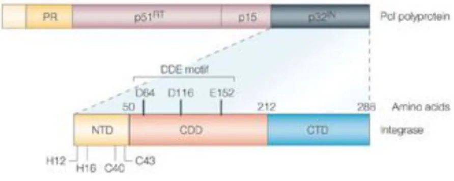
IN is composed of three domains: the N-terminal domain (residues 1-50), the Core domain (residues 51-212) and the C-terminal domain (residues 213-288) (Figure 5). The division of IN into these three domains is based on functional and protelolytic studies (Engelman and Craigie, 1992; Engelman et al., 1993; van Gent et al., 1993).
The N-terminal domain is a bundle of three alpha-helices and it characterized by a two His and two Cys motifs named HHCC domain, and a SH3 fold domain (Craigie, 2001). This region is important for the activity of IN both in vitro and in vivo (Khan et al., 1991; Schauer and Billich, 1992). Mutations in these conserved residues block the integration mechanism (Cannon et al., 1994). This domain binds to a zinc ion and the conserved HHCC motif seems to be important for this interaction. N-terminal domain is important for the multimerization of IN; in the absence of zinc this domain has a disordered structure while in the presence of this ion it adopts an ordered secondary structure and the tetramerization occurs more rapidly (Zheng et al., 1996).
The Core domain is responsible for the catalytic activity of IN and is the most conserved among all retroviral INs (Kulkosky et al., 1992). It consists of a central five-stranded $-sheet with six surrounding helices. The catalytic region contains the invariant triad of acidic residues, the D,D-35E motif. Mutagenesis of these aminoacids greatly impairs IN enzymatic activity and viral replication as a consequence (LaFemina et al., 1992; Shin et al., 1994; Taddeo et al., 1994). The structure of the Core domain is very similar to the Rnase H domain of RT enzyme, the RuvC protein of E.Coli and the bacteriophage Mu transposase, all enzymes that catalyses substitution reactions involving phosphodiester bonds (Hostomska et al., 1991; Ariyoshi et al., 1994; Rice and Mizuuchi, 1995). In addition to its catalytic activity, the core domain has also been proposed to be involved in DNA binding (Drelich et al., 1993).
The C-terminal domain is the most variable region among retroviral INs (Lutzke et al., 1994). This domain binds DNA in a non specific manner, and since integration into host DNA has been demonstrated to be non specific, the C-terminal domain was suggested to interact with the target DNA. However studies performed with chimeric INs demonstrated that the Core domain is responsible for targeted DNA binding (Katzman and Sudol, 1995). It appears more likely that the C-terminal domain binds to the very ends of the viral DNA (Esposito and Craigie, 1998; Jenkins et al., 1997).
Figure 5. Schematic rapresentation of the Pol gene and the IN domains.
IN (p32IN) is encoded at the 3'-end of the pol gene. HIV-1 IN is composed by three domains. The amino-terminal domain (NTD) that coordinate one zinc atom through H12, H16, C40 and C43. The catalytic core domain (CCD) containing the DDE motif: D64, D116 and E152. The carboxy-terminal domain (CTD) is involved in DNA binding (Pommier et al., 2005).
2.2 IN enzymatic activity
Integration is an essential step for all retroviruses and mutations that interfere with this process block their replication.
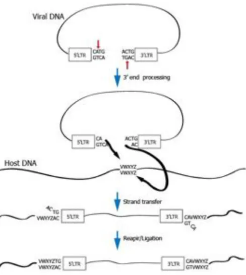
The integration mechanism can be divided into two distinct steps: 3’ processing and strand transfer.
The 3’ processing step occurs in the cytoplasm within the pre-integration complex. During this step, IN removes a GT dinucleotide from the 3’ end of each viral LTR. This dinucleotide is always adjacent to a highly conserved CA dinucleotide, and is crucial for recognition of viral DNA by IN. Mutations in this site cause severe defects in integration.
Following the nuclear entry of the PIC, IN catalyses the strand transfer reaction that consists in a nucleophilic attack by the 3’-hydroxyl residues of the viral ends on phosphodiester bridges located on either side of the major groove in the host DNA. Then IN catalyses a transesterification reaction, the processed CA-3’-OH viral DNA ends are ligated to the 5’-O-phosphate ends of the host DNA. At this point viral DNA is ligated to the cellular DNA by only one strand at each end. Through an unknown process, the gaps flanking the viral DNA are filled in by extending the free 3’ end of the target DNA, the mismatched viral 5’ end is trimmed and the resulting ends are ligated.
The identity of the protein that performs the gap repair remains unknown;, it is possible that viral proteins direct cellular enzymes to the viral DNA or even act directly to repair the gap (Coffin J.M., Hughes S.H., Varmus H.E. Retroviruses).