INTERNATIONAL PhD SCHOOL IN
Biomolecular and Biotechnological Sciences
Cycle XXVII
Subject: Clinical and Molecular Microbiology
Director: Professor Leonardo Sechi
POLYMORPHISMS OF cagA AND vacA GENES
IN HELICOBACTER PYLORI ISOLATED FROM
GASTRODUODENAL DISEASES PATIENTS IN
CENTRAL VIETNAM
PhD student: Nam Phan Trung
Tutor: Dr. Bianca Paglietti , Prof. Salvatore Rubino Co-Tutor: Prof. Tran Van Huy
Originality statement
I, Phan Trung Nam declare that the thesis for the award of a Doctor of Philosophy degree in Clinical and Molecular Microbiology of Ph.D School in Biomolecular & Biotechnological Science at University of Sassari, hereby submitted by me, has not been previously submitted for a degree at this or any other university and that it is my original work in design and execution, and that all the reference materials contained therein have been duly acknowledged.
Date:
Acknowledgments
I would like to express my deep gratitude to each of you who have supported me throughout these years and contributed to the accomplishment of this work. Particularly, I would like to thank:
Professor Salvatore Rubino and Bianca Paglietti, my main supervisors and Professor Tran Van Huy, my co-supervisor, for providing me with a great opportunity to work in Helicobacter pylori research and gastroenterology, for accepting me as a PhD student, for sharing me your broad world of science, for your trust, support and encouragement, for your great effort in revising my manuscripts and thesis. Especially, I would like to acknowledge Professor Salvatore Rubino and Bianca Paglietti for your concern with my situation, always finding time in your busy schedule for me when needed and taking care of me. You are the best supervisors I could ever have wished for.
Professor Piero Cappuccinelli for providing me with a great opportunity to have the PhD scholarship and work in Helicobacter pylori research, for introducing me to Clinical and Molecular Microbiology Department in University of Sassari, for your generous support and arrangement from my beginning.
Doctor Antonella Santona for sharing her knowledge and experience in laboratory work, for analyzing my samples, for revising my manuscripts and thesis, for willingness to listen and fruitful discussion.
Professor Bruno Masala, Professor Claudia Crosio and Professor Leonardo Sechi who facilitated my attendance to the PhD program at Sassari University. Ms Giustina Casu and Mr Giovanni Sini for coordination and administration during my study.
All members at Clinical & Molecular Microbiology Department and Laboratory in University of Sassari for supporting and creating extremely nice working atmosphere during my study.
Professor Cao Ngoc Thanh, Dean of Hue Medical University, for accepting me as a PhD student in Biomedicine, for your kindness, support.
Doctor Le Van An, Tran Thi Nhu Hoa and members at the Department of Microbiology and Carlo Urbani Center, Hue Medical University for supporting and sharing scientific knowledge, experience in laboratory work making this thesis possible.
All members at Center of Gastroenterology Endoscopy, Hue University Hospital for your interest in the study, practical helps, handling samples and friendship.
Pham Thi Hop Khanh, my wife, for your endless love, for your patience and support, for taking care of our precious children: Phan Khanh Phuong, Phan Nam Phuong, and standing by my side throughout these years.
My parents for giving me all your love, for always encouraging me to move forward, for your trust and for invaluable and endless support. My brother and sister in law for their support, encouragement during my study.
All my dear Vietnamese and Italian friends for their support, encouragement and for sharing the happiness and sadness during these years.
In particular, I would like to thank all patients who made this thesis possible by participating in the study.
This thesis is the result of a cooperation in doctoral education between Hue Medical University - Vietnam and Sassari University - Italy.
Abstract
Helicobacter pylori is an important pathogen in humans, with infection rates around 50% of the world‟s population globally but it varies geographically. H. pylori, in particular its virulence factors, plays a significant role in the pathogenesis of upper alimentary tract diseases, including peptic ulcer disease, gastric mucosa associated with lymphoid tissue lymphoma and gastric cancer. A successful eradication therapy is important not only to reduce the risk of developing gastric cancer but also to treat other H. pylori severe related disorders. Vietnam is a developing country with high prevalence of H. pylori infection and an intermediate risk of gastric cancer but there has been a little information about antibiotic resistance and virulotyping of H. pylori, especially in the Central region.
The aim of this thesis was to determine the antimicrobial susceptibility patterns and the molecular basis of the resistance pattern; to investigate cagA and vacA genotypes, the two most important virulence genes, and to determine a clinical relevance of cagA and vacA polymorphisms of H. pylori strains isolated from dyspeptic patients in Central Vietnam.
We examined 127 dyspeptic patients from 7 provinces in Central Vietnam who were referred to Hue University Hospital between July 2012 and January 2014 for upper gastrointestinal endoscopy, to clarify clinical diagnosis and check H. pylori infection with respect to age, sex, endoscopic diagnosis and medical history. Two gastric biopsies from antrum were collected, one for rapid urease test during the procedure, and another for culture. A total of 97 H. pylori strains were isolated on Columbia agar base plus sheep blood and selective supplement under microaerophilic condition, all of them were
identified by microscopic, biochemistry characteristics and further confirmed by PCR targeting 16S rRNA and/or ureA. Unfortunately, we failed to re-cultivate 5 strains from minus 80oC store, one of them was lost. Finally, on 92 strains antibiogram was performed and 96 extracted DNA samples from 96 strains were available for further analysis.
Antimicrobial susceptibility tests to 4 common antibiotics for H. pylori eradication treatment were conducted by E-test and disc diffusion methods. Point mutations related to clarithromycin and levofloxacin on 23S rRNA and gyrA genes were detected by sequencing their amplified PCR products. vacA and cagA genotyping of 96 strains were carried out using PCR and sequencing to determine their prevalence and polymorphism, then their clinical relevance was further analyzed.
The pattern of antimicrobial susceptibility testing showed that 42.4% were resistant to clarithromycin (primary–34.2%; secondary–73.7%), 41.3% to levofloxacin (primary–35.6%; secondary–63.2%), 76.1% to metronidazole and 1.1% to amoxicillin. Multidrug resistance was observed in 56.5% (primary–50.7%; secondary–79%) of isolates (p < 0.05). The rate of resistance to levofloxacin in females was significantly higher than in males (p < 0.05) and the resistance to clarithromycin and levofloxacin was increased according to the rise of age.
To define the breakpoints for disk diffusion as a feasible and cheap method for determining qualitative susceptibility based on E-test results as a gold standard test, we found that susceptible strains to clarithromycin had inhibition diameters ≥ 24mm and resistant to clarithromycin with inhibition diameters ≤ 18mm. To levofloxacin, strains with inhibition diameters ≥ 30mm
defined as susceptible, and those with inhibition diameters ≤ 26mm defined as resistant.
Most of the clarithromycin and levofloxacin resistant strains harboured resistance associated mutation with common position at A2143G, T2182C in 23S rRNA gene and at Asn-87 or Asp-91 in gyrA gene. MICs increased in strains carrying quadruple mutations in their 23S and in strains with Asn-87 gyrA mutation (p < 0.05). One high level levofloxacin (MIC = 32 mg/L) resistant strain had new mutations with combination of N87A, A88N and V65I.
Determination of virulence factors of 96 strains, cagA genotyping showed that cagA gene was detected in 85.4% of H. pylori strains. In cagA negative strains, 57.1% suggested the presence of a deviating cag-PAI. The cagA genes were further characterized EPIYA motif to classify cagA type showing 9.8% Western cagA strains (one EPIYA motif - ABCC and 7 had an EPIYA motif – ABC), whereas all 90.2% East Asian - cagA strains had an EPIYA motif – ABD. To confirm cagA genotyping by PCR method, sequencing analysis was performed exhibiting the consistent results with previously analysed PCRs. Clinical relevance of cagA genotype showing East-Asian cagA type was present in all strains isolated from gastric ulcer and gastric cancer patients, which tended to be higher than those from gastritis and duodenal ulcer. On the contrary, cagA negative and Western cagA type were only in strains from gastritis, duodenal ulcer patients.
Analysis of vacA polymorphism revealed that 97.9% of strains had vacAs1 genotype; 91.5% had vacAi1; vacAm1 and vacAm2 presented 56% and 44%, respectively. The combination of three regions of vacA showed that the
s1i1m1 type had a predominant percentage with 55.7%, followed by s1i1m2, s1i2m2 and s2i2m2 with 35.2%, 6.8% and 2.3%, respectively. All vacA s1m1 linked with i1, all s2m2 with i2 and s1m2 linked with i1 or i2. Clinical relevance of vacA polymorphism exhibiting vacAs1 and vacAi1 was present in almost all strains in different gastroduodenal diseases. On the other hand, vacAm1 was more predominant than vacAm2 in strains isolated from gastric ulcer patients (75% vs 25%) and it showed a higher frequency of vacAm1 than that in duodenal ulcer (44%) and gastritis (58.9%). Furthermore, in gastric ulcer strains, vacAs1i1m1 was more predominant than s1i1m2 and s1i2m2 polymorphic types (75% vs 12.5% and 12.5%) with a significant difference (p < 0.05). Moreover, vacAs1i1m1 was present in strains isolated from gastric ulcer patients with a higher frequency than in that from duodenal ulcer (41.7%) and gastritis (59.3%). All s2i2m2 types were present in gastritis and duodenal ulcer strains.
The relation of cagA and vacA genes showed that cagA-positive status was very closely associated with vacA s1i1m1 genotype with highest frequency (98%), followed by s1i1m2 (77%) and s1i2m2 (33%), none with s2i2m2. On the contrary, cagA-negative status was only present in vacA s2i2m2 and was predominant in vacA s1i2m2 (67%).
In conclusion, we highlight a very high resistance rate in Central Vietnam with 56.5% of multiple resistance and higher prevalence of secondary resistance versus primary resistance, in particular to clarithromycin and levofloxacin. Data on determinants of resistance to clarithomycin and levofloxacin (mutations in 23s RNA and gyrA genes) are new information in Vietnam. cagA positive status, East Asian cagA type and vacA s1i1m1 type
are the most predominant in gastric ulcer and gastric cancer. The prevalence of cagA negative status and Western cagA type is higher than that reported in previous studies in different areas of Vietnam.
Publications and Conference proceedings arising from this thesis Publications:
- Phan Trung Nam, Tran Van Huy, Tran Thi Nhu Hoa, Le Van An, Antonella Santona, Bianca Paglietti, Piero Cappuccinelli, Salvatore Rubino (2013). ”Diffusion methods for clarithromycin and levofloxacin susceptibility testing of Helicobacter pylori”, Journal of medicine and pharmacy, (18): 63-69. ISSN 1859 – 3836.
- Phan Trung Nam, Tran Van Huy, Tran Thi Nhu Hoa, Le Van An, Antonella Santona, Bianca Paglietti, Piero Cappuccinelli, Salvatore Rubino (2013). ”Antibiotic resistance of Helicobacter pylori with E-test in Central region of Vietnam in two years 2012-2013”, Vietnamese Journal of Gastroenterology, VIII(33):2122-2132. ISSN 1859 – 0640.
- Phan Trung Nam, Antonella Santona, Tran Van Huy, Tran Thi Nhu Hoa, Le Van An, Pietro Cappuccinelli, Salvatore Rubino, Bianca Paglietti (2014). “High rate of levofloxacin resistance in a background of clarithromycin and metronidazole resistant Helicobacter pylori in Vietnam”. (has been accepted for publication in International Journal of Antimicrobiol Agents).
Oral presentation:
Phan Trung Nam et al (2013), “Antibiotic resistance of Helicobacter pylori with E-test in Central region of Vietnam in two years 2012-2013” in 19th National Scientific Congress on Gastroenterology, Hanoi – Vietnam.
TABLE OF CONTENTS
Originality statement i
Ackownledgments ii
Abstract iv
Publications and Conference proceedings arising from this thesis ix
List of figures xvi
List of tables xviii
List of abbreviations xix
Contents:
1. Introduction 1
1.1. Helicobacter pylori 1
1.1.1. Introduction & history 1
1.1.2. Microbiology 2
1.2. Epidemiology 4
1.2.1. Prevalence of H. pylori infection 4
1.2.2. Molecular Epidemiology of H. pylori 6
1.2.3. Transmission of H. pylori infection 9
1.2.3.1. Oral-oral transmission 9
1.2.3.3. Faecal-oral transmission 10 1.3. H. pylori virulence and colonisation factors 11
1.3.1. Cytotoxin associated gene A (cagA gene) 11
1.3.1.1. Pathogenicity mechanism of CagA 12
1.3.1.2. CagA type: Western versus East Asian 14
1.3.1.3. Clinical importance of cagA 15
1.3.2. Vacuolating cytotoxin VacA 16
1.3.2.1. Effects of VacA on cellular 17
1.3.2.2. vacA polymorphism 17
1.3.2.3. Disease associations of VacA 19
1.3.3. Duodenal ulcer promoting gene (dupA) 20
1.3.4. Adhesins and outer membrane proteins (OMPs) 21
1.3.4.1. Blood group antigen binding adhesin (BabA) 21
1.3.4.2. Sialic acid-binding adhesin (SabA) 22
1.3.4.3. Outer inflammatory protein (OipA) 22
1.3.5. Induced by contact with epithelium A (IceA) 23
1.3.6. Acid resistance and motility 23
1.4. Diseases associated with H. pylori infection 24
1.4.1. Gastritis 24
1.4.2. Peptic ulcer disease: duodenal ulcer and gastric ulcer 25
1.4.4. Gastric MALT lymphoma (Mucosa associated lymphoid tissue) 27
1.4.5. Extra-gastrointestinal manifestations 28
1.5. Diagnosis of H. pylori infection 29
1.5.1. Invasive diagnostic methods 29
1.5.1.1. Histology 29
1.5.1.2. Rapid urease test (RUT) 30
1.5.1.3. Culture 31
1.5.2. Non-invasive diagnostic methods 31
1.5.2.1. Urea breath test (UBT) 31
1.5.2.2. Stool antigen assay 32
1.5.2.3. Serology 33
1.6. Treatment of H. pylori infection 34
1.6.1. First-line treatment 35
1.6.1.1. First-line treatment in areas with low clarithromycin resistance 35 1.6.1.2. First-line treatment in areas with high clarithromycin resistance 35
1.6.2. Second-line treatment 36
1.6.2.1. Second-line treatment in areas with low clarithromycin resistance 36 1.6.2.2. Second-line treatment in areas with high clarithromycin resistance 37
1.6.3. Third-line treatment 37
1.7. Antimicrobial susceptibility testing 37
1.7.1.1. Phenotypic methods 38
1.7.1.2. Genotypic detection of resistance 40
1.7.2. Relevance of H. pylori resistance to antibiotics 43 1.7.3. Impact antibiotic resistance on H. pylori eradication 44
2. Research objectives – Aims 45
3. Materials and methods 46
3.1. Study design 46
3.2. Study population 48
3.3. Methods 50
3.3.1. Biopsy specimens for culture of H. pylori 50
3.3.2. Preparation of blood agar plates for growth of H. pylori 50
3.3.3. Culture of H. pylori 51
3.3.4. Strain storage 53
3.3.5. Antimicrobial susceptibility testing 54
3.3.5.1. Disk diffusion 54
3.3.5.2. Epsilometer test 55
3.3.6. DNA extraction 56
3.3.7. Detection of point mutations of 23S rRNA and gyrA genes 56
3.3.7.1. PCR Amplification 56
3.3.7.2. DNA purification 57
3.3.7.4. Nucleotide sequencing 58
3.3.7.5. Sequence analysis 58
3.3.8. PCR for genotyping of cagA gene 58
3.3.9. PCR for genotyping of vacA gene 61
3.4. Statistical methods 63
4. Results 64
4.1. Study population 64
4.2. Antimicrobial resistance 65
4.2.1. Phenotypic methods 65
4.2.1.1. Prevalence of Antibiotic resistance of H. pylori strains 65 4.2.1.2. Age and sex distribution of antibiotic resistance 66 4.2.1.3. Distribution of MICs in 92 H. pylori strains 67 4.2.1.4. Primary and secondary resistance of H. pylori strains 70
4.2.1.5. Resistance patterns of H. pylori strains 70
4.2.1.6. Compare two susceptibility testing: E-test and disk diffusion 72
4.2.2. Genotypic detection of resistance 74
4.2.2.1. Sequencing of 23S rRNA gene related clarithromycin resistance 74 4.2.2.2. Sequencing of gyrA gene related levofloxacin resistance 76 4.3. Genotyping and clinical relevance of virulent factors 78
4.3.1. cagA gene 78
4.3.1.2. cagA genotype in relation to gastroduodenal diseases 80
4.3.2. vacA gene 81
4.3.2.1. Genotyping of vacA gene 81
4.3.2.2. vacA genotype in relation to gastroduodenal diseases 84 4.3.2.3. Association of vacA polymorphism and cagA status 85
5. Discussion 87
5.1. Characteristics of study population and clinical H. pylori strains 87
5.2. Antimicrobial susceptibility testing 89
5.2.1. Prevalence of Antibiotic resistance of H. pylori strains 89 5.2.2. Compare two susceptibility testing: E-test and disk diffusion 93 5.2.3. Point mutation in 23S rRNA gene related clarithromycin resistance 94 5.2.4. Point mutation in gyrA gene related levofloxacin resistance 96 5.3. Genotyping and clinical relevance of virulent factors: cagA and vacA 98
5.3.1. cagA status and cagA type 98
5.3.2. cagA genotype in relation to gastroduodenal diseases 101
5.3.3. Genotyping of vacA gene 103
5.3.4. vacA genotype in relation to gastroduodenal diseases 105 5.3.5. Association of vacA polymorphism and cagA status 106
6. Concluding remarks 108
List of figures:
Figure 1.1. Worldwide epidemiology of H. pylori infection 4 Figure 1.2. Geographic distribution of Helicobacter pylori populations 8 Figure 1.3. CagA structure and its effects in host cell 14 Figure 1.4. H. pylori VacA structure and functional effects 18 Figure 1.5. Commonlydiagnostic H. pylori infection tests 34
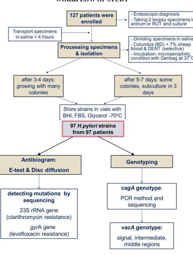
Figure 3.1. Workflow of study 47
Figure 3.2. Study population in the Central provinces 48 Figure 3.3. Endoscopically diagnostic features of diseases 49 Figure 3.4. Morphology of H. pylori and rapid urease testing 53
Figure 3.5. Disk diffusion method 54
Figure 3.6. Epsilometer test 55
Figure 3.7. Genotyping of cagA-EPIYA motifs 59
Figure 3.8. Genotyping of vacA-s/i/m regions 62
Figure 4.1. Distribution of gastroduodenal disease in 97 patients with H.
pylori positive culture 64
Figure 4.2. Age distribution of antibiotic resistance 66 Figure 4.3. Sex distribution of antibiotic resistance 67
Figure 4.4a. MIC distribution of amoxicillin 68
Figure 4.4b. MIC distribution of clarithromycin 68
Figure 4.4d. MIC distribution of metronidazole 69 Figure 4.5. Comparison of primary and secondary resistance 70 Figure 4.6. Correlation of inhibition zone diameter (ID) and MIC value of
clarithromycin in 56 H. pylori isolates 72
Figure 4.7. Correlation of inhibition zone diameter (ID) and MIC value of
levofloxacin in 56 H. pylori isolates 73
Figure 4.8. Disk diffusion and E-test for the same strain 73
Figure 4.9. Point mutations in 23S rRNA gene 74
Figure 4.10. Amino acid N87 and D91 mutations in gyrA gene 76 Figure 4.11. Distribution of cagA status and cagA type in 96 strains 78
Figure 4.12. cagA status by PCR 79
Figure 4.13. Typing cagA-EPIYA motif by PCRs 79
Figure 4.14. Sequences of CagA EPIYA motif 80
Figure 4.15. cagA status and cagA type in relation to diseases 81
Figure 4.16. Distribution of vacA genotype 82
Figure 4.17. vacAs/m multiplex PCR 82
Figure 4.18. vacAi PCR-typing 83
Figure 4.19. Distribution of polymorphic vacA gene structure 83 Figure 4.20. vacA gene polymorphism in relation to diseases 85 Figure 4.21. vacA gene polymorphism in relation to cagA status 86
List of tables:
Table 1.1. Genotypic methods used to detect macrolide resistance 41
Table 3.1. Primers for genotyping cagA 60
Table 3.2. Primers for genotyping vacA 62
Table 4.1. Antibiotic resistance of 92 H. pylori strains with E-test in different
gastric disorders 65
Table 4.2. Resistance Pattern of H. pylori 71
Table 4.3. Mutations of the 23S rRNA gene in clarithomycin –
resistant isolates 75
Table 4.4. Mutations of the gyrA gene in levofloxacine –
resistant isolates 77
List of abbreviations
AC Amoxicillin
cagA Cytotoxin associated gene A
CH Clarithromycin
CLSI Clinical and Laboratory Standards Institute
DU Duodenal ulcer
EPIYA Glutamic-Proline-Isoleucine-Tyrosine-Alanine E-test Epsilometer Test
EUCAST European Committee on Antimicrobial Susceptibility Testing
GAS Gastritis
GC Gastric cancer
GU Gastric ulcer
LE Levofloxacin
MIC Minimum Inhibitory Concentration MLST Multilocus Sequence Typing
MZ Metronidazole
PCR Polymerase Chain Reaction PPI Pronton Pump Inhibitor PUD Peptic Ulcer Diseases
QRDR Quinolone Resistance Determining Region
1. Introduction
1.1. Helicobacter pylori
1.1.1. Introduction & history
The presence of spiral shaped bacteria in the human stomach was first described in 1892 by the Italian pathologist Giulio Bizzozero [1]. However, proof for an infectious origin of diseases of the upper gastrointestinal tract was not provided until the early 1980s when Warren and Marshall isolated H. pylori from gastric biopsies and established the bacterium‟s association with gastritis and peptic ulceration and they were awarded the Nobel Prize in Physiology or Medicine for this discovery in 2005 [2]. The bacterium was first named Campylobacter pyloridis because of its location and some common properties with Campylobacter jejuni. When the difference between Campylobacter pylori and Campylobacter organisms were confirmed by Goodwin et al in 1989 [3], the name was changed to Helicobacter and Helicobacter pylori (H. pylori) became the first member of the new species. The name Helicobacter reflects the two morphological appearances of the organism, often rode-line in vitro and helical in vivo. More than 30 Helicobacter species have been isolated, some infecting occasionally also humans e.g. H. heilmannii, H. fenelliae and H. pullorum but the primary hosts for the non-H. pylori species are animals [4].
Following the discovery of H. pylori a large amount of research has been carried out over the last 2 decades, leading to the development of diagnostic tests and antibiotic treatment strategies for H. pylori infection. However, despite this intense investigation, H. pylori colonisation is still widespread in developing countries. Its route of transmission is poorly
understood and emerging antibiotic resistance has consequences for the efficacy of treatment. Research into H. pylori infection and disease is therefore important in developing novel prevention and treatment strategies. 1.1.2. Microbiology
H. pylori is a microaerophilic Gram negative bacterium which is able to colonise and persist in the mucus layer of the human stomach. Cells are from 2 to 4 µm in length and usually curved or spiral shaped with 2 – 6 unipolar sheathed flagella of approximately 3 µm in length which allow rapid motility through the viscous mucus layer. Morphological changes to a viable but non culturable coccoid form occur after prolonged culture or antibiotic treatment
[5].
H. pylori is commonly isolated from gastric biopsy samples of infected patients, occasionally from gastric juice, faeces and vomitus [6]. Some studies have reported detection of H. pylori in water but the relevance of these studies based on polymerase chain reaction (PCR) remains unclear [7]. H. pylori is a fastidious microorganism and requires complex growth media. The optimal environment for growth of H. pylori is microaerophilic at 37°C. Although the natural habitat of H. pylori is the acidic gastric mucosa because of its ability to produce acid-neutralizing ammonia, a more neutral pH between 5.5 and 8 is the optimal for bacterial grows. The agar plates used to culture H. pylori are supplemented with blood or serum and antibiotics such as vancomycin, trimethoprim, cefsoludin, and amphotericin B or polymyxin B. [8]. Isolation of H. pylori from biopsy samples is difficult and not always successful. H. pylori grows slowly and it may take from 3 to 7 days to achieve a good colony yield.
Prolonged culture is not only unable to increase colony size but may also lead to a transition to the non-culturable coccoid form [8].
Identification of H. pylori is based on colonies, microscopic morphology and biochemical characteristics including oxidase, urease and catalase positivity. Colonies of H. pylori from primary culture on supplemented blood agar at 37oC under microaerophilic condition usually take 3-5 days, are circular (1-2mm), convex, translucent in appearance. There is slight hemolysis in blood agar around colonies, which are greyish in colour. Microscopic examination of the cultured bacteria may show a morphology different from the bacteria present in the biopsy specimen, i.e., bacilli which are neither spiral shaped nor motile, but straight or curved [8]. Although the bacteria can be stained with common histological stains such as hematoxylin and eosin (H & E), silver-containing stains such as Warthin-Starry or Steiner are strongly recommended, particularly when H & E fails to reveal organisms in a biopsy specimen with chronic active inflammation [9]. Urease enzyme that hydrolyses urea to ammonia and carbon dioxide is one of the most important factors for the survival of H. pylori when colonizing the gastric mucosa [10].
The complete genome sequence of H. pylori consisting of a circular chromosome with a size of 1,667,867 base pairs and G+C content of 35-40% has been reported and the extent of molecular mimicry between of H. pylori and human has been fully explored [11]. H. pylori is genetically heterogeneous, which results in every infected individual carrying a distinct strain [12]. Genetic heterogeneity is thought to occur mainly via DNA rearrangement and the introduction and deletion of foreign sequences [13].
1.2. Epidemiology
1.2.1. Prevalence of H. pylori infection
Globally, H. pylori infection affects 50% of the population but prevalence of H. pylori shows large geographical variations [14]. Within geographical areas, the prevalence of H. pylori inversely correlates with socioeconomic status, in particular in relation to living conditions during childhood which is a period of major risk for H. pylori infection [15].
Figure 1.1. Worldwide epidemiology of H. pylori infection [16].
Study of the epidemiology of H. pylori in the Asia Pacific region reveals that there is a wide variation in the prevalence of H. pylori infection, both between countries and within countries. In general, the seroprevalence rates in developing countries are higher than in developed countries. In India, the reported overall seroprevalence rate was 79% [17]. In Vietnam, the H. pylori seroprevalence rate was over 75% [18]. On the other hand, the seroprevalence rates in more developed countries in the Asia Pacific region are generally lower. Among North-east Asian countries, the overall seroprevalence rate was 39% in Japan [19], 55% in Taiwan [20] and 60% in South Korea [21]. Among South-east Asian countries, the reported seroprevalence rate was 36% in Malaysia [22],
31% in Singapore [23] and 57% in Thailand [24].
In each country, differences in seroprevalence rates of H. pylori infection between different geographic regions and also between different ethnic groups have been reported. A cross-sectional study conducted in children residing in industrial and rural areas of Italy reported that the seroprevalence of H. pylori infection was significantly higher in children residing in rural areas compared with those residing in industrial areas [25]. The study also found that, in rural areas, children having shepherd dogs were at greatest risk for H. pylori acquisition. In China, seroprevalence rates have been noted to be higher in regions with higher gastric cancer incidence rates [26]. In Malaysia, the seroprevalence rate has been reported to be lower in west Malaysia (26–31%) than in east Malaysia (43–55%) [22]. In Taiwan, the highest seroprevalence rate (63%) has been reported in rural areas where the aborigines live and in which gastric cancer rates are highest. This compares with a prevalence rate of 40.5% in urban areas, where gastric cancer rates were lowest [20]. In Vietnam, differences in H. pylori seroprevalence rates have been reported between an urban area (Hanoi; 79%) and a rural area (Hatay; 69%) [18].
Generally, it is agreed that H. pylori infection has been declining in industrialized countries. In Japan, the overall seroprevalence rate declined from 73% in 1974 to 55% in 1984 and declined even further to 39% in 1994 [19]. In South Korea, the seroprevalence rate decreased from 67% in 1998 to 60% in 2005 [21]. In particular, lower seroprevalence rates have been observed in the younger population. In Singapore, the seroprevalence rate in those aged less than 3 years has been reported to be 3%, but rose to 71% among those aged more than 65 years [23]. The major decline in H. pylori seroprevalence is probably
associated with socio-economic development, including: improvement in public health measures, personal hygiene and living conditions [5]. Consequently, childhood infections have decreased, leading to a lower seroprevalence rate of H. pylori in the younger generations, thus lowering the overall seroprevalence rate in the population.
The decline in H. pylori infection has been matched by a decline in gastric cancer incidence and gastric cancer mortality. For example, in Japan, with the fall in H. pylori prevalence between 1986 and 1996 the fall in gastric cancer mortality has been greater in younger age groups (20–39 years) in comparison with the overall gastric cancer mortality rate [27]. However, a study in Singapore has demonstrated that Singaporean Chinese and Singaporean Indians have similar H. pylori seroprevalence rates but have significantly different gastric cancer incidence rates [28]. In this case, bacterial virulence factors, host genetic factors or dietary factors could influence carcinogenesis.
1.2.2. Molecular Epidemiology of H. pylori
Population structure analysis based on MLST has revealed seven modern population types of H. pylori, which derived from six ancestral human populations. Interestingly, the incidence of gastric cancer is closely related to the distribution of H. pylori populations. The different incidence of gastric cancer can be partly attributed to the different genotypes of H. pylori circulating in different geographic areas. Furthermore, population genetic studies based on MLST analysis help predict prehistoric human migration „„accompanied‟‟ by H. pylori [29].
Multilocus sequence typing of the seven core housekeeping genes (atpA, efp, mutY, ppa, trpC, ure1, yphC) of H. pylori isolates from different geographic
regions has revealed seven main geographic strains, termed hpEurope, hpEastAsia, hpAsia2, hpAfrica1, hpAfrica2, hpNEAfrica, and hpSahul [30], [31].
The hpEurope strain is common in Europe and countries colonized by Europeans, while hpEastAsia characterizes strains from East Asia. The hpEastAsia strain has been further classified into hspMaori (Polynesians), hspAmerind (native Americans) and hspEAsia (East Asia) subpopulations. The hpAsia2 strain was isolated from South and South-east Asia. hpAfrica1 includes two subpopulations, hspWAfrica (West Africans, South Africans, and Afro-Americans) and hspSAfrica (South Africans); hpAfrica2 is very distinct and has only been isolated in South Africa. hpNEAfrica is predominant in isolates from Northeast Africa. hpSahul strains are isolated from aborigines of Australia and highlanders in New Guinea. All these modern populations derived from six ancestral populations which were designated ancestral European 1 (AE1), ancestral European 2 (AE2), ancestral EastAsia, ancestral Africa1, ancestral Africa2[32], and ancestral Sahul [31]. H. pylori is predicted to have spread from East Africa over the same time period as anatomically modern humans (58,000 years ago) (Fig 1.2), and has remained intimately associated with their human hosts ever since[30], [31], [33].
Figure 1.2. Geographic distribution of Helicobacter pylori populations and predicted traces of prehistoric human migration [29]
(Colored circles illustrate the putative distribution of H. pylori populations before the „„Age of Exploration.‟‟ Black arrows and numbers represent predicted paths and times of migration)
It has been observed that populations with high gastric cancer rates correspond almost exactly to populations with hpEastAsia strains [34]. In South Asian countries where H. pylori seroprevalence rates are high but gastric cancer prevalence rates are low, H. pylori strains have been reported to be predominantly hpAsia2. Similarly, in Africa, most strains have been shown to be hpNEAfrica, hpAfrica1 or hpAfrica2 , and the gastric cancer rates are also correspondingly lower than in East Asia [34]. Interestingly, the phylogeny of most cag PAI genes was similar to that of MLST, indicating that cag PAI was probably acquired only once by H. pylori, and its genetic diversity reflects the isolation by distance which has shaped this bacterial species since modern humans migrated out of Africa [35]. In addition, strains without the presence of
cagA has been reported to be less virulent regardless MLST, thus, the cagA genotype rather than the phylogeographic origin is a better predictive factor of gastric cancer [36].
1.2.3. Transmission of H. pylori infection
Up to date, there is still an ongoing debate about the exact mode of transmission of H. pylori infection that has been lasting over two decades [37]. The majority of data support the notion that transmission is within families, close contact and level of household sanitation appear to be the important variables [38], [39]. In one study, H. pylori status was determined in 41 families; the results revealed that, if the index case (either the mother or father) was positive, the children and spouse in that family were also likely to test positive for H. pylori. If the index case was negative, then the children and spouse were likely to be negative as well[40]. The most probable transmission route is through oral-oral, gastro-oral and faecal-oral pathways.
1.2.3.1. Oral-oral transmission
The role of oral cavity as a reservoir of H. pylori infection has been controversial. Only one study found H. pylori by culture [6]. Several studies using PCR amplification from saliva and dental plaque have demonstrated the presence of H. pylori in the mouth [6], [41]. Close mouth to mouth contact has been identified as a risk factor for oral-oral transmission [42], [43]. Cultural and social differences such as pre-mastication of food and sharing chopsticks in Asian countries may be an explanation for oral-oral transmission route. The evidence for this transmission pathway is supported by a study from Bangladesh where the Hindu babies had higher prevalence of H. pylori infection as compared to Muslims assumed to be due to Hindu mothers
regularly coating their nipple by saliva before breastfeeding and feeding their babies by pre-mastication of food [43]. In addition, the data from one study conducted by Chow et al showed that the infection prevalence of people who used chopsticks to eat from communal dishes was significantly higher than in those who did not (64.8% versus 42.3%) [42].
1.2.3.2. Gastro-oral transmission
Vomitus has been suggested as an important vehicle for H. pylori transmission as this organism had been successfully cultured from gastric juice and vomitus [6]. Also, an increased acquisition of H. pylori in children during a gastroenteritis outbreak has been reported in one centre [44]. The gastro-oral transmission route seems to occur by either vomitus or regurgitation of stomach contents. Data from some studies have shown that endoscopists had a higher risk of acquiring H. pylori infection compared to the general population [45].
1.2.3.3. Faecal-oral transmission
The faecal-oral route is another potential route of transmission through exposure to contaminated food or water.
Firstly, evidence for a faecal-oral transmission route of H. pylori has been reported in several studies using DNA to detect H. pylori in stool of infected patients and nowadays it‟s become one of non-invasive tests to diagnose H. pylori infection [46]. In addition, the bacterium has been isolated by culture of faecal samples in several studies [6], [47].
Secondly, there is mounting evidence which suggests that the prevalence of H. pylori infection has a strong correlation with access to clean water. Several studies found contaminated water to be a risk factor for H. pylori
transmission [48], [49]. In a study from India that examined 500 adults, three biopsy samples were collected from each subject to assess H. pylori infection
[50]. Based on detection by PCR amplification of the gene encoding 16S rRNA from H. pylori, the prevalence of infection among people who drank water from wells was 92%, compared with 75% in those who drank tap water (P < 0.001). H. pylori infection prevalence was found to be higher in people with a low clean water index (88%) than in those with a higher clean water index (33%) (P < 0.001). The results of the study suggested that the risk of acquisition and transmission of H. pylori can be prevented to a large extent by regular boiling of water used for drinking purposes.
Therefore, faecal-oral transmission has been proposed to commonly occur in developing countries because of limitations in hygiene conditions [5]. The rapid decrease of the infection in developed countries has been speculated to be due to the decrease in gastrointestinal infections in children that are still very common in developing countries [51].
1.3. H. pylori virulence and colonisation factors 1.3.1. Cytotoxin associated gene A (cagA gene)
CagA protein was discovered in the early 1990s in a study with the strong association between serological responses to CagA and peptic ulcer disease [52]. Then, the cloning of the cagA gene [53] as well as identification of cag pathogenicity island (cagPAI) genes and their role in inflammation [54]. Recently, one study have firmly implicated that CagA plays as a bacterial oncoprotein by attenuating apoptosis in vivo and in vitro [55] and another study demonstrated that transgenic expression of CagA in mice leads to the development of aberrant gastric epithelial proliferation and gastric carcinoma[56].
The cagA gene, encodes 120 to 140 kDa CagA protein, is the most studied virulence factor of H. pylori, which is located at one end of the cagPAI, an approximately 40 kb region that is thought to have been incorporated into the H. pylori genome by horizontal transfer from an unknown source [57]. The cag PAI, contains 27 to 31 genes, encodes a type IV secretion system through which CagA is delivered into host cells (Fig 1.3) and cagA is recognized as a marker for the cagPAI region [58], [59].
1.3.1.1. Pathogenicity mechanism of CagA
CagA phosphorylation-dependent host cell signaling:
Following its injection into epithelial cells, CagA undergoes targeted tyrosine phosphorylation by Src and Abl kinases of host cell at motifs containing the amino acid sequence EPIYA, which are located within the 3‟ terminus of CagA [60]. Within epithelial cells, phosphorylated CagA activates a eukaryotic phosphatase (SHP-2) as well as ERK which results in the impairment of a variety of intracellular signaling systems leading to morphological aberrations (Fig 1.3) [60].
CagA phosphorylation-independent host cell signaling:
Nonphosphorylated CagA also exerts effects within the cell that contribute to pathogenesis. Translocation, but not phosphorylation, of CagA leads to aberrant activation of β-catenin, disruption of apical-junctional complexes, and a loss of cellular polarity [61]. Currently, there are at least 10 known phosphorylation-independent CagA host interaction partners [62]. In addition, CagA itself forms dimers in cells in a phosphorylation-independent manner, and the CagA multimerization (CM) sequence (FPLxRxxxVxDLSKVG) was
identified as the site responsible for dimerization [63]. This sequence is located within the EPIYA-C segment, but is just downstream of the EPIYA-D segment. The CM sequence is also essential for the formation of the CagA–PAR1 (MARK) complex-bound non phosphorylated CagA inhibits the kinase activity of PAR1 to promote the loss of cell polarity, mucosal damage, inflammation and carcinogenesis [63], [64].
The CM sequence has also been named as the MARK2/PAR1B kinase inhibitor (MKI) [65] or named as the conserved repeat responsible for phosphorylation independent activity (CRPIA) [66] that was reported to be responsible for the interaction of CagA with activated c-Met led to the upregulation of β-catenin and nuclear factor κB (NFκB) transcriptional activities which promoted proliferation and inflammation. Therefore, there are currently three acronyms that essentially correspond to the same sequence in CagA [63], [64].
Figure 1.3. CagA structure and its effects in host cell [67].
A: Type IV secretion system, encoded by cag PAI, deliver CagA into the host cells; B: CagA
phosphorylation motifs and cellular morphogenic alterations induced by intracellular CagA; C: Structural polymorphism in CagA. Western-type CagA contain EPIYA-A, EPIYA-B, and EPIYA-C segments. By contrast, East-Asian-type CagA contain the EPIYA-A, EPIYA-B and D segments. The sequence flanking of the D segment is EPIYA-TIDFDEANQAG, but that of EPIYA-C segment is EPIYATIDDLGGP.
1.3.1.2. CagA type: Western versus East Asian
cagA is a polymorphic gene. In particular, there are different numbers of repeat sequences located in the 3' region of the cagA gene of different H. pylori strains. CagA can be divided in two types, the East-Asian type and the Western type, according to the repeat sequences of the 3‟ region of cagA gene[67].
The repeat regions contain the Glu-Pro-Ile-Tyr-Ala (EPIYA) motifs of CagA in strains harvested from persons residing in Western countries have been termed A, B, or C based on sequences flanking the EPIYA motif. In contrast, phosphorylation sites within CagA proteins from East Asian H. pylori strains lack the EPIYA-C motif and, instead, contain a different motif, which is termed D [60]. As such, each CagA is assigned a sequence type consisting of the names of the EPIYA segments in its sequence that is, ABC, ABCC or ABCCC for Western-type CagA and ABD for East-Asian-type CagA due to strong conservation and a lack of duplication in the D region (Fig 1.3). Analyses of the repeat regions of CagA has demonstrated that, although not common, some strains isolated in East Asian countries have a Western-type CagA sequence. By contrast, none of the Western strains studied have an East-Asian-type CagA sequence[68], [69].
In vitro, Western CagA with increased the number of EPIYA-C motifs or East Asian CagA mostly with one EPIYA-D motif is associated with the intensity of CagA phosphorylation, epithelial cellular elongation, and induction of proinflammatory cytokines [70, 71]. CagA of East-Asian type, containing EPIYA-D segments, exhibits a stronger binding affinity for Src homology 2 containing protein-tyrosine phosphatase (SHP-2) and a greater ability to induce morphological changes in epithelial cells than Western-type CagA, which contains one EPIYA-C segments[72].
1.3.1.3. Clinical importance of cagA
CagA positive strains were reported to be associated with severe clinical outcomes not only in Western countries [67] but also in East Asian countries, however, the odds ratio in East Asian countries was smaller than that in Western
countries [73]. Additionally, H. pylori strains possessing more than three EPIYA-C motifs are more frequently associated with gastric atrophy, intestinal metaplasia, and gastric cancer [74, 75].Individuals infected with East-Asian type cagA strains were reported to have an increased risk of peptic ulcer or gastric cancer than those with Western-type cagA strains in Southeast Asian countries, such as Thailand, Malaysia, Singapore [76], [77]. However, in East Asia countries, it is difficult to differentiate between gastritis and gastric cancer simply by considering the number of repeated sequences because almost all strains contain East Asian type CagA with a single EPIYA-D segment [69].
1.3.2. Vacuolating cytotoxin VacA
VacA is the second most extensively studied H. pylori virulence factor. Unlike cagA, virtually all the H. pylori strains have a functional vacA which encodes a vacuolating cytotoxin[78].
In 1988, Leunk and colleagues discovered that cell free supernatants from broth cultures of H. pylori induced vacuolar degeneration of various cultured epithelial cell lines [79]. This effect was subsequently shown to be caused by a secreted protein toxin, designated VacA[80]. vacA gene, which was cloned from several strains of H. pylori, encodes the vacuolating cytotoxin, 140-kDa precursor protein [81][82].
Current models indicate that a 96-kDa VacA protein is secreted, which is then cleaved into an 88-kDa mature protein (p88) and a 10.5-kDa passenger domain (p10). The mature, secreted p88 subunit can undergo further proteolytic cleavage to yield two fragments, p33 and p55 [83], [78], which represent the two functional domains of VacA (Fig.4). Cell binding is mediated by the p55 fragment of VacA [84], but p33 and p55 can also exert multiple other effects.
VacA inserts into planar lipid bilayers to form anion selective membrane channels [85]. The p33 domain contains a hydrophobic sequence, which is involved in pore formation whereas the p55 fragment contains one or more cell-binding domains [84]. Since the p55 subunit contains the m1 and m2 alleles, delineation of protein sequences from unrelated H. pylori strains should allow identification of VacA structural features that are important for binding to host receptors [86].
1.3.2.1. Effects of VacA on cellular
In addition to inducing vacuolation, VacA can induce multiple cellular activities, including membrane channel formation, cytochrome c release from mitochondria leading to apoptosis, and binding to cell-membrane receptors followed by initiation of a proinflammatory response (Fig 4). VacA can also specifically inhibit T-cell activation and proliferation [67]. Studies indicate that VacA and CagA can even inhibit at least some of each other‟s signaling pathways, for example, CagA has been shown to promote the expression of the apoptotic suppressor Mcl1, and inhibit epithelial cell apoptosis caused by vacA
[55, 87]. These data again emphasize the importance of in vitro infection experiments in which interaction among H. pylori virulence factors can be taken into account.
1.3.2.2. vacA polymorphism
vacA gene is present in all strains of H. pylori and well conserved between strains but there is significant diversity in three distinct regions of the gene: the signal (s) region, the middle (m) and intermediate (i) regions (Fig. 4). Characterisation of these regions has revealed that allelic variation exists. Allelic diversity in vacA has allowed strains to be genotyped s1 or s2 for the signal
region and m1 or m2 for the middle region[88].
Figure 1.4. H. pylori VacA structure and functional effects [89].
The vacA genotype is an important determinant of toxicity. Type s2 VacA, carrying a hydrophilic N-terminal, is cleaved a 12 amino acid in the signal peptide sequence and turned into type s1 VacA with a hydrophobic N-terminus. This 12 residue extension to VacA has been shown to be responsible for the loss of vacuolation induction by s2 strains [88, 90]. The vacA middle region which encodes part of the p58 region has been shown to be associated with cell specificity, m1 forms vacuolate a variety of cell types but m2 forms are more limited in the cells they vacuolate [88, 91].
The recently identified intermediate region, is located between the signal and mid regions of vacA, can also be divided into two types i1 and i2 [92]. vacA s1/m1 type strains were nearly always type-i1 and that type s2/m2 strains were always type-i2. The s1/m2 strains varied in their intermediate region type, however, they found that strains that were s1/i1/m2 had vacuolating activity
whereas type s1/i2/m2 strains were non-vacuolating. The i region is thought to be involved in cytotoxin binding [92]. The vacA gene may comprise any combination of signal, intermediate and mid region types. Most of the vacA combinations have been detected in strains isolated from infected individuals, although the s2/m1 form of vacA is rarely found [88].
1.3.2.3. Disease associations of VacA
The clinical significance of VacA has been assessed from studies in animal models and from several observations in humans infected with H. pylori. Firstly, large quantities of purified VacA can induce ulcer-like erosions when administered into the mouse stomach [93]. Orally administered toxigenic H. pylori sonicates and purified VacA also induce epithelial vacuolation, loss of gastric gland architecture and infiltration of mononuclear cells into the lamina propria [94]. Among vacA type, s1m1 strains are the most cytotoxic, followed by s1m2 strains, whereas s2m2 strains have no cytotoxic activity and s2m1 strains are rare[67].
Several studies have investigated the association of vacA type and disease outcome. In Western populations such as the USA and Western Europe where vacA allelic diversity is common, s1 genotypes are more frequently associated with higher levels of inflammation in the gastric mucosa than s2 types [68, 95].
vacA s1m1 and s1m2 strains have been shown to be associated with peptic ulceration and s1m1 type strains have been associated with gastric carcinoma [68, 92, 95, 96]. The vacA i1 type is a risk factor for peptic ulcer disease and has been shown to be associated with duodenal ulcer disease as well as gastric cancer [92, 96]. Importantly Rhead et al found that although vacA s1, i1 and m1 alleles along with cagA positive status were all associated with gastric adenocarcinoma in an
Iranian population, the association was most pronounced with i1-type strains. Furthermore, only the i region status was independent of all other alleles studied. They concluded that the vacA i region is the best independent marker of toxicity and pathogenicity of a strain in Iranian[92].
In East Asia, however, most H. pylori strains have an s1-type, therefore the pathogenic difference cannot be explained by the type of s region present. [68]. Moreover, it is almost all cagA positive strains are classified as an vacA s1 strain, whereas cagA-negative strains are often combined with s2/m2 type [88]. With respect to the m region, however, there is variation within East Asia. Although m1 strains are common in parts of north East Asia, such as Japan and South Korea, m2 strains are predominant in parts of South-East Asia, such as Taiwan and Vietnam[67].
1.3.3. Duodenal ulcer promoting gene (dupA)
In 2005, the first disease specific H. pylori virulence factor that induced duodenal ulcer and had a suppressive action on gastric cancer was identified, and was named duodenal ulcer promoting gene A [97]. This gene is located within the plasticity zone of the H. pylori genome. Initial analysis of 500 H. pylori strains from Colombia, South Korea, and Japan showed an increased risk for duodenal ulcer and a decreased risk for gastric cancer in persons carrying dupA-positive strains [97]. In vitro, DupA increases IL-8 production [97]. However, a subsequent study focused on strains from Belgium, South Africa, and the United States found no significant relationships between dupA expression and duodenal ulcer but a significant association with gastric cancer [98]. Comparison of strains from Iran and Iraq indicates that dupA expression is significantly associated with duodenal ulceration in strains isolated from Iraq but not in Iranian isolates [99].
No association was found between dupA expression and gastric cancer or duodenal ulcer in strains from Japan [100] or Sweden [101], but correlations were observed between dupA and duodenal ulcer disease or gastric cancer in Indian and Chinese [102], [103], [101]. It seems likely that dupA may promote duodenal ulceration and prevent gastric cancer in some, but not all, populations. 1.3.4. Adhesins and outer membrane proteins (OMPs)
Adherence of H. pylori to the gastric epithelium facilitates initial colonization, persistence of infection, and delivery of virulence factors to host epithelial cells. Approximately 4% of the H. pylori genome is predicted to encode outer membrane proteins (OMPs), which is significantly more than that for other known bacterial species. OMPs expression has been associated with gastroduodenal diseases [61].
1.3.4.1. Blood group antigen binding adhesin (BabA)
BabA is encoded by the babA2 gene, which binds to fucosylated Lewisb antigen (Leb) on the surfaces of gastric epithelial cells and is the most well-described H. pylori OMP [104]. Transgenic mice that express Leb on pit and surface mucous cells demonstrated that H. pylori attaches to the surfaces of Leb expressing cells and induces more severe gastritis than in nontransgenic mice despite a comparable colonization density, suggesting that Leb mediated colonization may increase the pathogenic potential of H. pylori [105]. Analyses of binding specificities of H. pylori strains from across the world suggest that the BabA adhesin has evolved in response to host mucosal glycosylation patterns to permit H. pylori to adapt to its host and to maintain persistent colonization [106]. The presence of BabA2 is associated with duodenal ulcer disease and gastric cancer, and when found in conjunction with cagA and vacA s1 alleles, it is
associated with an even greater risk of developing more severe disease [107]. Recently, there are the conflicting data on the usefulness of BabA2 expression in predicting clinical outcome in different countries, for examples, BabA2 expression is associated with the severity of gastric disease in strains isolated from Germany or northern Portugal [107], [108] but not a biomarker for peptic ulcer disease or gastric cancer in ThaiLan strains [109].
1.3.4.2. Sialic acid-binding adhesin (SabA)
SabA is an H. pylori adhesin that binds to the carbohydrate structure sialyl Lewisx antigen expressed on gastric epithelium and is associated with an increased gastric cancer risk but a reduced risk for duodenal ulceration [110]. Sialyl-Lewisx expression is induced during chronic gastric inflammation, suggesting that H. pylori modulates host cell glycosylation patterns to enhance attachment and colonization [111]. Furthermore, SabA is regulated by phase variation, such that SabA expression can rapidly be switched “on” or “off” to adapt to changes exerted by the gastric niche [110].
1.3.4.3. Outer inflammatory protein (OipA)
OipA is an inflammation related outer membrane protein which functions as an adhesin and is reported to be involved in the attachment of H. pylori to gastric epithelial cells [67]. OipA expression is linked to increased IL-8 production in vitro [112]. Recent a experiment demonstrated a role for OipA in induction of the mucosal cytokines IL-1, IL-17, and tumor necrosis factor alpha (TNF-α) and in gastric mucosal inflammation [113]. OipA is also involved in upregulation of matrix metalloproteinase 1 (MMP-1) and in β-catenin translocation and accumulation in the nucleus that can influence carcinogenesis [89]. H. pylori contains either a functional or nonfunctional oipA gene, and the presence of a
functional gene is significantly associated with the presence of duodenal ulcers, gastric cancer, and increased neutrophil infiltration [110].
Interestingly, the funtional oipA and cagA positivity are closely linked with each other and the cagA status is also linked to the vacA s region type and it is further closely linked to the presence of the babA gene. The links among these factors should have a certain biological significance and they may somehow interact with each other. It might be more relevant to hypothesize that these factors interact synergistically with each other and induce serious diseases, rather than to discuss which factor is the most virulent [67].
1.3.5. Induced by contact with epithelium A (IceA)
IceA was highlighted by the isolation of mRNA transcripts from strains associated with peptic ulcer disease [114]. Two polymorphisms were identified: IceA1 for which transcription is up regulated following contact with epithelial cells and the inactive IceA2 which is not. The biological significance of these genes is unclear; IceA1 has been associated with peptic ulcer disease, but this association is not universal [115].
1.3.6. Acid resistance and motility Acid resistance
Urease, which converts urea into ammonia and carbon dioxide, is essential for the survival of H. pylori in the acidic gastric environment [5]. It allows H. pylori to maintain a constant periplasmic and internal pH which is required for transmembrane potential difference [5]. Urease is produced by all H. pylori strains although the level of urease activity differs significantly between H. pylori isolates [116]. Urease has been proposed to participate in tissue damage by
producing ammonia which is thought to be cytotoxic to epithelial cells [117]. Motility
Motility is essential for H. pylori colonization [118]. Flagellar motility is thought to be required for the initial stages of infection allowing the bacterium to move from the acidic environment of the gastric lumen into the less acidic mucus layer [119]. Mutants defective in the synthesis in either one of the two flagellins have impaired colonization efficiency and non-motile double mutants are completely non-virulent [120].
1.4. Diseases associated with H. pylori infection
Although gastric colonization with H. pylori induces histologic gastritis in all infected individuals and most of them has not any symptoms, only a minority develop related diseases of long-term carriage of H. pylori. It is estimated that infected patients have a 10 to 20% lifetime risk of developing ulcer disease and a 1 to 3% risk of developing distal gastric cancer [121], [122]. The risk of development of these disorders in the presence of H. pylori infection depends on a variety of bacterial, host, and environmental factors that mostly relate to the pattern and severity of gastritis [61].
1.4.1. Gastritis
The association between acute gastritis and H. pylori infection was first observed by Warren and Marshall when the developed acute gastritis several days after drinking a pure culture of H. pylori [123].
The natural history of H. pylori infection can be divided in two phases. The acute phase in which bacteria proliferate and cause gastric inflammation, hypochlorhydria develops and some gastrointestinal symptoms appear such as
fullness, nausea and vomiting. This phase often occurs during childhood and almost difficult to diagnose. After several weeks, the chronic phase begins in which the inflammatory response is reduced and the pH becomes normal, and the infected person becomes asymptomatic [124]. The colonization of H. pylori in gastric mucosa leads to infiltration of neutrophilic and mononuclear cells in both the antrum and the corpus that can result in chronic inflammatory [5]. When colonization becomes persistent, a close correlation exists between the level of acid secretion and the distribution of chronic gastritis. The most common phenotype is non-atrophic gastritis with normal acid secretion in asymptomatic subjects. [5]. Another phenotypic antral-predominant gastritis is associated with hyperchlorhydria and duodenal ulcer, whereas a corpus-predominant pangastritis leads to hypochlorhydria, gastric atrophy, intestinal metaplasia and an increased risk of distal gastric cancer [125], [126].
Gastritis is mostly diagnosed, classified and graded based on the histo-pathological criteria [9], [127]. However, endoscopic classification for gastritis is also used by endoscopists to describe the gastric mucosal changes and there are some relationship between endoscopic and histological characterizations [128], [129].
1.4.2. Peptic ulcer disease: duodenal ulcer and gastric ulcer
Peptic ulcers are defined as mucosal defects with a diameter of at least 0.5 cm penetrating through the muscularis mucosa. Gastric ulcers mostly occur along the lesser curvature of the stomach, in particular, at the transition from corpus to antrum mucosa [130]. Duodenal ulcers usually occur in the duodenal bulb, which is the area most exposed to gastric acid.
Approximately 10% to 20% of infected patients will develop peptic ulcers and ulcer relapse is prevented after cure of the infection [121], [131]. In initial reports from all over the world, approximately 95% of duodenal ulcers and 85% of gastric ulcers occurred in the presence of H. pylori infection, furthermore, the lifetime risk for ulcer disease in infected subjects is 3 to 4 times higher than in H. pylori-negative subjects [132]. Duodenal ulcer are more prevalent in younger individuals and gastritis in duodenal ulcer was primarily antral and spared the gastric corpus with high levels of acid secretion [133].
Although the same major causative factor (H. pylori infection), gastric ulcer is associated with a high risk, but duodenal ulcer with a low risk, of gastric cancer, and patients with gastric ulcers typically have atrophic gastritis and corpus-predominant gastritis leading to decreasing acid secretion, whereas patients with duodenal ulcers have antral-predominant gastritis, but few atrophic changes [134].
1.4.3. Gastric cancer
Gastric adenocarcinoma has been considered an infectious disease since 1994, when the International Agency for Research on Cancer categorized Helico-bacter pylori infection as a class I human carcinogen [135]. The pathogenesis of gastric adenocarcinoma represents a prototype for bacteria induced and inflammation driven malignancies [136].
Gastric cancer is a major cause of global morbidity and mortality. It is the fourth most common cancer worldwide, accounting for 9% of all new cancers, and ranking the second cause of cancer death [137]. Approximately two-thirds of the cases occur in developing countries, with a wide variation in incidence rates worldwide. The highest incidence rates are reported in Eastern Asia, Central and
Eastern Europe, and Central and South America [137].
Typically, the diagnosis of gastric cancer is delayed by a lack of early specific symptoms, and most patients are diagnosed after cancer has invaded the muscularis propria. The 5-year survival rate for gastric cancer is less than 15% even in the United States [138].
Histologically, two distinct variants of gastric carcinoma have been identified: diffuse-type gastric cancer and intestinal-type adenocarcinoma, which progresses through a series of well-defined histological steps and was first described in 1975 [139]. Intestinal-type adenocarcinoma is initiated by the transition from normal mucosa to chronic superficial gastritis; this is followed by atrophic gastritis and intestinal metaplasia, finally leading to dysplasia and adenocarcinoma [140], [141]. This process usually takes decades after persistent infection with H. pylori. Intestinal-type gastric cancer affects men twice times than women and commonly occurs in men above 50 years old [142]. Corpus-predominant gastritis predisposes individuals toward gastric cancer, which is thought to be due in part to decreased acid secretion. In contrast, antrum predominant gastritis results in increased acid production and predisposes individuals to duodenal ulcer disease, which is associated with a decreased risk of gastric cancer [126]. The loss of gastric acidity contributes to the promotion of the endogenous formation of N-nitroso compounds by the gastric bacterial flora with non-H. pylori species, may be the causative factors of gastric cancer [143]. Moreover, decreased acid in gastric juice results in low levels of ascorbic acid and a diminished ability to block the N-nitrosation process [134].
1.4.4. Gastric MALT lymphoma (Mucosa associated lymphoid tissue)
![Figure 1.1. Worldwide epidemiology of H. pylori infection [16].](https://thumb-eu.123doks.com/thumbv2/123dokorg/8349276.133455/24.918.156.812.387.614/figure-worldwide-epidemiology-h-pylori-infection.webp)
![Figure 1.2. Geographic distribution of Helicobacter pylori populations and predicted traces of prehistoric human migration [29]](https://thumb-eu.123doks.com/thumbv2/123dokorg/8349276.133455/28.918.160.801.107.469/figure-geographic-distribution-helicobacter-populations-predicted-prehistoric-migration.webp)
![Figure 1.4. H. pylori VacA structure and functional effects [89].](https://thumb-eu.123doks.com/thumbv2/123dokorg/8349276.133455/38.918.177.800.164.498/figure-h-pylori-vaca-structure-functional-effects.webp)
![Figure 1.5. Common diagnostic H. pylori infection tests [16].](https://thumb-eu.123doks.com/thumbv2/123dokorg/8349276.133455/54.918.195.764.185.445/figure-common-diagnostic-h-pylori-infection-tests.webp)

![Figure 3.7. Genotyping of cagA-EPIYA motifs [206]](https://thumb-eu.123doks.com/thumbv2/123dokorg/8349276.133455/79.918.153.794.527.839/figure-genotyping-of-caga-epiya-motifs.webp)
![Figure 3.8. Genotyping of vacA-s/i/m regions [210]](https://thumb-eu.123doks.com/thumbv2/123dokorg/8349276.133455/82.918.184.776.110.467/figure-genotyping-of-vaca-s-i-m-regions.webp)
