Alma Mater Studiorum - Università di
Bologna
Scuola di Dottorato di Ricerca
in Scienze Mediche e Chirurgiche
Dottorato di ricerca in Scienze Biomediche
Progetto formativo in Neurofisiologia
XXIV ciclo
Settore scientifico-disciplinare di afferenza: BIO/09
Sigla del settore concorsuale di afferenza: 05/D1
Tesi di Dottorato
Sonic Hedgehog pathway impairment in
Neural Precursor Cells of the Ts65Dn mouse,
an animal model of Down syndrome
Dott.ssa Valentina Maria Mitrugno
Coordinatore:
Tutor:
Prof. Lucio Ildebrando Cocco Dott.ssa ElisabettaCiani
Dipartimento di Fisiologia umana e generale
Esame finale A.A. 2011/2012
1. AIM OF THE STUDY
Pag. 52. INTRODUCTION
Pag .72.1. DOWN SYNDROME
Pag. 7
2.1.1 History of DS Pag. 7
2.1.2 Epidemiology Pag. 9
2.1.3 Etiology Pag. 10
2.1.4 Somatic traits and clinical features Pag. 12
2.1.5 Behavioural defects in DS Pag. 17
2.1.6 Neuroanatomy of DS Pag. 19
2.1.7 Causes of brain hypothophy in DS Pag. 20
2.1.8 The Human Chromosome 21, genotype-phenotype Pag. 23 correlations
2.2 MOUSE MODELS FOR DS Pag. 24
2.2.1 Mice trisomic for the entire MMU16 Pag. 25
2.2.2 Segmental MMU16 trisomic mice Pag. 28
2.2.3 Mice trisomic for HSA21 Pag. 32
2.2.4 Transgenic mice with triplication of individual genes Pag. 33 2.2.5 Segmental trisomic mice disomic for individual genes Pag. 35
2.3 NEUROGENESIS ALTERATIONS IN DS ANIMAL
MODELS Pag. 36
2.4 MOLECULAR MECHANISMS UNDERLYING
NEUROGENESIS IMPAIRMENT IN DS Pag. 38
2.5 AAP GENE AND NEUROGENESIS Pag. 40
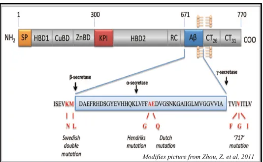
2.5.1 The human gene APP Pag. 40
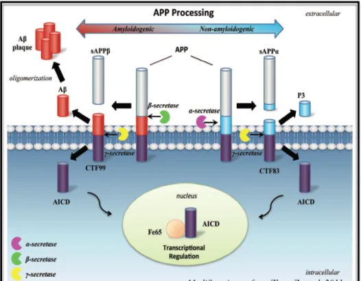
2.5.3 Role of APP in neurogenesis Pag. 43 2.5.4 Role of sAPPα in the positive regulation of neurogenesis Pag. 44 2.5.5 Role of AICD in the negative modulation of neurogenesis Pag. 45
2.5.6 APP and DS Pag. 48
2.6 HEDGEHOG SIGNALING Pag. 51
2.6.1 Sonic Hedgehog (Shh) signaling Pag. 52
2.6.2 Non-canonical Shh signaling Pag. 55
2.6.3 Shh signaling in the developing Central Nervous System Pag. 55 2.6.4 Shh signaling and Neural Stem Cells Pag. 56 2.6.5 Shh signaling and Neural Stem Cells in the SVZ and
in the Hippocampus Pag. 58
2.6.6 Shh and DS Pag. 60
3. MATERIALS AND METHODS
Pag. 623.1 TS65DN MICE COLONY AND TREATMENT Pag. 62
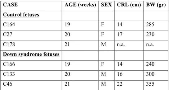
3.2 HUMAN FETUSES Pag. 62
3.3 HISTOLOGICAL PROCEDURES Pag. 63
3.4 CELL CULTURES AND TREATMENTS Pag. 64
3.5 NEUROSPHERES DIAMETER MEASURAMENTS Pag.66
3.6 BRDU IMMUNOCYTOCHEMESTRY IN
NEUROSPHERES Pag. 67
3.7 CELL CYCLE ANALYSIS IN NEUROSPHERES Pag. 67
3.8 IN VITRO DIFFERENTIATION ANALYSIS
OF NPCS Pag. 68
3.9 PTCH1 QUANTIFICATION IN NEUROSPHERES Pag. 68
3.10 BRDU IMMUNOHISTOCHEMESTRY Pag. 69
3.11 PTCH1 IMMUNOHISTOCHEMESTRY AND
QUANTIFICATION IN VIVO Pag. 69
3.12 WESTRN BLOTTING Pag. 70
3.14 ANTISENSE EXPERIMENTS Pag. 72 3.15 METHYLCYTOSINE IMMUNOPRECIPITATION
(mCIP)
Pag. 723.16 CHROMATIN IMMUNOPRECIPITATION
(ChIP)
Pag. 733.17 STATISTICAL ANALYSIS
Pag. 75
4. RESULTS
Pag. 76
4.1. Neuronal precursor cultures from Ts65Dn mice exhibit
the same proliferation impairment as the in vivo condition Pag. 76 4.2 Neuronal precursor cultures from Ts65Dn mice exhibit
the same defective neuronal differentiation as the in vivo
condition Pag. 82
4.3 Deranged expression of genes belonging to the Shh
pathway in neural precursors from Ts65Dn mice Pag. 84
4.4 Neural precursors from Ts65Dn mice do not respond
to Shh Pag. 87
4.5 Silencing of Ptch-1 expression restores proliferation in
neural precursors from Ts65Dn mice Pag. 89
4.6 Ptch1 promoter is highly acetylated in neural precursors
from Ts65Dn mice Pag. 93
4.7 Triplicated amyloid precursor protein increases Ptch1
expression through AICD Pag. 98
4.8 In vivo Ptch1 overexpression in the trisomic brain Pag. 102
5. DISCUSSION
Pag. 1065.1. Cultured trisomic NPCs exhibit the same neurogenesis
defects as the in vivo condition Pag. 106
5.2 Impairment of the Shh pathway characterizes trisomic
NPCs Pag. 106
Shh-induced mitogenic response of trisomic NPCs Pag. 107 5.4 Mechanisms underlying Ptch1 overexpression in
trisomic NPCs Pag. 109
6. CONCLUSIONS
Pag. 1101. AIM OF THE STUDY
Down syndrome (DS), caused by trisomy of human chromosome 21 (HSA21), is one of the most common genetic causes of mental retardation, occurring in one out 800-1000 live births (Hayes and Batshaw 1993; Roizen and Patterson 2003; Shin, Besser et al. 2009). Although a wide range of variable traits characterizes Dow syndrome, intellectual disability is the unavoidable hallmark of the pathology, with a heavy impact on public health. Mental retardation in DS is likely due to widespread brain hypoplasia and hypocellularity that start to be present from early developmental stages and are retained in adulthood. Recent evidence from our laboratory shows that the fetal DS brain is characterized by severe proliferation impairment that involves numerous brain regions (Contestabile, Fila et al. 2007; Guidi, Bonasoni et al. 2008; Guidi, Ciani et al. 2011). These findings clearly indicate that neurogenesis impairment is a key determinant underlying brain malfunctioning in DS.
Mouse models that mimic human pathologies as closely as possible are becoming invaluable tools because they can be exploited to identify the mechanisms underpinning a given pathology and to examine the outcome of targeted therapies. Various mouse models for DS are now available but the Ts65Dn mouse is one of the most widely used because it displays several neuroanatomical and behavioral defects that closely recapitulate the human condition (Reeves, Gearhart et al. 1986). In the Ts65Dn mouse neurogenesis is severely impaired in the embryonic ventricular zone and hippocampus (Chakrabarti, Galdzicki et al. 2007) and in the hippocampus, cerebellum and subventricolar zone (SVZ) of neonate and adult mice (Baxter, Moran et al. 2000; Contestabile, Fila et al. 2007; Contestabile, Fila et al. 2009). This evidence validates the use of the Ts65Dn mouse model to get insight into the mechanisms underlying proliferation impairment in DS.
The mechanisms by which trisomy 21 interferes with brain development are still largely unknown. It has been recently shown that the reduced proliferation
of cerebellar granule cell precursors from Ts65Dn mice is related to an attenuated response to sonic hedgehog (Shh) (Roper, Baxter et al. 2006), a potent mitogen that controls cell division during brain development (Ishibashi, Saitsu et al. 2005). The wide brain expression of the Shh pathway (Ishibashi, Saitsu et al. 2005; Arsic, Beasley et al. 2007), suggests that it may exert a widespread role in the regulation of neural precursor proliferation in different brain neurogenic zones. If so, a generalized attenuation of the response to Shh might account for the widespread neurogenesis impairment that characterizes the trisomic brain.
Although in vivo models reflect in a more realistic fashion the environment of the diseased brain, in view of the complexity of the mechanisms underlying neurological diseases, simplified in vitro approaches are often essential tools to better dissect the molecular mechanisms that take part in the pathology.
The overall goal of the present study was to create an in vitro model suitable to understand the mechanism/s underlying neurogenesis impairment in DS. The specific goals were to:
i) establish whether cultures of neural precursor cells (NPCs) from Ts65Dn mice exhibit the same proliferation defects as the in vivo condition;
ii) establish whether NPCs exhibit a defective response to Shh, similarly to cerebellar granule cell precursors;
iii) dissect the molecular mechanisms that underlie deregulation of the Shh pathway and, consequently, the proliferation impairment of trisomic NPCs.
2. INTRODUCTION
2.1 DOWN SYNDROME (DS)
2.1.1 History of DS
The history of the discovery and characterization of the DS has been summerized in very fine details by Pierre L. Roubertoux and Bernard Kerdelhué in a review of 2006 (Roubertoux, Bichler et al. 2006). They describe that in 1838, Jean-Etienne-Dominique Esquirol, specialized in ‘‘mental insanity’’, published the first handbook of psychiatry, entitled “Des maladies mentales considérées Les sous les rapports médical, hygiénique et médico-légal” [Mental Disease: medical, health/hygiene and medical-legal considerations] (Esquirol 1838). Esquirol, who had previously published “Des Passions considerées comme causes, symptomes et moyens curatifs de l’aliénation mentale” [The passions considered as causes, symptoms and means of curing cases of insanity] (Esquirol 1805), had also set up a course on ‘‘mental disorders’’ in 1811, at Salpétrière Hospital in Paris, where he was an ‘‘ordinary physician’’ working under Pinel. A large section of the 1838 book was devoted to ‘‘Idiocy’’, covering what is now referred to as ‘‘mental deficiency’’, ‘‘mental retardation’’ or ‘‘feeble mindedness’’. Esquirol described a particular category of patient characterized by oblique eye fissures, epicanthic eye-folds, a flat nasal bridge and protruding tongue. Esquirol noted their short, stocky stature with virtually no neck, with malformed limbs and mental retardation. The description clearly tallies with the modern symptomatology of trisomy 21. Eight years later, Edouard Séguin (1846) took up this description of the symptomatological group, adding a detailed description of the small nose and open mouth; he described the morphology of the tongue, that was thick and cracked, and the susceptibility of the lungs and integuments to infection. In two later papers, first published in English (Séguin 1856; Seguin 1866), Séguin wrote that in spite of ‘‘profound idiocy’’, these ‘‘good kids’’ had language and were able to gain some basic knowledge. He described the mental pathology reported by Esquirol in greater detail and named it ‘‘furfuraceous cretinism’’ because of the bran-like appearance of the skin of trisomic 21 persons.
A classification based on ‘‘races’’ devised by the German Blumenbach was popular in England in the mid-nineteenth century and featured five main groups: Mongolians, Aztecs, Caucasians, Malayans and Ethiopians. The British alienist, John Langdon Haydon Down, classified patients in the mental hospital where he was working and assigned them to these ethnic groups. ‘‘A very large number of congenital idiots are typical Mongols. So marked is this that, when placed side by side, it is difficult to believe the specimens compared are not children of the same parents.’’ (Down 1866). The description of what was later referred to as trisomy 21 was made using Down’s obsolete racial framework. ‘‘The Mongolian type of idiocy occurs in more than ten per cent of the cases which are presented to me. They are always congenital idiots and never result from accidents after uterine life. They are, for the most part, instances of degeneracy arising from tuberculosis in the parents. They have considerable power of imitation, even bordering on being mimics. They are humorous and a lively sense of the ridiculous often colors their mimicry. This faculty of imitation can be cultivated to a very great extent and a practical direction given to the results obtained. They are usually able to speak; the speech is thick and indistinct, but may be improved very greatly by a well-directed scheme of tongue gymnastics. The coordinating faculty is abnormal, but not so defective that it cannot be strengthened. By systematic training, considerable manipulative power may be obtained. The circulation is feeble and however much advance is made intellectually in the summer, some amount of retrogression may be expected in the winter. Mental and physical capabilities are, in fact, directly as the temperature. The improvement which training effects in them is greatly in excess of what would be predicted if one did not know the characteristics of the type. The life expectancy, however, is far below the average, and the tendency is to tuberculosis which I believe to be the hereditary origin of the degeneracy.’’ The term ‘‘mongolism’’ was adopted from Down’s classification and was in common usage by the late nineteenth century, but as it refers to clearly non-scientific etiology, it has been discarded. The term ‘‘mongolism’’ was gradually seen as an embarrassment and in 1961 the term ‘‘Down’s syndrome’’ was suggested by Allen (as reported by Ward, 1998).
It was not until 1959 that Dr. Jerome Lejeune, a French physician, made the discovery that Down’s Syndrome was the result of a chromosomal abnormality. His research led him to the fact that the cells of people with Down’s Syndrome had 47 chromosomes instead of 46. Dr. Jerome Lejeune identified in subjects with Down’s syndrome a triplication of the chromosome 21. In this way, the term ‘‘trisomy 21’’ (TRS21) was introduced.
2.1.2 Epidemiology
In the world, 1 of every 800-1000 live births has trisomy 21 (Winter, Ostrovsky et al. 2000). Worldwide about 220,000 infants with trisomy 21 are born each year with phenotypes collectively referred to as DS (Christianson, Howson et al. 2006). The prevalence of DS does not correlate to geographic location, socio-economic level or ethnicity (Lejeune 1959; Czarnetzki, Blin et al. 2003). The increasing probability of having a DS child correlates to the increasing age of the mother, as was first observed by Penrose (Penrose 1951).
2.1.3 Etiology
MATERNAL AGE INCIDENCE
< 30 years old 1 of 1500 30-34 years old 1 of 580 35-39 years old 1 of 280 40-44 years old 1 of 70 > 45 years old 1 of 38
Genomic aneuploidy, defined as an abnormal number of copies of a genomic region, is a common cause of human genetic disorders. Classically, the term aneuploidy was restricted to the presence of supernumerary copies of whole chromosomes (trisomy), or absence of chromosomes (monosomy), but now we can extend this definition to include deletions or duplications of subchromosomal regions (Antonarakis, Lyle et al. 2004). Trisomy 21 is a model of all human disorders that are the result of supernumerary copies of a genomic region (Antonarakis, Lyle et al. 2004).
Non-disjunction
Several hypotheses were formulated before Lejeune discovered the path to the genetic/chromosomal origin of what was then called ‘‘Mongolism’’, with an extra copy of one chromosome but two other advances were needed before the discovery could be made. Cytogenetic studies were needed: 1) to determine the exact number of human chromosomes; 2) for the individual cytological characterization of each chromosome. Tijo and Levan (Tijo 1956) established that the human genome has 46 chromosomes. Two years later, in 1958, Lejeune reported that a patient had 47 instead of 46 chromosomes, the hypothesis being either a translocation on chromosome 4 or an extra chromosome present. Lejeune confirmed the finding of an extra chromosome 21 in two patients and went on to report an extra chromosome 21 in 9 ‘‘mongoloid’’ patients (Lejeune 1959). The year after this discovery, the observation of a triple chromosome 21 in patients with the same clinical diagnosis was reproduced by other independent groups (Fig.1).
Figura 1
Figure 1 Karyotype for trisomy Down syndrome. G-banded
karyotype of a trisomy 21 female, showing three copies of human chromosome 21 (HSA21).
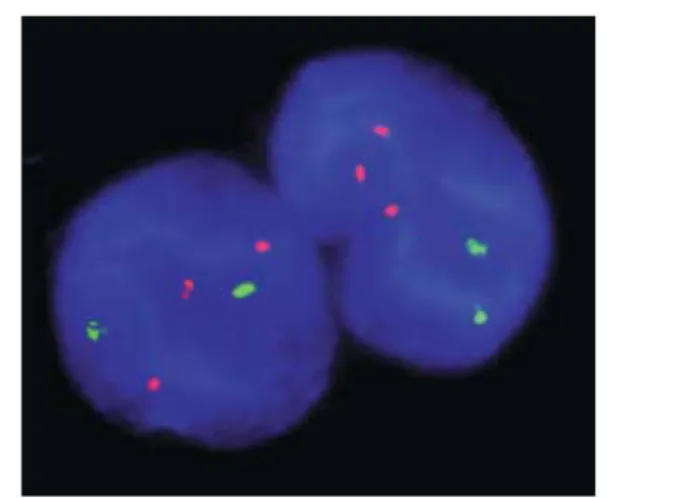
Figure 2: Fluorescent in situ hybridization (FISH) of interphase nuclei of a trisomy 21 fetus. In red the trisomic chromosome 21.
More recent analyses using molecular tools (Fig. 2) have confirmed that in DS, 95% of all cases are caused by full trisomy of chromosome 21, whereby all HSA21 genes are present in 3 copies, resulting from a chromosomal non-disjunction. One cell has two HSA21 instead of one, so the resulting fertilized egg has three HSA21 chromosomes. If a sperm or egg with an abnormal number of chromosomes merges with a normal mate, the resulting fertilized egg will have an abnormal number of chromosomes. Hence the scientific name, trisomy 21. Recent research has shown that in these cases, approximately 90% of the abnormal cells are eggs (Antonarakis 1996). The cause of the non-disjunction error remains still unknown. Research is currently aimed at trying to determine the cause and timing of the non-disjunction events.
Robertsonian Translocation
Three to four percent of all cases of trisomy 21 are due to Robertsonian Translocation. In this case, two breaks occur in separate chromosomes, usually the 14th and 21st chromosomes. There is a rearrangement of the genetic material so that some of the 14th chromosome is replaced by extra 21st chromosome. So while the number of chromosomes remains normal, there is a triplication of the 21st chromosome material. Translocations resulting in trisomy 21 may be inherited.
Mosaicism
The remainder of cases of trisomy 21 is due to mosaicism. In genetics, a mosaic or mosaicism denotes the presence of two or more populations of cells with different genotypes in one individual who has developed from a single fertilized egg. Mosaicism may result from a mutation during development which is propagated to only a subset of the adult cells.
2.1.4 Somatic traits and clinical features
Children with DS exhibit chatacteristic and peculiar physical traits:
• Short stature : children with DS usually have slow growth rate, and in
• Low muscle tone : children may have less muscle strength than other
children of the same age;
• Short neck, thick with fat and excess skin : usually this feature becomes
less obvious as the children grow;
• Short and stocky limbs, some children may have a wider space between
the thumb and second finger of the foot;
• One fold in the central part of the palm : it is called the simian line.
Facial features. They show typical facial features which include, for example, ears with modified form, usually small and with a low placement (Fig. 3 E). They present, also, abnormal mouth and tongue, with mouth often open (Fig. 3 F). It is typical to find children with exfoliative glossitis and tongue with scrotal appearance (in adolescents and adults). Furthermore, it is frequrent pseudomacroglossia (Fig. 3 F). It is very characteristic a flattened nasal bridge and this flat nose portion, located between the two eyes (nasal bridge), is frequently clogged (Fig. 3A). There are, often on their faces Brushfield’s spots. They are colored spots on the iris, without affecting the sight. Moreover, malformation of the teeth are very frequent: baby teeth may grow later and in an unusual way with, often, agenesis of lateral incisors.
Skeleton and skin features. Besides the short arms, they exhibit a finger clinodactyly of the fifth finger with a single flexion crease, flat foot and increased space between first toe and second toe.Tipically their skin is dried with xerosis. There are recurent cases, also, of hyperkeratotic lesions, alopecia, vitiligo, foliculitis and skin infections.
Psychomotor retardation. Hypotonia occurs soon at birth.
Congenital abnormalities associated with DS. Although DS is usually linked to intellectual disability, the morbidity from other associated congenital abnormalities is considerable. Many infants with DS have co-occurring congenital malformations (Fig. 4) requiring intensive surgical and medical management. To anticipate the care needed by these infants, providers and parents require accurate
information about birth defects that may be present. With this purpose in mind, many articles use different datasets to identify the rate at which structural birth defects are identified among children with DS. Overall, about one in five live born children with DS dies before the age of 5 years, and about two of five survivors have major health problems in addition to mental retardation in early childhood (Noble 1998).
Congenital heart diseases were the most frequently documented birth defects in live born infants with DS and approximately 46% of infants with DS were born with one or more heart defects. The three most common types of congenital heart defects were atrioventricular canal defects (45%), ventricular septal defects (35%) and patent ductus ateriousus (Roizen and Patterson 2003). The atrioventricular canal defect is the most serious of these defects. In the normal formation of the heart the endocardial cushions grow toward each other and leave openings between the atria and the ventricles, where the mitral and tricuspid valves form. The atrioventricular canal defect is caused by a failure of this process and results in the formation of a single valve structure with a septal defect above and below it. Survival can be improved with surgery, but the prognosis in children with these abnormalities is still poor because of the increased risk of heart failure. The ventricular septal defect, a defect in the ventricular septum, which divides the left and the right ventricles, may require surgery, though about 15% close spontaneously. The patent ductus is a congenital defect wherein a neonate's ductus arteriousus, the shunt connecting the pulmonary artery to the aortic arch, fail to close after birth. It is not a serious abnormality and can be corrected by a relatively simple surgical procedure. Assessment of all newborns with DS with an echocardiogram is the standard recommendation. Symptoms of serious heart disease may be absent or hidden due to the tendency of children with DS to develop pulmonary vascular resistance. Adolescents and young adults with no known intracardiac disease can develop mitral valve prolapse (46%) and aortic regurgitation (17%), and most experts recommend assessment also of adult patients.
After the co-occurring heart abnormalities, the second most common malformations in children with DS are those of the gastrointestinal tract (11%) (Fig. 4) like duodenal atresia, Hirschsprung's disease and tracheo-oesphaegal fistula (Noble 1998). Duodenal atresia is the congenital absence or complete closure of a portion of the lumen of the duodenum. Approximately 8% of all infants with DS show this disease and it’s 300 times more common in individuals
Figure 3: Down syndrome main features. A) Typical face, B) eye of a DS
subject. Note the oblique eye fissures and the epicanthic eye-folds , C)-D) hand (C) and feet (D). Note the shortened, incurved fifth fingers. E) small ear, F) protruding tongue. 2 se cr B C D E F
affected than in normal newborns. The Hirschsprung's disease or aganglionic megacolon, involves an enlargement of the colon by bowel obstruction resulting from absence of the enteric innervation in this section of bowel.
Other congenital abnormalities have been reported in association with DS: malformations in the genitourinary system (6%), including stenosis or atresia of the urinary tract at any location, malformations of the abdominal wall, limb defects (9%) (reduction of both the lower and upper extremities) and congenital cataract (1%) (Noble 1998).
2.1.5 Behavioural defects in DS
Mental retardation remains the invariable hallmark of DS and its more
Figure 4: Variable traits and clinical features in Down syndrome.
invalidating pathological aspect with a hard impact in the public health. In DS patients, the average IQ score is around 50, with individual values ranging from 30 to 70. (Chapman and Hesketh 2000; Vicari 2006). The early infancy is characterized by a delayed cognitive development, leading to mild-to-moderate mental retardation. The IQ declines from early in the first year to late childhood (Brown, Greer et al. 1990; Hodapp, Dykens et al. 1990) and the decline in cognitive performance that occurs in the adult DS patients has been considered as the consequence of accelerated aging (Devenny, Silverman et al. 1996; Lott and Head 2005). Mental retardation in DS is characterized by major neurological dysfunctions in the short- and long-term memory (Carlesimo, Marotta et al. 1997; Nadel 2003). It has been also demonstrated a decline with age on measures of long-term memory, providing behavioral evidence of hippocampal dysfunction by adolescence (Carlesimo, Marotta et al. 1997), consistent with the structural magnetic resonance imaging (MRI) finding of reduced hippocampal volumes in adolescents with DS (Jernigan, Bellugi et al. 1993; Pennington, Moon et al. 2003). There is also evidence for impairment of prefrontal cortex and cerebellar function (Nadel 2003) and speech and articulation are also particularly affected. The lower performances of DS in linguistic tasks may be partially explained in terms of impairment of the frontocerebellar structures involved in articulation and verbal working memory (Fabbro, Libera et al. 2002). Moreover, learning can be complicated by avoidance strategies when faced with cognitive challenges (Epstein 1995). Psychometric examinations of DS patients have shown that not all skills are affected in all persons or to the same extent. (Crnic 2002; Krinsky-McHale, Devenny et al. 2002; Brown 2003; Clark and Wilson 2003; Pennington, Moon et al. 2003; Vicari 2006). Although all domains of development follow the
usual sequence, a deficiency in language production relative to other areas of development often causes substantial impairment (Chapman, Seung et al. 1998). Children with DS have more behavioral and psychiatric problems than in other children, but fewer than in other individuals with mental retardation. In adult age, DS patients can have a similar prevalence of psychiatric problems to other people with intellectual disability. A raised frequency of psychiatric problems is also
related to the increased prevalence of depression in people with DS. However, they seem protected from some psychiatric disorders such as personality disorder, schizophrenia and anxiety. (Collacott, Cooper et al. 1998). Elsewhere, DS children show continuous but gradual improvement in mental age throughout childhood; intelligence quotients generally decline from early in the first year to late childhood (Hodapp, Dykens et al. 1990). Consequently, early implementation of special education programs results in improved cognitiveabilities in DS individuals. (Chapman, Seung et al. 1998).
By age 40 years, there is a ubiquitous occurrence of plaques and tangles suggestive of Alzheimer disease (AD), with an increase in dementia. The earliest manifestations of dementia in DS appear to reflect frontal lobe dysfunction with changes in sociability, emotional-based language, and depressive symptoms. However, physical–chemical dating of amyloid has suggested that it is first deposited in the frontal and entorhinal cortices. The amyloid burden in DS is, in part, related to increases in the expression of the amyloid precursor protein gene, but other factors are likely involved. Oxidative stress secondary to critical-regionmutations in mitochondrial DNA is associated with an increased brain concentration of oxidized β-amyloid.
2.1.6 Neuroanatomy of DS
The DS brain is characterized by numerous structural alterations that start to be present from early developmental stages and are retained in adulthood. Reduced thickness of cortical layers, diffuse hypocellularity, and astrocytic hypertrophy are a characteristic feature of the DS brain (Bartesaghi, Guidi et al. 2011). Autopsy data on the neuroanatomy of DS have been complemented by increasing studies from in vivo structural imaging using computer assisted tomography, voxel-based morphometry and to a larger extent MRI. The brain of DS subjects is characterized by several postmortem macroscopic features that are related to pre- and post-natal abnormalities in synaptogenesis leading to retardation of brain growth (Schmidt-Sidor, Wisniewski et al. 1990), such as lower brain weight and
brachycephaly, with a small cerebellum, frontal and temporal lobes reduced number and depth of the cerebral sulci, and a narrow superior temporal gyrus. (Coyle, Oster-Granite et al. 1986; Schmidt-Sidor, Wisniewski et al. 1990; Becker, Mito et al. 1991). MRI studies show regional brain abnormalities in young and non-demented adult DS subjects, consistent with postmortem data. Overall, brain volume is reduced in DS subjects, including cerebellar and cerebral gray and white matter. (Schapiro, Haxby et al. 1992; Pinter, Eliez et al. 2001). In particular, the reduced cerebellum shows a decreased volume of lobules VI–VIII (Avraham, Sugarman et al. 1991; Jernigan, Bellugi et al. 1993; Raz, Torres et al. 1995). In the temporal neocortex of fetuses with DS the emergence of lamination is both delayed and disorganized (Golden and Hyman 1994), total neuron number is reduced in the hippocampal dentate gyrus (DG), hippocampus and parahippocampal gyrus (Guidi, Bonasoni et al. 2008) and the cellular layers of the cerebellum are characterized by prominent hypocellularity and reduced thickness (Guidi, Ciani et al.).
In contrast, an increased volume is found in other brain areas, such as ventricles (Kesslak, Nagata et al. 1994; Pearlson, Breiter et al. 1998¸ Ikeda, 2002 #630; Schimmel, Hammerman et al. 2006) parahippocampal gyrus after adjustment for overall brain volume (Kesslak, Nagata et al. 1994; Raz, Torres et al. 1995; Teipel, Schapiro et al. 2003; Teipel and Hampel 2006) temporal, parietal and posterior cortex, lenticular nucleus, thalamus and hypothalamus (Jernigan, Bellugi et al. 1993; Pinter, Eliez et al. 2001), while the occipital lobe and superior temporal gyrus do not show volume changes after adjustment for overall brain volume (Frangou, Aylward et al. 1997; Pinter, Eliez et al. 2001). The second phase of cortical development and the emergence of lamination are delayed and disorganized in fetal DS brain. In addition to the known effects of the hippocampal formation in spatial memory, the altered cortical layer and cerebellum also may participate to cognitive and behavioral phenotypes in DS (Funahashi, Takeda et al. 2004).
2.1.7 Causes of brain hypotrophy in DS
The widespread hypoplasia of the DS brain is considered to be the main cause of mental retardation. Several lines of evidence show that two major determinants underlie brain hypotrophy in DS are neurogenesis reduction and dendritic hypotrophy (Coyle, Oster-Granite et al. 1986; Becker, Mito et al.
1991). In addition, an increase in cell death at more advanced life stages contributes to further reduce neuron number.
Neurogenesis alterations
The fact that brain and cerebellar volume reduction and cortical hypocellularity are already present in children and fetuses with DS (Sylvester 1983; Schmidt-Sidor, Wisniewski et al. 1990; Golden and Hyman 1994; Winter, Ostrovsky et al. 2000; Pinter, Eliez et al. 2001) strongly suggests that defective neuronogenesis during critical phases of brain development may be a major determinant of microencephaly.
Due to the obvious difficulties in obtained fetal material, very little information is available concerning neurogenesis in the fetal DS brain. Our group recently obtained evidence that in fetuses with DS cell proliferation is severely impaired in the DG (most likely due to alteration of the cell cycle), in the germinal matrix of the inferior horn of the lateral ventricle and in the germinal zones of the hippocampus proper and parahippocampal gyrus (Contestabile, Fila et al. 2007; Guidi, Bonasoni et al. 2008). Quantification of the number of mature neurons and astrocytes in the hippocampus and parahippocampal gyrus showed that in all these regions fetuses with DS had proportionally fewer neurons and a larger number of astrocytes compared with normal fetuses (Guidi, Bonasoni et al. 2008). In trisomic fetuses, our group found a defective neurogenesis in the EGL of the cerebellum, and in a region of the fifth lobe that is the remnant of the cerebellar VZ (Guidi, Ciani et al.). This evidence clearly shows proliferation impairment in numerous regions of the fetal DS brain. Importantly, these defects are shared by trisomic mice (see below) which validates the use of mouse models for DS and renders evidence obtained in mouse model transferable to the human condition.
Dendritic hypotrophy
Dendritic pathology is a typical feature of the DS brain and DS neurons exhibit patent alterations in dendriitic pattern and spine density and shape. In the visual cortex of fetuses with DS neuronal morphology is similar to that of control fetuses (Takashima, Becker et al. 1981). Likewise, the dendrites of layer IIIc pyramidal neurons of the prefrontal cortex in 2.5-month-old infants with DS is similar to that of normal cases (Vuksic, Petanjek et al. 2002). Dendritic abnormalities, however, begin to appear at later ages. A study in a 3 month-old infant with DS shows that interneurons in the motor cortex have a lower dendritic area (Prinz, Prinz et al. 1997). The pyramidal neurons of the visual cortex of newborns older than 4 months and older infants with DS have shorter basilar dendrites (Takashima, Becker et al. 1981). Dendritic hypotrophy is also present in pyramidal neurons of the parietal cortex of children with DS (Schulz and Scholz 1992). The dendritic hypotrophy seen in childhood continues into adulthood, with a marked decrease in dendritic branching and dendritic length in elderly adults with DS (Takashima, Ieshima et al. 1989). In the visual cortex of children with DS, the total dendritic length is above normal in the infantile period (6 months old or less) but drops steadily to below normal in juvenile subjects (older than 2 years) (Becker, Armstrong et al. 1986). This reduction contrasts with expanding dendritic arborization in normal children. These data indicate that in individuals with DS the dendritic tree atrophies in early childhood without a recovery at subsequent life stages.
While in the visual cortex of fetuses with DS spine counts (basilar dendrites) are similar to those of control fetuses, newborns and older infants with DS have a decreased number of spines and spines exhibit an altered morphology (Takashima, Becker et al. 1981). In normal subjects, spine density on the basal dendrites of cortical pyramidal neurons increases from neonate to 15 years of age and gradually decrease after 20 years. In contrast, spine density poorly increases in children and rapidly decreases in adults with DS (Suetsugu and Mehraein 1980; Takashima, Ieshima et al. 1989; Takashima, Iida et al. 1994). A reduced spine density has been found in the apical dendrites of pyramidal neurons of the
hippocampus and cingulate cortex and in both the apical and basilar dendritic arbors of CA1 and CA2-3 pyramidal neurons in patients with DS when compared to age-matched control (Suetsugu and Mehraein 1980; Takashima, Ieshima et al. 1989; Takashima, Iida et al. 1994). An additional decrease in spine density occurs in DS patients with associated AD, when compared to age-matched controls, and DS with no AD (Takashima, Ieshima et al. 1989; Ferrer and Gullotta 1990). The dendritic spines of the DS brain exhibit, in addition to reduced density, also aberrant morphology. Studies of infants with DS demonstrated that spines were small, had short stalks and were intermingled with unusually long spines (Marin-Padilla 1976; Purpura 1979). Considering that drebrin expression is reduced in DS individuals (Shim and Lubec 2002), a reduced excitatory-inhibitory ratio is expected in DS individuals, which may lead to neuron hypoexcitation.
Biochemical alterations
Biochemical alterations also occur in fetal DS brain, which could serve as substrates for the morphological changes (Engidawork and Lubec 2003). In addition to the cholinergic system, that undergoes alterations at advanced life stages, the glutamatergic, serotonergic, noradrenergic and GABAergic systems are profoundly altered in DS individuals and in trisomic mice (Wisniewski and Bobinski 1991; Risser, Lubec et al. 1997). Alterations involve neurotransmitter levels as well as defects of receptor expression/function.
Although young children with DS appear to be born with a normal septohippocampal cholinergic system (Kish, Karlinsky et al. 1989), an aging-dependent neurodegeneration of the basal forebrain cholinergic neurons (BFCNs) was observed (Yates, Simpson et al. 1983; Casanova, Walker et al. 1985). Because BFCNs provide the major cholinergic input to the hippocampus and neocortex, the degeneration of these neurons may have functional consequences at level of cholinergic receptors. These dysfunctions could produce additional learning and memory deficits in older individuals with DS (Yates, Simpson et al. 1983) and could be an outgrowth of the Alzheimer disease in these patients. In addition, an early onset of an Alzheimer disease-like neurohistopathology is
systematically observed by the fourth decade (Dalton and Crapper-McLachlan 1986).
2.1.8 The Human Chromosome 21, genotype–phenotype
correlations
Chromosome 21 (HSA21) is the smallest human autosome that represents around 1±1.5% of the human genome. Different hypotheses have been presented to account for the relationship between trisomy 21 and the occurrence of DS phenotypes. The effects of trisomy on brain phenotypes have been explained by two hypotheses: 1) the “gene-dosage effect” hypothesis claims that DS phenotypes are determined by increased dosage of a subset of dosage-sensitive genes, and of their encoded proteins, especially during development (Delabar, Theophile et al. 1993; Korenberg, Chen et al. 1994; Ait Yahya-Graison, Aubert et al. 2007; Lyle, Bena et al. 2009), and 2) the “amplified developmental instability” hypothesis holds that Hsa21 trisomy determines a general alteration in developmental homeostasis (Roizen and Patterson 2003; Antonarakis, Lyle et al. 2004). The gene-dosage hypothesis proposes that the 50% increase in expression at the RNA level of trisomic genes is the initiating cause of the DS phenotype. Recent experiments using microarrays and quantitative RT-PCR of human DS and mouse model samples indicate that the majority of trisomic genes in the majority of tissues indeed show increased expression, although not always by precisely 50% (Mao, Zielke et al. 2003; Amano, Sago et al. 2004; Kahlem, Sultan et al. 2004; Lyle, Gehrig et al. 2004; Dauphinot, Lyle et al. 2005). The triplicated genes could modulate directly or indirectly the expression of target genes on both the Hsa21 and the other chromosomes, determining a secondary gene dosage effect. Thus, the gene expression alterations in the brain, resulting from the different genetic mechanisms in response to the gene over-dosage in DS, may induce primary phenotypes at cellular level consisting in alterations in cellular processes, such as proliferation, differentiation, synaptogenesis, dendritogenesis and apoptosis in neuronal and glial cells. Even though several genes have been identified that are deregulated in DS brains, the challenge in DS research is to
establish a correlation between the functions of triplicated genes and features of the cognitive and behavioral phenotypes seen in DS.
2.2. MOUSE MODELS FOR DS
Although studies in DS cases with partial trisomy have helped to suggest chromosomal regions and HSA21 genes that may contribute to various phenotypes, the ability to resolve the basis of neurological and other phenotypes is limited. The use of animal models, particularly the mouse, is an invaluable tool for understanding underlying genetic and molecular mechanisms of DS. The advantages are: (1) the ability to study a large number of animals in a short time; (2) the availability of mice engineered to contain a specific gene(s); (3) the ability to control for genetic background; (4) the low cost of screening; (5) the ability to carry out studies on both developing and mature subjects; (6) the ability to design and execute a variety of therapeutic interventions; and (7) the lack of postmortem delay. The number and variety of these models is growing (Fig 5). At the genomic level, the long arm of the human chromosome 21 (Hsa21) is approximately 33.7 Mb in length and contains about 430 protein-coding genes of which 293 have a homolog in the mouse genome. These genes are found in syntenic regions localized on three different mouse chromosomes. From the centromeric to the telomeric end of the Hsa21, the first and largest region is found on Mmu16 (about 37 Mb in length with 224 orthologous genes), followed by a smaller region on Mmu17 (1.1 Mb in length with 22 orthologous genes) and finally a region on Mmu10 (2.3 Mb in length with 47 orthologous genes). In these three syntenic regions, relative order and orientation of the genes are preserved between the two species. With the introduction of new genetic engineering methods, it will be possible to produce additional models in which triplication of smaller genetic segment is readily accomplished.
2.2.1 Mice Trisomic for the entire MMU16
Ts16 Mice trisomic for the entire 16th chromosome (Ts16) were generated by spontaneous Robertsonian translocation. Unfortunately, these mice die in utero, thus limiting research during the postnatal period. Death probably occurs as the result of cardiovascular abnormalities and insufficient placenta function (Miyabara, Gropp et al. 1982). A number of studies focused on nervous system abnormalities in Ts16 fetuses showed similarities with DS. These changes ranged from delayed development to degenerative processes. There is delayed cortical development in Ts16 mice. The cross-sectional area of cortical layers appears normal at E13. However, at E16, the cortical thickness is significantly (12–48%) smaller when compared with that of disomic mice (Haydar, Blue et al. 1996). Interestingly, 2 days later (E18) no such a difference could be detected. In addition to delayed development, a degenerative process has been found in these mice. Sweeney et collaborators (Sweeney, Hohmann et al. 1989) quantified the number of acetylcholinesterase (AChE)-positive neurons in the basal forebrain of Ts16 mice and found a significant (40%) loss when compared with controls. In a recent study, Dorsey et al.(Dorsey, Bambrick et al. 2002) studied brain derived neurotrophic factor (BDNF) signaling in Ts16 cultured hippocampal neurons and found evidence supporting failed tyrosine receptor kinase (TrkB)-mediated BDNF signaling in these cells. Accordingly, TrkB phosphorylation was significantly (33%) reduced in response to BDNF in Ts16 mouse hippocampal neurons. Moreover, trisomic cells were characterized with increased levels of a truncated, kinase-deficient isoform (T1) of the TrkB (TrkB T1). Interestingly, disruption of a gene encoding this protein restored the survival of cultured cortical and hippocampal Ts16 neurons. Although Ts16 mice have been extremely helpful in directing our attention to the probability that mouse model scan be used to study DS, this model suffers from severe practical and theoretical limitations. Because they do not survive birth, we are largely prevented from studying age-dependent degenerative effects. Furthermore, in addition to carrying orthologues of HSA21, MMU16 harbors orthologues of genes on humanchromosomes 3, 16, and 22.
2.2.2 Segmental MMU16 trisomic mice
HS A 2 1 M M U 1 6 Ts 16 Ts 65 Dn Ts Cj e1 Ms 1T s6 5 Ts 1Rh r S T CH M R P L 39 AP P G RIK1 S O D1 O L IG 2 O L IG 1 DS CR1 CBR1 DY RK1 A BACE2 ORF 9 Z NF 29 5 Ts 1Rh r/ Ts 65 Dn Ts 2Cj e Tc 1 Tc 1Y u DSC R M M U 1 0 M ou se sy ntenicregions y/+ )1Ye ;Dp(17 Yey/+ 6)1 ;Dp(1 y/+ )1Ye Dp(10
HS A 2 1 M M U 1 6 Ts 16 Ts 65 Dn Ts Cj e1 Ms 1T s6 5 Ts 1Rh r S T CH M R P L 39 AP P G RIK1 S O D1 O L IG 2 O L IG 1 DS CR1 CBR1 DY RK1 A BACE2 ORF 9 Z NF 29 5 Ts 1Rh r/ Ts 65 Dn Ts 2Cj e Tc 1 Tc 1Y u DSC R M M U 1 0 M ou se sy ntenic
regions y/+ )1Ye ;Dp(17 Yey/+ 6)1 ;Dp(1 y/+ )1Ye Dp(10
Figure 5: Mouse models of Down syndrome. Schematic representation of Hsa21
and corresponding regions of Mmu10 (hatched region), Mmu16 (gray region) and Mmu17 (black regions). Different mouse models that carry different triplication of different sets of genes orthologous to those of Hsa21 are indicated.
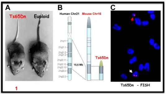
Ts65Dn Davisson et collaborators (Davisson, Schmidt et al. 1990), using irradiation, induced reciprocal translocation of T(16C3-4;17A2) and generated a mouse segmentally trisomic for MMU16 entitled Ts65Dn (Fig 6). The triplicated region of MMU16 in Ts65Dn extends from Znf295 to Mrp139 and contains at least 132 genes (Fig. 6). Ts65Dn mice recapitulate a variety of DS structural and functional changes. Similar to DS, basal forebrain cholinergic neurons (BFCNs) undergo age dependent degeneration in these mice (Cooper, Messer et al. 1999; Hunter, Bimonte et al. 2003; Salehi, Delcroix et al. 2004; Seo and Isacson 2005). The degeneration was linked to a marked decrease in the retrograde transport of nerve growth factor (NGF), a keytrophic factor for these neurons. By delivering NGF via intracerebro ventricular injection of NGF, and thus bypassing the retrograde transport, degenerative changes were rapidly reversed, even in very old Ts65Dn mice (Cooper, Messer et al. 1999). BFCNs project extensively to the hippocampus and cortex (Salehi, Delcroix et al. 2003). In a series of careful studies, marked synaptic structural abnormities were detected in these and other brain regions in Ts65Dn mice (Belichenko, Masliah et al. 2004; Kleschevnikov, Belichenko et al. 2004). Kurt and collaborators (Kurt, Kafa et al. 2004) studied neuronal density in the CA1 area of the hippocampus in old Ts65Dn mice and found a significant reduction in cell density. No such a difference was found at 3 months of age (Lorenzi and Reeves 2006). Furthermore, a significant reduction in the area and number of dendritic branches, spine density and in the layer III pyramidal cells in Ts65Dn mice has been reported (Kurt, Kafa et al. 2004). Alterations in spine size and density in Ts65Dn hippocampus were associated with failure to induce long-term potentiation (LTP) in the dentate gyrus and CA1 area (Kleschevnikov, Belichenko et al. 2004; Costa and Grybko 2005). In behavioral analyses, Ts65Dn mice showed significant spatial and non spatial learning disabilities, as shown by the hidden platform and probe tests in the Morris water maze (MWM) (Sago, Carlson et al. 2000; Hyde, Crnic et al. 2001; Hyde, Frisone et al. 2001). Ts65Dn mice are the most used model for DS.
Ts2Cje Since male Ts65Dn are infertile, only females can be used for breeding. Furthermore, the occurrence in progenyis ~20–25%. The Epstein Laboratory discovered a new mouse model called Ts2Cje that was generated by spontaneous Robertsonian fusion in which the triplicated segment in Ts65Dn was fused to mouse chromosome12 (Villar, Belichenko et al. 2005). Although the litter size in Ts2Cje mice is similar to those of Ts65Dn, the frequency of progeny bearing Rb (12.1716) has been reported to be higher than Ts65Dn mice (~43%). Furthermore, both male and female Ts2Cje mice are fertile. In terms of central nervous system changes, there was a significant decrease in spine density on the dendrites of dentate granule cells together with enlarged dendritic spines (~38%) in Ts2Cje mice (Villar, Belichenko et al. 2005), very similar to the changes described in Ts65Dn mice (Belichenko, Masliah et al. 2004). The functional consequences of these changes in Ts2Cje mice are yet to be explored.
Ts1Cje Crossing balanced T(12:16)1Cje to wild type mice produced trisomic mice for a fragment of MMU16 extending from Sod1 to Znf295, but not including
Figure 6: Ts65Dn mouse model. A) Ts65Dn and control mouse. B)
Ts65Dn mouse model shows segmental trisomy for a distal region of
chromosome 16, a region that shows perfectly conserved linkage with human chromosome 21. C) Fluorescent in situ hybridization (FISH) in Ts65Dn mouse.
a functional copy of Sod1 (Sago, Carlson et al. 1998; Sago, Carlson et al. 2000). Ts1Cje mice harbor three copies of ~78% of genes triplicated in Ts65Dn mice (Olson, Roper et al. 2004). There are important similarities between Ts1Cje and Ts65Dn mice in regards to CNS structural changes as enlargement of dendritic spines, decreased density of spines, and selective reorganization of inhibitory afferents (Belichenko, Kleschevnikov et al. 2007). Unlike hippocampal changes, no evidence of shrinkage and loss of markers in BFCNs in old Ts1Cje mice have been shown (Salehi, Delcroix et al. 2006). In accordance with this finding, NGF transport in the septo-hippocampal pathway in Ts1Cje mice was significantly increased relative to that in the Ts65Dn mouse suggesting that one or more genes in the segment that distinguishes Ts65Dn and Ts1Cje mice contribute significantly for the dramatic reduction of NGF transport.
Failure in synaptic plasticity as measured by LTP have been reported in the both CA1 area (less severe than Ts65Dn) (Siarey, Villar et al. 2005) and dentate gyrus (Belichenko, Kleschevnikov et al. 2007) of hippocampus in Ts1Cje mice. The T maze spontaneous alternation task is abehavioral paradigm known to reveal dysfunction of the hippocampal system (Gerlai 2001; Lalonde 2002). Comparing Ts65Dn and Ts1Cje mice, both mice differed significantly from that of the corresponding 2N mice (Belichenko, Kleschevnikov et al. 2007). In the context of Morrison water maze, while Ts1Cje mice do show abnormalities in spatial learning and memory, they are less severe than in Ts65Dnmice (Sago, Carlson et al. 1998; Sago, Carlson et al. 2000).
Ms1Ts65Dn Ms1Ts65Dn mice are produced by crossing Ts65Dn mice with T(12:16)1Cje mice (Sago, Carlson et al. 1998; Sago, Carlson et al. 2000). They are segmentally trisomic for the genetic segment from Mrp139 to Sod1, but not including a functional copy of Sod1. Unlike Ts65Dn, which are found to be hyperactive, and Ts1Cje, which are hypoactive, Ms1Ts65Dn mice were found to be normally active. Sago et al. (Sago, Carlson et al. 2000) compared the performance of these mice with Ts65Dn and Ts1Cje mice in the Morrison water maze and found that Ms1Ts65Dn mice did significantly better. In the reverse
probe test (preference score), and unlike Ts65Dn mice, neither Ts1Cje nor MS1Ts65Dn mice showed a significant difference with 2N mice (Sago, Carlson et al. 2000). Ms1Ts65Dn mice have not yet been studied extensively for changes in the structure and function of circuits involved in cognition.
Ts1Rhr Comparing DS patients with segmental trisomy 21 has suggested that triplication of a 5.4 Mb region ofHSA21 called DS critical chromosomal region (DSCR) is associated with various DS phenotypes (Korenberg, Chen et al. 1994). In 2004, Olson et al. (Olson, Roper et al. 2004) reported generation of a new mouse called Ts1Rhr. These mice have a 3.9-Mb duplication of DSCR. This region contains mouse orthologues of the 33 genes in the human DSCR (i.e., around 32% of triplicated genes in Ts65Dn mice), with boundaries at Cbr1 andMx2 genes. Surprisingly, it was found that, unlike Ts65Dn, Ts1Rhr mice do not show a smaller skull or mandible. This indicates that the genes causing facial features of DS are not located inthe DSCR. Relatively a very few studies have focused on brain abnormalities in Ts1Rhr mice. Recently, Aldridge et al. (Aldridge, Reeves et al. 2007) studied brain volume and found a significant (~20%) reduction in the entire brain as well as the cerebellum.
Ts1Rhr/Ts65Dn Using a similar strategy as for Ts1Rhr, Olson et al. (Olson, Richtsmeier et al. 2004) deleted DSCR to produce a mouse trisomic for most of the genes trisomic in Ts65Dn (70%) but with only the two copies of DSCR (Ts1Rhr/Ts65Dn). No results of nervous system function have yet been reported. Macroscopic analysis of Ts1Rhr/Ts65Dn mice showed ~18% reduction in the brain volume and abnormalities in around 80% of linear brain distances (Aldridge, Reeves et al. 2007) indicating a significant changes in the brain shape. Studies on craniofacial features showed that with the exception of brachycephaly, Ts65Dn and Ts1Rhr/Ts65Dn mice have similar craniofacial measurements. This suggests that DSCR is not sufficient to produce the phenotypes present in Ts65Dn. These studies are important because they chart the direction that future
studies will take in examining the contribution of gene segments and individual genes.
Tc1Yu Since Ts65Dn mice have three copies of a segment of MMU16 as well as a small portion of MMU17, Li and collegues (Li, Yu et al. 2007) recently reported generation of another DS mouse model with triplication of a larger segment of MMU16 without any contribution from MMU17. Using Cre/loxP-mediated chromosomal engineering, the Yu laboratory generated stem cells with duplication of MMU16 between D930038D03Rik and Zfp295genes (~22.9 Mb). Only 38% of Tc1Yu mice survive birth and no gross abnormality was detected through at least the age of 10 months. Around 37% of Tc1Yu mice suffer from cardiovascular abnormalities as early as E18.5. Furthermore, gastrointestinal abnormalities e.g., annular pancreas have also been reported. No studies on CNS related phenotypes have yet been reported.
Dp(10)1Yey/+;Dp(16)1Yey/+;Dp(17)1Yey/+ Recently a new mouse DS model that carries duplications spanning the entire Hsa21 syntenic regions on all Mmu10, Mmu16 and Mmu17 mouse chromosomes has been created (Dp(10)1Yey/+;Dp(16)1Yey/+;Dp(17)1Yey/+). This mouse mutant exhibits DS-related neurological defects, including impaired learning/memory and decreased hippocampal LTP (Yu, Li et al. 2010), very similarly to the Ts65Dn mouse. These results suggest that the critical genes associated with the DS brain phenotypes may reside within the Mrpl39–Zfp295 genomic segment of the Mmu16 and support the use of the Ts65Dn mouse as the best genetic murine DS model.
2.2.3 Mice trisomic for HSA21
Tc1 An additional category of mouse model for trisomy 21 resulted from the insertion of all or part of Hsa21. The Tc1 trans-species mouse strain, carrying an almost entire Hsa21, was developed using irradiation microcell-mediated chromosome transfer (O'Doherty, Ruf et al. 2005). This model exhibits several key phenotypes of DS (O'Doherty, Ruf et al. 2005; Morice, Andreae et al. 2008;
Galante, Jani et al. 2009). The most obvious CNS morphological changes found in Tc1 mice was a reduction in the density of cerebellar granule neurons in old Tc1 mice. Abnormalities in hippocampal dependent learning and memory as well as decreased LTP have been detected in physiological studies in the dentate gyrus of the hippocampus. Using a novel-object recognition task, Tc1 mice spent significantly shorter times exploring novel objects. Interestingly, no difference was found between Tc1 mice and their control littermates in the spontaneous alternation T-maze; another test of hippocampal-related cognition (O'Doherty, Ruf et al. 2005). One short coming of this model is a high degree of mosaicism within tissues. For instance, only approximatelyone-third of brain cells was found tocarry HSA21. Examining brain phenotypes will require careful attention to which cells maintain HSA21 and which do not. Furthermore, effective transmission of HSA21 is possible only by out crossing to achieve a mixed strain background (O'Doherty, Ruf et al. 2005). Thesefindings are evidence for the propensity of mouse cells to eliminate HSA21. Finally, the consequences of expression of a large number of human proteins in mouse micro-environment must be taken into account in interpreting the phenotypes detected.
2.2.4
Transgenic mice with triplication of individual genes
Several transgenic mice that express from one or a few genes to entire segments of HSA21 (YAC transgenic mice) have been produced. As expected, none of these recapitulates the entire phenotype of DS, but many have features that are reminiscent of aspects of the phenotype. Transgenic mice containing the human Cu-Zn Superoxide dismutase 1 (SOD1) gene were the first mice produced to contain a gene encoded on HSA21 (Epstein, Avraham et al. 1987). These mice have some features that are similar to those seen in people with DS (Avraham, Sugarman et al. 1991; Gahtan, Auerbach et al. 1998). Many investigators have produced transgenic mice that express normal and mutant forms of beta-amyloid peptide (APP), and have demonstrated learning and memory deficits in these mice, including performance decline with age. In a recent experiment, compound transgenic mice that express human SOD1 and APP were created (Harris-Cerruti,
Kamsler et al. 2004). Working memory and long-term memory are severely impaired in these double transgenic mice; they have defects in APP processing, lipofuscin accumulation and mitochondrial anomalies. These findings are intriguing in light of the increasing evidence for a link between mitochondrial dysfunction, oxidative stress, APP processing and DS (Busciglio, Pelsman et al. 2002). Other genes that cause phenotypes that are relevant to DS when overexpressed in transgenic mice include PFKL (Peled-Kamar 1998), the Drosophila minibrain homolog gene (DYRK1A) (Dierssen, Ortiz-Abalia et al. 2006), the neurotrophic factor S100 β (Krapfenbauer, Engidawork et al. 2003), the transcription factor ETS2 (Wolvetang, Wilson et al. 2003) and the transcription factor single-minded (SIM2) (Chrast, Scott et al. 2000). Sim2 is overexpressed in Ts1Cje mouse fetuses (Vialard, Toyama et al. 2000) and in trisomic tissues (Lyle, Gehrig et al. 2004).Variations of SIM2 expression level were found in the cerebellum, cortical layers, and hippocampus (Rachidi, Lopes et al. 2005), key human brain regions involved in learning and memory that also are altered in DS patients (Raz, Torres et al. 1995; Pinter, Eliez et al. 2001; Rachidi, Lopes et al. 2005). The transgenic mice overexpressing Sim2 (Tg Sim2) display reduced sensitivity to pain and mild impairment of learning (Ema, Ikegami et al. 1999; Chrast, Scott et al. 2000). These behavioral anomalies found in the Sim2 transgenic mice remembered some phenotypes observed in trisomic mouse models for DS, Ts65Dn and Ts1Cje (Coussons-Read and Crnic 1996; Sago, Carlson et al. 1998; Martinez-Cue, Baamonde et al. 1999) and in DS patients (Hennequin, Allison et al. 2005).
Two transgenic mouse models overexpressing DYRK1A have been generated. The first one, carried a human YAC 152F7, containing DYRK1A (Smith, Stevens et al. 1997); the second, carried the full-length DYRK1A cDNA (Altafaj, Dierssen et al. 2001). In the Morris water test, the transgenic lines carrying the YAC 152F7 showed lower performance in the probe test, in which the platform is removed. In the reverse learning paradigm, the transgenic mice showed the most severe deficits with no significant learning of the new platform position, indicating deficits in learning flexibility. Mouse line carrying a 152F7 YAC
fragment (152F7tel) containing only the DYRK1A gene, showed the same phenotype than the original YAC lines, indicating that the correct dosage of DYRK1A gene is crucial for brain function, learning and memory (Smith, Stevens et al. 1997). The two models show a significant impairment in spatial learning and memory, indicating hippocampal and prefrontal cortex function alteration. These transgenic mice showed increased brain weight and neuronal size (Branchi, Bichler et al. 2004) and dysfunction of reference memory (Smith, Stevens et al. 1997; Altafaj, Dierssen et al. 2001). They also exhibit neurodevelopmental defects, delayed craniocaudal maturation and motor dysfunction (Altafaj, Dierssen et al. 2001). All these alterations are comparable with those found in murine models of DS with trisomy of chromosome 16, and suggest a causative role of DYRK1A in mental retardation and motor anomalies in DS patients. Moreover, DYRK1A-deficient mice have defects in central nervous system development, and DYRK1A appears sensitive to gene dosage as heterozygous mutant mice show limited changes in central nervous system development (Fotaki, Dierssen et al. 2002). The YAC 230E8 mouse lines showed altered density of neurons in cerebral cortex (Rachidi, Lopes et al. 2005). This abnormal density of neurons may explain the learning and memory deficits of these animals in the Morris water test (Smith, Stevens et al. 1997).The YAC 230E8 contains the DOPEY2 gene, a new member of the Dopey family involved in morphogenesis that has been suggested as a candidate gene for the neurological alterations and mental retardation observed in these mice (Rachidi, Lopes et al. 2005). Ideally, transgenic mouse models of DS would contain a single extra copy of the relevant gene and regulatory elements that are similar enough to wild-type genes to assure typical spatio-temporal patterns of expression. These conditions are rarely achieved (Patterson and Costa 2005). Also, most transgenic mice contain human transgenes, and these might not be appropriately expressed in mice. Nonetheless, it is remarkable that these individual transgenes, and sometimes combinations of transgenes, have phenotypes that are reminiscent of DS. Construction of more transgenic mice seems well justified (Patterson and Costa 2005).
2.2.5 Segmental trisomic mice disomic for individual genes
Ts65Dn: App+/+/-. One critical question in understandingthe biology of DS is whether individual genes play a significant role in creating important phenotypes. To address this, Epstein et al. (Epstein, Berger et al. 1990) generated Ts65Dn mice with the normal two copies of specific genes (e.g., amyloidprecursor protein, App). This was achieved by breeding Ts65Dn female swith males in which one copy of the App gene was deleted. Ts65Dn: App+/+/- are viable and have no gross nervous system abnormalities with normal life span (Cataldo, Petanceska et al. 2003). Cataldo et al. (Cataldo, Petanceska et al. 2003) studied the endosomal system in Ts65Dn mice (with three copies of App) and found enlarged early endosomes in the basal forebrain as early as 2 months. Interestingly, these changes were not found in Ts65Dn mice withonly two copies of App. Significantly, they did not detect changes in endosomes in mice that are transgenic for mutant App. However, a recent report has documented an increase in early endosomes size and number (Laifenfeld, Patzek et al. 2007). Deleting one copy of App markedly improved NGF retrograde transport in Ts65Dn mice, reaching a level similar to that seen in Ts1Cje mice. Morphological analysis of BFCNs revealed that the increase in NGF retrograde transport was accompanied by a significant improvement in BFCN morphology in the medial septal nucleus (MSN). Indeed, this parameter did not distinguish the Ts65Dn: App+/+/- mice from their 2N controls (Salehi et al., 2006).
2.3 NEUROGENESIS ALTERATIONS IN DS ANIMAL
MODELS
During the last few years various groups have tackled the issue of neurogenesis impairment in the DS brain by taking advantage of various mouse models.
In Ts16 mice the number of precursors of the future somatosensory cortex just before neuronogenesis begins, is notably reduced. At each cell cycle during neuronogenesis, a smaller proportion of Ts16 progenitors exit the cell cycle and the cell cycle duration is longer in Ts16 than in euploid progenitors (Haydar,
Nowakowski et al. 2000). In the Ts65Dn mouse a reduced proliferation has been detected at all examined life stages. Reduced neural precursor proliferation is already present during embryonic development in the hippocampal region and VZ, with a reduction in the number of neocortical and hippocampal neurons (Chakrabarti, Galdzicki et al. 2007). The reduced neurogenesis appears to be due to elongation of the cell cycle. Interestingly, the neurogenesis defects of Ts65Dn mice lead to an imbalance between production of excitatory and inhibitory neurons In particular, Chakrabarty et al. found that the medial ganglionic eminence (MGE) (which gives origin to inhibitory neurons) of Ts65Dn mice at E14.5 undergoes divisions at a normal rate but gives a higher neuronal output due to a large progenitor population. The large output from the MGE explains the observation that in the neocortex and hippocampus of neonate and young Ts65Dn mice there are fewer excitatory but more inhibitory neurons (Chakrabarti, Best et al. 2010). The excessive production of inhibitory interneurons is due to over-expression of the triplicated genes Olig1 and Olig2, as normalization of their expression rescues the Ts65Dn phenotype. In Ts65Dn mice, the postnatal SVZ of the lateral ventricle, which is the largest neurogenic area of the adult brain, exhibits a remarkably reduced proliferation rate that starts in the perinatal period and continues up to senescence (Bianchi, Ciani et al. 2010; Bianchi, Ciani et al. 2010; Trazzi, Mitrugno et al. 2011). A severe neurogenesis reduction has been also documented in the embryonic neocortex and VZ and in the SVZ of adult Ts1Cje and Ts2Cje mice (Ishihara, Amano et al. 2010). In the adult Ts1Cje SVZ trisomy does not appear to affect the number of neural stem cells but results in reduced numbers of neural progenitors and neuroblasts (Hewitt, Ling et al. 2010). Analysis of differentiating Ts1Cje neural progenitors shows a severe reduction in number of produced neurons, whilst the number of astrocytes is increased (Hewitt, Ling et al. 2010).
A severe neurogenesis impairment characterizes the DG of the Ts65Dn mouse at all examined life stages (Insausti, Megias et al. 1998; Lorenzi and Reeves 2006; Contestabile, Fila et al. 2007; Bianchi, Ciani et al. 2010). The study of the phenotype acquired by differentiating neural progenitor shows a reduction in the