Claudio M. Pacella, MD
Giancarlo Bizzarri, MD
Fabrizio Magnolfi, MD
Piero Cecconi, MD
Bruno Caspani, MD
Vincenzo Anelli, MD
Antonio Bianchini, MD
Dario Valle, MD
Sara Pacella, MD
Guglielmo Manenti, MD
Zaccaria Rossi, MD
Index terms:Lasers, interstitial therapy, 761.1299 Liver, interventional procedures,
761.1299
Liver neoplasms, 761.323
Liver neoplasms, therapy, 761.1266, 761.1299, 761.323
Published online before print 10.1148/radiol.2213001501 Radiology 2001; 221:712–720 Abbreviations:
HCC⫽ hepatocellular carcinoma LTA⫽ laser thermal ablation PEI⫽ percutaneous ethanol injection
1From the Departments of Radiology and Diagnostic Imaging (C.M.P., G.B., V.A., A.B., D.V., S.P., G.M.) and Endo-crine, Metabolic, and Digestive Diseases (Z.R.), Regina Apostolorum Hospital, Via St Francesco 50, 00041 Albano Laziale, Rome, Italy; the Gastroenterology Unit, St Donato Hospital, Arezzo, Italy (F.M.); and the Diagnostic Radiology Service, St Anna Hospital, Como, Italy (P.C., B.C.). From the 2000 RSNA scientific assem-bly. Received September 6, 2000; sion requested October 20; final revi-sion received May 7, 2001; accepted June 5. Address correspondence to C.M.P. (e-mail: [email protected]). ©RSNA, 2001
Author contributions:
Guarantor of integrity of entire study, C.M.P.; study concepts, C.M.P., G.B., Z.R.; study design, C.M.P., G.B., P.C.; literature research, S.P., G.M.; clinical studies, F.M., Z.R.; data acquisition, A.B., S.P.; data analysis/interpretation, G.B., F.M., P.C., B.C., A.B.; statistical analysis, D.V., S.P., G.M., C.M.P., A.B.; manuscript preparation, C.M.P., S.P.; manuscript definition of intellectual content, all authors; manuscript editing, A.B., S.P.; manuscript revision/review, C.M.P., P.C., B.C., Z.R.; manuscript final version approval, all authors.
Laser Thermal Ablation in the
Treatment of Small
Hepatocellular Carcinoma:
Results in 74 Patients
1
PURPOSE: To evaluate the safety, local effectiveness, and long-term results of laser
thermal ablation (LTA) in the treatment of small hepatocellular carcinoma (HCC).
MATERIALS AND METHODS: Ninety-two biopsies proved small HCCs (range,
0.8 – 4.0 cm) in 74 patients who were treated percutaneously with LTA in an outpatient clinic. A laser at a power of 5.0 W was coupled with one to four fibers that were advanced through 21-gauge needle(s) for 6 –12 minutes. All lesions were evaluated with computed tomography (CT) for changes in size and vascular pattern, recurrence rates, and cumulative survival rates. Patients were examined for compli-cations.
RESULTS: No major complications occurred in 117 LTA sessions, with an average of
1.3 sessions per tumor. At 3 months, CT scans showed a nonenhancing area (complete necrosis) in 89 (97%) of 92 lesions. During follow-up (range, 6 – 66 months; mean, 25.3 months), 84 tumors (91%) decreased in size. The local recur-rence rates (range, 1–5 years) ranged from 1.6% to 6.0%. Recurrecur-rence rates (range, 12– 60 months) in other liver segments ranged from 24% to 73%. Cancer-free survival rates (range, 1– 4 years) ranged from 73% to 24%. Overall survival rates were 99%, 68%, and 15% at 1, 3, and 5 years, respectively. Twenty-one patients (28%) died.
CONCLUSION: LTA is a safe and effective treatment for small HCC.
Early detection strategies for hepatocellular carcinoma (HCC) have increased the number of small HCCs that are treatable with resection in countries where this disease is endemic (1,2). The survival rates reported (3,4) after partial resection or subsegmentectomy have improved in the past decade, mainly because of the ever smaller tumors undergoing resection. However, resection is possible in only a small proportion of patients because of the underlying advanced cirrhosis (4). Furthermore, it has been observed that HCC frequently recurs in the liver remnant after resection (5). Orthotopic liver transplantation is also an effective treatment for small unresectable HCCs when donors are available for potential recipients (6).
These problems in the management of HCC prompted the development of other potentially therapeutic modalities, such as transcatheter arterial embolization (7); trans-catheter arterial chemoembolization (8); percutaneous cryotherapy (9); and ultrasono-graphically (US) guided percutaneous injection of hot saline (10), acetic acid (11), or ethanol (12), all of which destroy cancer cells. Transcatheter arterial chemoembolization does not eliminate all malignant cells, and although extensive tumor necrosis can be observed, viable residual tumor cells remain, particularly beneath the tumor capsule, in intracapsular invasion, in daughter nodules, and in portal venous tumor thrombi (8,13). Percutaneous ethanol injection (PEI) is now widely used for small HCC (12,14). The effect of PEI is roughly comparable to that of surgical resection (5,12). However, a larger number of treatment sessions is required (12), and PEI does not always produce complete necrosis (13,14). Furthermore, recurrence after treatment is very common (12,15–17), and needle track implantation is not unusual, especially when HCCs are 2 cm or greater at their largest diameter (18); in addition, sclerosing HCCs respond poorly to PEI therapy (19).
To improve the results obtained with PEI, other percutaneous techniques have been developed. The most promising minimally invasive techniques are hyper-thermic methods of tissue destruction, such as radio-frequency electrocoagula-tion (20 –22), microwave coagulaelectrocoagula-tion (23), and laser thermal ablation (LTA) (24 –29). Each of these techniques gener-ates intralesional heat by a different mechanism, but the local hyperthermia is the unifying mechanism underlying their effectiveness. To date, all of these new percutaneous lesion-heating tech-niques are less well established than PEI. The purpose of our work was (a) to evaluate the safety of LTA in the treat-ment of small HCCs, (b) to investigate the local effectiveness of this technique, and (c) to analyze the long-term results in a series of 76 patients.
MATERIALS AND METHODS
Between March 1994 and December 1999, we enrolled 74 patients (92 lesions) with a single tumor 4 cm or smaller or with one to three nodules 3 cm or smaller, among 308 patients with HCC diagnosed and staged at the Regina Apos-tolorum hospital. All patients were poor surgical candidates or had unresectable
lesions. Exclusion criteria were age older than 80 years, evidence of extrahepatic metastases (based on the coincident find-ings of at least two imaging techniques) and/or lobar and local portal venous thrombosis, uncontrolled liver disease decompensation (gastrointestinal bleed-ing, encephalopathy, refractory ascites, bacterial infection), clotting impairment
(platelet count less than 40 ⫻ 109/L or
prothrombin time with international normalized ratio, or INR, greater than 1.99 seconds) (22), renal failure, or Child-Pugh class C cirrhosis. Patients who ful-filled the criteria gave their written in-formed consent to enter the study, which had been approved by the investigation and ethics committee of the hospital. The patients’ baseline characteristics are summarized in the Table.
Histologic grading of the neoplasm was confirmed in all cases at US-guided percutaneous biopsy with a continuous real-time scanner (AU 590 Asynchro-nous, AU 4 Idea; Esaote Biomedica, Genoa, Italy) by using an 18-gauge cut-ting needle (Biomol; Hospital Service, Rome, Italy). In all patients, we
per-formed an␣-fetoprotein assay. The size,
number and location of lesions in the liver, vascular and biliary ingrowth, and regional lymphatic involvement were
es-tablished at US and computed tomogra-phy (CT). CT was performed with a heli-cal scanner (HiSpeed Advantage; GE Medical Systems, Milwaukee, Wis). In all patients, triple-phase scans were ob-tained starting 25, 70, and 180 seconds after the powered injection (CT 9000; Liebel-Flarsheim, Cincinnati, Ohio) of a single bolus of a solution of 150 mL of iopromide (Ultravist 370; Schering, Ber-lin, Germany) in water at a rate of 5 mL/ sec (29).
The presence of a tumor capsule (com-plete hyperattenuating ring surrounding the lesion[s] during equilibrium phase) was established on pretreatment contrast material– enhanced CT scans. The lesions that were surrounded by a capsule or with smooth, well-circumscribed mar-gins without a capsule (absence of hyper-attenuating ring surrounding the le-sion[s] during equilibrium phase) were considered noninfiltrating. The lesions with irregular margins or encapsulated lesions with extranodular growth (irreg-ular capsule) and/or peripheral satellite nodules were classified as infiltrating.
On the basis of the behavior of the le-sions on pretreatment contrast-enhanced CT scans obtained during the arterial dominant phase, the tumors were classi-fied as hyperattenuating (presence of tu-mor stain), hypoattenuating (absence of tumor stain), or a mixed pattern (inho-mogeneous pattern). The US texture of the lesion(s), as compared with that of the surrounding liver parenchyma, was classified as hypoechoic, hyperechoic, iso-echoic with hypoiso-echoic rim, or mixed. The volume (the three largest perpendic-ular diameters of the lesion multiplied by 0.525 [ellipsoid volume]) of each tumor was assessed from the CT image by one radiologist (C.M.P.). Seven nodules were superficially located and/or were beneath the capsule; three, close to the gallblad-der; two, near the porta hepatis; ten, near the hepatic veins; two, near the medium-sized vessels; and none, adjacent to the vena cava. Two nodules in the third seg-ment deformed the liver margin (Table).
LTA Technique
Preparation for LTA was performed with the same guidelines as those used for large-core percutaneous biopsy. All patients were treated in our outpatient clinic. Percutaneous treatment is per-formed in the angiography suite by one radiologist in the simpler cases and by two radiologists in more complex and harder-to-reach cases, with one nurse who is experienced in dealing with
con-Baseline Characteristics of Patients
Patient Characteristic Value
No. of patients 74
No. of lesions 92
Age range (y) 52–80
Mean age (y)⫾ SD 67.5⫾ 7.7
Sex (M:F) 47:27
Origin of disease (VHC, VHB, VHC⫹ VHB, cryptogenetic, or alcohol-related) 48:14:3:2:7
Mean serum bilirubin (mol/L) ⫾ SD 18.8⫾ 10.3
Mean serum albumin (g/L)⫾ SD 32⫾ 5
Mean prothrombin activity (INR)⫾ SD 1.17⫾ 5.74
Child-Pugh score A or B 54:20
Tumor stage solitary or multinodular 58:16
Ratio of tumor of 0.8–1.0, 1.1–2.0, 2.1–3.0, 3.1–4.0 cm 5:24:51:12 Diameter of tumor (cm) Range 0.8–4.0 Mean⫾ SD 2.4⫾ 0.7 Volume of tumor (mL) Range 0.5–30 Mean⫾ SD 8.2⫾ 6.1
Tumor stain present, absent, or mixed 78:7:7
Tumor location unilobar or bilobar 87:5
Tumor segment location 2, 3, 4, 5, 6, 7, or 8 4:7:13:9:30:9:20 US pattern hypoechoic, hyperechoic, isoechoic with hypoechoic rim, or
mixed
74:5:7:6 Tumor capsule present, absent, or irregular 38:46:8 Histologic grade of tumor differentiation well differentiated, moderately
differentiated, poorly differentiated, undifferentiated, or mixed
43:39:8:1:1 No. of patients with serum AFP value⬍20, 20–200, or ⬎200 (g/L) 54:15:5 Note.—AFP⫽ ␣-fetoprotein, INR ⫽ international normalized ratio, VHB ⫽ virus hepatitis B, VHC ⫽ virus hepatitis C.
scious sedation during interventional proce-dures (30). Local anesthesia was achieved by using 5 mL of 2% xilocaine (Xilocaine 2%; AstraZeneca, Basiglio, Italy). Just be-fore needle insertion, both 3–7 mg of mi-dazolam maleate (Ipnovel 15; Roche, Mi-lan, Italy) and 4 mL of fentanyl citrate (Fentanest; Pharmacia & Upjohn, Milan, Italy) were administered intravenously while the cardiovascular and respiratory systems were continuously monitored. With US guidance and a 3.5-MHz convex transducer (CAB411A-EZU-PA3B4-1; Es-aote Biomedica), the needles were posi-tioned within the lesion in the area to be treated, and the tip positions were con-firmed with two-plane US images.
LTA was performed with a continuous-wave neodymium yttrium-aluminium-garnet, or Nd:YAG, laser (DEKA-M.E.L.A.; Florence, Italy) that operated at a
wave-length of 1.064m. This laser source was
used with an optical beam-splitting de-vice (SMART 1064 HCC; DEKA-M.E.L.A.) with four separate fibers that were always illuminated concurrently (simultaneous or multiple-fiber application) (31). A 21-gauge modified Chiba needle (Hospital Service) and a plane-cut tip fiberoptic with a 300-m quartz core (Medical En-ergy, Pensacola, Fla) were used. The nee-dles were placed within the tumor prior to starting treatment, and each stylet was replaced by a sterile optical fiber, which was advanced until at least 1 cm of the bare fiber tip was in direct contact with the deepest part of the tumor. Both the number of needles (one to four) and the arrangement were chosen in accordance with the size, shape, and location of the lesions.
To achieve regional coagulation with necrosis of the main tumor, capsular in-vasion, and neighboring intrahepatic metastases (peripheral satellite nodules and portal vein thrombi), up to four nee-dles were simultaneously placed in a square configuration with interneedle spacing of 1.0 –1.5 cm, encompassing the whole tumor and surrounding noncan-cerous parenchyma, with the intention of creating a volume of necrosis larger (a rim at least 5–10 mm of the rim of nor-mal liver around the tumor) than the original volume of the neoplasm. Once the laser was turned on and the sched-uled energy was delivered for the single treatment, if warranted by the size of the lesion, another laser treatment was per-formed by withdrawing the needles and fiber tips 2.0 cm from the treated area and carefully repositioning them in the contiguous, still untreated cranial area within the lesion. All laser illuminations
were monitored continuously with real-time US. At the end of the treatment, the optic fiber was slowly pulled out with the laser still on. Once the needles were pulled out, US was performed to reveal intra-and/or extrahepatic complications. LTA sessions were performed by one or two of five radiologists (C.M.P., G.B., F.M., P.C., B.C.) with at least 5 years experience in interventional abdominal procedures and fellowship training in our hospital.
After the procedure, patients were observed for 6 hours in the outpatient recovery room and, if no complications occurred, were discharged. Before the procedure and at 6 and 24 – 48 hours and 1 month after treatment, the following serologic values were measured: levels of transaminases, alkaline phosphatase, bil-irubin, electrolytes, hemoglobin, fibrino-gen, haptoglobin, and creatinine; pro-thrombin activity; and complete blood cell count. The degree of liver function impairment was estimated by means of routine biochemical parameters within 2 months after LTA. In addition, recorded data included Child-Pugh classification, growth of tumor, and occurrence of ex-trahepatic disease.
Study Design and Assessment of Treatment Effectiveness
Our aim was to achieve a rapid, safe, and complete destruction of small HCCs by producing a large area of necrosis while markedly reducing the number of sessions required to carry out a complete ablation. On the basis of our experience (32) and that reported by other authors (33), we used 5 W per fiber with an expo-sure time of 6 minutes to reach 1,800 J per fiber for each single tumor treatment. A laser illumination that lasted 6 minutes was considered a single tumor treatment. Up to two treatments were scheduled during any one session. We scheduled no more than two irradiation cycles for any one lesion, depending on the size of the tumor. The first irradiation cycle was composed of a maximum of two sessions and a maximum of two tumor treatments per session; and the second irradiation cycle, with the same design as the first cycle, took place after 3 months if there was incomplete necrosis.
We used one fiber for lesions less than
or equal to 1 cm (n⫽ 5), two fibers for
lesions less than or equal to 2 cm (n⫽
24), and four fibers for lesions with a
diameter of 2.1– 4.0 cm (n⫽ 63). Thus,
we determined the number of LTA tumor treatments and sessions, the total energy used per session and per tumor, the mean
volume of necrosis obtained and the en-ergy delivered per milliliter of tumor, and the mean total procedure time.
During and at 5– 6 hours and 24 – 48 hours after treatment, US was performed to evaluate any changes in the echotex-ture of the lesions treated and in ther-mally induced necrosis. After 1–2 weeks and every 6 months thereafter, US was performed to monitor changes in size and lesion detectability during follow-up.
CT was performed 24 – 48 hours after the first irradiation cycle to evaluate the short-term effects of the procedure, assess the extent of necrosis, and plan further intervention. Still-enhancing areas were assumed to indicate residual viable tu-mor, and an additional irradiation cycle (ie, one or two sessions of LTA) was per-formed. Thus, tumor necrosis was con-sidered complete when no areas of en-hancement were seen in the tumor or at the periphery on CT scans obtained 3 months after the first irradiation cycle or 24 – 48 hours after a possible second irra-diation cycle. If CT scans obtained after the second irradiation cycle showed re-sidual viable tissue, LTA was considered to have failed and was discontinued; in our hospital, patients then underwent segmental transcatheter arterial chemo-embolization within 30 – 60 days after LTA failure.
On the basis of the findings of this imaging technique, the response to LTA was defined in accordance with World Health Organization criteria as complete response, no evidence of neoplastic dis-ease; partial response, reduction greater than 50%; no change, reduction less than 50%; and progressive disease, increase of greater than 25% (34). Local recurrence was diagnosed when enhancing foci were seen close to, along the edge of, or within the treated area. CT was performed 3 months after the end of treatment and every 6 months thereafter. All CT scans were analyzed in consensus by two expe-rienced radiologists (C.M.P., D.V.). Ther-apeutic effectiveness was also assessed
with an␣-fetoprotein assay performed
af-ter the end of LTA and every 6 months thereafter. During follow-up, hepatic se-rum functional indexes were measured every 6 months.
Statistical Analyses
Statistical analysis was conducted ev-ery 6 months. The patients’ baseline char-acteristics and follow-up results are pre-sented as means plus or minus SDs. Rates of local recurrence, occurrence of new le-sions, death, cancer-free survival, and
cu-mulative survival were calculated from the time of initial treatment of the original HCC by using the Kaplan-Meier method. Comparison was made by performing the log-rank test. P values of less than .05 were considered to indicate a statistically signif-icant difference.
RESULTS
A total of 196 tumor treatments (range, one to six treatments; median, two; mean, 2.1) were performed during 117 sessions (range, one to three sessions; median, one; mean, 1.3). The total energy used at each session was 1,800 –15,900 J (median, 8,000 J). The energy used per tumor was 3,600 – 44,600 J (median, 12,500 J). The mean volume of necrosis obtained was
15.2 mL ⫾ 9.5 (SD) (range, 2–44 mL),
with a total energy deposition of 800 J/mL of tumor (range, 350 –1,600 J/mL; median, 700 J/mL). The mean total
pro-cedure time was 25.5 minutes⫾ 7.9 per
session (range, 15– 45 minutes).
Imaging
Laser illuminations were monitored continuously with real-time US. In every case, once LTA treatment had begun, continuous US monitoring revealed, after a delay of about 30 – 60 seconds, a slowly enlarging and coalescing hyperechoic zone around each fiber tip resulting from vaporization of fluid and formation of microbubbles of gas. This irregular and poorly defined hyperechoic area then
progressively increased to envelop the entire tumor. Vapor appeared as echo-genic linear foci radiating from the treat-ment area along tissue planes and/or he-patic vessels or coming up through the needle sheath and becoming visible as smoke in the needle hub. US performed after treatment depicted a decrease in the echogenicity of the treated area, and after 5– 6 hours, a heterogeneous lesion was surrounded by a 3–5-mm hypoechoic rim. At US performed after 24 – 48 hours, the thermal lesion was characterized by a small central hypoechoic area (zone of vaporization), which was surrounded by a thin hyperechoic rim (zone of carbon-ization) and a thick outer hypoechoic area (zone of coagulation) with associ-ated acoustic shadowing (32) (Fig 1). This US appearance was nearly identical to that on 1–2-week and 6- and 12-month follow-up images, apart from a decrease in size of both the central cavity and the entire heated area. These changes pre-cluded precise assessment of the coagula-tion size after LTA.
On precontrast CT scans, the entire treated tumor was replaced by an irregular area of hyperattenuation that was sur-rounded by a thin rim of lower attenuation (the “ghost” of the original neoplasm) (Fig 2). On contrast-enhanced CT scans ob-tained after treatment, the whole heated region appeared as a uniform area of low attenuation that was surrounded by a rim of higher attenuation (Fig 2), which disap-peared gradually during follow-up. In five
(5%) of 92 cases, the area of hyperattenua-tion that surrounded the coagulative ne-crosis had, after 24 hours, a segmental con-figuration (Fig 3).
All 84 (91%) successfully treated tu-mors had diminished in size by 65% and 87% at 6 and 12 months, respectively, after LTA. The nonenhancing areas at the site of the treated HCC remained detect-able as small hyperechoic areas with acoustic shadowing at US follow-up ex-amination (Fig 1) and as small areas of hypoattenuation during the arterial (Fig 2) and equilibrium phases on follow-up CT scans obtained 24 –36 months after treatment. The coagulation areas were no longer detectable at follow-up CT and on US images obtained 36 – 48 months after treatment. In our series, two lesions were still detectable after 48 and 60 months and had the characteristics just described.
Therapeutic Responses and Follow-up Findings
A single irradiation cycle was performed in 84 (91%) of 92 lesions. In eight (9%) tumors, a second irradiation cycle that consisted of only one session was per-formed (Fig 4). We obtained a volume of necrosis larger than that of the original le-sion in 77 (84%) of 92 tumors; in 12 (13%) tumors, we achieved a volume equal to the initial volume and corresponding to the original lesion in size and shape (Fig 4); in the remaining three (3%) tumors, we ob-served evidence of tumor persistence (par-tial response). Complete response (com-plete necrosis) was achieved in 89 (97%) of 92 tumors. Of the three tumors (two pa-tients) in which necrosis was incomplete, one was of grade 2 and located in the fifth segment and two were of grade 3 and lo-cated in the eighth segment. In both tu-mors, transcatheter arterial chemoemboli-zation was performed; in one after 32 and in the other 55 days after the last LTA treat-ment.
During follow-up (range, 6 – 66 months; mean, 25.3 months), five (6%) of 89 suc-cessfully treated lesions showed a local re-currence. The 1-, 2-, and 5-year local recur-rence rates were 1.6%, 6.0%, and 6.0%, respectively, and all local recurrences were observed within 2 years after LTA (range, 8 –22 months; mean, 14.8 months) (Fig 5). A new HCC (one or more lesions with di-ameter of 0.8 –3.0 cm; mean, 2.1 cm) oc-curred elsewhere in the liver in 36 (49%) of 74 patients within 3– 64 months (mean, 17.7 months) after LTA. The 12-, 24-, 36-, 48-, and 60-month recurrence rates in seg-ments other than that of the original site were 24%, 45%, 62%, 73%, and 73%,
re-Figure 1. (a) Transverse US image obtained 24 hours after LTA shows four thermal lesions characterized by a small central hypo-echoic area (zone of vaporization) that is surrounded by a hypere-choic rim (zone of carbonization) (arrowheads) and an outer broad hypoechoic area (zone of coagulative necrosis) (arrows). (b) Longitu-dinal US image obtained 24 months after LTA shows an ill-defined hyperechoic area (arrowhead) with associated acoustic shadowing (arrow).
spectively. These recurrences were usually treated with additional courses of LTA in
patients (n⫽ 20) with one to three new
lesions or with transcatheter arterial
che-moembolization in patients (n⫽ 5) with
more than three new lesions. The
remain-ing patients (n⫽ 11) did not undergo
fur-ther treatment because they developed
ei-ther severe liver decompensation (n⫽ 6),
disseminated neoplastic disease (n⫽ 3), or
were in poor general condition (n ⫽ 2).
Cancer-free survival rates, including both local recurrences and new lesions, were 73%, 50%, 36% and 24% at 1, 2, 3, and 4 years, respectively.
Cumulative survival rates were 99%, 95%, 68%, 45%, and 15% at 1, 2, 3, 4, and 5 years, respectively (Fig 6). In pa-tients with Child-Pugh class A, the sur-vival rates were 100%, 97%, 73%, 62%, and 31% at 1, 2, 3, 4, and 5 years, respec-tively. These figures show no statistically
significant difference (P ⫽ .052) from
those obtained in patients with Child-Pugh class B, who had survival rates of 95%, 89%, 68%, 29%, and 0% at 1, 2, 3, 4, and 5 years, respectively (Fig 7). Serum ␣-fetoprotein levels returned to normal after LTA in all patients who had had a
slightly increased serum ␣-fetoprotein
level and in four (80%) of five patients who had had a serum level over 200
ng/mL (200g/L) before therapy. In the
remaining patient, the serum
␣-fetopro-tein level remained high due to a new HCC elsewhere in the liver.
During follow-up, 21 (28%) of 74 pa-tients died. The cause of death was HCC in nine (43%) patients, hepatic failure in nine (43%), cardiovascular disease in two (10%), and primary lung neoplasm in one (5%). Lung metastasis occurred in one (11%) of nine cases of death due to HCC.
Side Effects and Complications
No major complications requiring in-tensive care were encountered during or after the procedure. Three minor compli-cations in the course of 120 LTA sessions occurred in three patients during the first 6 –24 hours after the procedure: self-re-mitting variceal bleeding (reduction of hemoglobin level by 8 g/dL [80 g/L]), subcapsular hemorrhage, and perirenal inflammation. The first two complica-tions required no treatment, although discharge from the outpatient recovery room was delayed, and hospital admis-sion was indicated. Neither transfuadmis-sion nor surgical repair was necessary for ei-ther the patient with variceal bleeding, since the hematocrit level returned to baseline values within 5 days, or the
pa-tient with intraparenchymal bleeding. In the third patient in whom the lesion was located peripherally, adjacent to the Glis-son capsule in the sixth segment, we ob-served perirenal inflammation (hypo-echoic irregular area at US and ill-defined area of hyperattenuation at contrast-en-hanced CT), which cleared up in 20 days; the perirenal disease required the admin-istration of 1 g of amoxicilline (Zimox; Pharmacia & Upjohn) once a day for 7 days but not hospitalization.
All patients experienced mild to moder-ate pain at the insertion site or the
epigas-tric level, or diaphragmal irritation with right shoulder tip pain during the proce-dure, especially when the tumor was super-ficial or contiguous to the porta hepatis. Sixty-one (82%) of 74 patients experienced moderate pain or slight discomfort in the right hypochondrium during the next few days. In only 36 (59%) of these cases did the pain require the administration of 500 mg of acetaminophen (Tachipirina 500; Angelini, Rome, Italy) twice a day; in five (8%) cases, 10 drops of ketorolac trometh-amine (Toradol; Recordati, Rome, Italy) was required three times a day in the
fol-Figure 2. CT scans show complete response after LTA (energy delivered, 28,800 J). (a) Transverse arterial phase CT scan obtained before LTA shows a hyperattenuating 3.7-cm-diameter nodular lesion (arrow) in segment 8. (b) Transverse precontrast CT scan obtained 24 hours after LTA shows that the entire tumor has been replaced by an irregular and ill-defined area of hyperat-tenuation (the “ghost” of the original neoplasm) (arrowhead) surrounded by a rim of lower attenuation (arrows). The small zones of vacuolation resulted from vaporization of fluid and formation of microbubbles of gas. (c) Transverse arterial phase contrast-enhanced CT scan obtained 24 hours after LTA shows the entire heated region characterized by a homogeneous area of hypoattenuation, due to coagulation, surrounded by a thin rim of hyperattenuation (arrow-heads) due to hyperemia. The area of necrosis is larger than the original nodular lesion. (d) Trans-verse arterial phase CT scan obtained 24 months after LTA shows a marked reduction in size of the area of necrosis (arrow).
lowing 3– 4 days. Seventy (95%) of 74 pa-tients had a mild or moderate increase in body temperature (37°–38°C) without a fo-cus infection, which returned to normal within 3– 4 days. In 19 (50%) of 38 patients in whom lesions were located in the dome of the liver, pleural effusion was observed on CT scans after 24 hours. None of these minor complications required treatment, and all cleared up after a few weeks. In all patients, transaminase levels were two to 10 times higher after treatment than at baseline, but they decreased to normal baseline values within 3–20 days. No changes in the serum level of total biliru-bin and unconjugated bilirubiliru-bin, complete
blood cell count, or platelet count were observed. At the time this article was writ-ten, no evidence of needle-route seeding had been seen.
DISCUSSION
To our knowledge, our patient population was the first series of patients with small HCC to be treated percutaneously with LTA. Our study findings demonstrate that treat-ment with LTA can result in complete tumor necrosis in a high percentage of tumors. We hypothesized that in the three nodules with incomplete response, treatment failure could be explained by improper placement of the needles in the lesion. When simultaneous multiple fiber application is used, the dis-tance at which the fibers are positioned de-termines whether the synergistic thermal ef-fect between the fibers will occur (31,35). Even a small deviation from the linearity of the insertion tract can cause either diverging fibers with insufficient tissue coagulation or converging channels with an accumulation of light and heat, causing subsequent tissue carbonization and fiber breakdown. If the optical fibers are correctly placed within the tumor, a thermal lesion that is larger than that obtained with PEI (5,12,17,22) or with other heating percutaneous techniques (22,23) invariably occurs.
An advantage of LTA is that it requires fewer treatment sessions for a complete treatment than does PEI (12,22). Our re-sults are comparable to those achieved with radio-frequency electrocoagulation (22), but with the latter method, the in-vestigators excluded patients who had le-sions adjacent to the hepatic hilum or confluence of the hepatic veins (22). The use of thin fibers and fine needles with LTA makes it possible to reach the entire lesion safely, even if it is very deep, diffi-cult to get at, near the gallbladder or the hepatic hilum, or superficial. Moreover, LTA treatment seems to be extremely well tolerated and produces no severe side effects or major complications.
Our study findings show that local re-currence (Figs 5, 8) does occur in small HCCs treated with LTA, but this draw-back is infrequent. Other authors (5,11,14 – 16), by using PEI, report variable values that range from 4% to 30%. In our opin-ion, this difference can be explained, at least in part, by the more complete ne-crosis that is achieved with LTA in most cases.
It has been shown that PEI therapy cannot be effective for intracapsular and extracapsular nodules. In fact, viable tu-mor tissue remains in portions isolated
by septa, along the edge of the lesion, and in small nodules around the main lesion (14). PEI can eliminate the main tumor, but it cannot destroy any neigh-boring neoplastic or preneoplastic nests (36). Thus, HCC may recur after PEI treat-ment, not only because of incomplete necrosis of the main nodule but also be-cause of the progressive growth of the neighboring nodules (36). Furthermore, the large number of treatment sessions required for PEI (5,15–17,22) may in-crease the risk of tumor cell spread. In contrast, with one to two LTA treatment sessions, one can obtain a volume of ne-crosis larger than the original neoplasm with an adequate margin of security (sur-gical margin) around the lesion. More-over, few authors (37) who reassessed preoperative diagnoses by examining re-sected liver specimens have observed that even the latest imaging techniques are not sensitive enough to depict all small HCCs. Thus, in our study, even the latest imaging techniques did not depict all of the small intratumoral nodules, in-tracapsular invasions, and exin-tracapsular nodules, which will still be viable.
Cancer cells of this sort, which eluded percutaneous therapy, are perhaps the main cause of local recurrence. Most un-recognized intratumoral nodules, that is, intracapsular and extracapsular cancer cells that are undetectable at imaging and are not identified at pretreatment staging, may have a chance of being de-stroyed with LTA. In this way, in our opinion, LTA may be equivalent to lim-ited hepatic resection and may influence
long-term survival, achieving results
comparable to those of segmentectomies. Confirmation can come only from con-trolled prospective trials in which both treatments are compared and the pa-tients are followed up for at least 5 years. It is difficult to compare reliably radio-frequency ablation with other heating techniques, as different approaches have been used to increase the diameter of co-agulation. Moreover, the relatively short follow-up time reported for some of the systems does not facilitate helpful com-parison. However, some preliminary con-siderations should be stressed. Radio-fre-quency ablation, like PEI, demonstrated less effectiveness in the treatment of con-tiguous multinodular HCC, probably be-cause of the fibrosis surrounding the small neoplastic nodules, which may in-fluence heat diffusion and thereby limit treatment effectiveness (19,22). More-over, by using this method in neoplastic tissue of the most common type (ie, nod-ular), one can generally reproduce the
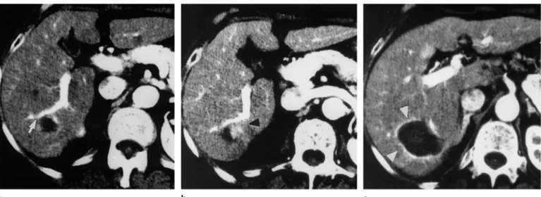
Figure 3. CT scans show complete response after LTA (energy delivered, 28,800 J). (a) Trans-verse arterial phase CT scan obtained before LTA shows a hyperattenuating 3.5-cm-diame-ter nodular lesion (arrow) in segment 4. (b) Transverse arterial phase contrast-en-hanced CT scan obtained 24 hours after LTA shows a homogeneous area of hypoattenua-tion surrounded by a segmental area of hyper-attenuation (arrowheads).
size and shape of the necrotic area so that it is similar to that of the tumor, as if a mold of the tumor had been made (oven effect) (22). The same authors (38) con-firmed these results in another study, ex-plaining that with HCC it is generally sufficient to treat just the tumor itself, and that the oven effect may protect sur-rounding structures.
More recently, Rossi et al (39), by using the radio-frequency interstitial tumor abla-tion, or RITA, with a four-pronged umbrella needle system (RITA Medical Systems, Mountain View, Calif) after occlusion of the tumor blood supply, confirmed again that lethal temperatures are reached just inside the HCC nodule, whereas nonle-thal temperatures are detected in the he-patic tissue immediately surrounding the nodule, and therefore, the resultant ne-crosis selectively involves only the HCC nodule and reproduces the nodule shape. The same authors (39) reported that in their study, the local recurrences may have been due to the unsatisfactory dif-fusion of heat within the HCC nodule, as well as extracapsular invasion or daugh-ter nodules outside the original nodule. These results seem to indicate that radio-frequency ablation is unable to induce a volume of necrosis larger than the origi-nal lesion and thus obtain the complete destruction of neighboring neoplastic or preneoplastic nests. However, a longer follow-up is needed for a better under-standing of the outcomes of these tech-niques, and more valid data are needed to compare the different options.
Recurrence of new HCCs in locations far from the original tumor was frequent dur-ing follow-up; this was probably due to multiple metachronous or unnoticed syn-chronous lesions or to intrahepatic metas-tases that were not detected at imaging (37). Such new lesions are part of the natural history of HCC in cirrhosis. The entire cirrhotic liver contains regenerative nodules, and hepatocarcinogenesis is prob-ably a stepwise process from a regenerative nodule to an adenomatous hyperplasia and then to HCC (40). Thus, the entire liver is ready for the development of mul-tiple HCCs, and the first lesion to be de-tected is only the beginning of a
multi-focal disease with several tumor clones (40,41).
The survival rates in our study were com-parable to those of other studies (12,17). However, we stress that some reports in-volved patients who received diagnosis and were staged a few years ago when im-aging techniques were less accurate. In our view, the use of these less accurate imaging techniques to verify local control of the disease may have caused an underestima-tion of the rates of local recurrence and of the short-, mid-, and long-term results with PEI. Hence, this limitation must also be taken into account when our results are compared with those of other authors.
Figure 4. CT scans show residual viable tissue after the procedure. (a) Transverse arterial phase CT scan obtained before LTA shows a hypoat-tenuating 2.5-cm-diameter nodular HCC (arrow) in segment 6. (b) Transverse arterial phase CT scan obtained 3 months after the first treatment session (energy delivered, 14,400 J) shows an area of residual viable tissue (arrowhead) within the original lesion contiguous to the coagulation. The size and shape of the area of necrosis produced by LTA together with the residual neoplastic tissue are the size and shape of the original neoplasm. (c) Transverse CT scan obtained after the second treatment session (energy delivered, 14,400 J) shows complete coagulation of the residual viable tissue with an area of necrosis (arrowheads) larger than the original lesion.
Figure 5. Graph shows 5-year local recurrence rates, calculated with the Kaplan-Meier method, for 74 patients with small HCCs treated with LTA. All recurrences were observed within 2 years.
Thus, there is a need for prospective, ran-domized, controlled trials with a greater number of patients with predefined char-acteristics to facilitate the analysis and comparison of the different treatment op-tions offered to patients with HCC.
The best way to ascertain the effective-ness of LTA is to assess the presence of vascular enhancement after contrast ma-terial administration at CT, considering that nonenhancement represents ab-sence of viable tissue and thus successful necrosis of the tumor (20 –22,25–27). The use of triple-phase helical CT is, in our opinion, essential for assessing treatment effectiveness and completeness and for early detection of tumor recurrence (42). The relationship between the enhancing peripheral halo and LTA treatment
de-serves some comment, and in our view, the attenuation behavior, with its seg-mental appearance after bolus injection, can be explained. The hyperattenuating halo around the coagulated tissue that is thought to be related to hyperemia is probably due to a rapid change in hepatic arterial flow in response to thermal in-jury. Thermal ablation induces a marked increase in arterial perfusion in the sur-rounding liver within a few minutes after the start of treatment. The heated area of necrosis can cause hepatic segmental ve-nous occlusion, and in this condition, the portal veins become draining veins and the occluded region is supplied with arterial blood alone (43). This phenome-non causes hyperattenuation in the oc-cluded area at CT since arterial flow itself increases and the contrast medium is less diluted there. CT acquisition with rapid sequential sections makes it possible to evaluate these changes.
Determination of the volume of the area to be heated is complex and de-pends on the laser power, the laser irra-diation time and wavelength of light involved, how the energy reaches the tissue, and such tissue factors as optical and thermal properties (22–27,29,44). Since different types of tissue have dif-ferent optical characteristics, the choice of a specific laser treatment must be related to the tissue type involved (44). Because of these factors, according to a few authors (27), it is not practical to establish a fixed deposition of laser en-ergy for any given tumor size; LTA of tumors of the same size may result in
variable necrosis, despite the use of the same amount of laser energy. On the basis of our experience, however, we believe that the therapeutic parameters are 5 W of laser power administered so as to reach simultaneously a total en-ergy of 7,200 J to an ellipsoid volume
ranging from 8 to 18 mL⫾ 3.9 (mean,
13.4 mL) for a single tumor treatment
and ranging from 12 to 40 mL ⫾ 11.5
(mean, 21.2 mL) for two tumor treat-ments with a total energy deposition of 14,400 J (32) and a procedure time of 6 and 12 minutes, respectively.
Although no formal cost-effectiveness analysis was performed as part of this work, it should be pointed out that for LTA, the laser generator costs were $40,000 –$45,000 and the costs for a set of fibers were only $1,000 –$1,500, but this set can be used to treat up to 50 patients (45).
In conclusion, LTA is a safe and effec-tive percutaneous technique for the treat-ment of primary liver tumors. However, further research in this field is needed to elucidate (a) the exact correlation be-tween histopathologic structure and laser energy and (b) the correlation between hepatic blood flow and the actual extent of the area of necrosis (46).
Acknowledgment: We thank Manuela Latini and Laura Morelli for their help in the care of patients during conscious sedation.
References
1. Oka H, Kuroda N, Kim K, et al. Prospec-tive study of early detection of hepatocel-Figure 6. Graph shows 5-year cumulative survival curves, calculated
with the Kaplan-Meier method, for 74 patients with small HCCs treated with LTA. The survival rates were 99%, 68%, and 15% at 1, 3, and 5 years, respectively.
Figure 7. Graph shows 5-year cumulative survival curves, calculated with the Kaplan-Meier method, in accordance with the degree of liver dysfunction on the basis of Child-Pugh classification. The difference was not statistically significant (P⫽ .052). The survival rates were 100%, 73%, and 31% in patients with Child-Pugh class A (■) and 95%, 68%, and 0% in patients with Child-Pugh class B (Œ) at 1, 3, and 5 years, respectively.
Figure 8. Transverse arterial phase CT scan obtained 17 months after LTA shows hyperat-tenuating viable tissue (arrowheads) around and within the lesion due to local recurrence.
lular carcinoma in patients with cirrhosis. Hepatology 1990; 12:680 – 687.
2. Zoli M, Magalotti D, Bianchi G, et al. Ef-ficacy of a surveillance program for early detection of hepatocellular carcinoma. Cancer 1996; 78:977–985.
3. Makuuchi M, Kosuge T, Takayama T, et al. Surgery for small liver cancers. Semin Surg Oncol 1993; 9:298 –304.
4. Bruix J, Cirera I, Clavet X, et al. Surgical resection and survival in western patients with hepatocellular carcinoma. J Hepatol 1992; 15:350 –355.
5. Castells A, Bruix J, Bru C, et al. Treatment of small hepatocellular carcinoma in cirrhotic patients: a cohort study comparing surgical resection and percutaneous ethanol injec-tion. Hepatology 1993; 18:1121–1126. 6. Rolles K. Transplantation of liver cancer.
In: Okuda K, Tabor E, eds. Liver cancer. New York, NY: Churchill Livingstone, 1997; 531–535.
7. Matsui O, Kadoya M, Yoshikawa J, et al. Small hepatocellular carcinoma: treatment with subsegmental transcatheter arterial embolization. Radiology 1993; 188:79 – 83. 8. Bronowicki JP, Vetter D, Doffoel M. Che-moembolization of hepatocellular carci-noma. In: Okuda K, Tabor E, eds. Liver cancer. New York, NY: Churchill Living-stone, 1997; 463– 469.
9. Silverman SG, Tuncali K, Adams DF, et al. MR imaging– guided percutaneous cryo-therapy of liver tumors: initial experi-ence. Radiology 2000; 217:657– 664. 10. Honda N, Guo Q, Uchida H, et al.
Percu-taneous hot saline injection therapy for hepatic tumors: an alternative to percuta-neous ethanol injection therapy. Radiol-ogy 1994; 190:53–57.
11. Ohnishi K, Yoshioka H, Ito S, et al. Pro-spective randomized controlled trial comparing percutaneous acetic acid in-jection and percutaneous ethanol injec-tion for small hepatocellular carcinoma. Hepatology 1998; 27:67–72.
12. Livraghi T, Giorgio A, Marin G, et al. Hep-atocellular carcinoma and cirrhosis in 746 patients: long-term results of percu-taneous ethanol injection. Radiology 1995; 197:101–108.
13. Higuchi T, Kikuchi M, Okazaki M. Hepa-tocellular carcinoma after transcatheter hepatic arterial embolization: a his-topathologic study of 84 resected cases. Cancer 1994; 73:2259 –2267.
14. Shiina S, Tagawa K, Unuma T, et al. Per-cutaneous ethanol injection therapy for hepatocellular carcinoma: a histopatho-logic study. Cancer 1991; 68:1524 –1530. 15. Ishii H, Okada S, Nose H, et al. Local recurrence of hepatocellular carcinoma after percutaneous ethanol injection. Cancer 1996; 77:1792–1796.
16. Ebara M, Ohto M, Sugiura N, et al. Percu-taneous ethanol injection for the treat-ment of small hepatocellular carcinoma: study of 95 patients. J Gastroenterol Hepatol 1990; 5:616 – 626.
17. Shiina S, Tagawa K, Niwa Y, et al. Percu-taneous ethanol injection therapy for the treatment of hepatocellular carcinoma: results in 146 patients. AJR Am J Roent-genol 1993; 160:1023–1028.
18. Ishii H, Okada S, Okusaka T, et al. Needle tract implantation of hepatocellular car-cinoma after percutaneous ethanol injec-tion. Cancer 1998; 82:1638 –1642. 19. Yamashita Y, Matsukawa T, Arakawa A, et
al. US-guided liver biopsy: predicting the effect of interventional treatment of hep-atocellular carcinoma. Radiology 1995; 196:799 – 804.
20. Rossi S, Di Stasi M, Buscarini E, et al. Percutaneous RF interstitial thermal abla-tion in the treatment of hepatic cancer. AJR Am J Roentgenol 1996; 167:759 –768. 21. Solbiati L, Goldberg SN, Ierace T, et al. Hepatic metastases: percutaneous radio-frequency ablation with cooled-tip elec-trodes. Radiology 1997; 205: 367–373. 22. Livraghi T, Goldberg SN, Lazzaroni S, et
al. Small hepatocellular carcinoma: treat-ment with radio-frequency ablation ver-sus ethanol injection. Radiology 1999; 210:655– 661.
23. Murakami R, Yoshimatsu S, Yamashita Y, et al. Treatment of hepatocellular carci-noma: value of percutaneous microwave coagulation. AJR Am J Roentgenol 1995; 164:1159 –1164.
24. Matthewson K, Coleridge-Smith P, O’Sul-livan P, et al. Biological effects of intrahepatic neodymium:yttrium-aluminium-garnet la-ser photocoagulation in rats. Gastroen-terology 1987; 93:550 –557.
25. Huang GT, Wang TH, Sheu JC, et al. Low-power laserthermia for the treatment of small hepatocellular carcinoma. Eur J Cancer 1991; 27:1622–1627.
26. Dowlatshahi K, Bhattacharya AK, Silver B, et al. Percutaneous interstitial laser therapy of a patient with recurrent hepa-toma in a transplanted liver. Surgery 1992; 112:603– 606.
27. Amin Z, Donald JJ, Masters A, et al. He-patic metastases: interstitial laser photo-coagulation with real-time US monitor-ing and dynamic CT evaluation of treatment. Radiology 1993; 187:339 –347. 28. Vogl TJ, Muller PK, Hammerstingl R, et al. Malignant liver tumors treated with MR imaging-guided laser-induced thermo-therapy: technique and prospective re-sults. Radiology 1995; 196:257–265. 29. Pacella CM, Bizzarri G, Ferrari FS, et al.
Interstitial photocoagulation with laser in the treatment of liver metastases. Ra-diol Med 1996; 92:438 – 447.
30. Kraft M, Arellano RS, Mueller PR. Con-scious sedation for the non-anesthesiolo-gist: a primer. Semin Intervent Radiol 1999; 16:89 –99.
31. Steger AC, Lees WR, Shorvon P, Walmsley K, Bown SG. Multiple-fibre low-power in-terstitial laser hyperthermia: studies in the normal liver. Br J Surg 1992; 79:139 –145. 32. Pacella CM, Rossi Z, Bizzarri G, et al.
Ul-trasound-guided percutaneous laser abla-tion of liver tissue in a rabbit model. Eur Radiol 1993; 3:26 –32.
33. Heisterkamp J, van Hillegersberg R, Sinofsky EL, et al. Heat resistant cylindrical diffuser for interstitial laser coagulation: comparison with a bare-tip fibre in ex vivo porcine liver. Lasers Surg Med 1997; 20:304–309. 34. Miller AB, Hoogstraten B, Staquet M, et
al. Reporting results of cancer treatment. Cancer 1981; 47:207–214.
35. Heisterkamp J, van Hillegersberg R, Ijzermans JNM. Interstitial laser coagulation for hepatic tumours. Br J Surg 1999; 86:293–304. 36. Vilana R, Bruix J, Bru C, et al. Tumor size
determines the efficacy of percutaneous injection for the treatment of small hep-atocellular carcinoma. Hepatology 1992; 16:353–357.
37. Utsunomiya T, Matsumata T, Adachi E, et al. Limitations of current preoperative liver imaging techniques for intrahepatic metastatic nodules of hepatocellular car-cinoma. Hepatology 1992; 16:694 –701. 38. Livraghi T, Goldberg SN, Lazzaroni S, et
al. Hepatocellular carcinoma: radio-fre-quency ablation of medium and large le-sions. Radiology 2000; 214:761–768. 39. Rossi S, Garbagnati F, Lencioni R, et al.
Percutaneous radio-frequency thermal ablation of nonresectable hepatocellular carcinoma after occlusion of tumor blood supply. Radiology 2000; 217:119 –126. 40. Takayama T, Makuuchi M, Hirohashi S, et
al. Malignant transformation of adeno-matous hyperplasia to hepatocellular car-cinoma. Lancet 1990; 336:1150 –1153. 41. Takenaka K, Adachi E, Nishizaki T, et al.
Possible multicentric occurrence of hepa-tocellular carcinoma: a clinicopathologi-cal study. Hepatology 1994; 19:889 – 894. 42. Pacella CM, Anelli V, Bizzarri G, et al. Evaluation of the vascular pattern of hep-atocellular carcinoma with dynamic computed tomography and its use in identifying optimal temporal windows for helical computed tomography. Eur Radiol 1998; 8:30 –35.
43. Murata S, Itai Y, Asato M, et al. Effect of tem-porary occlusion of hepatic vein on dual blood supply in the liver: evaluation with spi-ral CT. Radiology 1995; 197:351–356. 44. van Hillegersberg R, Pickering JW,
Aalders M, et al. Optical properties of rat liver and tumor at 633 nm and 1064 nm: photofrin enhances scattering. Lasers Surg Med 1993; 13:31–39.
45. Dodd JD, Soulen MC, Kane RA, et al. Mini-mally invasive treatment of malignant he-patic tumors: at the threshold of a major breakthrough. RadioGraphics 2000; 20:9–27. 46. Heisterkamp J, van Hillegersberg R, Mul-der PGH, et al. Importance of eliminating portal flow to produce large intrahepatic lesions with interstitial laser coagulation. Br J Surg 1997; 84:1245–1248.