1
Università degli studi di Roma “La Sapienza”
Dipartimento di Fisiologia e Farmacologia
“Vittorio Erspamer”
Dottorato di ricerca in Neuroscienze Clinico-Sperimentali
E Psichiatria (XXXI ciclo)
Curriculum: Neurofisiologia
Coordinatore: Prof. Marco Salvetti
Extracellular mild acidosis reduces the Ca
2+permeability
of human NMDA receptors
PhD student Supervisor
3
Abstract
……….. 51. Introduction
……….. 61.1. Human NMDA Receptor (hNMDARs)……….. 6
1.1.1. Structure………. 6
1.1.2. Physiological functions……….. 6
1.1.3. Excitotoxicity………. 12
1.1.4. Pharmacological modulation………. 14
2. Ca
2+permeability of hNMDARs
………... 172.1. Ca2+ permeability of hNMDARs and its modulation……….. 17
2.2. Study of Ca2+ permeability of ligand gated ion channels……… 18
3. Extracellular pH alterations in brain
………. 193.1. Extracellular pH alterations in Central Nervous System (CNS)………. 19
3.2. pH modulation of ligand gated ion channels ……….. 20
3.3. NMDARs and extracellular mild acidosis in brain ischemia……….. 21
4. Materials and methods
……… 224.1. Whole cell currents in transfected HeLa cells………. 22
4.2. Single channel recordings on transfected HeLa cells……….. 23
4.3. Whole-cell currents in primary cortical neurons………. 24
4.4. [Ca2+]I measurements in cortical neurons in culture……….. 24
4.5. Fractional Ca2+ current (Pf)………. 25
4.6. Statistical analysis………... 25
5. Results
……….. 265.1. Mild acidosis reduces Ca2+ permeability of human NMDARs………... 26
5.2. Mild acidosis reduces the single channel conductance of human NR1/NR2A NMDARs……… 32
4
5.3. Extracellular acidosis differentially modulates NMDA evoked whole cell
current and Ca2+ entry in mouse cortical neurons in culture………... 37 5.4. In search of the other cations able to modulate Pf of human NMDARs………. 41
6.
Discussion
……….. 427.
Conclusions
………. 465
Abstract
N-methyl-D-Aspartate receptors (NMDARs) are glutamate gated ion channels involved in excitatory synaptic transmission and in others physiological processes such as synaptic plasticity and development. The overload of Ca2+ ions through NMDARs, caused by an excessive activation of receptors, leads to excitotoxic neuronal cell death, which occurs in several neurological disorders. For this reason, the reduction of Ca2+ flux through NMDARs has been a central focus in finding therapeutic strategies to prevent neuronal cell damage. Extracellular H+ are allosteric modulators of NMDARs. Starting from previous studies which demonstrated that extracellular mild acidosis reduces NMDA evoked whole cell current, we studied the effect of extracellular mild acidosis on the NMDARs Ca2+ permeability measured as “fractional calcium current” (Pf, i.e. the percentage of the total current carried by Ca2+ ions) for human NMDARs NR1/NR2A and NR1/NR2B transiently transfected in HeLa cells. Here, we report that extracellular mild acidosis significantly reduces Pf of both human NR1/NR2A and NR1/NR2B NMDARs, decreasing also the single channel conductance in outside out patches for NR1/NR2A receptor. Reduction of Ca2+ flux through NMDARs is also confirmed in cortical neurons in culture. Furthermore, analysis of both NMDA evoked Ca2+ transients and whole cell currents in extracellular mild acidosis showed that extracellular H+ differentially modulate the ionic permeation (Na+ and Ca2+) through NMDARs.
Here we show that extracellular H+ limit Ca2+ entry through NMDA receptors, highlighting a protective mechanism by which the NMDARs Ca2+ flux is regulated during acidic shifts of extracellular pH.
6
1. Introduction
1.1. Human NMDA Receptor (hNMDARs)
1.1.1. Structure
Excitatory synaptic transmission in central nervous system is mediated mostly by amino acid glutamate activating both ionotropic and metabotropic receptors.
Ionotropic glutamate receptors (iGluRs) are ligand gated ion channels that induce excitatory glutamate-evoked currents, whereas metabotropic glutamate receptors (mGluRs) are G protein coupled receptors that control cellular responses through the activation of G protein pathways. IGluRs and mGluRs modulate each other using various mechanisms on different levels in both, physiological and pathological conditions (Reiner and Levitz, 2018).
IGluRs are divided with respect to their pharmacological properties into: GluA (AMPA) generating the rapid excitatory synaptic current , GluK (kainate) with modulatory role in both pre and post synaptic sites and GluN (NMDA) involved in several neuronal functions and pathological conditions for their high Ca2+ permeability (Mayer, 2006; Traynelis et al., 2010). All iGluRs are integral membrane proteins sharing a common structure consisting of four subunits assembled to form a central pore through which pass cation fluxes (Na+, K+ and Ca2+). AMPA and kainite receptors are more permeable to Na+ and K+, whereas NMDA receptors (NMDARs) have a higher Ca2+ permeability (Chaffey and Chazot, 2008).
To date have been identified three families of NMDAR subunits with different splice variants due to posttranscriptional RNA processing: GluN1 with eight isoforms, four GluN2 subunits (A-D) and two GluN3 (A-B). Functional receptors are formed by two identical obligatory GluN1 subunits and two identical GluN2 or GluN3 subunits, all assembled in tetrameric complexes (Fig.1A). Have also been described native triheteromeric receptors containing two GluN1 and two different GluN2 (or combination of GluN2 and GluN3) having different brain distribution, biophysical, pharmacological and signalling properties compared to diheteromeric receptors, suggesting the presence of specific receptor subpopulations for specific neuronal functions (Paoletti, 2011; Stroebel et al., 2018).
All NMDARs subunits have a typical modular organization consisting of four domains (Fig.1B): an extracellular region amino-terminal domain (ATD) involved in subunit assembly; an agonist binding domain (ABD) which binds glycine on GluN1 subunit and glutamate on GluN2 subunit; a transmembrane domain (TMD) with three transmembrane segments (M1,M3 and M4) plus a short re-entrant loop (P loop or M2) which forms the ion channel and an intracellular C-terminal domain (CTD) involved in receptor trafficking, anchoring and coupling
7
to signalling complexes with a variable length depends on subunit. (Mayer, 2006; Stephenson, 2001).
Fig.1Molecular structure of NMDAR.
A, Diheteromeric NMDA receptor complex consisting of two GluN1 and two GluN2 subunits and corresponding crystal structure. B, Domains organization of a single NMDAR subunit. Amino-terminal domain (ATD), agonist binding domain (ABD) composed of two segments (S1 and S2) folded in two lobes (D1 and D2), transmembrane domain (TMD) with three transmembrane helices (M1, M3 and M4) plus a short re-entrant loop (M2) and the C-terminal domain (CTD). Figure adapted from (Hansen et al. 2018)
The distribution of NMDARs in brain regions depends on subunit composition and stages of development. NR1 subunit isoforms are expressed throughout the brain (Paoletti, 2011), NR2B and NR2D subunits are predominantly expressed in neonatal brain and, partially, replaced in adult brain by NR2A and NR2C leading to changes in functional properties of receptor (Cull-Candy et al., 2001; Monyer et al., 1992, 1994). NR2A subunit shows an ubiquitous expression, NR2B is expressed in cortex, hippocampus, amygdala, ventral nuclei of thalamus and olfactory bulb, whereas the NR2C subunit is almost present in the granule cell layer of cerebellum (Laurie et al., 1997).
8
Within the neuron NMDARs are present in synaptic, presynaptic and postsynaptic sites (Paoletti 2011; Köhr 2006) but have also been detected in astrocytes and oligodendrocyte where are involved in myelination process (Köhr, 2006)
9
1.1.2. Physiological function
At resting membrane potential (-70mV) NMDARs are blocked by extracellular Mg2+ preventing the ions permeation (Mayer et al., 1984; Nowak et al., 1984). During excitatory neurotransmission glutamate, released in presynaptic sites, binds AMPA, kainite and NMDA receptors in postsynaptic locations generating an excitatory postsynaptic potential (EPSP). If the synaptic inputs cause a postsynaptic depolarization of sufficient amplitude and duration to remove the Mg2+ block, NMDARs which have bound glutamate on NR2 subunit, and glycine on NR1 or NR3 will open allowing the flow of permeant ions (Fig.2) (Dingledine et al., 1999; Kleckner and Dingledine, 1988).
The activation of receptor requires different conformational changes in all transmembrane helices (M1, M3 and M4) and re-entrant pore forming loop (M2). NMDARs exhibit Ca2+ and glycine dependent desensitization (Clark et al., 1990)(Mayer et al., 1989) and a deactivation kinetic which depends on subunit composition, values span a ~ 50 fold range (from 100ms to 4s), with NR2C-containing receptors displaying rapid deactivation kinetic similar to NR1/NR2B receptors but slower than NR2A-containig receptors, whereas NR1/NR2D-containing receptors exhibit a decay time constant of 4-5s (Cull-Candy and Leszkiewicz, 2004). Subunit composition influences also single channel properties and sensitivity to Mg2+ block: NR1/NR2A and NR1/NR2B containing receptors have “high conductance” (~ 50 pS) and a high Mg2+ sensitivity, whereas NR1/NR2C and NR1/NR2D containing receptors are described as “low conductance” (~ 40 and 20 pS) and exhibit a lower sensitivity to Mg2+ block (Fig.3) (Cull-Candy, 2007).
10
Fig.2 activation of NMDAR.
Left: At resting membrane potential NMDAR channel pore is blocked by Mg2+ ions. Right: After a depolarization and binding of glycine and glutamate, the Mg2+ block is removed, and the channel allows the ions flow. Figure adapted from (Zito and Scheuss, 2009)
11
Fig.3 NMDARs functional properties influenced by subunit composition.
A, Single channel recordings showing high conductance openings for NR2A-NR2B containing receptors, and low conductance openings for NR2C-NR2D containing receptors. B, Whole cell recordings from recombinant NMDARs showing different time constants of deactivation after 1ms pulse of 1mM glutamate: NR2A, 100ms; NR2B 250ms; NR2C 250ms; NR2D 4s. Figure adapted from (Cull-Candy and Leszkiewicz, 2004).
12
For their high Ca2+ permeability NMDARs are involved in several neuronal functions: Ca2+ ions act as second messenger translating the synaptic input in biological responses. Mice knockout for the obligatory NR1 subunit died after 8-15 hours after birth, indicating a vital function for NMDARs which control neuronal survival, migration and differentiation during neurodevelopment (Forrest et al. 1994, Ikonomidou et al. 1999) (Scheetz and Constantine-Paton, 1994). In adult brain NMDARs are involved in excitatory synaptic transmission contributing, with AMPA receptors, to the excitatory postsynaptic currents (EPSCs) and play an essential role in brain plasticity through the induction of long term potentiation (LTP) (Artola and Singer 1987; Collingridge and Bliss 1987; Massey et al. 2004) and long-term depression (LTD)(Bear and Malenka, 1994). These processes “measure” the ability of brain to change with external stimuli, such as learning and memory and require Ca2+ flux through NMDARs with consequent activation of different Ca2+ dependent proteins such as Ca2+/calmodulin-dependent protein kinase II (Fan et al., 2014) (Cull-Candy and Leszkiewicz, 2004).
Furthermore, Ca2+ flux through NMDARs promotes neuronal survival leading to activation of transcription factor cAMP-response-element-binding-protein (CREB) which regulates the expression of pro-survival genes such as brain-derived neurotrophic factor (BDNF) (Hardingham and Bading, 2003)
13
1.1.3. Excitotoxicity
In physiological conditions, the increase of intracellular Ca2+ ionsdue to activation of NMDARs is crucial to cell functioning but a Ca2+ ions overload for excessive release of glutamate or for a chronic exposure could induce cell death for “excitotoxicity”. Excitotoxicity causes neuronal dysfunctions and degeneration, occurring in both acute Central Nervous System (CNS) insults such as ischemia and brain injury and in several neurodegenerative disorders such as amyotrophic lateral sclerosis, Parkinson’s disease and others (Parsons and Raymond, 2014). This mechanism of neuronal cell death was explained for the first time by Lucas and Newhouse who observed degeneration of inner layers of retina after injections of glutamate in infant mice (Lucas and Newhouse, 1957). Later, Olney coined the term “glutamate excitotoxicity” describing intracranial lesions after injections of glutamate in adult and infant mice (Olney, 1969).
The key role of Ca2+ ions in glutamate neurotoxicity was demonstrated by Choi in cortical neurons: cell death was potentiated in calcium-rich extracellular solution, whereas the neuronal loss was attenuated with removal of extracellular calcium (Choi, 1985); use of NMDARs antagonists reduced neurotoxicity, indicating that neuronal cell death was mediated primarily by these receptors (Choi, 1987; Choi et al., 1988).
Excitotoxic Ca2+ overload through NMDARs impairs the regulatory cellular mechanisms activating several pro-neuronal cell death pathways. First of all, neurons lose their Ca2+ buffering capacity, intracellular Ca2+ ions are sequestrated by mitochondria leading to metabolic acidosis, oxidative free radicals generation(Arundine and Tymianski, 2003) and activation of different Ca2+ dependent proteases, and consequent cell death (Lau and Tymianski, 2010).
Several studies suggested that glutamate-mediated excitotoxicity is related to NMDARs localization and subunit composition (Fig.4). An activation of extrasynaptic receptors leads to neuronal cell death pathway triggering to a CREB shut off and block of pro-survival genes expression (Hardingham and Bading 2010; Walton and Dragunow 2000; Mantamadiotis et al. 2002), whereas activation of these receptors in synaptic location promotes neuronal survival (Hardingham and Bading, 2003; Hardingham et al., 2002; Parsons and Raymond, 2014). But, other lines of evidence suggest that silencing of synaptic NMDARs offers neuroprotection against neurotoxic insult which is only in part repressed by use of memantine, an extrasynaptic NMDARs specific antagonist (Papouin et al., 2012; Wroge et al., 2012), demonstrating an excitotoxic contribute of synaptic NMDARs. It was hypothesized that NR2A and NR2B containing receptors have differential role in promoting neuronal survival or death, for their
14
mostly expression in synaptic locations NR2A containing receptors are associated with cell survival, whereas NR2B containing receptors, more abundant in extrasynaptic sites, are thought to be responsible for cell death signaling (Liu et al., 2007). Taken together, all these studies do not solve the long-lasting paradox that NMDARs can promote both neuronal survival and death.
Fig.4 Excitotoxicity depends on localization and subunit composition of NMDARs.
NR2A subunits-containing receptors are more abundant in synapses and associated with
neuronal survival, whereas NR2B subunit-containing receptors are more expressed at extrasynaptic sites and associated with the activation of neuronal cell-death pathway.
15
1.1.4. Pharmacological modulation
For their involvement in several neurological disorders, NMDARs have attracted attention in pharmacological industry in order to develop therapeutic strategies able to reduce the excessive Ca2+ flux through receptors without impairing their physiological function.
NMDARs have distinct binding sites for endogenous or synthetic compounds and can be modulated both positively and negatively (Fig.5).
Competitive agonists and antagonists bind the agonist binding domain of receptors (ABD). Glutamate and glycine, physiologically present in brain, bind the ABD on NR2 subunit and NR1 subunit respectively. NMDA is a selective agonist for NMDAR and binds ABD on NR2 subunits. Several agonists and antagonists binding NR2 subunits are derived from structure modifications of glutamate: one of the first discovered compounds was the (R)-2-amino-5-phosphonopentanoate (R)-AP5 , homologous ( R) -AP7 and (R)-CPP (Lehmann et al., 1987; Morris et al., 1986; Roman et al., 1989), with last two compounds showing higher subunit selectivity compared to R-AP5 for steric effects due to their larger structures (Paoletti and Neyton, 2007). To date no glutamate binding site antagonists developed shows a high NR2 subunit selectivity, and this would be important because each isoform of NR2 subunit confers distinct properties to NMDARs.
The modification of the structure of glycine, a coagonist required for NMDAR activation binding the ABD of NR1 subunit, led to develop of NR1 antagonists showing little selectivity for NR1 subunits isoforms (Gonda, 2012; Paoletti and Neyton, 2007).
Different organic compounds act like uncompetitive antagonists, require the activation of receptor and block NMDARs by occluding the channel pore. Usually, open channel blockers poorly discriminate different NMDAR subunits except MK-801 (dizocilipine) showing higher affinity for NR2A and NR2B containing receptors rather than NR2C-NR2D containing receptors (Wong et al., 1986). Several dissociative anaesthetics act as NMDAR open channel blockers such as phencyclidine (PCP) thienylcyclohexylpiperidine (TCP) and ketamine (Anis et al., 1983). Memantine, a NMDAR channel blocker used in Alzheimer’s disease to treat cognitive deficits, occludes channel during long lasting depolarization typical of chronic excitotoxic conditions preventing neurotoxicity (Parsons et al., 1999a).
NMDARs are also modulated by endogenous and synthetic compounds, which bind extracellular sites N-terminal domain (NTD) of NR2 subunits. Ifenprodil and derivates are the only known compounds with high NR2B selectivity (Williams, 1993) resulting relevant as therapeutic tools.
16
Polyamines are NR2 subunit selective positive modulators, received much attention for their potential as therapeutic tools. Spermine and spermidine are endogenous polyamines present in high concentrations in central nervous system, acting by at least three different mechanisms: 1. an inhibition of NMDARs in voltage dependent manner, acting mostly on NR2A and NR2B subunits;
2. a glycine dependent potentiation of receptors acting on NR2A and NR2B-containing receptors and not controlled by NR1 subunit RNA splicing;
3. a voltage and glycine independent potentiation of NR2B-containing receptors through a relief of tonic proton inhibition at physiological pH (Dingledine et al., 1999).
Also extracellular Zn2+ ions act as allosteric modulators binding the N-terminal domain of NR2A subunit. Zn2+ ions are present in synaptic vesicles and act in voltage dependent or independent manner. The voltage dependent inhibition is weaker than Mg2+ block and reduces the single channel amplitude, whereas voltage independent inhibition involves both a reduction in opening frequency and a decrease in open duration (Chen et al., 1997; Harrison and Gibbons, 1994). Zn2+ ions bind with high affinity NR2A with an IC
50 value in nanomolar range, whereas the affinity is lower for NR2B subunit with IC50 in low micromolar range (Hansen et al., 2018).
17
Fig.5 Ligand binding sites in NMDAR.
Competitive agonists and antagonists bind the ABD domain on NR1 and NR2 subunit. Allosteric modulators bind receptor in a specific site with selective affinity for NR1 or NR2 subunit. Channel blockers act in pore region. Figure adapted from (Paoletti and Neyton, 2007).
18
1.2. Ca
2+permeability of hNMDARs
1.2.1. Ca2+ permeability of hNMDARs and its modulation
Glutamate receptor ion channel is formed by a water filled pore with internal and external cavities separated by a narrow constriction at which pore has the smallest dimension. Both, permeation properties and conductance of iGluRs are controlled by the residues of QRN site, located at the apex of M2 loop. The site is formed by Gln (Q) or Arg (R) in AMPA and kainite receptors (Hume et al., 1991) followed by an Asn(N) in NR1 and NR2 subunits and a Gly(G) in NR3 subunit for NMDARs (Traynelis et al., 2010). Residues of QRN site affects the single channels conductance (Swanson et al., 1996, 1997), Mg2+ sensitivity (Burnashev et al., 1992; Mori et al., 1992) and Ca2+ permeability of receptors (Burnashev et al., 1992).
Among iGluRs, NMDARs show the higher Ca2+ permeability, due to the presence of multiple Ca2+ binding sites located within the pore and at the external mouth of the channel. The residues located at the external entrance of channel seem to be critical for the Ca2+ permeability of receptor (Premkumar and Auerbach, 1996), but the structural elements are still not well understood. A motif in the external vestibule of the NR1 subunit and formed by a cluster of charged residues and a proline (DRPEER) represents a key determinant of the high Ca2+ permeability of NMDARs (Beck et al., 1999; Watanabe et al., 2002). Neutralization of negative charges of DRPEER motif with mutagenesis experiments leads to a reduction of Ca2+ permeability of NMDARs, showing the importance of negatively charged side chains for the interaction with Ca2+ ions (Watanabe et al., 2002).
Ca2+ permeability of NMDARs changes with the subunit composition: NR2A/NR2B containing receptors show the higher Ca2+ permeability, that can be reduced by a replacement of Asn with Gln in NR1 or NR2B (Burnashev et al., 1992), leading to different conductance levels of receptor (Premkumar et al., 1997), whereas NR2C and NR3 containing receptors have a reduced Ca2+ permeability (Traynelis et al., 2010). Ca2+ permeability of glutamate receptors can be modulated by different factors such as protein kinase A (PKA) phosphorylation (Skeberdis et al., 2006), mutations of residues lining QRN site and structure alterations of M2 loop (Traynelis et al., 2010). Though Piccari et al (2011) demonstrated the possibility of a pharmacological reduction of the Ca2+ permeability of a ligand-gated ion channel, to date, despite the pathological role of overload of Ca2+ ions through NMDARs, no pharmacological tool has been shown to reduce their Ca2+ permeability maintaining the ability to allow Na+ influx.
19
1.2.2. Study of Ca2+ permeability of ligand gated ion channels: experimental approaches
Ca2+ flux through NMDA receptors has been directly demonstrated for the first time using Ca2+ indicator dyes in voltage-clamped spinal cord neurons (MacDermott et al., 1986; Mayer et al., 1987). To quantify the Ca2+ permeability through ion channels different experimental approaches have been proposed: the first method was based on the measurement of Ca2+ -induced reversal potential shifts changing the extracellular Ca2+ ions concentration to have a relative Ca2+ permeability PCa/Pmonovalent. The permeability ratio of Ca2+ ions respect to various monovalent cations (Na+, K+ and Cs+) for NMDARs was obtained for cultured neurons, neurons in brain slices and recombinant NMDARs and was found to be in the range of 3 to 5 , basing on the Goldman-Hodgking-Katz (GHK) constant field assumptions extended for divalent cations (Schneggenburger, 1996). Neher developed an approach which allowed direct determination of Ca2+ influx under physiological conditions using Ca2+ indicator dye Fura-2 (Neher, 1995). The method was based on the simultaneous recordings of fluorescent signals and whole-cell currents allowing to estimate the fraction of the total current carried by Ca2+ ions, also called “fractional Ca2+ current” or P
f ( Z. Zhou and Neher 1993; Vernino et al. 1994). This experimental approach does not depend on any theoretical assumption, requires the absence of any form of Ca2+ induced Ca2+ release and a perfect voltage control of the cell. Schneggenburger et al (1993) measured the Pf values of NMDA and AMPA/kainite receptors in septal neurons. Experiments were performed with a concentration of extracellular Ca2+ ions of 1.6 mM and a membrane potential of -80mV and the Pf values were 7% and 1.4%, respectively for NMDA and AMPA/kainite receptors. In 1995, Burnashev and colleagues (1995) measured the Pf values for different types of recombinant NMDARs (NR1-NR2A and NR1-NR2C), AMPA and kainite receptors expressed in a heterologous system, the human embryonic kidney cells (HEK cells). The results showed that fractional Ca2+ currents through iGluRs was dependent of subunit composition or editing of QRN site. In particular, for NMDARs the fractional calcium current was independent of extracellular Mg2+ with value of 8.2% for NR1-NR2C NMDARs and 11% for NR1-NR2A receptors.
20
1.3. Extracellular pH alterations in brain
1.3.1. Extracellular pH alterations in the central nervous system (CNS)
Regulation of extracellular pH (pHe) in the central nervous system (CNS) is a key factor for maintaining normal cellular function. pH microelectrode studies, in different isolated CNS preparations, have indicated that interstitial brain pH changes occur during synaptic transmission, release of neurotransmitter, neuronal excitability and pathological state. Shifts range from long-lasting global to fast and more localized transients. (Ruusuvuori and Kaila, 2014)
Changes in extracellular H+ gradient can impact on the release of synaptic vesicles containing neurotransmitters, and the rise of presynaptic intracellular Ca2+ concentration depends on the activity of voltage-gated calcium channels that shows a high pHe sensitivity (Sinning and Hübner, 2013; Tombaugh and Somjen, 1996).
In different regions throughout the CNS, stimulated neuronal activity, seizure or spreading depression are associated with an increase of extracellular H+ followed by an acidosis that could persist for minutes. H+ ions exert their effect acting on calcium-sensing non selective cation channels mediating calcium flux which accompanies neuronal excitation (Chu et al., 2003; Chesler and Kaila, 1992). Furthermore, extracellular H+ ions act modulating conductance of voltage-, ligand- and proton-gated ion channels showing their important role in control of synaptic transmission. Whole-cell recordings performed in rat hippocampal CA1 showed pHe sensitivity for neuronal Na+, K+ and Ca2+ channels: Na+ and Ca2+ currents were decreased by moderate acidosis and enhanced by alkaline exposure, whereas K+ currents were unaffected by pH exchange (Tombaugh and Somjen, 1996). Rapid extracellular acidification activates H+ gated channels, the acid sensing ion channels (ASICs), belonging to the family of amiloride sensitive cation channels andknown to play a role in hippocampal long-term potentiation (LTP; Sinning and Hübner, 2013; Wemmie et al., 2002) and involved in pain transmission as demonstrated by their expression in sensory neurons (Waldmann et al., 1997).
21
1.3.2. pH modulation of ligand gated ion channels
Both GABAA and NMDARs show high pH sensitivity, demonstrating the important role of pHe changes in keeping the balance between excitatory and inhibitory synaptic transmission. Experiments with recombinant GABAA receptors demonstrated that an acidification of pHe enhanced GABAA currents depending on subunit composition and stage of development (B. J. Krishek et al. 1996; Belinda J. Krishek and Smart 2001).
Extracellular H+ ions are allosteric modulators of NMDARs with an inhibitory concentration close to physiological pH (S. F. Traynelis and Cull-Candy 1990). External acidosis suppresses NMDA-evoked currents whereas an extracellular alkalosis enhances NMDA evoked currents. The effect of H+ ions on these receptors is voltage independent and decreases the open frequency of channels without affecting unitary conductance or individual dwell times (Banke et al., 2004; Traynelis and Cull-Candy, 1990, 1991; Vyklický et al., 1990). Furthermore, Chen et al (1998) demonstrated that, also for presynaptic NMDARs alkalinization potentiated the frequency of spontaneous synaptic current (SSC), which is reduced by an acidification.Similarly to several biophysical and pharmacological properties, also the pHe modulation of NMDARs shows a dependence on subunit composition: NR2A-B and D-containing receptors have an IC50 near physiological pH, whereas NR2C-containing receptors are less sensitive to changes in pHe with an IC50 near pH 6.0. H+ sensitivity can be modulated by RNA splicing on NR1 subunit and by polyamines which potentiate receptor function through a relief of proton inhibition (S. F. Traynelis, Hartley, and Heinemann 1995). Structural determinants of NMDARs H+ binding site are not well understood but mutations in extracellular end of the second transmembrane domain (M3) and adjacent linker to the S2 portion of the glycine-binding domain in NR1 subunit or mutations on glutamate binding domain in NR2 subunit affect the H+ ions permeation suggesting the hypothesis that the proton sensor and receptor gate may be functionally and structurally integrated (Banke et al., 2004; Dravid et al., 2007; Low et al., 2003) .
22
1.3.3. NMDARs and extracellular mild acidosis in brain ischemia
A reduction of blood flow in brain due to a thrombosis, embolism or systemic hypo-perfusion, can cause an ischemic stroke characterized by the activation of several pathways including: excitotoxicity, acidotoxicity, ionic imbalance, oxidative stress inflammation and apoptosis. All these conditions lead to cell death involving neuron, glia and endothelial cells (Doyle, Simon, and Stenzel-Poore 2008). One of the first event after ischemic stroke is the membrane depolarization, caused by impairment of mitochondrial ATP synthesis, which leads to a release of glutamate increasing its concentration in synaptic space. As result, activation of NMDA and AMPA receptor increases the level of intracellular Ca2+ ions with consequent stimulation of several Ca2+ dependent proteases and enzymes and the known cytotoxic effects. The rise of intracellular Ca2+ ions also derives from the activation of homomeric ASIC1a, a subtype of ASICs particularly permeable to Ca2+ ions, which mediates cell death for acidotoxicity (Wang and Xu, 2011). For these reasons, Ca2+ permeability of NMDARs and Ca2+-mediated cell death are most studied therapeutic targets for ischemic stroke.
Several studies suggest that extracellular mild acidosis protects neurons against NMDARs-mediated excitotoxicity during ischemia (Giffard et al., 1990; Tombaugh and Sapolsky, 1990) demonstrating a reduced NMDAR activation due to extracellular H+ but no one investigated the modulation of their potentially neurotoxic Ca2+ flux. Also, to date no clinical trial using NMDARs antagonists for stroke produced neuroprotective effects, because of their side effects at therapeutic doses and the impairment of physiological functions of receptor (Wu and Tymianski, 2018), suggesting the importance to study a mechanism through which reduce the NMDARs Ca2+ permeability without affect the excitatory synaptic transmission.
23
2. Materials and methods
2.1. Whole-cell currents in transfected HeLa cells
Human epithelial HeLa cells were grown in Dulbecco’s Modified Eagle Meadium (DMEM) supplemented with 10% heat-inactivated FBS and 1% penicillin-streptomycin, at 37° C in a 5% CO2 humidified atmosphere. Cells were plated on cover slides (8 * 104 cells/ml) and transiently transfected 24 hours later using Magnetofection (Neuromag; OZ Bioscences, Mairselle, France) according to the manufacturer’s protocol, adding 0.5 g human NR1, NR2A or NR2B cDNA subtype per well. Recordings were carried out 24-36 hours following transfection. Whole-cell currents were recorded at room temperature using borosilicate glass patch pipettes having a tip resistance of 3-5 MΩ filled with the internal solution: for Pf measurements, 140 mM CsCl, 10mM HEPES and 0.5 mM Fura-2 (pH 7.3); otherwise, 140mM CsCl, 10mM HEPES, pH value was adjusted at 7.3 with CsOH 1M. The patch series resistance was compensated by 80-95%, and measurements were performed at holding potential of -70mV. Cell capacitance was routinely compensated using the amplifier function and value used to estimate cell surface. Membrane currents were filtered at 3KHz upon the acquisition with HEKA EPC 800 (HEKA Elektronik, Germany) and analyzed offline. During recording, cells were continuously super fused using a gravity-driven perfusion system consisting of independent tubes for normal and NMDA and Glycine containing external solutions: 140mM NaCl, 2.8 mM KCl, 2mM CaCl2, 10mM glucose, and 10mM HEPES, pH values were adjusted with NaOH 1M at 7.3, 6.8 and 6.5. Perfusion tubes were connected to a fast exchanger system (RSC-100; Bio-logic,Claix, France). Data sampling and analysis were performed using pClamp10 software (Molecular Devices, Sunnyvale, CA, USA).
24
2.2. Single channel recordings on transfected HeLa cells
Transfected HeLa cells, with 0.5 M human NR1 and NR2A cDNA as described above, were used for single channel recordings 36-48 hours following transfection. Single channel recordings were performed from excised outside-out patches with HEKA EPC 800 (HEKA Elektronik, Germany) at room temperature using borosilicate glass patch pipettes having a tip resistance of 3-5 MΩ filled with the internal solution: 140mM CsCl, 10mM HEPES, pH value was adjusted at 7.3 with CsOH 1M. The holding potential was -70 mV. External solution contained: 140mM NaCl, 2.8 mM KCl, 2mM CaCl2, 10mM glucose, and 10mM HEPES, pH values were adjusted with NaOH 1M at 6.5 and 7.3. NR1/NR2A channels in patches were activated by addition of NMDA 200M and glycine 50M to the external solution. Responses were recorded during continuous perfusion of the agonist-containing external solutions at different pH values (7.3, 6.5) using a gravity-driven perfusion system consisting of independent tubes connected to a fast exchanger system (RSC-100; Bio-Logic, Claix, France). Voltage-clamp recordings were filtered at 5 kHz and digitized at 20 kHz (pClamp 10). Distributions of all-point amplitudes were fitted by multiple Gaussian curves, and then chord conductance values were calculated.
25
2.3. Whole-cell currents in primary cortical neurons
Cortical neuronal cultures were prepared from the brain of newborn C57BL/6 mice (P0-P1). Cerebral cortices were chopped and digested in 2.5% (wt/vol) trypsin for 20 min at 37° C. Cells (10 x 104 cells/cm2) were seeded on dishes coated with poly-L-lysine (100 μg/ml) in MEM with Earle’s BSS supplemented with 100 mM sodium pyruvate, 20% (wt/vol) Glucose, 10% FBS, 100 U/ml penicillin, 0.1 mg/ml streptomycin, and 200 mM Glutamine. After 4 hours, the medium was changed with Neurobasal medium supplemented with 200 mM glutamine, 1% B-27, 100 U/ml penicillin, and 0.1 mg/ml streptomycin. Twice a week, half Neurobasal medium was changed and replaced with fresh medium. Cell cultures were used after 10-11 days. Whole-cell patch-clamp recordings were performed on cortical neurons in culture at room temperature, at a holding potential of -70 mV, using borosilicate glass patch pipettes having a tip resistance of 3-5 MΩ filled with internal solution: 140mM CsCl, 10mM HEPES, pH value was adjusted at pH 7.3. The patch series resistance was compensated by 10-30%. Cell capacitance was routinely compensated, membrane currents were filtered at 3KHz upon the acquisition with HEKA EPC 800 (HEKA Elektronik, Germany) and analyzed off-line. During recordings, cells were continuously superfused using a gravity-driven perfusion system consisting of independent tubes for normal and NMDA and glycine containing external solutions: 140 mM NaCl, 10 mM HEPES, 2.8 mM KCl, 2 mM CaCl2, 2 mM MgCl2,10 mM glucose, pH values were adjusted with NaOH at 6.5 and 7.3. Perfusion tubes were connected to a fast exchanger system (RSC-100; Bio-Logic, Claix, France). Data sampling and analysis were performed using pClamp 10 software (Molecular devices, Sunnyvale, CA, USA).
2.4. [Ca2+]i measurements in cortical neurons in culture
Measures of [Ca2+]i were obtained by time-resolved digital fluorescence microscopy using Ca2+ indicator Fura-2 (excitation 340 and 380 nm, emission 510 nm). Cortical neurons in culture were incubated with the cell-permeant Fura-2 acetoxylmethylester (2M; Molecular probes, Life technologies) for 45 minutes at 37° C in culture medium. Ca2+ transients were elicited by applying NMDA 200 M and glycine 50 M for 3 second with a preapplication of normal external solution at pH 6.5 or 7.3 for 10 seconds without agonist.
26
2.5. Fractional Ca2+ current (Pf)
The procedure used for Pf measurements follows the method proposed by Zhou and Neher (Zhou and Neher, 1993), which has the advantage to be independent of any assumption on ion-permeation properties (Burnashev et al., 1995; Neher, 1995). All measurements were performed in transfected HeLa cells, (which have a membrane capacitance rangeing between 10 and 20 pF) and allow almost perfect voltage clamp, thus being an ideal preparation for determination of Pf, which requires that current flows uniquely through the channel under study. Cells were loaded with cell-impermeant Fura-2 through the patch pipette used to measure NMDA-evoked currents. Recordings of fluorescence signals and whole-cell membrane currents were synchronized, and images were acquired and analysed off-line. All optical parameters and digital camera settings were maintained throughout this study to avoid nonhomogeneous data. The changes of [Ca2+]I were expressed as F/F (i.e. basal fluorescence), using only one excitation wavelength, 380 nm, to increase the temporal resolution. Determinations were carried out after the basal fluorescence had reached a stable value. Cells displaying a low-basal F380 values were discarded. In order to evaluate Pf the F:Q ratio between the fluorescence increase (F) and total charge that had entered the cell at each fluorescence acquisition time (Q) was defined as F/Q = (F/F)/ Q. For each cell, we used the F/Q points that, immediately after the onset of the NMDA evoked response, exhibited a linear relationship, indicating that the Ca2+-buffering capability of Fura-2 was not saturated. The F/Q ratio value was then measured as the slope of the linear regression best fitting the F-Q plot. Finally, Pf was determined by normalizing the ratio obtained in standard medium (F/Q) to the calibration ratio, measured when Ca2+ ions were the only permeant species (F/QCa): Pf = (F/Q) / (F/QCa). Calibrations were performed on different days throughout the whole experimental period, using NMDA-evoked Ca2+ currents. Only the first NMDA application to each cell was used for analysis, in order to avoid data variation observed with repetitive applications.
2.6. Statistical analysis
All data were expressed as means ± S.D. and analysed using paired t-test or one-way ANOVA. All performed tests gave a power > 0.8. Significance for all tests was set at P < 0.05.
27
3. Results
3.1. Mild acidosis reduces the Ca2+ permeability of human NMDARs
To investigate the effect of acidosis on the NMDAR Ca2+ permeability we measured the fractional Ca2+ current (i.e. the percentage of the total current carried by Ca2+ ions; Pf) of human NR1/NR2A NMDAR expressed in HeLa cells, simultaneously recording NMDA-evoked whole-cell currents and variations of intracellular free Ca2+ concentration ([Ca2+]
i; Fig. 6). Selective agonist NMDA (200 M) and glycine (50 M) were coapplied for two seconds at three different pHe values (6.5, 6.8, 7.3). At pHe 7.3 the Pf value of NR1/NR2A NMDAR exhibited was 11.3 ± 4.4 % in agreement with Burnashev et al. (1995). Lowering pHe from 7.3 to 6.8, the Pf value was 8.5 ± 3.1 %, not significantly different from the value at physiological pHe, whereas when pHe was reduced to 6.5 the Pf was significantly decreased to 7.6 ± 1.9 % (Fig. 7).
28
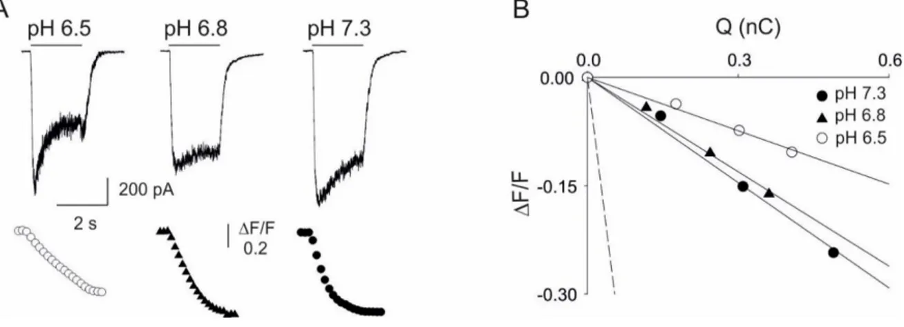
Fig. 6. Acid pHe reduces the Ca2+ permeability of human NR1/NR2A NMDAR.
A, typical traces obtained by simultaneous recordings of whole-cell currents (top) and Ca2+ transients (bottom) evoked by coapplication of NMDA and glycine (200 M and 50 M, respectively) at the indicated pHe values in HeLa cells transiently transfected with human cDNA encoding for NR1 and NR2A subunits. Experiments were performed in Mg2+ free normal external solution to unmask NMDA currents at negative potentials. Holding potential, -70 mV. At excitation wavelength of 380 nm, [Ca2+]
i increase corresponds to a downward deflection of Fura-2 fluorescence emission. Traces are aligned and share the same temporal scale. B, linear relationships between F/F and Q obtained from the same cells as in A. The dashed line represents the mean slope obtained from calibration experiments (5.54 nC-1; n = 9).
29
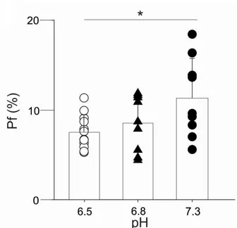
Fig. 7 Pf value of human NR1/NR2A NMDAR is significantly reduced at pHe 6.5.
The Pf valueswas 7.6 ± 1.9 % (n=12), 8.5 ± 3.1 % (n=9), and 11.3 ± 4.4 % (n=9) for pHe value of 6.5, 6.8 and 7.3, respectively. * P=0.036 (ANOVA). Symbols represent the Pf value of the individual cells. Error bars represent SD.
30
To have a more general view about the effects of acidosis on the Ca2+ fluxes mediated by NMDARs, we extended our study to the human NR1/NR2B NMDAR subtype, mainly expressed at extrasynaptic locations and exhibiting distinct functional properties (Cull-Candy and Leszkiewicz, 2004). We analyzed the human NR1/NR2B NMDARs as above described for NR1/NR2A receptor (Fig. 8), again finding that low pHe values could alter the Ca2+ permeability. In particular, Pf value of NR1/NR2B receptor was 9.1 ± 2.3 % at pHe 7.3 and 8.1 ± 2.5 % at pHe 6.8 Pf (not significantly different), whereas the Pf value fell to 4.6 ± 1.2 % at pHe 6.5 (Fig. 9). Our data demonstrated that extracellular acidosis is able to reduce significantly and selectively the Ca2+ flow through human NMDARs.
31
Fig. 8. Acid pHe reduces the Ca2+ permeability of human NR1/NR2B NMDA receptor.
A, typical traces obtained by simultaneous recordings of whole-cell currents (top) and Ca2+ transients (bottom) evoked by coapplication of NMDA and glycine (200 M and 50 M, respectively) at the indicated pHe values in HeLa cells transiently transfected with human cDNA encoding for NR1 and NR2B subunits. Experiments were performed in Mg2+ free normal external solution to unmask NMDA currents at negative potentials. Holding potential, -70 mV. At excitation wavelength of 380 nm, [Ca2+]
i increase corresponds to a downward deflection of Fura-2 fluorescence emission. Traces are aligned and share the same temporal scale. B, linear relationships between F/F and Q obtained from the same cells as in A. The dashed line represents the mean slope obtained from calibration experiments (5.54 nC-1; n = 9).
32
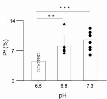
Fig. 9. Pf value of human NR1/NR2B NMDAR is significantly reduced at pHe 6.5.
The Pf valueswere 4.6 ± 1.2 % (n=9), 8.1 ± 2.5 % (n=6), and 9.1 ± 2.3 % (n=7) when measured at pHe value of 6.5, 6.8 and 7.3, respectively. *** P<0.001, **P=0.006 (ANOVA). Error bars represent SD. Symbols represent the Pf value of the individual cells.
33
3.2. Mild acidosis reduces the single channel conductance of human NR1/NR2A NMDARs
The above reported Pf data led us to hypothesize that the selective reduction, due to acidosis, of the Ca2+/Na+ ratio of the current flowing through NMDARs could affect the conductance of these receptor-channels, even though previous reports did not find significant variations of this parameter in similar conditions (Banke et al., 2005; Tang et al., 1990; Traynelis and Cull-Candy, 1990, 1991; Vyklický et al., 1990). We recorded single channel activity in outside-out patches excised from HeLa cells transiently expressing human NR1/NR2A NMDARs. Coapplication of NMDA and glycine (200 M and 50M, respectively) elicited a high number of channels openings, with a relatively high open probability, drastically reduced upon shifting the pHe from 7.3 to 6.5, as expected from previous studies (Fig. 10) (Banke et al., 2005; Traynelis and Cull-Candy, 1990, 1991; Vyklický et al., 1990)).
34
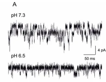
Fig. 10. Extracellular acidosis modulates single channel activity of human NR1/NR2A NMDAR in outside-out patches.
Typical traces recorded from the same outside-out patch excised from a HeLa cell expressing human NR1/NR2A NMDARs upon coapplication of NMDA and glycine (200 M and 50 M, respectively); pHe values, 7.3 (top) and 6.5 (bottom). Holding potential, -70mV.
35
The single channel chord conductance values were obtained from all-point histograms best fitted with multiple Gaussian distributions (Fig. 11; -70 mV): all outside-out patch exhibited a reduction in chord conductance going from physiological pHe to acidosis (pH 6.5). The mean chord conductance values were 43.3 ± 5.0 pS and 40.6 ± 4.6 pS for pHe values of 7.3 and 6.5, respectively (Fig. 12; P <0.001). Though the conductance value at physiological pH is similar to the values previously reported, the small but significant conductance reduction upon acidosis, to our knowledge, has never been recognized before (Banke et al., 2005; Traynelis and Cull-Candy, 1990, 1991; Vyklický et al., 1990).
36
Fig. 11. Extracellular acidosis down regulates both open probability and conductance of human NR1/NR2A NMDA receptor.
All-point amplitude histograms from the outside-out traces shown in A at physiological pHe (left) or during acidosis (right). Distributions were obtained from the same outside-out patch and were best fitted by multiple Gaussian curves. The indicated chord conductances (-70 mV) were calculated from the Gaussian fit.
37
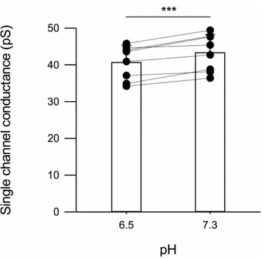
Fig. 12. Extracellular acidosis produces a small but significant reduction of single channel chord conductance of human NR1/NR2A NMDAR.
Mean (bar) and individual (symbols) single channel chord conductance values at two different values of pHe 6.5, 7.3 (n=8) *** P<0.001 paired t-test.
38
3.3. Extracellular acidosis differently modulates NMDAR-evoked whole-cell currents and Ca2+ entry in mouse cortical neurons in culture.
Basing on our findings, we hypothesized that also in native neurons an acid pHe should reduce to a greater extent the Ca2+ entry due to NMDAR activation, rather than the NMDAR-mediated whole-cell currents. To investigate this hypothesis, considering that in neurons is not possible to reliably measure the Pf due to diffusion and clamp limitations, we decided to measure disjointedly Ca2+ transients and whole-cell currents evoked by NMDA application on cortical neurons in culture (Fig. 13 A,B). Experiments were performed coapplying NMDA and glycine as above, at pHe values of 7.3 and 6.5. As expected from previous studies (Tang et al., 1990; Traynelis and Cull-Candy, 1990, 1991; Vyklický et al., 1990) we observed a significant reduction of both NMDA-evoked Ca2+ transients (Fig 13 A) and NMDA-evoked whole-cell currents (Fig 13 B) when pHe was decreased from 7.3 to 6.5. It is relevant to note that the extracellular acidosis did not affect [Ca2+]
i and currents at the same extent: the NMDA-evoked Ca2+ influx decreased significantly more than NMDA-evoked whole-cell currents (35.7 ± 19.7 % vs 53.4 ± 10.0 %; Fig. 14), confirming our hypothesis and suggesting that the proton-induced modulation of NMDAR Pf could exert significant effects on the accumulation of Ca2+ ions in native neurons. The reduction of whole-cell current amplitudes is very similar to what observed in HeLa cells transfected with human NR1/NR2A NMDARs in the same kind of experiments (52 ± 20 %, not shown), suggesting that mouse and human receptors are modulated in the same way by acidosis.
Interestingly, in the absence of any agonist the application of external solution at pH 6.5 evoked small but evident Ca2+ transients in neurons, likely due to the opening of proton-gated ion channels (Fig. 13 A; (Waldmann et al., 1997)). However, neurons exhibited acid-evoked Ca2+ entry at pHe 6.5 rather small and transient if compared with the Ca2+ entry through NMDARs, with a completely different distribution (Fig. 15), suggesting a negligible proton-induced neurotoxicity at this level of pHe.
39
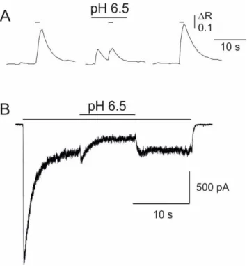
Fig. 13. Extracellular acidosis reduces both NMDA-evoked Ca2+ transients and NMDA-evoked whole-cell currents in cortical neurons in culture.
A, Ca2+ transients recorded from a mouse cortical neuron in culture elicited by coapplication of NMDA and glycine (200M and 50M, respectively; short horizontal bars). pHe was transiently shifted during agonist application from 7.3 to 6.5 (3 second application, as indicated). Please note the Ca2+ transient elicited by the application of the external solution at pH 6.5 immediately before the application of agonists. B, whole-cell current elicited by coapplication of NMDA and glycine (200M and 50M, respectively; long horizontal bar) recorded from a different cortical neuron. pHe was transiently shifted during agonist application from 7.3 to 6.5, as indicated. Please note the small and transient inward current elicited by protons and the succeeding reduction of the NMDA-evoked current.
40
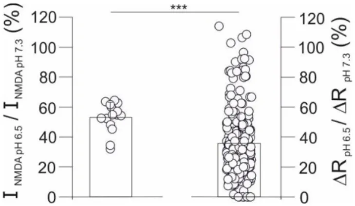
Fig. 14. Extracellular acidosis reduces significantly more NMDA-evoked Ca2+ transients than NMDA-evoked whole cell currents in cortical neurons in culture.
Percentage reduction of whole-cell currents (left, 53.4 ± 9.8 %; n=15) and Ca2+ transient amplitudes (right, 35.7 ± 19.7 %; n=318) recorded from individual mouse cortical neurons in culture when pHe was shifted from 7.3 to 6.5. Please note that Ca2+ transient amplitudes were significantly more reduced than currents. *** P <0.001 (ANOVA ).
41
Fig. 15. Ca2+ transients evoked by pHe 6.5 are smaller than NMDA-induced ones in mouse
cortical neurons in culture.
Distributions of the Ca2+ transient amplitudes elicited by application of an acid external solution (white bars, pHe 6.5) or by the coapplication of NMDA and glycine (200 M and 50 M, respectively; black bars, pHe 7.3).
42
3.4. In search of other cations able to modulate the Pf of human NMDARs
The reduction of the Pf of human NMDARs could be exploited to reduce glutamate neurotoxicity in pathological conditions. For this reason we looked for the action of different cations, starting with the effect of the presence of extracellular Mg2+ on the NMDAR Ca2+ permeability. Despite the strong reduction of the total current elicited in the presence of Mg2+ (2 mM) by NMDA in HeLa cells transfected with human NR1/NR2A receptors, it was possible to measure the Pf value: 9.9 ± 2.9 % (n=11, Tab. 1), not different from the same value in the absence of Mg2+. Furthermore, we measured the Pf of the same receptor in the presence of other larger organic cations: spermine (100 M), an intracellular polyammine known to block cation fluxes through ligand-gated channels (Benveniste and Mayer, 1993); memantine (1 M), an open channel blocker of NMDAR used in patients wit Alhemer’s disease (Parsons et al., 1999b); IEM 1754 (1 M) and IEM 1460 (100 M), two open channel blocker of NMDARs (Antonov and Johnson, 1996; Antonov et al., 1995).Unfortunately, none of these molecules was able to reduce the Pf value of human NMDA NR1/NR2A receptors (Tab. 1), suggesting the need of future studies to identify different kinds of cationic modulators.
Table 1.
Effect of different extracellular cations on Pf value of human NR1/NR2A NMDAR.
Cations Pf (mean ± SD) N + Mg2+ 9.9 ± 2.9 % 11 - Mg2+ 11.3 ± 4.4 % 9 Spermine 8.6 ± 3 % 14 Memantine 16.4 ± 2.8 % 4 IEM 1754 9.3 ± 1.7 % 4 IEM 1460 9.2 ± 3 % 11
43
4. Discussion
Extracellular acidosis and overactivation of NMDARs, with consequent Ca2+ ions overload, are two pathogenic markers of brain ischemia (Doyle et al., 2008). NMDARs represent a suitable target for a pharmacological strategy against ischemic damage, but to date no drug show to be useful for neuroprotection, considering that most molecules interfere with the normal neuronal function and cause substantial side effects at potentially therapeutic doses (Ikonomidou and Turski, 2002).
Here we report that mild extracellular acidosis (pHe 6.5; (Tombaugh and Sapolsky, 1990)) reduces the Ca2+ permeability of human NMDARs, contributing to limit the Ca2+ entry through these receptors. Using simultaneous recordings of whole-cell currents and Ca2+ transients elicited by NMDA application, we showed that the fractional Ca2+ current (Pf, i.e. the percentage of total current carried by Ca2+) of both NR1/NR2A and NR1/NR2B human NMDARs is significantly reduced during extracellular acidosis compared to Pf measured at physiological pHe . This reduction of Ca2+ flux has been also confirmed for NMDARs expressed in native mouse cortical neurons. Taken together, these results show a new molecular mechanism underlying the neuroprotective effect of extracellular mild acidosis (Giffard et al., 1990; Tombaugh and Sapolsky, 1990), providing an important contribute in the search of a new strategy against glutamate-mediated neurotoxicity, mainly due to the an excess of Ca2+ ions flowing through NMDARs (Hardingham and Bading, 2003).
Acidification of pHe occurs in CNS during normal neuronal functions and modulates ligand-,voltage- and proton-gated ion channels (Obara et al., 2008), but is also involved in several brain acute pathologies and neurodegenerative disorders causing acidosis-mediated cell death (Obara et al., 2008; Wang and Xu, 2011). Acidotoxicity and excitotoxicity are two major cell death mechanisms in ischemic brain, and for this reason several studies focused their attention to the modulation of rat NMDARs by extracellular mild acidosis (Giffard et al., 1990; Tang et al., 1990; Traynelis and Cull-Candy, 1990, 1991; Vyklický et al., 1990). These studies clearly showed that extracellular acidosis reduced NMDARs open probability with a consequent reduction of NMDA-evoked currents. Our results confirm the acidosis-induced reduction of NMDA activation also for human receptors, with a relevant novelty: extracellular acidosis significantly and selectively reduces the Ca2+ flux through open NMDA receptor-channels. The high Ca2+ permeability of NMDARs is due to several Ca2+ binding sites within the pore as well as in external vestibule, depends on subunit composition with receptors containing NR2A and NR2B showing the highest Ca2+ permeability (Burnashev et al., 1995; Traynelis et al.,
44
2010). Previous studies demonstrated that the Pf of NMDARs and others receptor channels can be physiologically and pharmacologically modulated (Piccari et al., 2011; Skeberdis et al., 2006; Sobczyk and Svoboda, 2007). Our work reports a significant reduction of Pf of human NR1/NR2A and NR1/NR2B NMDARs in extracellular acidosis, adding new evidence about the possibility to modulate the Ca2+ permeability of ligand-gated ion channels.
Previous studies stated that an acidification in pHe strongly decreased the NMDA channel opening frequency without changing unitary conductance (Tang et al., 1990; Traynelis and Cull-Candy, 1991). Thus, the H+ binding site on NMDARs is thought to be closely associated to gating site, but the exact mechanism of action is still unclear (Banke et al., 2005; Low et al., 2003). Our analysis of single-channel activity of human NR1/NR2A NMDARs in outside-out patches confirms a reduction in frequency of channel openings, highlighting a small but significant decrease of the single channel conductance. This finding is in good agreement with the reduction of ion fluxes expected from the observed reduction of Pf. In particular, combining our Pf and single channel data, it is possible to estimate that, upon shift of the pHe from 7.3 to 6.5, the Na+ flux through human NMDAR is almost unaffected (from 38.5 pS to 37.6 pS, 98%), while the Ca2+ flux is much more reduced (from 4.8 pS to 3.0 pS, 63%). The apparent discrepancy with previous studies is likely due to different experimental conditions: rat vs human NMDARs, low extracellular concentration of Ca2+ ions different pHe values (Traynelis and Cull-Candy, 1990, 1991; Vyklický et al., 1990). The reduction of single channel conductance for human NR1/NR2A NMDARs and the reduction of the Pf values for both NR1/NR2A and NR1/NR2B receptors upon acidosis, suggest that extracellular H+ modulate Ca2+/Na+ permeation of receptors binding negative charged residues on NR1 subunit. One of possible H+ binding site could be a cluster of charged residues (DRPEER) located at extracellular vestibule in NR1 subunit that represents a key determinant of high Ca2+ permeability of NMDARs (Watanabe et al., 2002) but further studies will be necessary to identify the exact mechanism of the H+-induced reduction the NMDAR Ca2+ permeability. Extracellular acidosis occurs during normal neuronal functioning in CNS and modulates several ligand-, voltage- and proton-gated ion channels(Obara et al., 2008). As expected from previous studies (Tang et al., 1990; Traynelis and Cull-Candy, 1990, 1991), our experiments in cultured cortical neurons show that both NMDA-evoked Ca2+ mobilization and whole-cell current amplitudes are strongly reduced upon mild acidification. However, the two reductions are not proportional, with NMDA Ca2+ mobilization significantly more reduced than NMDA currents, further supporting the hypothesis of a differential effect of protons on Ca2+ and Na+ fluxes. All the experimental data show that mild acidosis exert a dual action on NMDARs: a reduction of
45
open probability plus a selective reduction of Ca2+ permeability. Combining these two effects, it is possible to quantitatively estimate the overall reduction of NMDAR-mediated Ca2+ influx, passing from pHe 7.3 to pHe 6.5 to 35 %, due to the decrease of both NMDA currents and Pf (to 52 % and 67 %, respectively, for human NMDA NR1/NR2A receptor; Fig.16).
Fig. 16 Percentage of decrease of NMDA evoked whole cell current and Pf due to pHe 6.5 in
human NR1/NR2A NMDAR transfected in HeLa cells.
The extracellular acidosis is known to activate acid sensing ion channels (ASICs; (Waldmann et al., 1997)), proton-gated channels expressed throughout the nervous system. To date seven isoforms have been identified, but only 1a, 2a and 2b subtypes are present in CNS neurons (Wemmie et al., 2006) The homomeric ASIC1a channels are highly permeable to Ca2+, whereas the other subtypes are only permeable to Na+ (Yermolaieva et al., 2004). Activation of ASICs1a is due to extracellular pH ranging from 6.9-5.0 with a pH50 of 5.8 (Hesselager et al., 2004) and increases the probability of action potential initiation (Vukicevic and Kellenberger, 2004). A coupling between NMDARs and ASIC1a channels worsen the neuronal conditions during ischemic damage, leading to cell death (Simon, 2006). In our experiments we show that in the absence of agonist, the pHe 6.5 solution elicited small Ca2+ transients likely due to ASIC activation. These Ca2+ transients were much smaller than those evoked by NMDA at pHe 7.3, suggesting a negligible contribution of ASICs to neuronal toxicity at pHe 6.5, in agreement with their known pH50 values (Hesselager et al., 2004).
As expected, extracellular Mg2+, present in all our experiments on neurons, is able to strongly decrease the channel open probability at hyperpolarized potentials (Nowak et al., 1984), but we show here that it does not affect the Ca2+ permeability. Hence, the residual NMDA-evoked current, observed in neurons in the presence of Mg2+ at a holding potential -70 mV, is sufficient to mediate a relevant Ca2+ influx, which can contribute to neurotoxicity in the prolonged
46
presence of extracellular glutamate. Besides Mg2+, we tried to identify cationic molecules able to decrease the Pf of human NMDARs, to be characterized as new pharmacological tools against excitotoxity due to excessive glutamate-induced Ca2+ entry. Unfortunately, our effort were not successful. Further studies will be necessary to unveil possible pharmacological tools, as well as to explain the exact molecular mechanism by which extracellular H+ modulate NMDARs ion selectivity.
48
5. Conclusions
The data presented in this study showed that extracellular mild acidosis: 1) reduces the Ca2+ permeability of human NMDARs;
2) slightly but significantly reduces the single-channel conductance of human NMDARs, as expected quantitatively from the reduction of Ca2+ influx;
3) reduces the NMDA-induced Ca2+ mobilization in cultured neurons, more than expected from the observed reduction of NMDA inward currents.
These data confirm the results of previous studies, according to which extracellular mild acidosis decreases the effect of glutamatergic neurotransmission reducing NMDA-evoked whole cell currents, and adds new evidence demonstrating that the neuroprotective action of mild acidosis is also due to a reduction of Ca2+ flux compared to Na+ fluxthrough NMDARs, Thus, this study provides a proof of concept of the hypothesis that it is possible to reduce Ca2+ overload in neurons, and hence excitotoxicity, modulating the NMDAR Ca2+ permeability, without blocking these receptor channels. Although to date several tested molecules have failed in attaining this goal, this therapeutic strategy will remain the focus of future studies, aiming to unveil the mechanisms and the sites of the H+ modulation of human NMDARs, thus allowing a targeted pharmacological design.
49
References
Anis, N.A., Berry, S.C., Burton, N.R., and Lodge, D. (1983). The dissociative anaesthetics, ketamine and phencyclidine, selectively reduce excitation of central mammalian neurones by N-methyl-aspartate. Br. J. Pharmacol. 79, 565–575.
Antonov, S.M., and Johnson, J.W. (1996). Voltage-dependent interaction of open-channel blocking molecules with gating of NMDA receptors in rat cortical neurons. J. Physiol. 493, 425–445.
Antonov, S.M., Johnson, J.W., Lukomskaya, N.Y., Potapyeva, N.N., Gmiro, V.E., and Magazanik, L.G. (1995). Novel adamantane derivatives act as blockers of open ligand-gated channels and as anticonvulsants. Mol. Pharmacol. 47, 558–567.
Artola, A., and Singer, W. (1987). Long-term potentiation and NMDA receptors in rat visual cortex. Nature 330, 649–652.
Arundine, M., and Tymianski, M. (2003). Molecular mechanisms of calcium-dependent neurodegeneration in excitotoxicity. Cell Calcium 34, 325–337.
Banke, T.G., Dravid, S.M., and Traynelis, S.F. (2005). Protons trap NR1/NR2B NMDA receptors in a nonconducting state. J. Neurosci. Off. J. Soc. Neurosci. 25, 42–51.
Beck, C., Wollmuth, L.P., Seeburg, P.H., Sakmann, B., and Kuner, T. (1999). NMDAR Channel Segments Forming the Extracellular Vestibule Inferred from the Accessibility of Substituted Cysteines. Neuron 22, 559–570.
Benveniste, M., and Mayer, M.L. (1993). Multiple effects of spermine on N-methyl-D-aspartic acid receptor responses of rat cultured hippocampal neurones. J. Physiol. 464, 131– 163.
Burnashev, N., Schoepfer, R., Monyer, H., Ruppersberg, J.P., Günther, W., Seeburg, P.H., and Sakmann, B. (1992). Control by asparagine residues of calcium permeability and magnesium blockade in the NMDA receptor. Science 257, 1415–1419.
Burnashev, N., Zhou, Z., Neher, E., and Sakmann, B. (1995). Fractional calcium currents through recombinant GluR channels of the NMDA, AMPA and kainate receptor subtypes. J. Physiol. 485 ( Pt 2), 403–418.
Chaffey, H., and Chazot, P.L. (2008). NMDA receptor subtypes: Structure, function and therapeutics. Curr. Anaesth. Crit. Care 19, 183–201.
Chen, N., Moshaver, A., and Raymond, L.A. (1997). Differential sensitivity of recombinant N-methyl-D-aspartate receptor subtypes to zinc inhibition. Mol. Pharmacol. 51, 1015–1023. Chen, Y.H., Wu, M.L., and Fu, W.M. (1998). Regulation of presynaptic NMDA responses by external and intracellular pH changes at developing neuromuscular synapses. J. Neurosci. Off. J. Soc. Neurosci. 18, 2982–2990.
Chesler, M., and Kaila, K. (1992). Modulation of pH by neuronal activity. Trends Neurosci.
50
Choi, D.W. (1985). Glutamate neurotoxicity in cortical cell culture is calcium dependent. Neurosci. Lett. 58, 293–297.
Choi, D.W. (1987). Ionic dependence of glutamate neurotoxicity. J. Neurosci. 7, 369–379. Choi, D.W., Koh, J.Y., and Peters, S. (1988). Pharmacology of glutamate neurotoxicity in cortical cell culture: attenuation by NMDA antagonists. J. Neurosci. Off. J. Soc. Neurosci. 8, 185–196.
Chu, X.-P., Zhu, X.-M., Wei, W.-L., Li, G.-H., Simon, R.P., MacDonald, J.F., and Xiong, Z.-G. (2003). Acidosis decreases low Ca(2+)-induced neuronal excitation by inhibiting the activity of calcium-sensing cation channels in cultured mouse hippocampal neurons. J. Physiol. 550, 385–399.
Clark, G.D., Cufford, D.B., and Zorumski, C.F. (1990). The effect of agonist concentration, membrane voltage and calcium on n-methyl-d-aspartate receptor desensitization.
Neuroscience 39, 787–797.
Collingridge, G.L., and Bliss, T.V.P. (1987). NMDA receptors - their role in long-term potentiation. Trends Neurosci. 10, 288–293.
Cull-Candy, S.G. (2007). NMDA Receptors. In Encyclopedia of Life Sciences, John Wiley & Sons, Ltd, ed. (Chichester, UK: John Wiley & Sons, Ltd), p.
Cull-Candy, S.G., and Leszkiewicz, D.N. (2004). Role of Distinct NMDA Receptor Subtypes at Central Synapses. Sci STKE 2004, re16–re16.
Cull-Candy, S., Brickley, S., and Farrant, M. (2001). NMDA receptor subunits: diversity, development and disease. Curr. Opin. Neurobiol. 11, 327–335.
Dingledine, R., Borges, K., Bowie, D., and Traynelis, S.F. (1999). The Glutamate Receptor Ion Channels. Pharmacol. Rev. 51, 7–62.
Doyle, K.P., Simon, R.P., and Stenzel-Poore, M.P. (2008). Mechanisms of ischemic brain damage. Neuropharmacology 55, 310–318.
Dravid, S.M., Erreger, K., Yuan, H., Nicholson, K., Le, P., Lyuboslavsky, P., Almonte, A., Murray, E., Mosely, C., Barber, J., et al. (2007). Subunit-specific mechanisms and proton sensitivity of NMDA receptor channel block. J. Physiol. 581, 107–128.
Fan, X., Jin, W.Y., and Wang, Y.T. (2014). The NMDA receptor complex: a multifunctional machine at the glutamatergic synapse. Front. Cell. Neurosci. 8, 160.
Forrest, D., Yuzaki, M., Soares, H.D., Ng, L., Luk, D.C., Sheng, M., Stewart, C.L., Morgan, J.I., Connor, J.A., and Curran, T. (1994). Targeted disruption of NMDA receptor 1 gene abolishes NMDA response and results in neonatal death. Neuron 13, 325–338.
Giffard, R.G., Monyer, H., Christine, C.W., and Choi, D.W. (1990). Acidosis reduces NMDA receptor activation, glutamate neurotoxicity, and oxygen-glucose deprivation neuronal injury in cortical cultures. Brain Res. 506, 339–342.