1
Università degli studi di Catania
Dottorato di ricerca
Basic and Applied Biomedical Sciences
XXIX ciclo
Synthetic vs biological meshes:
can in vitro cellular responses predict the outcome in patients?
Literature review, experimental study and clinical experience in Day
Surgery and inpatients related to the improvement of quality of life
Dottoranda: Dott.ssa Marta Cavalli Tutor: Prof. Antonio Biondi
2
Sommario
Background ... 3
Literature review ... 6
The ideal mesh ... 6
Biological graft ... 6
Collagen cross-linking ... 8
Mesh integration ... 8
Foreign body response ... 9
Neocellularization and Neovascularization ... 10
Matrix Remodeling ... 10
Experimental study... 12
Materials and methods ... 12
Results ... 13
Morphologic analyses by SEM ... 13
Cell counting ... 18
Discussion ... 19
Conclusion ... 20
Clinical experience... 21
Materials and methods ... 21
Results ... 24
Discussion ... 25
Conclusion ... 30
References... 31
3
Background
Abdominal wall hernia surgical repair is a common procedure (1) (2). Over time, hernia repair has evolved from predominantly primary suture repairs to tension-free repairs with mesh (3). Open suture ventral hernia repairs are associated with recurrence rates even of 63% at 10 years’ follow-up (4). The advent of mesh repair has led to a significant decrease in this unacceptably high recurrence rate; one recent Cochrane meta-analysis of three studies demonstrated a pooled recurrence rate of 16% (5). About one million prostheses are used worldwide for abdominal wall repair each year (6) and, since the first description of use of a synthetic mesh for abdominal wall repair (3), a plenty of new materials have been introduced, leading to a considerable reduction in recurrence rates.
Synthetic meshes induce a strong foreign inflammatory reaction and a scar tissue is provided as a permanent repair to the abdominal wall defect with enough mechanical strength. Despite a substantial decrease in the recurrence rate after tension-free repair, the use of mesh is not without its own complications.
Infection is the most important and difficult to treat because bacteria avidly adhere to synthetic polymers and synthesize biofilms. These biofilms provide resistance to host immunologic defenses and hematogenous delivery of antibiotics leading to chronic infection of the tissue repair site. Typically, infections necessitate explantation of the mesh and the patient is left with a colonized abdominal wall defect (2).
Seroma formation, adhesions, chronic pain, erosion into bowel, fistula formation and bowel obstruction are other known complications of abdominal wall repair with mesh (7) (8) (9) (10) (11). So, the consequences of synthetic mesh failure can be catastrophic, especially in complex hernia repair in contaminated or clean–contaminated tissue, with bowel or fistula exposition and significant loss of domain (9).
Designed in response to these complications, composite meshes had should help eliminate some of the complications observed with synthetic mesh: a barrier layer on the visceral side of the mesh should decrease adhesion formation between the viscera and the mesh. However, several studier reported adhesion formation even with the use of composite meshes (9) (12) (13) (4) (14) (15).
4
In the attempt to reduce the possibility of these fearsome complications, the use of biological meshes in abdominal wall hernia repair was introduced (16).
Biologic meshes are derived from humans and animals and become incorporated into the wound, acting as a scaffold for tissue repair, leading to a strong, well-healed, vascularised wound (17). Due to the nature of biologics, the adhesions associated with synthetic mesh should not occur and neovascularization should soon allow delivery of immune cells and antibiotics (11).
Although biological meshes are gaining popularity, the exact molecular mechanism leading to host reaction and biological graft integration remains unclear and poorly understood by the surgical community.
However, even biologic grafts are not free from failure: inadequate mesh incorporation or degradation of implanted meshes have been reported (18) (19).
To date, in the literature there are no clear, uniform indications on usage, reasons to choose one type mesh over another or specific clinical indications.
Retrospective consideration of biologic mesh efficacy is compromised due to the compounding variables induced throughout their application: the variety of available products, the techniques used in hernia repair (onlay, inlay, sublay placement, component separation accomplished or not, double-mesh technique), the complex nature of hernias (size, site and potential or confirmed tissue contamination or infection), the body mass index (BMI), diabetes mellitus, smoking status (20) (21). If we could identify a cellular and molecular response towards the different type of mesh, it would afterwards be possible to understand if the body response could be foreseen and managed proactively rather than only reactively and try also to tailor therapies in case of secondary infections or adherences. Moreover, this type of knowledge could be exploited to optimize pretreatments both for artificial and biological meshes.
Without this knowledge, the improvement of mesh use will be mainly driven by ex-post studies, that are first based on human trials, very long, extremely costly and always not optimized in terms of homogeneity of data samples. Other studies use animal models, procedure that again presents elevated costs and drawbacks both from the ethical point of view and in terms of translation to human application.
5
Any information that can help to foresee an advantage in using a specific mesh in a specific setting or in a technical improvement of a specific mesh may provide an advantage both in terms of health care and economically.
In the first part of this thesis a review of all data available now about biological grafts and their interaction with tissue is presented.
In the second part our laboratory results concerning interaction between different matrices (synthetic and biological) used in abdominal wall repair surgery and primary fibroblast cultures are reported. Finally, in the third part, our clinical experience with biological meshes is described.
6
Literature review
The ideal mesh
Cumberland (22) and Scales (23) originally described the characteristics of the ideal prosthetic in the 1950s, and Hamer-Hodges and Scott (24) adapted the description in 1985.
The ideal synthetic mesh possesses 8 characteristics: non-carcinogenic, chemically inert, resistant to mechanical strain, capable of being sterilized, inert to body and tissue fluids, capable of limiting foreign body reaction in the host, amenable to fabrication in the necessary for, and unlikely to produce allergy or hypersensitivity reactions (24). With the introduction of biological mesh, additional criteria for the ideal prosthetic have been proposed: namely, the material must resist infection, provide a barrier to adhesions on the visceral side, and must respond in vivo similarly to autologous tissue (11). Additionally, an ideal material should be associated with very little surgical morbidity, such as seroma, easy to handle in open and laparoscopic instances, and cost effective.
Biological graft
Biologic mesh was introduced in clinical surgery as an alternative to synthetic mesh for abdominal incisional hernia repair in the late 1990s (25), as development of the success of autologous tensor fascia lata grafts for necrotizing abdominal wall infections, enteric fistulae, or exposed prosthetic material after ventral hernia repair. (11)
Biological meshes are considered as a scaffold for the binding of growth factors and other cellular elements necessary for healing response. The subsequent healing response and its strength are dependent on invasion of the extracellular matrix (ECM) of the biologic graft by patient cells, including endothelial cells.
The balance between ECM synthesis and degradation contributes to the ultimate success of the hernia repair.
Current biological meshes present in the market (tab. 1) differ in the mammalian source (animal or human), tissue of origin (dermal, pericardial, bladder or intestinal submucosa), as well as their processing method and sterilization.
7
Human cadaveric tissues offer the advantage of using allograft (within species) sourcing and thus lacking interspecies rejection risk. The source of such tissues is donor dependent, with variability in composition, health, thickness, and age of the tissue.
Alternatively, animals can be raised to precise specifications to achieve a more consistent product. The risk of allergic response to their ECM is low because of the high homology with similar human proteins. With nonhuman tissues, the risk of tissue rejection remains despite decellularization, as does the rare possibility of disease transmission.
Biological grafts vary in their tissue of origin. The dermis remains the preferred tissue source, though products made from alternative tissues, such as the pericardium, stomach, bladder, and intestinal submucosa, are also available.
While tissue source may certainly affect how a given mesh is reacted upon by the recipient, differences in tissue reaction are likely a result of the different methods of processing, decellularization, and sterilization used. Manufacturers utilize various proprietary methods and processing solvents that likely influence the innate biochemical and biomolecular structure of the collagen scaffold. Subsequently, these matrix alterations likely influence “foreign body” recognition and antigen presentation. In fact, it has been suggested that the manufacturing process for each mesh may be more critical to implant function than the source and the species from which the mesh is derived (26).
Table 1 Bioactive prosthetic materials
Mesh name Vendor Source Cross-linking
Alloderm LifeCell-Acelity Human dermis No
AlloMax Bard/Davol Human dermis No
CollaMend Bard/Davol Porcine dermis Yes
FlexHD Ethicon Human dermis No
FortaGen Organogenesis Porcine intestine Yes
MatriStem ACell Porcine bladder No
Peri-Guard Synovis Bovine pericardium Yes
Permacol Covidien Porcine dermis Yes
Strattice LifeCell-Acelity Porcine dermis No SurgiMend TEI Biosciences Fetal bovine dermis No Surgisis-Biodesign Cook Medical Porcine intestine No Tutopatch Tutogen Medical Bovine pericardium No
8
Veritas Synovis Bovine pericardium No
XCM Biologic Tissue Matrix® Synthes Porcine dermis No
XenMatrix Bard/Davol Porcine dermis No
Collagen cross-linking
Of all the various processing techniques used by manufacturers, collagen cross-linking is likely to have the most profound effect on tissue responses to biological meshes. Crosslinking is a biochemical process that, by using hexamethylene diisocyanate, carbodiimide, glutaraldehyde, or photo-oxidizing agents (27) (28), results in the creation of bonds between the collagen triple helices of the biologic scaffold and is believed to enhance the strength and durability of the scaffold (29), Even non-intentionally cross-linked products may undergo molecular and structural changes, such as collagen cross-linking, from gamma irradiation during the sterilization process (30). In addition, incomplete removal of chemical cross-linking agents could result in cytotoxicity from residues leaching from the mesh itself, which may induce prolonged toxic effects and heightened cellular responses (27). While clinical circumstances, requiring long-term tissue reinforcement, may provide some utility for a linked graft, numerous investigators have recently reported disadvantages of chemical cross-linking in both translational animal models and the clinical setting (31) (19) (32) (33).
Cross-linked grafts seem to be typically associated with encapsulation or prolonged inflammatory response characterized by foreign body giant cell reaction (34) (31) (33) (19) (32).
Mesh integration
Biological mesh integration remains an important and desirable outcome. However, this process is poorly understood and is often difficult to identify and quantify/measure the integration rate.
It appears that a cascade of events follows mesh implantation. After the placement of a mesh into the host, an acute inflammatory response takes place. This is a necessary event in wound healing and, obviously, it is highly influential in biologic mesh performance. While meshes with diminished biocompatibility do not allow for such colonization, grafts that are positively recognized by a host will have host cells migrating from the periphery of the mesh inward, thanks to ECM degradation by metalloprotease.
9
There are three main cellular players that participate in mesh ECM reabsorption/degradation, colonization and new ECM deposition: mononuclear cells (macrophages and mast cells), endothelial cells and fibroblasts. Once mononuclear cells populate the graft, the typical sequence of wound healing events likely takes place. Mononuclear cells secrete cytokines and other signaling factors to attract other cells, such as endothelial cells and fibroblasts. Endothelial cells ensure that new blood vessels are formed. Once fibroblasts are on site, new collagen synthesis and deposition takes place. Importantly, this process has to occur not only at the mesh/host interface but also within the graft itself. This would predispose a biological graft for ingrowth, incorporation, and new collagen deposition within the mesh (35).
Foreign body response
Inflammation appears to be a common component of host response to implanted biologic prosthetics (26) (32) (29) (36). This reaction may either aid in the integration of the mesh via normal wound healing mechanisms or induce a disproportionate inflammatory response. Such an exaggerated reaction will likely result in excessive scarring, graft encapsulation, and/or degradation (26) (32) (18). The balance between appropriate wound healing and detrimental sequelae is largely controlled by cytokines, growth factors, and other chemical signaling molecules produced by host macrophages at the site of host/mesh interface. Orenstein et al. were the first to evaluate the immunogenic potential of various human-derived and porcine-derived biologic meshes in vitro (32) (37). Regarding the former, AlloDerm appeared to induce the smallest degree of cytokine production, indicative of superior biocompatibility (37). Regarding porcine meshes, non-cross-linked porcine dermis mesh and, to a lesser degree, porcine intestinal submucosa–derived mesh, were associated with a markedly diminished cytokine production as compared with the cross-linked porcine dermis materials. While the exact clinical importance of the excessive macrophage activation in vitro is unclear, that early evidence of adverse effects of chemical cross-linking has subsequently been corroborated by a number of in vivo studies (26) (33) (18) (38).
Most recently, Petter-Puchner et al. reported a pronounced foreign body response to intraperitoneal implantation of Collamend and Surgisis in rats. It was observed that both meshes were surrounded by a broad rim of foreign body giant cells and granulomas (31). In contrast, another recent industry-sponsored study surprisingly found no evidence of inflammatory or immune response to Permacol (18).
10 Neocellularization and Neovascularization
Early cellular and vascular infiltration of a biologic matrix is critical for mesh integration. Monocyte/macrophage penetration of the graft from the surface inward is paramount for fibroblast proliferation and new collagen deposition. In the absence of angiogenesis, remodeling will not occur and the matrix will be replaced by scar. Butler et al. found that non-cross-linked porcine dermis promoted early cellular and vascular infiltration and likely contributed to a stronger mesh/musculofascia interface (33). Xu et al. reported that functional blood vessels paralleled host cell repopulation with clearly delineated channels lined with endothelial cells in human dermis by 1 month after implantation in primates (36). These findings were confirmed more recently in a sublay biologic mesh study in rats (18). The authors found that the AlloDerm group was associated with 100 percent neocellularity by 3 months after implantation. Neovascularization was clearly noted to support the cells. Finally, a normal, nondenatured collagen pattern was seen, indicative of remodeling and new collagen deposition (18). Of note, these findings were not seen in the cross-linked porcine dermis groups.
It is also important to point out, however, that Deeken et al. have recently reported that although cross-linking affected biological meshes with regard to cellular infiltration and neovascularization early on, those histologic features were no longer affected by cross-linking at a 1-year time point. While these isolated results are intriguing, it is unclear whether the findings by Deeken et al. are true representations of what happens in humans or just one of the limitations of long-term ventral hernia/biologic mesh investigation in resilient animal models such as the minipig.
Matrix Remodeling
The final and most important step in biologic mesh placement is graft integration and remodeling with new collagen deposition and tissue regeneration. Melman et al. suggested that when scaffold degradation is accompanied by cellular infiltration, ECM deposition and neovascularization, it can be viewed as remodeling (39). At times, however, when ECM deposition/neovascularization does not occur, mesh is likely replaced by a scar with a resultant detriment to a hernia repair. One of the other key factors that influence remodeling may be the rate of scaffold degradation (39). Almost uniform failure of absorbable meshes may be due to a fairly rapid degradation of the graft without proper support for new extracellular matrix components deposition. In fact, a gradual remodeling of an implanted tissue graft seems to be essential for abdominal wall repair because degradation or
11
absorption of a scaffold not balanced with deposition of new collagen would predispose to mesh failure (36)
Deeken et al. reported that non-cross-linked grafts exhibited more favorable remodeling characteristics. However, remodeling in their study was not associated with stronger reinforcement of native tissue repairs in the long term (29). These paradoxical results may be a consequence of the animal model used. Another recent study revealed essential lack of matrix absorption and absence of remodeling of cross-linked graft 6 months after implantation (18). Given its lack of integration into the host and likely resultant fibrous encapsulation, crosslinked grafts often act as permanent foreign body materials, similar to GoreTex-based synthetics (35).
It appears that the balance between extracellular matrix deposition and mesh degradation is critical for mesh remodeling and effective tissue reinforcement. Finally, while many investigators reported deposition of “new” matrix at the site of biologic mesh implantations, a typical scar plate developed as a part of normal wound healing could mimic regeneration. Distinguishing regenerated collagen within degraded scaffold versus fibrotic scar formation remains a challenge, even for experienced tissue histopathologists (35).
It is worth mentioning that one of the major limitations of biologic mesh research is the common animal models used. Most investigations are reported in rodents, guinea pigs, or minipigs. Those animals are chosen due to relative ease of implantation, low cost, and possible availability of genetic variants (mice) for future studies. However, tissue responses in those animals probably do not directly compare with those of humans, especially in the long term. Several investigators, on the other hand, have utilized the Old-World primates (26) (36). Those animals are highly homologous to humans in key components of the immune system. However, even though primates appear to be the most suitable animal model for translational biologic mesh research, ethical and financial constraints preclude widespread use of the primates for basic mesh investigations. Moreover, the most limiting aspect of animal research is not only in the chosen species but also that most investigations are performed under essentially ideal conditions. Typical patients undergoing biologic mesh repairs are likely to have multiple comorbidities, large abdominal wall defects, obesity, and various degrees of wound contamination. However, essentially no comparative investigations have been performed in anything but healthy animals.
12
Experimental study
The research has been performed under the direction of Prof. Paola Campomenosi of the Biotechnology and Life Science Department (DBSV) at University of Insubria.
Biotechnology and Life Science Department received a grant to conduct this research by LifeCell-Acelity.
This project aimed to understand aspects of the interaction between different matrices used in abdominal wall repair surgery and host cells, by using in vitro cell culture.
Materials and methods
The following four different types of matrix were used:
- Strattice, non-cross-linked acellularized porcine derma, LifeCell-Acelity,Branchburg, NJ, - Permacol, cross-linked acellularized porcine derma, Covidien, Mansfield, MA,
- Surgisis-Biodesign, non-cross-linked porcine intestinal submucosa, Cook Medical, Bloomington, Ind,
- Prolene, polypropylene highweight, monofilament, Somerville, NJ.
We used two different washing steps for different types of mesh: 1) for Strattice and Permacol:
- meshes were cut in small trapezoidal pieces for SEM (Scanning Electron Microscopy) analysis and in 1,5x1,5 cm squares for all other applications;
- mesh pieces were placed into a 50-ml conical tube containing 30 ml of sterile phosphate buffered saline (PBS);
- mesh pieces were washed extensively by placing the tube horizontally on an orbital shaker (about 100 rpm) for 2 hours at room temperature;
- PBS was substituted with 30 ml of fresh PBS; - washing was repeated as above three more times;
mesh pieces were placed in 12 wells tissue culture plates containing complete medium (RPMI 1650 + Fetal Bovine Serum + glutamine) and incubated overnight in a CO2 incubator at 37°C;
13
2) for Surgisis-Biodesign and Prolene
meshes were cut in small trapezoidal pieces for SEM analysis and in 1,5x1,5 cm squares for all other applications;
mesh pieces were washed for 5 minutes in PBS at room temperature, repeat for 5 times mesh pieces were washed (for equilibration) in complete medium (RPMI 1650 + Fetal Bovine
Serum + glutamine) for 5 minutes
mesh pieces were placed in 12 wells tissue culture plates containing complete medium and incubated over night as described for Strattice and Permacol;
the following day cells were seeded in fresh medium
Cells were seeded at 5x10⁴ cells/cm2 in a small volume on the matrices prepared as described above.
After two days from seeding, meshes were transferred on a new tissue culture plate, adding fresh medium. Two replicates for each type of matrix and each type of analyses were prepared and processed. Three days before each time point, medium was substituted with standard medium supplemented with Lyset. Analyses were performed at three time point: 10, 20 and 30 days.
The aim of SEM analyses was to analyze morphologically cell growth on each mesh surface.
Cell counting was performed with two different methods: a non-enzymatic method (Cell Titer Glo, Promega) (method 1) and an enzymatic detachment using a protease cocktail (composed of Accutase, trypsin and collagenase), followed by manual cell counting (method 2).
Gene expression analyses from RNA extraction are ongoing and further experiments with monocytes are planned.
Results
Morphologic analyses by SEM
Examination and acquisition of SEM images were performed in collaboration with Dr. Annalisa Grimaldi and Prof. Terenzio Congiu in the SEM facility of University of Insubria.
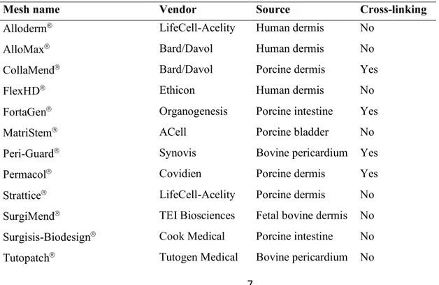
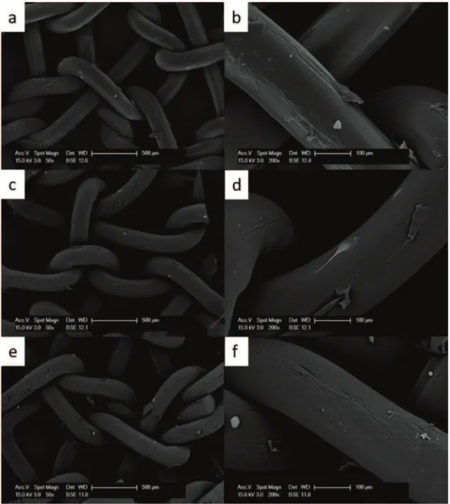
Strattice matrix (fig. 1) showed several cells adhering on the matrix surface on the upper surface, where cells were seeded. The number of cells was gradually increasing with time of incubation, indicating that they were healthy and proliferating. Cells were present also in the bottom surface,
14
although at lower numbers compared to the upper side. Cell distribution was uniform and cells were strongly interacting with the matrix.
Figura 1Growth of primary fibroblasts on the Strattice matrix analyzed at the SEM at 50x (left side) and 200x magnification (right side) at 10 days (a,b), 20 days (c,d) and 30 days (e,f).
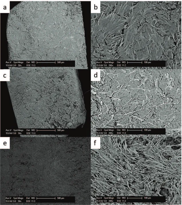
In the Permacol matrix at 10 days after seeding, cells were distributed unequally on the surface. The 50x magnification picture (fig. 2) shows a part of the matrix colonized by cells, while another part is free of cells. Unexpectedly, in some replicates at 20 days the number of cells on the surface was lower
15
than that at 10 days, whereas at 30 days the sample showed a monolayer of cells. Very few cells were found on the bottom of the matrix (opposite side respect to seeding).
Fig. 1 Growth of primary fibroblasts on the Permacol matrix analysed at the SEM at 50x (left side) and 200x magnification (right side) at 10 days (a,b), 20 days (c,d) and 30 days (e,f).
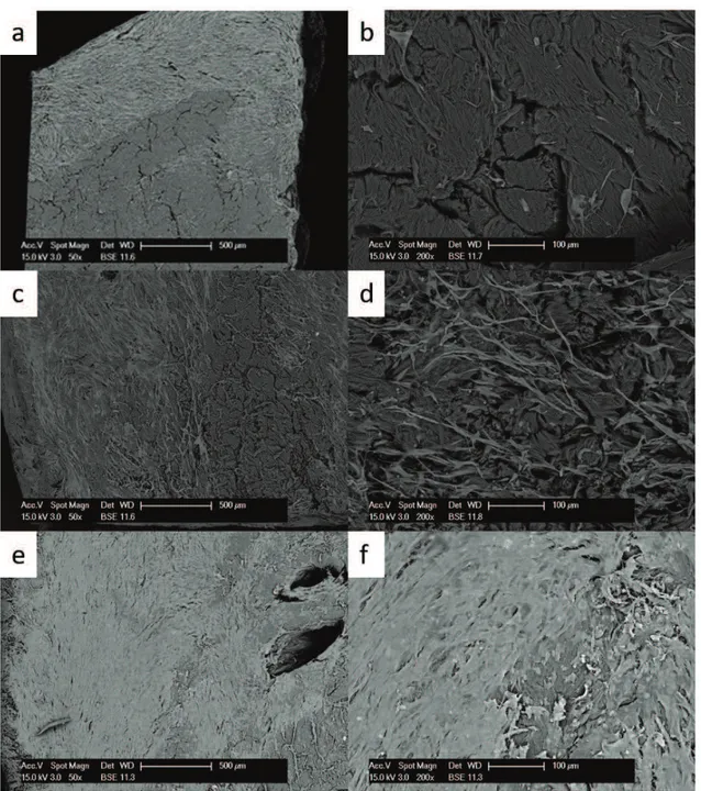
The Surgisis-Biodesign matrix (fig. 3) presented the largest number of cells, in particular at early times. At 10 days, cells covered the surface of the matrix and some cells were found also on the
16
bottom side. At 20 days, cells began to peel off. They were apparently arranged in different layers, but did not appear to really penetrate the matrix layers. At 30 days, unexpectedly a lower number of cells was observed.
Fig. 2 Growth of primary fibroblasts on the Surgisis-Biodesignl matrix analysed at the SEM at 50x (left side) and 200x magnification (right side) at 10 days (a,b), 20 days (c,d) and 30 days (e,f).
17
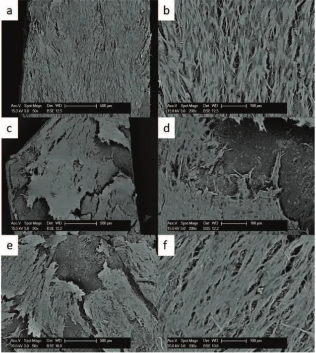
Cells seeded on this matrix passed through its large pore structure and did not seem to attach and grow easily on this material.
Fig. 3 Growth of primary fibroblasts on the Prolene mesh analysed at the SEM at 50x (left side) and 200x magnification (right side) at 10 (a,b), 20 (c,d) and 30 days (e,f).
18 Cell counting
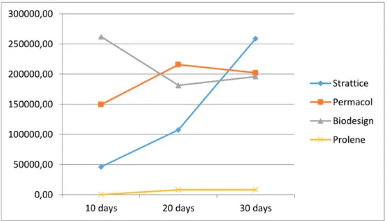
The data regarding cell counts obtained at each time point on the different types of matrix are presented on Figure 5: the mean between the numbers given by the two different cell counting methods are presented.
Fig. 5 Cell counting at 10, 20 and 30 days for each matrix
On the Strattice matrix a lower number of cells was counted at 10 days and 20 days compared to those on other biological matrixes at the same time point. However, the number of cells increased steadily at each time point on Strattice and at 30 days it was the highest among all types of mesh.
The number of cells growing on the Permacol matrix at each time point seemed to be quite variable, depending on the mesh piece examined. However, the mean cells number seemed to be higher than the cells counted on Strattice matrix only at 10 and 20 days, but less at 30 days.
The cells counted on the Surgisis-Biodesign at 10 days was the highest among all types of mesh at that time point. However, the number was reduced in the following time points, suggesting that this matrix was supporting adhesion, but the contacts made by cells did not strengthen with time and cells detached from the surface at the high densities reached in the following time points.
It is apparent that whatever the method used to evaluated cell growth on the Prolene synthetic mesh, almost no cells are retrieved from this matrix, observation that is corroborated by visual inspection by SEM analysis. 0,00 50000,00 100000,00 150000,00 200000,00 250000,00 300000,00
10 days 20 days 30 days
Strattice Permacol Biodesign Prolene
19
Cell counting recapitulated quite well the morphological observations about cell growth obtained at the SEM.
Discussion
Despite the rapid acceptance of biological material in surgical community, there are many answered questions as to the mechanism of action, ideal source material and effect of post-harvesting process methods. In order to better understand the cellular and molecular response towards different types of mesh, we seeded human fibroblast cells on different types of mesh, synthetic and biological meshes. Seeding cells onto surgical meshes, especially those with a large pore configuration, can be challenging given that only a very small number of cells would be in contact with mesh fibers. Gao et al. (40) approached this issue by placing their mesh on top of a near confluent monolayer of cells and by holding it in place with glass beads. With this method, cells tended to cover the whole culturing surface. Subsequently, they were forced to grow onto the mesh to avoid contact inhibition. Furthermore, new cells were dripped onto the mesh at every passage. Their results revealed that fibroblasts prefer Parietex (polyester multifilament mesh covered by a collegen-based hydrogel film) and Strattice, but their method failed to cover Marlex mesh. Marlex is a polypropylene mesh, very similar to Prolene mesh, that we used in our experiments. So, even if Gao et al. used a different protocol to cover the mesh, we obtained similar results. Gao et al. explained the cell-loading affinity of Parietex, a synthetic mesh too, considering its multifilament structure and the presence of collagen fiber.
To our knowledge this is the first study comparing cell-loading capacity in different biological meshes.
Our SEM analyses showed an interesting different behavior of fibroblasts on different types of biological mesh.
Cells growing on Strattice matrix were initially in lower numbers than those growing on other biological meshes. However, the cell numbers continued to consistently increase at each time point and, at 30 days, they were superior to those on other biological matrixes. Fibroblasts seemed to be healthy and proliferating and their distribution was uniform. Cells appeared to be adhere strongly to the matrix surface as confirmed by the number of cells retrieved for cell counting (cells were not lost during washes).
20
On Permacol matrix, cells were present in larger number than on Strattice mesh at early time point but they were non homogeneously distributed on the matrix surface and each replicate differed from the others. Cells seemed to interact weakly to the mesh surface, so that they were lost during sample preparation for SEM analysis and for cell counting.
The Surgisis-Biodesign matrix presented the largest number of cells, in particulary at early times, but cells seemed to interact strongly to each other, and more loosely to the matrix surface, so that they detached from it during matrix preparation for SEM analyses and for cell counting.
Gene expression analyses are in progress and maybe they can give us more details in cells and matrix interaction.
Conclusion
Our experimental study showed clear differences in behavior in vitro among synthetic prosthesis and different biological matrixes examined.
For understanding the reason of these differences and moreover their clinical implication, more preclinical studies are mandatory.
21
Clinical experience
After a large experience with more than 8000 surgical procedures for abdominal wall pathology, we set up a real “tailored” approach. In simple words, we try to find for every single patient the more suitable approach (laparoscopic or open, anterior, posterior or combined), anaesthesia, kind of mesh (synthetic, composite or biological,), fixation of the mesh (absorbable or not absorbable suture, fibrin glue, suturless) and component separation (anterior or posterior) if necessary.
We introduced biological mesh in our procedures 9 years ago, since that we used them with different clinical indications and now we reported our experience.
Materials and methods
We collected all the data about our experience in biological meshes in abdominal wall repair, in the last nine years.
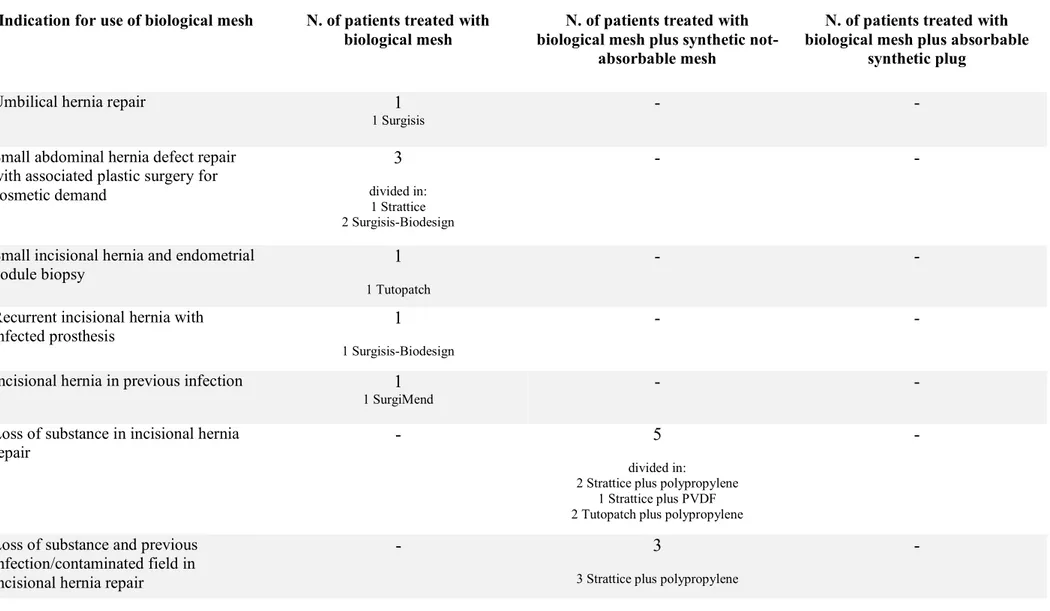
Since November 2007 to July 2016, we implanted 56 biological prosthesis in 54 patients: 24 Surgisis-Biodesign Cook (non-cross-linked porcine intestinal submucosa), 16 Strattice LifeCell-Acelity (non-cross-linked acellularized porcine derma), 12 Tutopatch Tutogen (non-(non-cross-linked bovine pericardium), 2 PeriGuard Synovis cross-linked bovine pericardium), 2 SurgiMend Tei (non-cross-linked fetal bovine derma). Patients age ranged between 7 and 81 years.
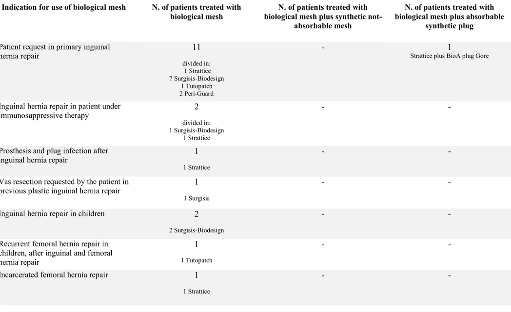
Indications for use of biological mesh are listed in tab. 2.
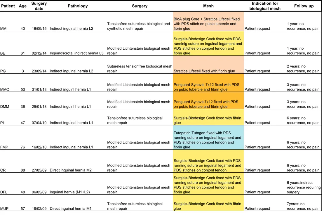
Limited to groin hernia repair, we collected 14 primary inguinal hernia repairs in adults, 2 of these were patients under immunosuppressive therapy (one after liver transplant and the second under corticosteroid therapy for rheumatoid arthritis). In the other 12 patients, we placed a biological mesh for specific request of the patient to have a non-synthetic prosthesis. Just in one of these patients with an indirect inguinal hernia (L2, according EHS classification (41)), we placed, in addition to the biological mesh on the posterior inguinal floor, a BioA® plug Gore (absorbable synthetic plug, polyglycolic acid-trimethylene carbonate) in the internal ring to fill the indirect defect.
We treated one prosthesis infection after plug and mesh inguinal hernia repair, removing the both infected prosthesis by a combined anterior and posterior approach to the inguinal channel throughout a high transversal inguinal incision and then placing a biological mesh in the preperitoneal space.
22
In a patient with a previous pure tissue inguinal hernia repair, we placed a biological mesh during surgery for vas resection for the reinforcement of the posterior inguinal floor.
We implanted also 3 biological meshes in three children: 2 in primary inguinal hernia repairs and one in recurrent femoral hernia, after a previous inguinal and femoral hernia tissue repair).
We chose a biological mesh for the repair of an incarcerated intravascular femoral hernia in a young female: the sac reduction was completed by a combined femoral and transinguinal approach and a biological mesh was placed in the preperitoneal space.
We reported 11 Pubic Inguinal Pain Syndrome (42) treatments, the so called “sports” hernia. In this kind of patient, we are used to propose a tailored open approach, under local anaesthesia, including nerve release, partial calibrated tenotomy of adductor longus and rectus abdominal muscles and reinforcement of the inguinal channel posterior wall with biological or lightweight synthetic mesh, as described in previous papers (42) (43). Choice between synthetic or biological mesh is usually made during surgery considering the degree of bulging of the posterior inguinal floor and the patient life style and physical activity.
We collected 7 post-operative inguinal chronic pain treatments with biological prosthesis in 6 patients: as reported in previous paper (44), in patient suffering by a chronic post-operative inguinal pain, we are used to approach the preperitoneal space in order to do the triple neurectomy (ilio-inguinal, ilio-hypogastric and genitofemoral nerve) and remove the plug previously placed, if present; then, through the same incision, we approach the anterior region to remove the mesh and the stitches, previously placed. Even if patient does not complain a recurrent hernia, inguinal region could be weakened by the removal of the previous repair, for this reason we normally complete surgery with the placement of an ultralight or biological mesh in the preperitoneal space. Once again, the choice between biological and synthetic mesh takes into account the degree of destruction of the inguinal floor and patient life style.
Regarding abdominal wall hernia repair, we collected 18 biological meshes in 17 patients.
In one umbilical hernia repair in a very thin female with defect size of 2 cm without rectus diastasis, we placed a flat biological mesh in the preperitoneal space.
In the three patients, complaining little abdominal wall defect and requiring a concomitant plastic surgery, the surgical steps were the following:
23
- first step, by plastic surgeon: suprapubic transversal incision between the two anterior superior iliac spines, round incision around the umbilicus and elevation of the abdominal subcutaneous until the xiphoid region;
- the second step, by us: opening of the rectus sheet and of the abdominal cavity, isolation and reduction of hernia, preparation of a retromuscolar-preperitoneal space until the retroxiphoid appendix, the Cooper ligament and the lateral edge of rectus muscle, accomplished of posterior component separation, closure of posterior rectus sheet, placement of the biological mesh fixed with fibrin glue sprayed on all the mesh surface (the umbilicus passed through a small incision in the mesh) and closure of the anterior rectus aponeurosis, keeping the umbilicus outside of the suture;
- The third step, by plastic surgeon: abdominoplasty with the excision of skin excess and replace of the umbilicus;
In one incisional hernia repair, after Pfannenstiel incision, after the biopsy of an endometrial nodule, a biological mesh was placed in the preperitoneal space.
We collected 9 patients with incisional hernia treated with biological mesh. In two patients, the peritoneal sheet seemed too thin and incomplete, so a biological mesh was placed to cover it, like a separating layer between the weak peritoneal and the synthetic not-absorbable mesh placed in the retromuscolar-preperitoneal space. In seven patients with loss of substance of the posterior compartment, despite a component separation done, the biological mesh was bridged to the border of the peritoneal defect, then a synthetic not-absorbable mesh was placed in the retromuscolar-preperitoneal space. Three patients of these suffered also a previous infection and contaminated field, while two of these had a contaminated field at the moment of surgery for bowel lesion during adhesiolysis in one case and for an enterocutaneous fistula in the other.
In one patient, presenting a recurrent incisional hernia and previous infected mesh, a biological mesh was placed in the retromuscolar-preperitoneal space.
In another patient, presenting a recurrent incisional hernia and infected mesh, after the removal of the infected mesh, a biological was placed in the retromuscolar-preperitoneal space.
We placed a biological mesh in retromuscolar-preperitoneal space after the removal of a synthetic mesh in a patient suffering for a postoperative chronic pain.
24
Follow up ranges from 4 to 108 months. Outcomes were evaluated by a medical visit at 6 and 12 months after surgery and then we invited all patients to come back in hospital for a visit once a year. If they did not present, we contacted them and interviewed by phone call.
We asked patients to report any kind of pain, at rest and during mild or heavy exercise, using a VAS (Visual Analogic Scale) score, ranging from 0 (no pain) to 10 (worst pain).
Results
No complications during intraoperative time were reported.
During the hospital stay, two complications were reported. One patient, underwent to abdominal repair with Strattice mesh and synthetic for an incisional medial and iliac hernia (site of previous ileostomy), required surgery in 11th day for a dehiscence of bowel lesion and both meshes were removed.
One deep wound infection requiring negative pressure wound therapy for two months was reported in a patient underwent to removal of previous infected mesh, component separation and biological repair (Surgisis-Biodesign).
Two inguinal recurrences were recorded in two patients underwent to Lichtenstein modified repair with Surgisis-Biodesing (in the group of patients requiring the use of biological mesh for their specific will), respectively two and four years after surgery.
Patients suffering with PIPS treated with biological mesh reported 100% pain relief and complete PIPS resolution.
Regarding patients treated for post-operative inguinal chronic pain, pain relief was obtained in 4 (4/6) patients, while one patient reported hernia recurrence 3 years after surgery (Tutopatch) and one patient complained again chronic inguinal pain (VAS: 4 at rest, 8 during activity).
The patient with postoperative ventral chronic pain reported a seroma, not requiring surgical revision neither aspiration, and at 1 month after surgery she had complete pain relief.
One patient with incisional hernia with loss of substance, treated with biological plus synthetic not-absorbable mesh, died 3 years after surgery for abdominal aorthic aneurysm rupture.
25 Discussion
Despite our experience includes 56 procedures in 54 patients, the variety in indications and kind of surgeries makes difficult to compare our results with those from other series.
Limited to inguinal hernia repair, in literature two meta-analysis (45) (25) of respectively three (46) (47) (48) and four (48) (47) (46) (49) RCTs, were recently publicated.
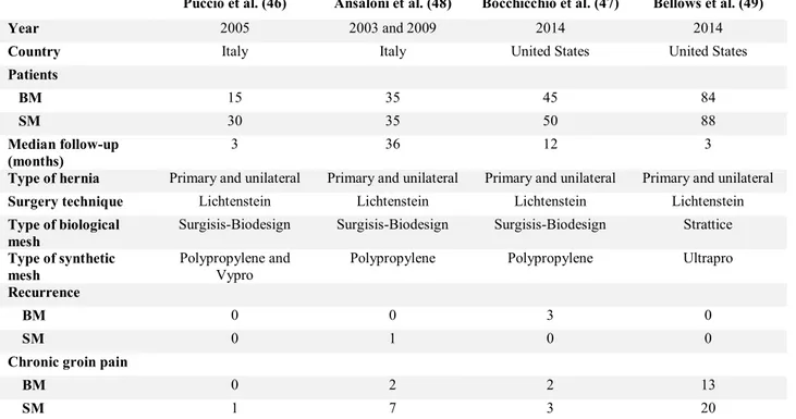
From the data of these meta-analyses, no statistical difference in overall post-operative recurrence between biologic mesh and synthetic mesh was found. However, Bochicchio et al. (47) reported a higher recurrence rate (3/45 patients, 6,7%) in biologic mesh (Surgisis-Biodesign) group at 1-year follow-up. They attributed the high recurrence rate to the inexperience of the surgeons and suggested that a biologic repair might require some added technical skill and experience.
Table 6 RCTs in inguinal hernia repair comparing biological mesh (BM) with synthetic mesh (SM)
Puccio et al. (46) Ansaloni et al. (48) Bocchicchio et al. (47) Bellows et al. (49)
Year 2005 2003 and 2009 2014 2014
Country Italy Italy United States United States
Patients BM 15 35 45 84 SM 30 35 50 88 Median follow-up (months) 3 36 12 3
Type of hernia Primary and unilateral Primary and unilateral Primary and unilateral Primary and unilateral
Surgery technique Lichtenstein Lichtenstein Lichtenstein Lichtenstein
Type of biological mesh
Surgisis-Biodesign Surgisis-Biodesign Surgisis-Biodesign Strattice
Type of synthetic mesh
Polypropylene and Vypro
Polypropylene Polypropylene Ultrapro
Recurrence
BM 0 0 3 0
SM 0 1 0 0
Chronic groin pain
BM 0 2 2 13
SM 1 7 3 20
In two retrospective case series with respectively 11 (50) and 38 (51) patients, inguinal hernias were repaired in an endoscopic technique (respectively TAPP and TEP) with Surgisis-Biodesign. During the follow-up period (mean 14,5 and 13 months, respectively), a recurrence rate of 9.1 and 2% was observed, respectively.
26
We reported two recurrences in adult patients underwent to inguinal hernia repair with Surgisis-Biodesign mesh, respectively at two and four years after surgery.
As previously said, it is not possible to compare our results considering the limited number in our series, but anyway our experience is proof that more extensive study with longer follow-up is necessary.
The RCTs identified in meta-analyses (25) (45) demonstrated areduced pain at rest, on coughing or on movement. We did not record chronic pain in our experience in primary inguinal hernia repair with biological mesh. However, in a recent review (52) of our experience in suturless tensionfree inguinal hernia repair with synthetic mesh, we reported a chronic post-operative pain rate rather low (2,89%), so probably our experience in biological mesh is too limited to see patients with chronic pain.
Catena et. al (53) reported their experience in inguinal hernia repair with Surgisis-Biodesign mesh in ten immunosuppressed patients with good results: no wound infection, no recurrence. We confirmed that the use of biological mesh is a good indication in immunosuppressed patients.
Frankling et al. (54) proposed successfully in a retrospective case series the use of biological meshes (Surgisis-Biodesign) even in a potentially contaminated setting, i.e., with incarcerated/strangulated bowel within the hernia or coincident with a laparoscopic cholecystectomy/colectomy as well as in a grossly contaminated field (i.e., gross pus or fecal spillage).
In a recent review (55) in evidence for replacement of an infected synthetic with biological mesh in abdominal wall hernia repair, a total of 47 patients (considering two different series of 15 (56) and 32 (57) patients) with inguinal infected prosthesis were treated by complete mesh removal and neither a synthetic nor a biological mesh was implanted to replace the explanted mesh. In the first series, infection resolved successfully in all patients, but one patient developed a recurrent hernia. In the second series, one recurrence and one fistula happened. To avoid recurrence after removal of the infected mesh, we preferred to place a biological mesh in the preperitoneal space after the complete plug and mesh removal and, until now, at 6 months after surgery, infection seem to be resolved and recurrence did not develop. Of course, a longer follow up is necessary.
In a patient with a previous pure tissue inguinal hernia repair, we placed a biological mesh during surgery for vas resection, even if a real hernia defect was not present at surgery time, but just for the reinforcement of the inguinal posterior wall, in consideration of the opening of the external oblique aponeurosis to reach the cord. In literature, no similar indication was reported.
27
Our choice of biological mesh in children was due in two cases to patient body appearance, that was not typically of children, despite physical development was not already completed, so a pure tissue repair seemed not suitable. In the third children, we preferred a biological mesh repair because it was a recurrent after a tissue inguinal and femoral hernia repair but synthetic mesh once again was not indicated in consideration of the young age. No complication reported in any children. No data about biological mesh use in similar cases were available in literature.
We reported one case of incarcerate femoral hernia: the crural sac passed between femoral vein and arthery and, after sac reduction by a combined femoral-transinguianl preperitoneal approach, we preferred to place a biological prosthesis in direct contact with femoral vessels, rather synthetic mesh, in order to prevent potential complication, like chronic pain, adhesion and vein compression (58). No intra or post-operative complication were reported. At 18 months after surgery, patient did not complain chronic pain neither recurrence.
Regarding patients affected by Pubic Inguinal Pain Syndrome (42), the so called ”sports” hernia, we proposed a tailored open approach (42) (43), including nerve release, partial calibrated tenotomy of adductor longus and rectus abdominal muscles and reinforcement of the inguinal channel posterior wall with biological mesh. Edelman proposed a laparoscopic TEP approach and the placement of a biological Surgisis-Biodesign mesh fixed with absorbable tacks and, through a separate skin incision along the inguinal crease, micro-cuts of the tendon of the adductor muscle and the placement of a biological mesh tacked to the inferior pubis and sutured to the adductor muscle. The use of a biological mesh was indicated by the absence of a real hernia defect. Just a bulge of the posterior wall and a small indirect sac (M1, L1, according EHS classification (41)) were present, contributing to compress the nerves against the hypertrophic rectus muscle. Our total experience in PIPS treatment include 105 patients and, among these, 11 with biological mesh placement. In the rest of patients an ultralight not-absorbable synthetic mash was used. We obtained complete pain relief of pain in both groups. We preferred an open approach because, by a single incision in inguinal region, we could reach inguinal channel, rectus muscle and adductor longus muscle and so act on all the reason of pain. Moreover, our open approach under local anaesthesia permitted us to ask the patient to do easily exercises during surgery and so to calibrate the partial tenotomy. In that way, partial tenotomy did not elicit in functional deficiency or limitation and so did not require the placement of a biological insertion.
We adopted the use of biological mesh also in the treatment of the post-operative inguinal pain: in these patients, the removal of the prosthesis previously placed could cause a weakness of the
28
transversalis fascia and, sometimes, of the external oblique aponeurosis in absence of a real hernia, so the placement of a mesh had a precautionary aim. We were used to prefer a biological mesh or an ultralight one to reduce the discomfort that a heavyweight mesh could cause, even if in literature there is still a controversial about whether using light-weight mesh instead of heavyweight mesh in could reduce the incidence of chronic groin pain (59) (60) (61). We chose a biological mesh in the 15% of the total of our patients treated for chronic pain. Results in term of chronic pain was similar (16,7% in biological group and 20% in synthetic group). We recorde one hernia recurrence in biological group and none in synthetic group.
We adopted a biological mesh in 17 patients undergoing to ventral hernia repair.
Our patient complained a small umbilical hernia with a defect size of 2 cm, so we chose a mesh repair, but with the fear of chronic discomfort and in consideration also of her thinness, we preferred a biological one.
Based on results of a large cohort with minimum 2 years of follow-up (62), even in small-sized umbilical and epigastric hernias, mesh-reinforcement can be used to avoid recurrence (recurrence rate of 2,2% in mesh group versus 5,6% in sutured group). The biologic mesh seemed to be a safe and reliable device for repairing primary umbilical hernia (defect size < or = 3 cm) in a prospective cohort study, reporting a recurrence rate of 2,8% (63). We were used to reserve suture repair just for hernia defect inferior to 1 cm and for larger defect (without rectus diastasis) we adopted absorbable synthetic mesh (less expensive) or biological mesh (more expensive).
In ventral hernia repair with loss of substance or a thin and weak peritoneal sheet, we adopted biological mesh to fill the loss of substance and isolate the viscera from synthetic mesh placed in the retromuscolar-preperitoneal space. Campanelli et al. (64), when biological mesh use was at beginning and very restricted, reported good results in the “double mesh technique”, with the use of vicryl (absorbable polyglactin mesh) mesh bridged to peritoneum and posterior sheet aponeurosis to fill the loss of substance. In a recent paper by Liu (65), utilization of the absorbable polyglactin mesh, as a separating layer between a synthetic mesh and viscera in a porcine model, was found to be associated with similar adhesion formation as unprotected synthetic meshes grossly. Histologically, however, visceral adhesions formed not against the synthetic mesh, but against a fibrous capsule that replaced Vicryl mesh and probably this capsule could prevent intestinal erosions into retromuscular synthetic meshes. For this reason, since biological scaffolds became more popular, we preferred them to vicryl mesh because adhesions should not occur with biological scaffold.
29
We successfully used the double mesh technique (biological + synthetic) also in previous infection, previous contaminated and contaminated field. We chose to implant just biological mesh (without additional synthetic mesh) in all those patients reporting small ventral hernia, in absence of abdominal wall relaxation and with a complete posterior compartment (peritoneal sheet and posterior rectus fascia).
The recent review (55) about treatment of infected synthetic mesh demonstrated that there were three approaches that could be taken depending on the individual patient situation in ventral infected mesh. The first option was to remove the infected mesh without a new implant. However, Tolino et al. (57) reported on a recurrence rate of 23% after removal of an infected mesh following incisional hernia operation without reimplantation of a new mesh. The second option was to replace the infected polypropylene mesh with a new polypropylene mesh (66). The short-term results showed a relative uneventful postoperative course after mesh replacement in 27 patients. Six (22%) patients developed a minor wound infection and were treated with dressings and antibiotics, five (19%) patients had wound infections requiring debridement and one required complete mesh removal. On follow-up, there were three hernia recurrences, one with an enterocutaneous fistula. The third option was to replace the explanted synthetic mesh with a biological mesh (67) (17) (68) (69) (70) (71). Long-term results were successful only if bridging was omitted (67) (17) (68). An unacceptably high recurrence rate was observed following bridging with biological meshes (67) (17) (68). When bridging was avoided, good results were obtained for replacement of an infected synthetic mesh with a biological (67) (17) (68).
In fact, for this reason, both in contaminated or clean field, in case of loss of substance we used the biological mesh just to fill the defect of the posterior compartment and then we placed a synthetic mesh to repair the abdominal wall function. Until now, we did not record recurrence in our experience.
About recurrence rate, Beale (72) proposed a systematic review on all biological mesh repairs used in open ventral incisional hernia repair and to allow additional comparisons between biologic products with respect to hernia recurrence and other surgical site complications (seroma, hematoma, infection). Mean recurrence rates for the devices Alloderm, Permacol, and Surgisis were 21%, 11%, and 8%, respectively, and similarly mean surgical site occurrence rates for the same devices 31%, 25%, and 40%. When comparing these products, patient population and surgical technique (inlay, onlay or sublay placement of the mesh and component separation accomplished or not) should be a factor.
30
A study by Huntington (73) examined long-term outcomes of ventral hernia repair with Strattice, AlloDerm, AlloMax, FlexHD, and Xenmatrix. After controlling for confounding factors such as tobacco use, medical comorbidities, hernia defect size and bridging mesh cases, Strattice mesh had significantly lower odds of postoperative hernia recurrence compared with AlloMax, FlexHD, and XenMatrix. The highest rates of recurrence were seen in XenMatrix mesh, a porcine acellular dermal mesh like Strattice, which had a 59.1% recurrence rate (at 11.0 months average follow-up) compared with 14.7% in Strattice mesh (with 17.6 month mean follow-up; P < .05 for recurrence rate comparison). The significant differences between XenMatrix and Strattice, both porcine acellular dermal meshes, may underscore the variation in tissue processing and design in biomesh engineering. AlloDerm, AlloMax, and FlexHD had similar rates of recurrence, 34.8–37.1%. Additionally, in the same study, Strattice had the lowest rates of seroma (15.2%) compared with the other meshes. Mesh and wound infection were similar across the biologic mesh groups; it is important to note that though the cases were complicated and the wound complication rates demonstrate that, the biologic meshes did not require explantation. Significant variation between meshes seems to underscore the real diversity in the market and the need for further research.
One more consideration about biological is due: charges and cost are important and often discussed and considered factors when using biomesh, which are frequently more expensive than synthetic meshes. One cost analysis (74) demonstrated that biologic mesh use more than doubled the direct cost of ventral hernia repair. This is also the reason why, in our experience, biological mesh use is limited to less than 1% of the total of our procedures, in very selected patients, and even in literature studies often reported its use in complex situation, not in routinary surgery.
Conclusion
Regarding primary inguinal hernia repair, large number, well-designed and long follow-up period RCTs and cost effectiveness analyses are still needed to assess the use of biologic mesh and the equivalence of biological meshes and synthetic meshes about recurrence rate and chronic pain. Studies, and our experience too, show that biological meshes can be used as an alternative in a potentially contaminated field as well as in a setting grossly contaminated field, both for inguinal and ventral hernia repair.
We successfully use biological mesh also in selected inguinal and femoral repair in children and in the treatment of Pubic Inguinal Pain Syndrome and of post-operative chronic pain after both inguinal and ventral repair.
31
In ventral hernia repair, an unacceptably high recurrence rate was observed following bridging with biological meshes, for this reason The Author suggest to bridge biological mesh to the border of the defect and place a synthetic not absorbable mesh in the retromuscolar-preperitoneal space.
Even if there is no still clear evidence in biological mesh indications, certainly in a specialized abdominal wall center, biological mesh should be available and in selected case is a good alternative to synthetic mesh.
Significant differences in clinical behavior, not only among different biological meshes, but also between biological with the same tissue source but different tissue processing and design in engineering, underscore the real need for further preclinical and clinical research.
Current knowledge in cells-matrix interaction in vitro is still so limited that does not allow to foresee
in vivo behavior of different biological matrices. We could deduce that synthetic prosthesis, not
interacting with fibroblastic cells, are not integrated in the surrounding tissues and therefore reinforce abdominal wall just inducing scar tissue. Integration and remodeling of biological meshes depends not only from tissue source, tissue processing and design in engineering, but also from type of surgery, type of abdominal defect and individual patient situation.
References
1. Rutkow IM. Surgical procedure in the United States. Then (1983) and now (1994). Arch Surg 1997, 132(9):983–990.
2. Engelsman AF, van der Mei H, Ploeg RJ, Busscher HJ. The phenomenon of infection in abdominal wall reconstruction. Biomaterials 2007,28(14):2314–2327.
3. Uscher FC. Hernia repair with marlex mesh. An analysis of 514 cases. Arch Surg 1962, 84:325–328. 4. Burger JW, Halm JA, Wijsmuller AR, et al. Evaluation of new prosthetic meshes for ventral hernia repair. Surg Endosc 2006;20:1320–1325.
5. den Hartog D, Dur AH, Tuinebreijer WE, Kreis RW. Open surgical procedures for incisional hernias. Cochrane Database Syst Rev 2008:CD006438.
6. Ansaloni L, Catena F, Coccolini F, Negro P, Campanelli G, Miserez M. New"biological" meshes: the need for a register, The EHS Registry forBiological Prostheses: call for participating European surgeons. Hernia 2009, 13(1):103–108.
7. Robinson TN, Clarke JH, Schoen J, Walsh MD. Major mesh-related complications following hernia repair: events reported to the Food and Drug Administration. Surg Endosc. 2005; 19(12):1556– 1560.
8. Halm JA, de Wall LL, Steyerberg EW, Jeekel J, Lange LW. Intraperitoneal polyproylene mesh repair complicates subsequent abdominal surgery. World J Surg 2007: 31:423–431.
32
9. Gray SH, Vick CC, Graham LA, Finan KR, Neumayer LA, Hawn MT. Risk of complications from
enterotomy or unplanned bowel resection during elective hernia repair. Arch Surg 2008: 143(6):582–586. 10. Kissane N, Itani KMF. A decade of ventral incisional hernia repairs with biologic acellular dermal matrix: what have we learned? Plast Reconstr Surg 2012: 130:194S–202S.
11. Cevasco M, Itani KM. Ventral hernia repair with synthetic composite and biologic mesh: characteristics, indications and infection profile. Surg Infect 2012: 13:209–215.
12. Jacob BP, Hogle NJ, Durak E, et al. Tissue ingrowth and bowel adhesion formation in an animal comparative study: Polypropylene versus Proceed versus Parietex composite. Surg Endosc 2007;21:629– 633.
13. Schreinemacher MHF, Emans PJ, Gijbels MJJ, et al. Degradation of mesh coatings and intraperitoneal adhesion formation in an experimental model. Br J Surg 2009;96: 305–313. .
14. Emans PJ, Schreinemacher MH, Gijbels MJ, et al. Polypropylene meshes to prevent abdominal herniation: Can stable coatings prevent adhesions in the long-term? Ann Biomed Eng 2009;37:410–418. . 15. Baptista ML, Bonsack ME, Felemovicius I, Delaney JP. Abdominal adhesions to prosthetic mesh evaluated by laparoscopy and electron microscopy. J Am Coll Surg 2000; 190:271–280.
16. Hall-Allen RTJ. Porcine dermal collagen repair of inguinal hernias. J Roy Coll Surg 1984; 29:154. 17. Rosen JM, Krpata DM, Ermlich B, Blatnik JA. A 5-year clinical experience with single-staged repairs of infected and contaminated abdominal wall defects utilizing biologic mesh. Ann Surg (2013) 257:991–6. 18. De Castro Bras LE, Shurey S, Sibbons PD. Evaluation of crosslinked and non-crosslinked biologic prostheses for abdominal hernia repair. Hernia 2012;16:77–89.
19. Pierce LM, Rao A, Baumann SS Glassberg JE, Kuehl TJ, Muir TW. Long-term histologic response to synthetic and biologic graft materials implanted in the vagina and abdomen of a rabbit model. Am J Obstet Gynecol. 2009;200:e541–e548.
20. Slater NJ, van der Kolk Hendriks T, van Goor H, Bleichrodt RP. Biologic grafts for ventral hernia repair: a systematic review. Am J Surg 2013: 205:220–230.
21. Muysoms F, Campanelli G, Champault GG, DeBeaux AC, Dietz UA, Jeekel J, Klinge U, Kockerling F,
Mandala V, Montgomery A, Morales Conde S, Puppe F, Simmermacher RKJ, Smietanski M, Miserez M.
EuraHS: the development of an international online platform for registration and outcome measurement of ventral abdominal wall hernia repair. Hernia 2012: 16:239–250.
22. VH, Cumberland. A preliminary report on the use of a prefabricated nylon weave in the repair of ventral hernia. Med J Aust. 1952;1:143–144.
23. Scales JT. Tissue reactions to synthetic materials. Proc Royal Soc Med. 1953;46:647–652.
24. Hamer-Hodges DW, Scott NB. Surgeo ’s orkshop:repla e e t of a a do i al all defe t usi g expanded PTFE sheet (GORE-TEX). J Royal Coll Surg Edin 1985: 30:65–67.
25. Fang Z, Ren F, Zhou J, Tian J. Biologic mesh versus synthetic mesh in open inguinal hernia repair: system review and meta-analysis. ANZ JSurg (2015).doi:10.1111/ans.13234.
26. Sandor M, Xu H, Connor J, et al. Host response to implanted porcine-derived biologic materials in a primate model of abdominal wall repair. Tissue Eng Part A. 2008;14:2021–2031.
33
27. Khor E. Methods for the treatment of collagenous tissues for bioprostheses. Biomaterials 1997;18:95– 105.
28. Schmidt CE, Baier JM. Acellular vascular tissues: Natural biomaterials for tissue repair and tissue engineering. Biomaterials 2000;21:2215–2231.
29. Deeken CR, Melman L, Jenkins ED, Greco SC, Frisella MM, Matthews BD. Histologic and biomechanical evaluation of crosslinked and non-crosslinked biologic meshes in a porcine model of ventral incisional hernia repair. J Am Coll Surg. 2011;212(5):880–8.
30. Gouk SS, Lim TM, Teoh SH, Sun WQ. Alterations of human acellular tissue matrix by gamma irradiation: Histology, biomechanical property, stability, in vitro cell repopulation, and remodeling. J Biomed Mater Res B Appl Biomater. 2008; (1):205-217.
31. Petter-Puchner AH, Fortelny RH, Silic K, Brand J, Gruber-Blum S, Redl H. Biologic hernia implants in experimental intraperitoneal onlay mesh plasty repair: The impact of proprietary collagen processing methods and fibrin sealant application on tissue integration. Surg Endosc. 2011;25:3245–3252.
32. Orenstein SB, Qiao Y, Klueh U, Kreutzer DL, Novitsky YW. Activation of human mononuclear cells by porcine biologicmeshes in vitro. Hernia 2010;14:401–407.
33. Butler CE, Burns NK, Campbell KT, Mathur AB, Jaffari MV, Rios CN. Comparison of cross-linked and non-cross-linked porcine acellular dermal matrices for ventral hernia repair. J Am Coll Surg. 2010;211:368– 376.
34. Harth KC, Rosen MJ. Major complications associated with xenograft biologic mesh implantation in abdominal wall reconstruction. Surg Innov. 2009;16(4):324–9.
35. Novitsky YM, Rosen MJ. The Biology of Biologics: Basic Science and clinical concepts. Plast. Reconstr. Surg. 130 (Suppl. 2): 9S, 2012.
36. Xu H, Wan H, Sandor M, et al. Host response to human acellular dermal matrix transplantation in a primate model of abdominal wall repair. Tissue Eng Part A. 2008; 14:2009–2019.
37. Orenstein SB, Qiao Y, Kaur M, Klueh U, Kreutzer DL, Novitsky YW. Human monocyte activation by biologic and biodegradable meshes in vitro. Surg Endosc. 2010;24:805–811.
38. Burns NK, Jaffari MV, Rios CN, Mathur AB, Butler CE. Non cross-linked porcine acellular dermal matrices for abdominal wall reconstruction. Plast Reconstr Surg. 2010;125:167–176.
39. Melman L, Jenkins ED, Hamilton NA, et al. Early biocompatibility of crosslinked and non-crosslinked biologic meshes in a porcine model of ventral hernia repair. Hernia 2011;15:157–164.
40. Gao y, Liu l, Blatnik JA, Krpata DM, AndersonJM, Criss CN, Posielski N, Novitsky YW. Methodology of fibroblast and mesenchymal stem cell coating of surgical meshes: A pilot analysis. J Biomed Mater Res Part B 2014, 102(4),797-805.
41. Miserez M, Alexandre JH, Campanelli G, Corcione F, Cuccurullo D, Pascual MH, Hoeferlin A,
Kingsnorth AN, Mandala V, Palot JP, Schumpelick V, Simmermacher RK, Stoppa R, Flament JB. The
European hernia society groin hernia classification: simple and easy to remember. Hernia. 2007 Apr;11(2):113-6.
42. G., Campanelli. Pubic inguinal pain syndrome: the so-called sports hernia. Hernia. 2010 Feb;14(1):1-4. 43. Cavalli M, Bombini G, Campanelli G. Pubic inguinal pain syndrome: the so-called sports hernia. Surg Technol Int. 2014 Mar;24:189-94.
34
44. Campanelli G, Bertocchi V, Cavalli M, Bombini G, Biondi A, Tentorio T, Sfeclan C, Canziani M. Surgical treatment of chronic pain after inguinal hernia repair. Hernia. 2013 Jun;17(3):347-53.
45. Nie X, Xiao D, Wang W, Song Z, Yang Z, Chen Y, et al. Comparison of porcine small intestinal submucosa versus polypropylene in open inguinal hernia repair: a systematic reciew and meta-analysis. PLoSOne 2015 10(8):e13507.
46. Puccio F, Solazzo M, Marciano P. Comparison of three different mesh materials in tension-free inguinal hernia repair: Prolene versus Vypro versus Surgisis. Int. Surg. 2005; 90: S21–3.
47. Bochicchio GV, Jain A, McGonigal K et al. Biologic vs synthetic inguinal hernia repair: 1-year results of a randomized double-blinded trial. J Am. Coll. Surg. 2014; 218: 751–7.
48. Ansaloni L, Catena F, Coccolini F, Gazzotti F, D’Alessandro L, Pinna AD. Inguinal hernia repair with porcine small intestine submucosa: 3-year follow-up results of a randomized controlled trial of
Li hte stei ’s repair ith pol prop le e esh ersus Surgisis I gui al Her ia Matri . Am. J. Surg. 2009; 198: 303–12.
49. Bellows CF, Shadduck P, Helton WS, Martindale R, Stouch BC, Fitzgibbons R. Early report of a
randomized comparative clinical trial of Strattice reconstructive tissue matrix to lightweight synthetic mesh in the repair of inguinal hernias. Hernia 2014; 18: 221–30.
50. Agresta F, Bedin N. Transabdominal laparoscopic inguinal hernia repair: is there a place for biological mesh? Hernia (2008) 12:609–12.
51. AP, Fine. Laparoscopic repair of inguinal hernia using surgisis mesh and fibrin sealant. JSLS (2006) 10:461–5.
52. Campanelli G, Bruni PG, Cavalli M, Morlacchi A. A Complete Sutureless, Hernia Repair for Primary Inguinal Hernia The Trabucco Repair: A Tribute to Ermanno Trabucco. Surg Technol Int. 2016 Apr;28:141-6. 53. Catena F, Ansaloni L, Leone A, De Cataldis A, Gagliardi S, Gazzotti F, Peruzzi S, Agrusti S, D'Alessandro
L, Taffurelli M. Lichtenstein repair of inguinal hernia with Surgisis inguinal hernia matrix soft-tissue graft in
immunodepressed patients. Hernia. 2005 Mar;9(1):29-31.
54. Franklin ME, Gonzalez JJ, Glass JL. Use of porcine small intestinal submucosa as a prothetic device for laparoscopic repair of hernias in contaminated fields: 2-year follow-up. Hernia (2004) 8:186–9.
55. Montgomery A, Kallinowski F, Köckerling F. Evidence for Replacement of an Infected Synthetic by a Biological Mesh in Abdominal Wall Hernia Repair. Front Surg. 2016 8;2:67.
56. Akyol C, Kocaay F, OrozaKunov E et al. Outcome of the patients with chronic mesh infection following open inguinal hernia repair. J Korean Surg Soc (2013) 84:287–91.
57. Tolino MJ, Tripoloni DE, Ratto R, Garcia MI. Infections associated with prosthetic repairs of abdominal wall hernias: pathology, management and results. Hernia 2009 13:631–7.
58. DeBord, J.R. Vascular injury during hernia repair. R. Bendavid (Ed.), Abdominal wall hernias, Springer-Verlag, New York (2001), pp. 690–699.
59. Currie A, Andrew H, Tonsi A, Hurley PR, Taribagil S. versus heavyweight mesh in laparoscopic inguinal hernia repair: a metaanalysis. Surg. Endosc. 2012; 26: 2126–33.
60. Smietanski M, Smietanska IA, Modrzejewski A et al. Review and meta-analysis on heavy and lightweight polypropylene mesh in Lichtenstein inguinal hernioplasty. Hernia 2012; 16: 519–28.