Sensitivity and specificity of mastoid vibration test in
detection of effects of vestibular neuritis
Sensibilità e specificità del test della vibrazione mastoidea nella
individuazione degli esiti della neurite vestibolare
D. NUTI, M. MANDALÀ
ENT Department, University of Siena Medical School, Italy
Key words
Vestibular neuritis • Bedside vestibular examination • Ma-stoid vibratory test
Parole chiave
Neurite vestibolare • Esame vestibolare clinico • Test vi-bratorio mastoideo
Summary
Aim of this study was to determine sensitivity and speci-ficity of the mastoid vibration test in patients who had suffered an attack of vestibular neuritis. Results were compared with the caloric test and two bedside tests of vestibular function (head shaking test and head thrust test). Results are reported in 28 patients who had a residual vestibular deficit 6 months after acute neuritis and in 25 healthy subjects. Mastoid vibration nystagmus was evoked in 21 patients but not in controls. In these patients, mastoid vibration test had a sensitivity of 75% and specificity of 100%. Since one patient had inverted mastoid vibration nystagmus, specificity of identification on the pathological side was 95%. Sensitivity of the test increased with increasing severity of the vestibular lesion. Indeed, mastoid vibration nystagmus was induced in 93% of patients with caloric paralysis and in 58% of those with caloric paresis. Nystagmus could usually be modulated or elicited by stim-ulation of either mastoid. In the few patients in whom mastoid vibration nystagmus was elicited only from one side, or when there was a clear difference in intensity of the nystagmus induced on the two sides, the stimulated side was more often the affected side. Four patients still showed spontaneous nystagmus. The caloric test was abnormal in 26/28 patients (93%) with paralysis in 16 and paresis in 12; 71% of patients had a head shaking induced nystagmus: 64% had an asymmetrical response in head thrust test. In conclusion, mastoid vibration test was overall more sensitive than head thrust test. Mastoid vibration test was slightly less sensitive than head shaking test in patients with severe residual deficit and more sensitive in patients with partial deficit. Mastoid vibration test, a valid, low cost clinical screening test for rapid detection of asymmetrical vestibular function, does not cause patient discomfort. It is suggested that this test be included in the diagnostic workup of all patients with suspected vestibular dysfunction.
Riassunto
Lo scopo di questo studio è stato quello di determinare la sen-sibilità e la specificità del test vibratorio mastoideo (MVT) in pazienti affetti da esiti di neurite vestibolare. I risultati sono stati confrontati con quelli ottenuti dalla stimolazione calori-ca e da due test dinamici della funzione vestibolare: l’head shaking test (HST) e l’head thrust test (HTT). Riportiamo in questo studio i risultati di 28 pazienti che presentavano un de-ficit residuo della funzione vestibolare a distanza di sei mesi dalla fase acuta, confrontati con 25 soggetti normali. Il ni-stagmo da vibrazione mastoidea (MVN) è stato evocato in 21 pazienti ed in nessuno dei soggetti del gruppo di controllo. Pertanto, nei nostri pazienti il MVT ha una sensibilità del 75% e una specificità del 100%. Poiché un paziente ha presentato un MVN invertito, la specificità nella identificazione del lato leso è del 95%. La sensibilità del test aumenta con la gravità della lesione vestibolare. È stato infatti possibile rilevare MVN nel 93% dei pazienti con paralisi calorica e nel 58% di quelli con paresi calorica. In genere il nistagmo vibratorio viene evocato o modulato dalla stimolazione di entrambe le mastoi-di. Nei pochi pazienti in cui il MVN compariva solamente con la stimolazione di una mastoide o quando era rilevabile una evidente differenza di intensità del nistagmo tra le due stimo-lazioni, il lato stimolato corrispondeva più frequentemente a quello affetto. A distanza di sei mesi dall’esordio, quattro pa-zienti presentavano ancora un nistagmo spontaneo mentre il test calorico risultava alterato in 26/28 pazienti (93%), con 16 paralisi e 12 paresi. Il 71% dei pazienti presentava un head shaking nystagmus. Nel 64% dei casi era evidente una rispo-sta asimmetrica al HTT.
In conclusione il MVT è risultato costantemente più sensibile dell’HTT. Rispetto all’HST, il MVT si è rivelato leggermente meno sensibile in pazienti con un deficit residuo severo e più sensibile nei soggetti con deficit parziali. I nostri risultati con-fermano la validità del MVT quale test di screening per una rapida individuazione di una asimmetria della funzione vesti-bolare. Il test non causa alcun disagio per il paziente, è di fa-cile esecuzione ed interpretazione e richiede una strumenta-zione di basso costo. Per tali motivi suggeriamo di includerlo all’interno delle procedure diagnostiche di routine per tutti i pazienti con una possibile disfunzione vestibolare.
Introduction
Vibration on the bone behind the ear may elicit nys-tagmus in patients with vestibular problems. The mas-toid vibratory test (MVT) was first introduced in 1973 by Lücke as a simple bedside test to detect static anomalies in peripheral vestibular function1. In 1993,
Hamann demonstrated that MVT elicited nystagmus in 75% of patients with unilateral vestibular deficit and 10% of patients with central vestibular disorders2.
The usefulness of MVT in the identification of unilat-eral vestibular loss was confirmed in later studies3 4.
A vibratory stimulus has been applied not only to the mastoids but also to the vertex and neck4 5. In 1999,
Hamann and Schuster6found that the sensitivity of
the test was increased using a frequency of 60 Hz in-stead of 100 Hz. In 2004, Dumas et al.7 reported
complete concordance between MVT, the head shak-ing test (HST) and the caloric test for detectshak-ing the side with total vestibular loss. It was also suggested that MVT elicits vibratory nystagmus (VN) in healthy subjects5 8.
It is not clear how mastoid vibration causes nystag-mus in patients with peripheral vestibular deficit. Vi-bration has been shown to excite semicircular canals and otolithic afferents in various animal species9-11
and in humans4 6 12 13, indicating that the effects on
eye movement can be attributed to direct stimulation of intact vestibular receptors. Kalberg et al., in par-ticular, showed that vibration to the mastoid bone in-duces eye movements similar to those seen acutely, without vibration, after unilateral vestibular loss and that the eye rotation axis of vibration-induced nys-tagmus is related to the extent of the unilateral vestibular deficit12. The vibratory test, according to
some authors, is not unlike the simultaneous caloric test7. Vibration also seems to increase afferent
activi-ty from stimulated muscles14, suggesting that the
ef-fects may be due to interaction between neck proprio-ceptors and the vestibular system15 16.
The aim of the present study was to determine the sensitivity and specificity of MVT in patients with damage caused by vestibular neuritis, comparing re-sults with those of the caloric test, the current gold standard for identifying unilateral deficit in vestibu-lar function. MVT was also compared with the head shaking test (HST) and with the head thrust test (HTT), other conventional bedside tests used in the detection of vestibular asymmetry.
Methods SUBJECTS
The cohort used in this study was a patient-based clinical population seen between January 2002 and January 2004. Patients were seen in the acute stage of
the disease (1-3 days after the beginning of symp-toms) and all were eligible for inclusion in the study. Inclusion criteria were: 1) acute vertigo lasting at least 24 hours, 2) horizontal unidirectional sponta-neous nystagmus lasting at least 24 hours, 3) no hear-ing loss, 4) no additional neurological signs or symp-toms (and normal brain imaging, if performed), 5) unilateral caloric weakness (caloric paresis or paral-ysis). Overall, 57 patients met these criteria.
Patients were asked to return for follow-up after 1, 3 and 6 months. Approximately 50% of the patients re-covered, since no spontaneous nystagmus was de-tectable, caloric response was balanced and no asym-metry was seen with bedside vestibular tests. Results of the vibratory test performed in 28 patients with residual deficit in vestibular function, six months after onset of the disease, are reported. Vestibular function was considered impaired if pa-tients had a caloric weakness and/or head shaking nystagmus and/or heat thrust sign and/or sponta-neous nystagmus.
CLINICAL TESTING
At each clinical examination, patients underwent a complete bedside clinical examination. Spontaneous nystagmus (SN), head-shaking-induced nystagmus (HSN) and mastoid vibration-induced nystagmus (MVN) were sought with fixation removed using Frenzel goggles.
The dynamic performance of the vestibulo-ocular re-flex was evaluated using manually-induced head thrusts: abrupt, rapid, high acceleration head rota-tions of about 20 degrees17. The patients were
in-structed to fix on the examiner’s nose. The HTT was considered abnormal if there was an obvious corrective saccade supplementing an inadequate slow phase and only the corrective movement was seen, or was con-siderably larger, with rotation towards one (the af-fected) side (Head Thrust Sign).
HST was performed according to Kamei et al.18. The
patients were placed in a sitting position with the head flexed down to 30°, the eyes closed and covered with Frenzel glasses. The head was then shaken horizontal-ly by the examiner around the jaw axis, 20 times for about 10 sec (2 Hz) with amplitude of about 45 de-grees to either side. When the head movement was stopped, patients were instructed to open their eyes and the examiner recorded the direction, frequency and duration of any nystagmus. HSN was considered present if at least three or more consecutive beats of nystagmus appeared in patients without SN. In pa-tients with SN, the test was considered positive when an evident increase in frequency was observed. Pat-terns of nystagmus response were classified as paretic HSN when the fast phase was directed towards the healthy ear and reversed HSN if it was directed to-wards the affected side19 20.
Caloric testing was performed with hot, cold and ice water. Maximum speed of the slow-phase of nystag-mus evoked by each ear was analyzed for unilateral weakness and directional preponderance as deter-mined by Jongkees formulae21. Caloric paresis was
defined when there was some response on both sides but the difference between the two ears was 30% or more. Caloric paralysis was defined when there was no response to ice water irrigation on one side. Pa-tients were considered to have recovered when asym-metry dropped to <30%.
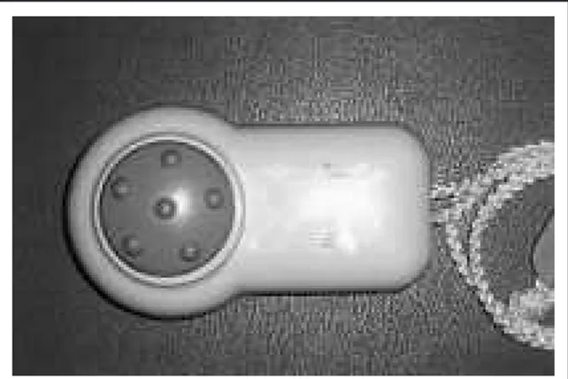
MVT was applied with a battery-powered device de-signed for physiotherapy. It has a circular contact surface, 4 cm in diameter (Fig. 1), which is placed perpendicular to the skin just behind the ear. Funda-mental frequency, measured with the aid of a phonometer, was 100 Hz (60-120 Hz). Vibratory stimulation was applied in the mastoid region for about 10 sec, first on one side and then on the other. The test was considered positive when persistent hor-izontal or horhor-izontal torsion nystagmus was evoked for the duration of the stimulus and if the phenome-non was repeatable. In patients with spontaneous nystagmus, the test was regarded as positive when nystagmus showed an evident increase in frequency. We also performed MVT in the control group of 25 normal subjects.
Results
Mean age of the 28 patients (23 male, 5 female) was 57 years. The right side was affected in 14 patients and the left in 14. Results of the tests are shown in Table I.
Spontaneous nystagmus was still present in four pa-tients, with the fast phase always directed towards the healthy ear. The caloric test was positive in 26/28 patients (93%), 12 of whom had caloric paresis, 14
caloric paralysis and two symmetrical caloric re-sponse. These two patients were considered still pathological due to the presence of HSN in both. In one, paretic HSN was detectable, in the other, re-versed nystagmus, beating towards the affected ear. The patient with a paretic HSN also showed MVN beating towards the affected ear.
HSN was evoked in 20/28 patients (71%), being paretic in 18, with the fast phase towards the healthy ear, while in two it was reversed.
The head thrust sign was evident in 18 patients (64%), always congruent with the affected ear. MVN was elicited in 21 patients (75%), being re-versed in one, with the fast phase beating towards the affected ear. In 16/21 patients, MVN was elicited by vibration of both mastoids. In three of these patients, MVN was stronger, with vibration of the mastoid on the affected side. In the other 13, no difference in nystagmus intensity was seen. In 5/21 patients, MVN was evoked only unilaterally. In three of these cases, this was provoked by vibrating the affected side, in the other two with contralateral stimulus. In none of these cases was the direction of the nystagmus re-versed by changing the side of stimulation. Mastoid vibration did not evoke nystagmus in any of the sub-jects in the control group.
MVN was evoked by MVT in 21/28 patients, thus a sensitivity of 75%. Since none of the normal subjects manifested MVN, the test has a specificity of 100% for patients with effects of vestibular neuritis. One patient experienced reversal MVN indicating that the specificity of identifying the pathological side is 95% (20/21 positive vibration tests).
When the study population was divided into three groups on the basis of the results of the caloric test, MVN was evoked in 13/14 patients with caloric paralysis (93%), 7/12 patients with caloric paresis (58%) and one of the two patients without caloric im-balance i.e., the patient with reversal MVN. Results are outlined in Table II.
Fig. 1. Mastoid vibrator.
Table I. Results of bedside vestibular tests and caloric test.
Present Absent SN 4 (14%) 24 (86%) MVN 21*(75%) 7 (25%) HSN 20†(71%) 8 (29%) HTS 18 (64%) 10 (36%) Caloric weakness 26 (93%) 2 (7%)
SN: spontaneous nystagmus; MVN: mastoid vibration-induced nystagmus; HSN: head shaking-induced nystagmus; HTS: head thrust sign
* One patient showed reversed MVN † Two patients showed reversed HSN
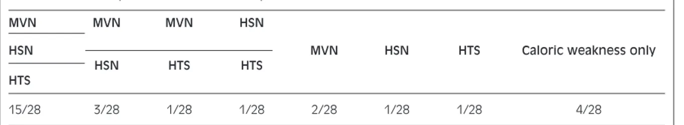
When the probability of identifying residual deficit in vestibular function was determined only by means of bedside clinical tests, without the caloric test, 15/24 patients (54%) were found to be positive with all three tests (MVT, HST, HTT), 5/28 (18%) with at least two and 4/28 (14%) with only one. The tests were negative in 4/28 patients (14%) with an abnor-mal caloric test (Table III).
Discussion
Vestibular neuritis may cause sudden unilateral loss of vestibular function. Approximately 50% of pa-tients recover this function spontaneously or after treatment22 23. While clinical and/or other signs of
deficit persist in the remaining 50%. The caloric test is still the method of choice to detect vestibular loss with a sensitivity of 93%. Dynamic tests of peripher-al vestibular deficit, such as HST and HTT, are sim-ple and rapid to perform and do not require specific instruments. Mastoid vibration is another simple method used in the detection of unilateral vestibular deficit.
In the present study, 28 patients were selected with
vestibular neuritis who still had evidence of residual vestibular deficit, as indicated by caloric weakness and/or dynamic vestibular signs, 6 months after the acute episode. In these patients, MVN was detected in 75%, HSN in 71%, HTS in 64%, and caloric weak-ness in 93%. These results confirm those of other studies, namely that the vibratory test shows good sensitivity for identifying compensated vestibular loss2 4 5 7. Considering only patients with vestibular
neuritis, MVT sensitivity, in the literature, ranges from 75 to 90%, with differences probably due to the fundamental frequency of the applied stimulus (60-100 Hz) and the method used in the detection of nys-tagmus (Frenzel glasses or videonystagmography)4 6 7.
The sensitivity of MVT increases with increasing severity of the vestibular lesion. Indeed, it was possi-ble to induce MVN in 93% of our patients with caloric paralysis. This finding is in keeping with those of Michel et al.5and Dumas and Michel4who
obtained sensitivity of MVT of 100% and 96%, re-spectively, in patients undergoing vestibular neurec-tomy. When we considered patients with caloric paresis, sensitivity of MVT was only 58%.
Comparison with the other two bedside vestibular tests showed that MVT was consistently more sensi-tive than HTT. Compared with HST, MVT was slightly less sensitive in patients with severe residual deficit (caloric paralysis) and more sensitive in those with partial deficits. Our results are in contrast to those of Hamann and Schuster6who reported a much
higher sensitivity of MVT with respect to HST (80%
vs. 35%) for identifying peripheral vestibular deficit.
This difference could be due to the different patient populations and timing of the tests.
In our patients, only one with vibration-induced nys-tagmus beating towards the affected side was identi-fied. Reversed MVN has been reported in other stud-ies with a higher incidence than in our study7. As
with HST, identifying the affected side with MVT is not always reliable. Though a rare possibility in vestibular neuritis, and without any satisfactory pathogenetic explanation, it should, nevertheless, be borne in mind as a limit of this method.
The side stimulated by vibration does not seem to
af-Table II. Relationship between bedside vestibular tests and caloric results.
Paralysis Paresis Symmetric response Caloric test 14 12 2 SN 3 (21%) 1 (8%) / MVN 13 (93%) 7 (58%) 1*(50%) HSN 14 (100%) 4†(33%) 2†(100%) HTS 13 (93%) 5 (42%) /
SN: spontaneous nystagmus; MVN: mastoid vibration-induced nystagmus; HSN: head shaking-induced nystagmus; HTS: head thrust sign
* One patient showed reversed vibration-induced nystagmus † One patient showed reversed head shaking-induced nystag-mus
Table III. Number of patients with one or more positive bedside tests.
MVN MVN MVN HSN
HSN MVN HSN HTS Caloric weakness only
HSN HTS HTS
HTS
15/28 3/28 1/28 1/28 2/28 1/28 1/28 4/28
fect the capacity to generate or modulate nystagmus. In most of our patients, MVN was elicited by stimu-lating on the right and left without any significant difference in response. In 8 patients, who showed MVN when only one mastoid was stimulated, or when there was an evident difference in intensity of the vibration-induced nystagmus, the side stimulated was more often the affected side (6/8 patients). Nys-tagmus changing direction was not detected in any of these patients as occurs in other labyrinth disorders, such as Menière’s disease.
We did not find MVN, in any of the 25 control sub-jects, that persisted for the duration of the vibration or was repeatable. This is in keeping with the find-ings of Hamann and Schuster6but not with those of
others5 8. One reason could be that our vibratory
stimulus was applied exclusively to the mastoids and that nystagmus was observed only with Frenzel glasses and not recorded by videonystagmography. In this case, our results indicate that simpler methods of stimulation and detection make the test more spe-cific and less subject to false positives. We prefer to apply the stimulus to the mastoids, avoiding stimula-tion of the neck and vertex muscles, to simplify the test and avoid possible interactions or interference with cervical afferents.
The mechanism underlying MVN appears to reside in the capacity of the vibratory stimulus to excite the semicircular canals and otolithic afferents9 10.
The effects caused by eye movements appear, there-fore, to be due to direct stimulation of intact vestibular receptors on the healthy side4 6 12 13. Some
Authors compare it to a simultaneous caloric test which exploits the peculiar stimulation. However, it is not clear why the response is not more intense when vibration is applied to the mastoid on the healthy side.
Table III illustrates an important practical aspect. Us-ing exclusively clinical tests, without the aid of the caloric test, it was possible to identify a vestibular deficit in 86% of patients. Thus for patients with a perforated ear drum or who refuse the caloric test, there is still a high probability of making a diagnosis. Moreover, the few cases in which MVT, HST and HTT were not useful for diagnosis, were all found to have a partial, rather than severe, vestibular deficit. On the other hand, the negative caloric tests in two patients who certainly still had a vestibular deficit emphasizes the need to carry out also other clinical tests.
In conclusion, the present results suggest that MVT is a rapid, comfortable and valid clinical screening test for detecting the presence of asymmetric vestibu-lar function. We propose that it be included, together with dynamic vestibular and positional tests, in the diagnostic work-up of patients with vertigo.
Acknowledgements
Study supported by Grant from University of Siena (PAR 2002). Authors are grateful to David S. Zee for reviewing the manuscript and Helen Ampt for lan-guage correction.
References
1 Lücke K. Eine Methode zur Provokation eines
pathologis-chen Nystagmus durch Vibrations-reize von 100 Hz. Z Laryngol Rhinol 1973;52:716-20.
2 Hamann KF. Le nystagmus de vibration: un signe de
per-turbation vestibulaire periferique. San Remo: Compte
ren-du des seances de la Societé d’Otoneurologie de langue française, XXVIIe symposium, San Remo 1993, Edition IPSEN.
3 Michel J. Nystagmun de vibration de Hamann. J Fr
Otorhi-nolaryngol 1995;44:339-40.
4 Dumas G, Michel J. Valeur semiologique du test de
vibra-tion osseux cranien. Liege: Compte rendu des seances de la Societé d’Otoneurologie de langue française, XXXIe sym-posium, Liege 1997, Edition IPSEN.
5 Michel J, Dumas G, Lavielle JP, Characon R. Diagnostic
value of vibration-induced nystagmus obtained by com-bined vibratory stimulation applied to the neck muscles and skull of 300 vertiginous patients. Rev Laryngol Otol Rhinol
2001;2:89-94.
6 Hamann KF, Schuster EM. Vibration induced nystagmus –
a sign of unilateral vestibular deficit. J Otorhinolaryngol Relat Spec 1999;61:74-9.
7 Dumas G, Lavielle JP, Schrmerber S. Vibratory test and
head shaking test and caloric test: a series of 87 patients. Ann Otolaryngol Chir Cervicofac 2004;121:22-32.
8 Perez N. Vibration induced nystagmus in normal subjects
and in patients with dizziness. A videonystagmography study. Rev Laryngol Otol Rhinol 2003;2:85-90.
9 Young ED, Fernandez C. Responses of squirrel monkey
vestibular neurons to audio frequency sound and head vi-bration. Acta Otolaryngol 1977;84:352-60.
10 Christensen-Dalsgaard J, Narins PM. Sound and vibration
sensivity of VIIIth nerve fibres in frogs Leptodactylus abi-labris and Rana pipiens pipiens. J Comp Physiol A 1993;172:653-62.
11 Wit HP, Bleeker JD, Mulder HH. Responses of pigeons
vestibular nerve fibers to sound and vibration with au-diofrequencies. J Acoust Soc Am 1984;75:202-8.
12 Karlberg M, Aw S, Black R, Todd M, MacDougall H,
Hala-mgyi M. Vibration-induced ocular torsion and nystagmus
after unilateral vestibular deafferentation. Brain 2003;126,956-64.
13 Lackner JR. Elicitation of vestibular side effects by
region-al vibration of head. Aerospace Med 1974;45:1267-72.
in-duced by tendon vibration in man: a microneurographic study. Exp Brain Res 1989;76:213-22.
15 Strupp M, Arbusow V, Dieterich M, Brandt T. Perceptual
and oculomotor effects of neck muscle vibration in vestibu-lar neuritis: ipsilateral somatosensory substitution of vestibular function. Brain 1998;121:677-85.
16 Popov KE, Lehkel H. Visual and oculomotor responses
in-duced by neck vibration in normal subjects and labyrinthine-defective patients. Exp Barin Res 1999;128:343-52.
17 Halmagyi GM, Curthoys IS. A clinical sign of canal
pare-sis. Arch Neurol 1988;45:737-9.
18 Kamei T, Kimura K, Kaneko H, Noro H. Revaluation of the
head shaking test as a method of nystagmus provocation. Jpn J Otol 1964;67:1530-4.
19 Hain TC, Splinder J. Head Shaking nystagmus. In: Sharpe
JA, Barber HO, editors. The vestibular-ocular reflex and
vertigo. New York: Raven; 1993. p. 217-28.
20 Hain TC, Fetter M, Zee DS. Head-shaking nystagmus in
unilateral peripheral vestibular lesions. Am J Otolaryngol 1987;8:36-47.
21 Jongkees LB. The caloric test and its value in evaluation of
the patient with vertigo. Otolaryngol Clin North Am 1973;6:73-93.
22 Nuti D, Mandalà M, Broman AT, Zee DS. Acute vestibular
neuritis: prognosis based upon bedside clinical tests (thrust and heaves). Ann NY Acad Sci 2005;1039:1-9.
23 Nuti D, Mandalà M, Gabbrielli M, Nati C. Il Compenso
nel-la neurite vestibonel-lare. Il Valsalva 2005;1,81:7-12.
n Received: April 14, 2005 Accepted: May 24, 2005
n Address for correspondence: Prof. D. Nuti, Dipartimento di Otorinolaringoiatria, Università di Siena, Policlinico “Le Scotte”, viale Bracci 4, 53100 Siena, Italy - Fax: +39 057 747940 - E-mail: [email protected]