Università degli Studi di Ferrara
DOTTORATO DI RICERCA IN
FARMACOLOGIA E ONCOLOGIA MOLECOLARE
CICLO XXII
COORDINATORE Prof. Pier Andrea Borea
Nociceptin/orphanin FQ and motor activity:
behavioural, biochemical and electrophysiological studies
in models of Parkinson’s disease
Settore Scientifico Disciplinare BIO/14
Dottorando Tutore
Doctoral dissertation
Nociceptin/orphanin FQ and motor activity:
behavioural, biochemical and electrophysiological
studies in models of Parkinson’s disease
by
Abstract
Nociceptin/orphanin FQ (N/OFQ) is an opioid-like neuropeptide which activates the NOP receptor. N/OFQ exerts an inhibitory control on locomotion through inhibition of dopamine (DA) neurons located in the substantia nigra (SN), which degenerate in Parkinson’s disease (PD). In the present study, we demonstrated that NOP receptor antagonists facilitated and inhibited motor behavior in 1-methyl-4-phenyl-1,2,5,6-tetrahydropyridine (MPTP)-treated mice and nonhuman primates depending on dose. In naïve mice, we found that dual response to NOP receptor antagonists was DA-dependent and mediated by D2 postsynaptic (facilitation) and D2 presynaptic receptors
(inhibition). Consistently, inhibition induced by high doses of NOP receptor antagonists in MPTP-treated mice was reversed by D2 receptor blockade, leading to a widening of
their therapeutic window. Evidence that endogenous N/OFQ not only sustains symptoms but also contributes to neurodegeneration in PD was also provided. In fact, NOP receptor knockout mice were found to be partially resistant against MPTP-induced loss of nigral DA cells. In order to understand the mechanisms underlying motor effects of endogenous N/OFQ, we investigated the role of nigral NOP receptors in the control of motor cortex (M1) output. Motor inhibition induced by exogenous N/OFQ was associated with reduction in M1 excitability while the opposite was true for motor facilitation induced by NOP receptor antagonists. Finally, we investigated M1 reorganization in parkinsonian conditions and found that M1 excitability was decreased after 6-OHDA lesioning in rats. We concluded that endogenous N/OFQ controls motor activity via NOP receptors located in SN and through modulation of DA transmission, leading to changes in activity of the basal ganglia-thalamo-cortical pathway and M1 output. Moreover, we provide evidence that NOP receptor antagonists may represent a novel approach for symptomatic and neuroprotective therapy of PD.
Table of content
Introduction 8
Parkinson,s disease 8
Motor circuitry 9
Treatment options for PD 12
Animals models of PD 14
The nociceptin/orphanin FQ neuropeptide 16
Purpose 20
Materials and methods 21
Animals 21
Lesion of the dopaminergic system 22
Pharmacological treatments 22
Behavioural studies in rodents 24
Behavioural studies in nonhuman primates 28
Intracortical microstimulation in rats 29
Neurotransmitter release 30
Histological analysis 31
Data presentation and statistical analysis 34
Materials 34
Results 35
Part I. Different subpopulations of D2 receptors mediate dual motor
responses of NOP receptor antagonists in mice 35
Dose-response curves of NOP receptor antagonists 35
DA receptor subtypes differentially modulate motor actions of
NOP receptor antagonists 36
DA receptor antagonists precented motor inhibition induced by
L-dopa and PPX 43
Part II. NOP receptor antagonists attenuate parkinsonism in
MPTP-treated mice 47
Characterisation of the experimental model 47
Responsiveness to classical antiparkinsonian compounds 50
Interaction between DA receptor agonists and NOP receptor antagonists 53 D2 receptor blockade prevented paradoxical inhibition induced by
DA receptor agonists and NOP receptor antagonists 55
Part III. NOPreceptor antagonists attenuate MPTP-induced parkinsonism
in nonhuman primates 57
J-113397 in naïve macaques 57
J-113397 in MPTP-treated macaques 58
Part IV. Deletion of the NOPreceptor confers resistance to MPTP-induced
toxicity 60
NOP-/- mice are partially resistant to MPTP-induced motor deficits 60 NOP-/- mice are partially resistant to MPTP-induced neurodegeneration 63 Part V. N/OFQ modulates cortical output and motor activity in rats
through nigral NOP receptors 64
I.c.v. injections of NOP receptor ligands affect motor activity 64 M1 injections of NOP receptor ligands did not affect motor activity 67 SNr injections of NOP receptor ligands affect motor activity 68
Effects of NOP receptor ligands on M1 output 70
Part VI. Cortical progressive changes in 6-OHDA-treated rats 75 Time-course of the M1 reorganization in 6-OHDA-treated rats 75 Biochemical mechanism underlying M1 changes in 6-OHDA-treated rats 82
General discussion 90
Part I. Endogenous DA mediates motor responses of NOP receptor
antagonists 90
Part II. NOP receptor blockade attenuates parkinsonism in mice 91 Part III. NOP receptor blockade attenuates parkinsonism in nonhuman
primates 93
Part IV. NOP receptor deletion protects against MPTP-induced toxicity 94 Part V. Nigral NOP receptors modulate M1 excitability and motor activity 95 Part VI. Cortical progressive changes in parkinsonian conditions 96
Acknowledgments 99
References 100
Introduction
Parkinson’s disease
Classified as the second most common neurodegenerative disorder, Parkinson’s disease (PD) is a disabling motor disease that affects approximately 0.1% of the world population and 1% of the population over 60 years of age. Clinical symptoms of PD are related to impairment of motor function such as akinesia (absence of movement or temporary paralysis), bradykinesia (abnormal slowness of movement execution), resting tremor, rigidity an abnormalities in gait and posture. Many parkinsonian patients also display non motor symptoms such as sleep disturbance, depression, anxiety, cognitive impairment and autonomic dysfunctions.
Neuropathology
Postmortem analysis of brains from PD patients demonstrated that the key pathological characteristics are loss of melanin-pigmented cells in the substantia nigra pars compacta (SNc) and presence of insoluble proteinaceous cytoplasmic inclusions termed Lewy bodies in the remaining cells (Irizarry et al., 1998). SNc, a nucleus located in the ventral mesencephalon, contains DA-producing neurons that project to the striatum, where they form synaptic connections with the GABAergic medium spiny neurons. Nevertheless, motor symptoms appear only when the level of degeneration exceeds the critical threshold of ~70% of nerve terminals and ~50% of midbrain DA neurons. However, neurodegeneration in PD extends beyond DA neurons; it is also detected in noradrenergic locus coeruleus, serotonergic raphe nuclei, cholinergic nucleus basalis of Meynert, cerebral cortex, olfactory bulb and autonomic nervous system. In recent times, a new concept has emerged in the understanding of PD pathology (Braak et al., 2003) proposing that PD neurodegeneration progresses from lower brainstem nuclei to cerebral cortical areas. The first appearance of disease-related symptoms correlates with functional deficits in the lower brainstem and olfactory bulb, then dysfunction impacts the brainstem to produce classical PD motor symptoms (Braak et al., 2003). Changes in other nuclei are observed and are thought to be secondary to the primary disease (Henderson et al., 2005). The disease progresses until cortical and cognitive changes develop.
Etiology
The cause of PD is poorly understood. Epidemiological studies have shown that while sporadic PD usually occurs at an average of 60 years of age, familial onset tends to develop at a younger age (<50 years) and occurs in approximately 1% of all PD cases (Polymeropoulos et al., 1996). Pathogenic mutations in specific genes (PARK loci) were observed in familial PD giving emphasis to the genetics of parkinsonism. Mutations in the alpha-synuclein (α-syn), Parkin, PTEN-Induced putative Kinase 1 (PINK1), DJ-1, Leucine-rich repeat kinase 2 (LRRK2), UCH-L1 and ATP13A2 genes have all been shown to be associated with familial PD (George et al., 2009). Although mutations in LRRK2 have also been detected in sporadic PD, genetic mutations alone cannot explain the majority of disease cases. The discovery of 1-methyl-4-phenyl— 1,2,3,6-tetrahydropyridine (MPTP; Langston, 1983) which causes DA neuron degeneration and parkinsonism in humans, suggested that environmental factors may play a pivotal role, most likely through toxicity pathways involving oxidative stress, mitochondrial dysfunction and inflammation (Przedborski et al., 2000). Various environmental toxins such as drugs of abuse (amphetamine, ecstasy, cocaine), agricultural chemicals (paraquat, rotenone), transition metals (iron, copper, manganese, aluminium) are known to cause the disease (George et al., 2009). In addition, proteasome dysfunction has been observed in DA neurons in PD patients, suggesting that the failure to clear damaged and cytotoxic protein aggregates contributes to neurodegeneration (Olanow and McNaught, 2006).
Motor circuitry
Motor information is generated in the cerebral cortex, processed in the basal ganglia and transmitted back to the motor cortex via the thalamic relay. This forms a functional loop which regulates movement initiation and execution (i.e. the so-called “cortico-basal ganglia-thalamo-cortical” loop; Albin et al, 1989; Alexander and Crutcher, 1990). The disruption of coherent information flow, (mediated by different neurotransmitters), between these structures results in disturbed motor activity. Primary neurotransmitters in the CNS are γ-amino butyric acid (GABA), glutamate (GLU) and dopamine (DA).
one and DA, depending on the receptor subtype activated, causes inhibitory (D2-like) or
excitatory (D1-like) responses.
The basal ganglia
The basal ganglia are a subcortical interconnected group of nuclei, comprised of the striatum, the globus pallidus (GP), the subthalamic nucleus (STN) and the substantia nigra (SN). Basal ganglia are involved in motor, cognitive and limbic functions (Alexander et al., 1986; Albin et al., 1989; DeLong, 1990; Obeso et al., 2000). In primates, the striatum is further divided into the caudate and the putamen, while in rodents these two structures are not separated from each other. In primates, also the GP is divided an external (GPe) and an internal (GPi) portion, which are referred to as the GP and the entopeduncolar nucleus, respectively, in rodents. At last, SN is separated in the SNc and the SNr. The striatum is the major input nucleus and receives excitatory (glutamatergic) afferents mainly from the cerebral cortex and the thalamus. The striatum also receives a dopaminergic input from the SNc. DA exerts a modulatory effect on GABAergic striatal medium spiny neurons which, in turn, regulate the BG output structures (GPi and SNr) via two distinct neuronal pathway: the so-called direct and indirect pathways. The neuronal populations originating the two pathways can be identified based on distinctive patterns of DA receptor subtypes and opioid co-transmitters. Neurons of the direct pathway predominantly express excitatory D1
receptors and prodynorphin mRNA, while neurons of the indirect pathway mainly express inhibitory D2 receptors and preproenkephalins mRNA (Gerfen et al., 1990). The
direct pathway makes monosynaptic contacts with GPi/SNr neurons, whereas the indirect pathway connects to the GPi/SNr via GPe and STN. Both GPi and SNr send inhibitory projections to the thalamus which, in turn, sends excitatory projections back to the cortex. Activation of the direct pathway inhibits GPi/SNr neurons, leading to disinhibition of thalamo-cortical glutamatergic projections and facilitation of movement initiation and execution. Conversely, activation of the indirect pathway, causes GPe inhibition and STN disinhibition. This will excite GPi/SNr neurons leading to thalamic inhibition and movement suppression, In PD, the lack of DA leads to an imbalance between the two pathways (Wichmann and DeLong, 1996). The lack of D1 receptor
stimulation leads to hypoactivation of the direct pathway while the lack of D2 receptor
causes hyperactivation of the inhibitory GPi/SNr neurons, which leads to thalamic inhibition and motor impairment (Obeso et al., 2000).
Motor cortex
In mammals, the hierarchical organization among the cortical motor areas is under investiagtion. The primary motor cortex (M1) was identified based on its agranular cytoarchitectonic (Brecht et al., 2004). The division between M1 and premotor cortex is notoriously fuzzy: it may be more of a gradient than a border (Graziano et al., 2002). Premotor cortex projects to and controls M1, which in turn projects to and controls the spinal cord. Damage to M1 does not cause a general loss of the ability to move; instead, it results in a specific deficit in fine manual coordination. Many new motor areas have been described, including the supplementary motor area, the cingulated motor areas and many subdivisions of the premotor cortex. However, M1 is the most important region for movement control. It contains a somatotopic representation of the major subdivisions of the body musculature and predominantly controls the limb muscles on the contralateral side of the body albeit only at the level of head, limbs and trunk. Within representations of body parts, M1 map appears to be organized in mostly distributed and overlapping patches (Schieber, 2001). The cortical map of movements is thought to be an emergent property of distributed, horizontal, modifiable network within the cortex (Donoghue, 1995). In recent years, evidence has been provided that PD is a complex network disorder in which abnormal activity of BG neurons strongly affects the excitability, oscillatory activity, synchrony responses of those areas of the cerebral cortex involved in planning and execution of movement (Galvan and Wichmann, 2008). However, the nature and the time at which these abnormalities appear are unknown. Previous studies indicate that M1 activity is reduced (Buhmann et al. 2003; Escola et al. 2002, 2003; Parr-Brownlie and Hyland, 2005; Lefaucheur, 2005; Brown et al., 2009), increased (Sabatini et al., 2000; Pelled et al., 2002; Seiss and Praamstra, 2004, Lefaucheur, 2005) or normal (Dick et al., 1984; Metz et al., 2004) under parkinsonian conditions in animals and humans. This variability could be attributed to precise experimental factors (i.e. methods, time and region of interest) since more recent studies performed in PD patients (Thickbroom et al., 2006; Huang et al., 2007) indicate a biphasic reorganization into M1 (i.e. increase and decrease of excitability). Indeed,
process of M1 reorganisation with progressive increases and decreases in regional metabolism at key nodes of the motor and cognitive networks.
Treatment options for PD
PD is presently incurable. Alleviation of parkinsonian symptoms to improve the quality of life for patients is the principal goal of PD management in clinical practice. Most patients in early stages of PD will improve in response to medications that are directed at correcting DA biochemical deficit and enhancing DA transmission with the DA precursor L-3,4-dihydroxyphenylalanine (L-dopa) or with DA receptor agonists.
L-dopa
Administration of the immediate DA precursor, L-dopa, is the gold standard of PD therapy (Carlsson, 2002). L-dopa is a small neutral molecule (3,4-dihydroxyphenylalanine) that crosses the blood brain barrier using the large aminoacid transporter and can be converted to DA in (spared) DA (and also serotonergic) neurons. Indeed, these neurons contain the enzyme that convert L-dopa to DA, the L-aromatic aminoacid decarboxylase (AADC). In order to avoid peripheral decarboxylation of L-dopa, the drug is combined with a decarboxylase inhibitor that does not significantly cross the blood brain barrier, such as carbidopa or benserazide (Hornykiewicz, 2002). L-dopa effectively alleviates PD symptoms in the early stages of disease. The current “storage hypothesis” holds that at this stage of PD the available DA neurons and pre-synaptic DA terminals maintain the capacity to process exogenous L-dopa and carry out physiological handling of synthesized DA (Obeso et al., 2004). It has been suggested that the benefits of L-dopa wear off with disease progression and ongoing death of DA neurons (Lee et al., 2008) This view may be misleading due to the inability to discriminate between treatment effects and natural progression of the disease. According to the “storage hypothesis”, in the absence of DA neurons, L-dopa is metabolized to DA by neural cells that lack “dopaminergic machinery” (George et al., 2009). As a result, DA release becomes pulsatile rather than continuous, and eventually leads to changes in post-synaptic receptor signalling and development of motor complications (Agid et al., 1985). Therefore, as the disease progresses, leading to fewer and fewer functional DA terminals in the striatum that can convert the L-dopa to DA
and store it, higher and more frequent doses are required to obtain the same efficacy as in the early phases of the disease. Inevitably, the long-term use of high doses and the increased frequency of administration, leads, in the majority of the PD patients, to the development of debilitating motor effects such as motor fluctuations and abnormal involuntary movements (dyskinesias) and non motor effects such as hallucinations and psychosis (due to overdosing of “normosensitive” DA receptors).
DA receptors agonists
The rationale for using direct DA receptor agonists was the delivery of continuous stimulation of DA receptors, thought to be necessary to prevent development of motor fluctuations in long-term (Radad et al., 2005). This approach was put forward as an alternative to L-dopa treatment, based on the hypothesis that L-dopa treatment set pulsatile stimulation of postsynaptic DA receptors, promoting the development of motor fluctuations (Obeso et al., 2000). DA agonists such as pramipexole (PPX), ropinirole and rotigotine bypass the degenerating neurons and directly stimulate the intact, although denervated, postsynaptic receptors in the striatum (Factor et al., 2008). These agonists are not as effective as L-dopa in relieving PD motor symptoms, however, their addition to L-dopa therapy can significantly reduce dyskinesias (Calne, 1993). Numerous in vitro and in vivo pre-clinical studies have established the neuroprotective potential of DA agonists, that can be mediated via several mechanisms including free radical scavenging and anti-oxidative properties (George et al., 2009). However, data from human trials are still not conclusive in this respect.
Other therapeutic options
Alternative treatments have been used with varying degrees of success such as GLU antagonists (Kornhuber et al., 1991), monoamine oxidase inhibitors (Birkmayer et al., 1975), catechol-O-methytransferase inhibitors (Ruottinen and Rinne, 1998), A2A
receptor antagonists (Jenner et al., 2009) and cholinergic antagonists (Duvoisin, 1967). Beside pharmacological treatment, some surgical procedures have been performed on PD patients, among which ablative surgery (i.e. pallidotomy or thalamotomy) or deep brain stimulation (DBS) of the thalamus, GPi or STN. The aim of pallidotomy and DBS is to reduce the excessive inhibitory output from GPi and SNr (Ashkan et al., 2004).
investigators continue to search for alternative therapies with an emphasis on neuroprotective agents to slow or halt the progression of the disease. However, the majority of PD patients will gradually deteriorate. It is thought that an ongoing apoptotic death of DA neurons in SN underpins this relentless natural history of PD. Novel restorative therapies for PD under investigation are transplantation of fetal DA neurons or stem cells, and gene therapy based on viral-mediated delivery of enzymes critical for DA metabolism or neurotrophic factors.
Animal models of Parkinson’s disease
Until now, very little is known about why and how the PD neurodegenerative process begins and progresses. Consequently, investigators still rely heavily on animal models to obtain greater insights into PD pathogenesis, and, in particular, to predict the clinical efficacy of new treatments. Whereas recent genetic discoveries have led to a number of genetic models of PD, none of these shows the typical degeneration of DA neurons. Therefore, neurotoxins such as 6-OHDA and MPTP still remain the most popular tools to produce selective neuronal death both in in vitro and in vivo systems.
MPTP
The protoxin MPTP, which is structurally similar to a number of commonly used herbicides and pesticides, can induce specific loss of substantia nigra neurons in many vertebrate species, from human to mouse. After systemic administration, MPTP rapidly enters the brain where it is processed into glial cells. MPTP first is metabolized by the enzyme MAO-B to 1-methyl-4-phenyl-2, 3-dihydropyridium (MPDP+) that then deprotonates to generate the corresponding pyridium species (MPP+). MPP+ enters in DA cells and accumulates in the mitochondria, where it inhibits cellular respiration through the blockade of the electron transport enzyme NADH:ubiquinone oxidoreductase (complex I). Blockade of this complex inhibits ATP production and stimulates superoxide radical formation (Przedborski et al., 2000). The produced superoxide radicals react with nitric oxide (NO) to produce peroxynitrite, a highly reactive tissue-damaging species that damages proteins by oxidation and nitration. Among these proteins is found tyrosine hydroxylase (TH), the rate limiting enzyme in DA synthesis. The process of nitration inactivates TH and, consequently DA
production. Peroxynitrite also nicks DNA, which, in turn, activates poly(ADP-ribose) polymerase (PARP). PARP activation consumes ATP, and thus acutely depletes cell energy stores. This latter event aggravates the preexisting energy failure due to MPP+ -induced mitochondrial respiration blockade and precipitates cell death (Smeyne and Jackson-Lewis, 2005). Studies using MPTP have led to the development of useful animal models of PD. MPTP-treated mouse is commonly used to investigate the neurotoxicity pathways underlying PD and test the neuroprotective potential of antiparkinsonian drugs. Nonetheless, this model has failed to reproduce the motor impairment seen in PD patients or consistently replicate the phenotype observed in other parkinsonism models. Indeed, mice treated with MPTP can display no change in motor behavior (Miller et al., 1991; Itzhak et al., 1999), transient (Nishi et al., 1991; Sedelis et al., 2000) or sustained (Fredriksson et al., 1994; Haobam et al., 2005) motor impairment, and even hyperlocomotion (Colotla et al., 1990; Chia et al., 1996). Though this variability could be attributed to precise experimental factors (Sedelis et al., 2001; Jackson-Lewis and Przedborski, 2007), it has prevented the MPTP mouse model from being used to screen for symptomatic antiparkinsonian drugs, essentially limiting its applications to neurotoxicity studies. Nonhuman primate models of Parkinson's disease are a key tool to unravel PD pathophysiology and evaluate therapeutic strategies for the disease. The motor and cognitive skills of nonhuman primates as well as their neuroanatomical complexity closely resemble those of humans and thus can provide insight on issues that have clinical impact (Emborg, 2007). MPTP-treated monkeys develop a parkinsonian motor syndrome that replicates key PD features such as rigidity, bradykinesia and postural instability and that is responsive to conventional DA replacement treatments (Stephenson et al., 2005). Similar to PD patients, MPTP-treated monkeys may also present nonmotor signs, including frontostriatal cognitive deficits (Taylor et al. 1999) and changes in sleep pattern (Barcia et al., 2003). Researchers have also documented temporary autonomic disturbances affecting noradrenergic cardiac innervation (Goldstein et al. 2003). Biochemical analysis of striatal DA in PD monkeys reveals differences depending on the method of administration and, to a lesser degree, the individual (Emborg, 2007). Intramuscular, subcutaneous or intravenous injections are appropriate methods of systemic administration in monkeys. Intraperitoneal injections are not advisable as they increase the risk of infection or gastrointestinal
the systemic model. Administration of the neurotoxin continues as needed to develop the desired level of disability, for this reason, it is important to evaluate animals after each dosing for PD signs and general effects of MPTP intoxication in order to stop administration if necessary.
6-OHDA
The most extensively used animal model of PD is the 6-OHDA rat (Ungerstedt, 1968). 6-OHDA is a toxin that enters DA neurons through the high affinity DA transporter (DAT) and accumulates in the cytosol. It then inhibits the mitochondrial complexes I and IV (Glinka and Youdim, 1995; Glinka et al., 1996) and simultaneously forms free radicals causing oxidative stress (Olanow, 1993), which synergistically leads to degeneration of DA neurons (Glinka et al., 1997). The toxin does not cross the blood barrier brain and, therefore, has to be injected directly into the brain, in an area containing DA fibers. Following 6-OHDA-induced DA degeneration, the rat exhibits many of the disease-related symptoms observed in PD, including motor deficits and development of L-dopa-induced dyskinesias. In order to obtain an almost complete destruction of the nigrostriatal DA pathway, 6-OHDA is injected into the medial forebrain bundle (MFB), in close vicinity to the SNc DA cell bodies (Ungerstedt, 1968). Following the injection, a rapid loss of DA neurons occurs within the first days, leading to a severe depletion (>97%) of striatal DA content and loss of SNc DA neurons (Ungerstedt, 1968; Kirik et al., 1998). The MFB lesion also depletes the VTA by up to 80% (Kirik et al., 1998). This lesion is most commonly performed unilaterally. Indeed, animals receiving bilateral injections develop aphagia and adipsia, requiring extensive monitoring and care to be kept alive (Ungerstedt, 1971; Zigmod and Stricker, 1972). In addition, the unilateral model has the advantage that behavioural deficit affects muscles at the body side contralateral to the lesion. The intact side can therefore be used as an internal control.
The nociceptin/orphanin FQ neuropeptide
The identification of nociceptin/orphanin FQ (N/OFQ; Meunier et al., 1995; Reinscheid et al., 1995) represented the first successful use of reverse pharmacology, and led to deorphanization of a particular G-protein coupled receptor, namely opioid receptor like
1 (ORL-1; Mollereau et al., 1994) or, more recently, N/OFQ peptide receptor (NOP; Cox et al., 2000).
The N/OFQ-NOP receptor system
NOP receptor activation inhibits the formation of cyclic AMP, closes voltage-gated Ca2+ channels and opens inwardly rectifying K+ channels. The net effect at cellular level is the reduction of neuronal excitability and neurotransmitter release (Mogil and Pasternak, 2001). Although the N/OFQ-NOP receptor system shares considerable structural and localization features with the classical opioid system, N/OFQ does not bind classical opioid receptors, and NOP receptor activity is insensitive to the opioid antagonist naloxone, an important property used (especially when selective antagonists were not available) to discriminate N/OFQ actions with those of classical opioids . For these reasons, the N/OFQ-NOP receptor system is considered as “a nonopioid branch of the opioid family” (Cox et al., 2000). The N/OFQ-NOP receptor system is widely expressed in cortical and subcortical areas (Darland et al, 1998; Neal et al, 1999) and is involved in a wide range of physiological responses, with effects noted in the nervous system (central and peripheral), the cardiovascular system, the airways, the gastrointestinal tract, the urogenital tract and the immune system (Lambert, 2008). A wide range of neurotransmitter systems, including glutamate, catecholamines and tachykinins, are modulated by N/OFQ (Lambert, 2008). Understanding of the complex biological roles played by endogenous N/OFQ was dependent upon the generation of useful research tools, particularly transgenic animals and selective ligands, especially antagonists. Knockout mice for the N/OFQ precursor (ppN/OFQ-/-; Köster et al., 1999) or the NOP receptor (NOP-/-; Nishi et al., 1997) gene are available. Recently, NOP-/- rats were also generated (Homberg et al., 2009). From an experimental point of view, basic pharmacological properties of selective NOP receptor ligands have been characterized. From a clinical perspective, NOP receptor agonists can be developed as an innovative drug class for the treatment of pain, water-retaining disease, anxiety, urinary incontinence, drug addiction and cough while NOP receptor antagonist may represent a novel approach for memory loss, depression, movement disorders and sepsis (Calo’ et al., 2010). For all this reason, the NOP receptor is an emerging target with broad therapeutic potential.
N/OFQ and motor activity
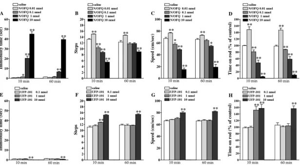
Endogenous N/OFQ exerts a physiologically inhibitory control over motor function (Marti et al., 2004a; 2008). Indeed, NOP receptor selective antagonists such as the peptide [Nphe1,Arg14,Lys15]N/OFQ-NH2 (UFP-101; Calo’ et al., 2002) and nonpeptide
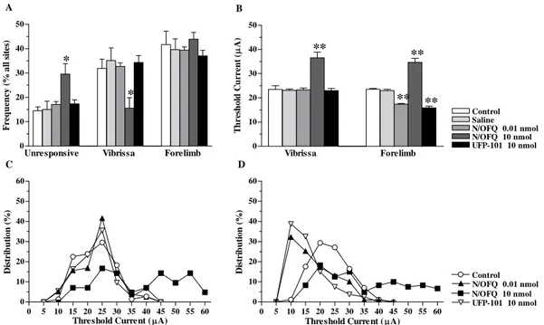
1-[(3R,4R)-1-cyclooctylmethyl-3-hydroxymethyl-4-piperidyl]-3-ethyl-1,3-dihydro-2H benzimidazol-2-one (J-113397 or Compound B; Kawamoto et al., 1999) and its achiral analogue 1-[1-(cyclooctylmethyl)-1,2,3,6-tetrahydro-5-(hydroxymethyl)-4-pyridinyl]-3-ethyl-1,3-dihydro-2H-benzimidazol-2-one (Trap-101; Trapella et al., 2006) increased stepping activity, run speed and rotarod performance in naïve rats (Marti et al., 2004a; 2008, 2009). J-113397 and Trap-101 also elevated motor performance in naïve mice (Viaro et al., 2008; Marti et al., 2008), while J-113397 increased arm movement speed in nonhuman primates (Viaro et al., 2008). The view of N/OFQ as a physiological constraint over motor activity was also corroborated by the finding that NOP-/- mice had greater stepping activity and rotarod performance than wild-type mice (Marti et al., 2004a, 2005; Viaro et al., 2008). Recent data, however, suggested that endogenous N/OFQ may play a more complex role in motor control. Indeed, J-113397 and Trap-101 facilitated motor activity at low doses and impaired it at higher ones through NOP receptor blockade in naïve rodents (Viaro et al., 2008; Marti et al., 2008). A similar dual response was also reported after i.c.v. administration of N/OFQ, low doses facilitating (Florin et al., 1996; Jenck et al., 1997; Higgins et al., 2001; Kuzmin et al., 2004, Marti et al., 2009) and higher ones inhibiting (Reinscheid et al., 1995; Devine et al., 1996; Rizzi et al., 2001; Higgins et al., 2001; Kuzmin et al., 2004; Marti et al., 2009) spontaneous locomotion. Importantly, we found that N/OFQ-induced motor facilitation was a true motor response (Marti et al., 2009) and not a result of an anxiolytic effect of N/OFQ as previously thought (Florin et al., 1996; Jenck et al., 1997). In fact, motor improvement induced by low N/OFQ doses given i.c.v. or injected into SNr was associated with enhanced motor cortex excitability and motor output, while motor impairment induced by higher N/OFQ doses was accompanied by opposite electrophysiological changes (Marti et al., 2009). Interestingly, NOP receptor antagonists replicated the electrophysiological and behavioral changes induced by low N/OFQ doses, overall suggesting that dual motor responses to NOP receptor ligands are mediated by NOP receptors in SNr and NOP receptor antagonists and N/OFQ (at low doses) activate common pathways. Evidence that mesencephalic DA neurons transduce motor actions of NOP receptor ligands has been presented. Indeed, N/OFQ and NOP
receptor antagonists, given systemically or into SNr, inhibited and facilitated DA release in dorsal striatum, respectively (Marti et al., 2004a). Moreover, N/OFQ inhibited DA release in limbic striatum (Murphy et al., 1999; Narayanan et al., 2004) while J-113397 elevated it, although via NOP-unrelated mechanisms (Koizumi et al., 2004). Finally, even the hyperlocomotive response to N/OFQ was reported to be DA-dependent (Florin et al., 1996; Kuzmin et al., 2004).
NOP receptor antagonists and PD
Endogenous N/OFQ also seems to play an important inhibitory role on motor behaviour in pathological conditions. Indeed, we collected evidence that N/OFQ sustains symptoms and neurodegeneration associated with PD (Marti et al., 2005). Thus, NOP receptor antagonists attenuated motor impairment in rats made hypokinetic with haloperidol (Marti et al., 2004b) or 6-OHDA lesioning (Marti et al., 2005, 2007, 2008) as well as in mice and nonhuman primates treated with MPTP (Viaro et al., 2008; Visanji et al., 2008). NOP receptor antagonists act via blockade of NOP receptors in SNr causing reduction of glutamate and increase of GABA release in SNr and, consequently, overinhibition of the nigro-thalamic pathway. To support pharmacological studies, genetic deletion of the NOP receptor (NOP-/- mice) conferred partial protection from haloperidol-induced akinesia (Marti et al., 2005), a phenomenon linked to the reduced ability of haloperidol to elevate nigral GLU release in NOP-/- mice (Mabrouk et al., 2010). Interestingly, DA depletion causes up-regulation of N/OFQ synthesis (Marti et al., 2005; Brown et al., 2006; Di Benedetto et al., 2009) and release (Marti et al., 2005) in SNr, thereby exacerbating negative influence of N/OFQ on DA cells and motor output. The role of N/OFQ in parkinsonism, however, may go beyond phenotype modulation. Indeed, mice with deletion of the N/OFQ precursor (ppN/OFQ) were found to be partially resistant to MPTP toxicity as shown by the reduced loss of nigral DA cells and striatal DA terminals observed following MPTP treatment in comparison with wild-type controls (Marti et al.,. 2005; Brown et al., 2006). However, different peptides (i.e. N/OFQ, N/OFQ II and nocistatin) are generated by cleavage of the ppN/OFQ precursor (Okuda-Ashitaka and Ito, 2000), questioning the view that endogenous N/OFQ modulates MPTP-induced neurotoxicity.
Purpose
The overall goal of this interdisciplinary study was to investigate the involvement of N/OFQergic transmission in motor activity under physiological and pathological conditions, in particularly PD. Specific aims of the study were as follows:
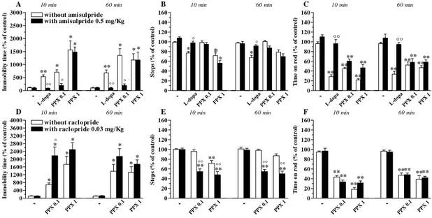
1) In order to dissect out the role of endogenous DA and the contribution of specific DA receptor subtypes to motor responses to NOP receptor antagonists, subtype-selective DA receptor antagonists were challenged against motor facilitating and inhibitory doses of N/OFQ and NOP receptor antagonists. This behavioural and neurochemical study was performed in naïve mice.
2) In order to prove the concept that blockade of nociceptinergic transmission is a new approach for symptomatic therapy of PD, we tested the effectiveness of NOP receptor antagonists in mice intoxicated with MPTP. This behavioural study was performed by testing NOP receptor antagonists alone or in combination with classical antiparkinsonian compounds.
3) In order to confirmed the efficacy of NOP receptor antagonists also in animals models that showed symptoms that closely resemble those observed in parkinsonian patients, we tested drugs in nonhuman primates. This behavioural study was performed in naïve and then in MPTP-treated macaques.
4) In order to demonstrate that endogenous N/OFQ sustains not only symptoms but also neurodegeneration associated with PD, we tested the impact of MPTP in NOP-/- mice by using behavioural tests and stereological counting.
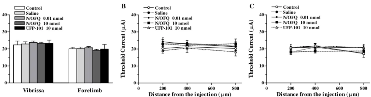
5) In order to investigate whether N/OFQ controls motor behaviour through NOP receptors located in SNr and modulation of motor cortex output, we used behavioral testing and intracortical microstimulation technique. NOP receptor ligands were locally injected in SNr (and M1, for a comparison) of naïve rats.
6) In order to study the progressive reorganization of motor cortex under parkinsonian conditions, the intracortical microstimulation technique was used in rats made hemiparkinsonians with 6-OHDA at different time points after lesion.
Materials and methods
Animals
All animals used in the study were housed with free access to food and water and kept under environmentally controlled conditions (12-h light/dark cycle with light on between 07:00 and 19:00). The experimental protocols were approved by the Italian Ministry of Health (licenses n. 94/2007B and 194/2008B) and Ethical Committee of the University of Ferrara. Adequate measures were taken to minimize animal pain and discomfort. After surgery, the skin was closed using surgical sutures and the wound was cleansed with an antibiotic solution (Rifamicina SV, Lepetit, Milano).
Mice
Young adult male (20-25 g; 10-12 weeks old) Swiss, C57BL/6J, NOP+/+ and NOP -/-mice were used in this study. Swiss and C57BL/6J -/-mice were purchased from Harlan Italy (S. Pietro al Natisone, Italy), while NOP-/- and NOP+/+ mice (Nishi et al., 1997) were raised at the vivarium of the Section of Pharmacology of the University of Ferrara. NOP-/- mice were grown on a C57BL/6J background.
Rats
Young adult male (150-300 g; 12-16 weeks old) Sprague-Dawley and Wistar rats were used in this study. Sprague-Dawley rats were purchased from Harlan Italy (S. Pietro al Natisone, Italy), while Wistar rats were generated at the vivarium of the Section of Human Physiology of the University of Ferrara.
Nonhuman primates
Young adult male macaques (Macaca fascicularis) were included in the study. Animals were housed individually at the New England Primate Research Center. All the studies were done following NIH guidelines and were approved by the IACUC at Harvard Medical Area and the New England Regional Primate Research Center.
Lesion of the DA system
In order to obtain a destruction of the DAergic system, different protocols were used, depending on the type of lesion to obtain (partial or total, unilateral or bilateral) and the species of animal to be treated.
6-OHDA lesion in rats
Unilateral lesion of DA neurons was induced in isoflurane-anaesthetised rats (Marti et al., 2005). Eight micrograms of 6-OHDA (in 4 µl of saline containing 0.02% ascorbic acid) were stereotaxically injected in the right MFB according to the following coordinates from bregma: AP= -4.4 mm, ML= -1.2 mm, VD= -7.8 mm below dura (Paxinos and Watson, 1982).
MPTP lesion in mice
Bilateral lesion of DA neurons was induced in C57BL/6J mice using an acute protocol for MPTP administration (4 x 20 mg/Kg, 90 min apart; Marti et al., 2005; Viaro et al., 2008, 2010).
MPTP lesion in nonhuman primates
Bilateral lesion of DA neurons was induced in nonhuman primates by subcrhonic administration of MPTP (0.3 mg/kg/week, i.v. for 7.5 ± 2.5 weeks) as described by Jenkins et al. (2004) and Sanchez-Pernaute et al. (2007).
Pharmacological treatments
For systemic administration (i.p.), the volume injected was 10 µl/g body weight. For local (central) administration, different methods are used in according to stereotaxic coordinates of rat brain (Paxinos and Watson, 1982) and mouse brain (Paxinos and Franklin, 2003).
Microinjection technique in mice
The injections in the lateral cerebral ventricle (LCV) of mice were given according to the procedure described by Laursen and Belknap (1986). Briefly, the syringe was held
at an approximate 45° angle to the skull. Bregma was found by lightly rubbing the point of the needle over the skull until the suture was felt. Once found, care was taken to maintain the approximate 45° angle and the needle was inserted about 2 mm lateral to the midline. The skull is relatively thin at this point, so only mild pressure was required to insert and remove the needle. Drugs were slowly injected (0.5 µl in about 5 sec) and to prevent the substance from refluxing, the needle was withdrawn from the skull 5 sec later.
Microinjection technique in awake rats
A guide cannula (outer diameter 0.55 mm, inner diameter 0.35 mm) was stereotaxically implanted under isoflurane anesthesia (1.4 % in air delivered at 1.2 ml/min) 1 mm above the right or left LCV, M1 or SNr, according to the following coordinates from bregma: LCV, AP -0.9, ML ±1.4, VD -2; M1, AP +2, ML ±2, VD -0.5; SNr, AP -5.5, ML ±2.2, VD -7.3. The cannula was secured to the skull by acrylic dental cement and metallic screws. A stainless steel obturator (outer diameter 0.30 mm) was left in place inside the guide. After a 7 day recovery period, each rat was opportunely handled and trained before behavioral tests. The day of the experiment, the obturator was removed and drugs were injected (volume 0.5 µl) through a stainless-steel injector (outer diameter 0.30 mm; inner diameter 0.15 mm) protruding 1 mm from the cannula tip.
Microinjection technique in anaesthetized rats
The animal was anaesthetized (ketamine hydrochloride, 50 mg/kg, i.p.) and placed in a Kopf stereotaxic apparatus and a large craniotomy was performed over the frontal cortex. The dura remained intact, and was kept moist with a 0.9 % saline solution. Drug injections were performed using a Hamilton syringe connected to the injection cannula with polyethylene tubing, in the left or right LCV (AP= 0.8 mm; ML= ±1.5 mm; VD= -3.5 mm below the pial surface), layer V of central M1 (AP= 2-3 mm and ML= 2.5-3 mm; VD= -1.5 mm) and SNr (AP= -5.5 mm; ML= ±2.2 mm; VD= -7.6 mm). Drugs were slowly injected and, to prevent the substance from refluxing, the needle was withdrawn from the cortex 120 sec later.
following coordinates from bregma (AP= 1 ML= 3; AP= 2 ML= 3; AP= 3 ML= 3). The cannula was lowered into the selected site 1 mm below the pial surface and the lidocaine was slowly injected (4 µl/min) and, to prevent the substance from refluxing, the needle was withdrawn from the cortex 120 sec later. For the duration of the experiment, cortical inactivation was maintained by supplementary injections of lidocaine (1-2 times for each animal) at the same sites when the forelimb movement at these sites was ICMS-evoked at the highest current used under the present experimental condition (60µA). The injected sites were remapped every 30 minutes to confirm that sites were inactivated.
Cortical bicuculline application
Under surgical stereomicroscopy, the dura on the M1 was removed and a 30 µl solution of 50 µM bicuculline was applied to the cortical surface (Stojic et al., 2008), by using a Gilson micropipette. The temperature of the solution was 35-37°C and the volume was maintained by supplementary application (1-2 times for each animal).
Behavioural studies in rodents
Motor activity in rodents was evaluated by means of different behavioural tests specific for different motor abilities, as previously described (Marti et al., 2004a, 2004b, 2005, 2007, 2008, 2009; Viaro et al., 2008, 2010). The different tests are useful to evaluate motor functions under static conditions or dynamic conditions, as an integration of coordination, gait, balance, muscle tone and motivation to run, and wre performed in a fixed sequence In the case of evaluation of pharmacological treatment, tests were performed 10 min before drug injection (control session) and 10 and 60 min after drug injection. With this protocol, both facilitatory/inhibitory and short/long term effects can be detected (Marti et al., 2004a).
Drug-induced rotation
The rotational model (Ungerstedt and Arbuthnott, 1970) was used to select the rats which had been successfully lesioned with 6-OHDA. Two weeks after lesion, rats were injected with amphetamine (5 mg/Kg i.p., dissolved in saline) and only those rats
performing >7 ipsilateral turns/min were enrolled in the study. This behaviour has been associated with >95% loss of striatal extracellular DA levels (Marti et al., 2002).
Bar test
This test, also known as the catalepsy test (Sandberg et al., 1988), measures the ability of the animal to respond to an externally imposed static posture. Each rodent was placed gently on a table and the right and left forepaws were placed alternatively on blocks of increasing heights (1.5, 3 and 6 cm for mice and 3, 6 and 9 cm for rats). The immobility time (in sec) of each forepaw on the block was recorded (cut-off time 20 sec per step, 60 sec maximum). Akinesia was calculated as total time spent on the blocks (mean between the two forepaws).
Reaction time test
This test measures motor reactivity of the animal in a open field. Mice were allowed to habituate to the center of a square arena (150x150 cm) for 5 min, then elevated 3 cm above the surface (lifting from the tail), and finally left to fall. When the animal touched the floor, the latency time for the first forelimb movement was recorded.
Drag test
This test (modification of the ”whelbarrow” test; Schallert et al., 1979), measures the ability of the animal to balance its body posture using forelimbs in response to an externally imposed dynamic stimulus (backward dragging; Marti et al., 2005). Each rodent was gently lifted from the tail (allowing the forepaws on the table) and dragged backwards at a constant speed (about 20 cm/sec) for a fixed distance (100 cm). The number of touches made by each forepaw was counted by two separate observers (mean between the two forepaws).
Elevated body swing test
The test was conducted in a dark plastic box (40×40×50 cm). Initially, each rat was allowed to habituate in the box for 5 min then it was elevated 3 cm above the ground by holding its tail. When the animal turned with its body (i.e. head and trunk) more than 30° to either side of the vertical axis a swing was counted. The direction and the number
Speed test
This tests essentially measure animal speed in an open field. The rodent was allowed to habituate in a square arena (150x150 cm) for 5 min then elevated 3 cm about the ground (by holding its tail) and finally positioned in the centre of the arena. When the animal touched the floor it started running. Behavior was scored online using the “correct walking” criteria (see Bouwman et al, 2005). Data acquisition was stopped when the rat changed its acceleration, velocity or direction. Run speed was calculated as distance traveled (cm/sec).
Stair climbing test
This test (modification of the SCA test; Kumar and Sehgal, 2007) analyzed the motivation and the motor skill during a climb-walk. Each mouse was positioned on the first step (2 cm height, 2 cm long, 5 cm wide) of a 45°-sloping staircase. At the top of the staircase a food pellet was lodged in a small dark goal box. The speed needed for climbing 20 consecutive steps (50 cm) was recorded. Steps made at the beginning and the end of the climb were excluded because of velocity changes or obvious acceleration/deceleration.
Grip test
This test was used to evaluate the skeletal muscular strength in mice (Meyer et al., 1979). The grip-strength apparatus (ZP-50N, IMADA, Japan) is comprised of a wire grid (5 × 5 cm) connected to an isometric force transducer (dynamometer). In the grip-strength test mice were held by their tails and allowed to grasp the grid with their forepaws. The mice were then gently pulled backward by the tail until the grid was released. The maximal and the average force exerted by the mouse before losing grip was recorded. The mean of 10 measurements for each animal was calculated and the maximal and mean force was determined. The skeletal muscular strength in mice was expressed in grams force (gf) and was recorded and processed by IMADA ZP-Recorder software.
Rotarod test
This test analyzes the ability of the rodents to run on a rotating cylinder (diameter 8 cm) and provides information on different motor parameters such as coordination, gait, balance, muscle tone and motivation to run (Rozas et al., 1997). The fixed-speed rotarod
test was employed according to a previously described protocol (Marti et al., 2004a; Viaro et al., 2008, 2010). Briefly, animals were tested at stepwise increasing speeds (180 sec each) and time spent on the rod calculated (in sec). Drug effect was calculated by monitoring motor activity within a limited speed range (four speed) causing a progressive decrement of performance to ~40% of the maximal response.
Footprinting test
This test provides information on gait patterns (Klapdor et al., 1997). Mice paws were marked with ink and gait patterns (stride length and width, foot angle, overlap, speed) analyzed after walking over a sheet of paper (Fig. 2F). The apparatus was composed of a white runway (5 cm wide, 70 cm long, with borders of 10 cm height) arranged to lead out into a dark goal box (20 x 20 x 30 cm). The parameters were measured by wetting forepaws and hindpaws with commercially available pencil nontoxic ink (the paws were painted with different colored inks) and allowing the mice to trot on a strip of paper (5 cm wide, 70 cm long) onto the runway. Pawprints made at the beginning and the end of the run were excluded because of velocity changes or obvious acceleration/deceleration. Stride length is the average distance (in mm) of forward movement between each forepaw and hindpaw footprint. Stride width is the average lateral distance (in mm) between opposite left and right forepaw and opposite left and right hindpaw. It has been calculated by measuring the perpendicular distance of a given step to a line connecting its opposite preceding and succeeding steps. Foot angle is the angle of hindpaw (in degree) with respect to main direction. It has been calculated by measuring the amplitude of angle between the direction of run and each direction of the hindpaw (line starting at center of paw to the third finger). Placement of paws is the footprint overlap (in mm) and is calculated by measuring the distance between the center of the forepaw footprint and the ipsilateral hindpaw footprint, taken from successive steps. Speed of run (in cm/sec), was calculated by the ratio between the length of runway and time spent along the runway. After the run, animals were placed in a cage filled with 0.5 cm warm water for 1 min in order to wash off the dye.
Spontaneous activity
analysed by two separate observers. Global, horizontal and fine movements were quantified together with immobility (i.e. freezing) time. Step parameters such as time for movement preparation and initiation, step speed and length were also evaluated. Activity measurements were conducted between 11:00 and 15:00 during the light phase.
Behavioural studies in nonhuman primates
In order to monitor parkinsonian phenotype and the effectiveness of drug treatments, motor activity in nonhuman primates were analysed by the movement analysis panel (MAP) test and neurologist evaluation.
Movement analysis panel (MAP)
Animals were trained to perform a computerized timed reaching task that measures the speed of arm movements (Jenkins et al., 2004). Training was carried out for an average of 6 days for the platform task and 8-12 days for the straight rod task, until the performance (time to retrieve the treats) was stable. For pharmacological evaluation, animals were tested 30 min after the administration of vehicle (saline) for two days and then with either saline or the active drugs. In addition, global motor activity data was obtained using activity monitors (Actiwatch) for a week at the naïve and parkinsonian stages (Table 1). These tests provide objective measures of bradykinesia and hypokinesia, respectively.
Parkinsonian rate scale (PRS)
For motor evaluation, animals were transferred to a Plexiglas observation cage where they were videotaped. Motor behavior was rated according to a scale based on the motor subscale of the UPDRS (Unified Parkinson Disease Rating Scale), as described (Jenkins et al., 2004; Sanchez-Pernaute et al., 2007). The following signs were scored from 0 to 3: bradykinesia in the left and right arms (L/R), tremor L/R, rigidity L/R, hypokinesia, posture/balance (for a total score from 0 to 24). Scores were obtained at 30-45 min after each drug or vehicle administration.
Intracortical microstimulation in rats
The intracortical microstimulation (ICMS) technique is commonly used to quantitatively evaluate plastic changes in the motor cortex following motor output disconnection because it is useful in evaluating the strength of functional connection between the motor cortex and the target motoneuron pool (Sanes et al., 1990). It presumes that after motor output disconnection/manipulation, the motor cortical changes in the types of evoked movements and in the minimal level of current necessary to evoke movements, reflect the adapting changes of cortical circuits. If so, the difference changes in cortical evoked movements and in their current threshold over time could reveal different underlying mechanisms in adaptive remodeling of disconnected/manipulated cortical circuits. In each animal, the movements evoked by ICMS in the frontal agranular cortex were mapped.
Surgical procedure
The anaesthetized animal was placed in a Kopf stereotaxic apparatus and a large craniotomy was performed over the frontal cortex of both hemispheres. The dura remained intact and was kept moist with an 0.9% saline solution. The electrode penetrations were regularly spaced out over a 500µm grid. Alteration in the coordinate grid, up to 50µm, was sometimes necessary to prevent the electrode from penetrating the surface blood vessels. These adjustments in the coordinate grid were not reported in the reconstructing maps. When the adjustment was over 50µm, the penetration at this site was not performed. Glass insulated tungsten electrodes (0.6-1MΩ impedance at 1kHz) were used for stimulation. The electrode was lowered perpendicularly into the cortex to a depth of 1.5 mm below the cortical surface and adjusted ±200 µm so as to evoke movement at the lowest threshold. In preliminary experiments this depth was found to correspond to layer V of the frontal agranular cortex (Franchi, 2000).
Mapping procedure
The mapping procedure was similar to the one described by Donoghue and Wise(1982) and Sanes et al. (1990), and detailed elsewhere (Franchi, 2000). Briefly, monophasic
movement without knowledge of the actual current intensity and was unaware of which group the particular rat belonged to. The other observer changed the level of current. Starting with a 60µA current, intensity was decreased in 5µA steps until the movement was no longer evoked; then the intensity was increased to a level at which approximately 50% of the stimulations elicited movement. This level defined the current threshold. If no movements or twitches were evoked with 60µA, the site was recorded as negative (ineffective site). Mapping was initiated at a high current because the initial polysynaptic recruitment of remote neurons optimizes the detection of movements in this 500µm step grid mapping. Body parts activated by ICMS were identified by visual inspection and/or muscle palpation. When eye movement was observed, the current threshold was determined under optical microscope. A normal component of the output organization of rat M1 is the presence of some sites along the border region between two movement representations from which movement of both body parts can be evoked simultaneously. At such sites, both movements were recorded for that position regardless of the individual thresholds (which were determined separately for each). Hereafter such movements are identified as ‘threshold-movement’ and ‘over-threshold movement’, respectively, according to value of the individual threshold. In some sites along the border region between two movement representations, both movements can be evoked simultaneously at the current threshold level (dual movement site). Forelimb movements evoked by threshold current typically consisted of brief twitches of the elbow or shoulder (proximal limb movement), wrist and digit (distal limb movement), or simultaneous twitches of both muscles groups. Forelimbs and hindlimbs were approximately half-way between flexion and extension and were alternately flexed and extended, particularly at the representational borders.
Neurotransmitter release
The pharmacological profiles of presynaptic DA receptors modulating monoamine release from mice striatal synaptosomes were studied and compared using different dopaminergic ligand.
Synaptosome preparation
To minimize pain and discomfort, mice were decapitated under light ether anesthesia and the striatum was quickly excised to prepare synaptosomes, as previously described (Morari et al., 1998). Briefly, striata were homogenized in ice-cold 0.32 M sucrose buffer at pH 7.4 then centrifuged for 10 min at 2,500 gmax (4°C). The supernatant was
then centrifuged for 20 min at 9,500 gmax (4°C) with the synaptosomal pellet being
resuspended in oxygenated (95% O2, 5% CO2) Krebs solution (mM: NaCl 118.5, KCl
4.7, CaCl2 1.2, MgSO4 1.2, KH2PO4 1.2, NaHCO3 25, glucose 10) containing ascorbic
acid (0.05 mM) and disodium EDTA (0.03 mM). Synaptosomes were pre-loaded with [3H]-DA by incubation in medium containing 50 nM [3H]-DA (specific activity 27.8 Ci/mmol, NEN DuPont, Boston, MA, USA.) for 25 min. One milliliter aliquots of the suspension (~0.35 mg protein) were slowly injected into nylon syringe filters (outer diameter 13 mm, 0.45 µM pore size, internal volume of about 100 µl; MSI, Westporo, MA, USA) which were then connected to a peristaltic pump. Filters were maintained at 36.5 °C in a thermostatic bath and superfused at a flow rate of 0.4 ml/min with a pre-oxygenated Krebs solution. Under these experimental conditions, spontaneous [3H]-DA
efflux was essentially unaffected by reuptake. Sample collection (every 3 min) was initiated after a 20 min period of filter washout. The effect of drugs was evaluated on both spontaneous and K+-stimulated neurotransmitter outflow. In this case, drugs were added to the perfusion medium 6 (agonist) or 9 (antagonist) min before a 10 mM KCl pulse (120 sec) and maintained until the end of the experiment.
[3H]-DA analysis
[3H]-DA levels in the samples were measured by liquid scintillation spectrophotometry. Sample superfusate (1.2 ml/sample) and filter retained (dissolved with 1 ml of 1 M NaOH followed by 1 M HCl) were opportunely mixed with Ultima Gold XR scintillation fluid (Packard Instruments B.V., Groningen, The Netherlands) and radioactivity was determined by a Beckman LS 1800 β-spectrophotometer.
endoplasmic reticulum and ribosomes, and occurring in nerve cells body and dendrites. Because of the RNA content, Nissl substance is very basophilic and will be very sharply stained with cresyl violet acetate. This aspecific staining was used for check the placement of the probes or electrode. The tracks was verified by microscopic examination and the animals in which the tracks were not correctly positioned were discarded from the study. On contrary, tyrosine hydroxylase (TH; tyrosine 3-monooxygenase) is a selective marker, because this enzyme is content only in the cytoplasme of dopaminergic cells. TH is responsible for catalyzing the conversion of the amino acid L-tyrosine to dihydroxyphenylalanine (dopa). Dopa is a precursor for dopamine which in turn is a precursor for norepinephrine (noradrenaline) and epinephrine (adrenaline).
Tissue processing
Mice were deeply anesthetized with ketamine and xylazine (85+15 mg/Kg; i.p.) and rats were deeply anaesthetised with Zoletil 100® (10 mg/Kg, i.m.; Virbac Laboratories,
Carros, France), transcardially perfused at room temperature with phosphate-buffered saline (PBS; 20 mM, pH 7.4) and fixed with cold 4% paraformaldheyde in PBS. Brains were removed, post-fixed overnight, transferred to 20% sucrose solution in PBS for cryoprotection (until they sunk) and stored at -80°C. We used a cryostat at −18°C to cut 40 µm coronal sections, which were collected free floating (in PBS) for all analysis.
Nissl staining
Sections at levels of cortex and SNc were mounted on gelatine-coated slides, stained with cresyl violet, dried in escalating alcohol concentration (50-70-90-95-100%), cleared in xylene and coverslipped with mounting medium.
TH immunohistochemistry
Sections at levels of striatum and SNc, were rinsed 3 times in PBS and incubated for 15 min in 3% H2O2 and 10% methanol in PBS to block the endogenous peroxidase activity.
After washing in PBS, the sections were preincubated in blocking serum (5% normal horse serum and 0.3% Triton x100 in PBS) for 60 min, followed by incubation in anti-TH mouse monoclonal antibody solution (1:2000, Chemicon, Temcula, CA) for 16 hr at room temperature. The sections were then rinsed in PBS and incubated for 1 hr in biotinylated horse anti-mouse IgG secondary antibody (1:200; Vector Laboratories,
Burlingame, CA). After rinsing, sections were incubated with avidin-biotin-peroxidase complex (Vector Laboratories) for 30 min at room temperature. After rinsing with PBS, immunoreactivity was visualized by incubating the sections in a solution containing 0.05% 3,3-diaminobenzidine (DAB) in 0.013% H2O2 in PBS for about 1 min. The
sections were rinsed in PBS, mounted on chrome-alum-coated slides, eventually counterstained with cresyl violet, dried with escalating alcohol concentration, cleared in xylene and coverslipped with mounting medium.
Optical density evaluation
The sections were viewed with a Zeiss Axioskop (Carl Zeiss, Germany). Sections were acquired (AxioCam ICc3, Carl Zeiss, Germany) and TH-immunoreactive fiber density analyzed using ImageJ software (Wayne Rasband; NIH, USA). For each animal, optical density was calculated as the mean of the 5 striatal levels and corrected for non-specific background, measured in the corpus callosum.
Stereological cell counting
For counting of TH-immunoreactive neurons (phenotypic marker) and cresyl violet stained cells (structural marker) in SNc, an unbiased stereological sampling method was used (West and Gundersen, 1990), based on optical dissector stereological probe (Bezard et al., 2003; Gross et al., 2003). Stereological analysis was performed using an Leica DMRE microscope with a motorized Z and X-Y stage encoders linked to a computer-assisted stereological system (Mercator Digital Imaging System, Explora Nova, La Rochelle, France). For each animal, SNc boundaries were delimited at low magnification (2.5×) by examining the size and shape of the different groups of TH-immunoreactive neurons and their axonal projections, as well as nearby fibre bundles according to the mouse brain atlas. SNc boundaries were drawn on every fourth section and the first was randomly chosen. SNc volume was calculated using the formula V(SNc)
=∑S td; where ∑S is the sum of surface areas (µm2), t the average section thickness and d the distance between the sections (Theoret et al., 1999). The average section thickness (t) was estimated to 12 µm after immunohistochemistry processing and guard zones of 2 µm were used to ensure that top and bottom of sections are never included in the analysis. Eight sections were used for each animal. From a random start position, a
between each counting frame. Within each frame, all cell nuclei which came into focus (40× immersion oil objective; Gundersen et al., 1988) were counted. A neuron was counted if more than half the cell body was inside the two consecutive boundaries taken into account. To estimate the number of TH-immunoreactive neurons we used: N=
V(SNc) (∑Q−/∑V(dis)); where N is the estimation of the number of TH-immunoreactive
neurons, V the volume of SNc, ∑Q− the number of cells counted in the frames and
∑V(dis) is the total volume of frames (Theoret et al., 1999).
Data presentation and statistical analysis
Data are expressed as means ± SEM of n determinations per group. Different statistical analysis was performed, as appropriate: Student’s t-test, χ2 test presented in a two-way contingency table, one-way ANOVA followed by the Newman-Keuls or PLSD test, and two-way repeated measure (RM) ANOVA followed by contrast analysis and the sequentially rejective Bonferroni test. P values <0.05 were considered to be statistically significant. In order to facilitate the readership, we reported the results of statistics performed on data in a separate section (appendix I).
Materials
6-OHDA bromide, amphetamine methylester, benserazide hydrochloride, bicuculline methochloride, L-dopa methyl ester and MPTP hydrochloride were purchased from Sigma (St. Louis, MO, USA). Amisulpride, domperidone, GBR12783, raclopride and SCH23390 were purchased from Tocris (Bristol, UK). PPX was purchased from McTony Bio&Chem (Vancouver, Canada). S33084 was provided by Institut de Recherches Servier (Croissy-sur-Seine, France). Lidocaine hydrochloride was purchased from S.Anna Hospital (Ferrara, Italy). N/OFQ, J-113397, Trap-101 and UFP-101 were synthesized in the laboratories of the Department of Pharmaceutical Chemistry at the University of Ferrara. All drugs were freshly dissolved in the vehicle just prior to use.
Results
Part I Different subpopulations of D2 receptors mediate dual motor responses of
NOP receptor antagonists in mice
Dose-response curves of NOP receptor antagonists
The motor profiles of three NOP receptor antagonists were investigated in C57BL/6J mice by using static and dynamic tests providing complementary information on motor parameters: the bar, drag and rotarod tests. The non peptide antagonist J-113397 and its achiral analogue Trap-101 were administered systemically while the peptide antagonist UFP-101 was given i.c.v. Basal activity in absolute values was 0.8 ± 0.1 sec (immobility time in the bar test), 16.5 ± 0.9 steps (drag test) and 937.9 ± 62.1 sec (time on rod). Motor activity was not different at the right and left paw so data were pooled together.
J-113397
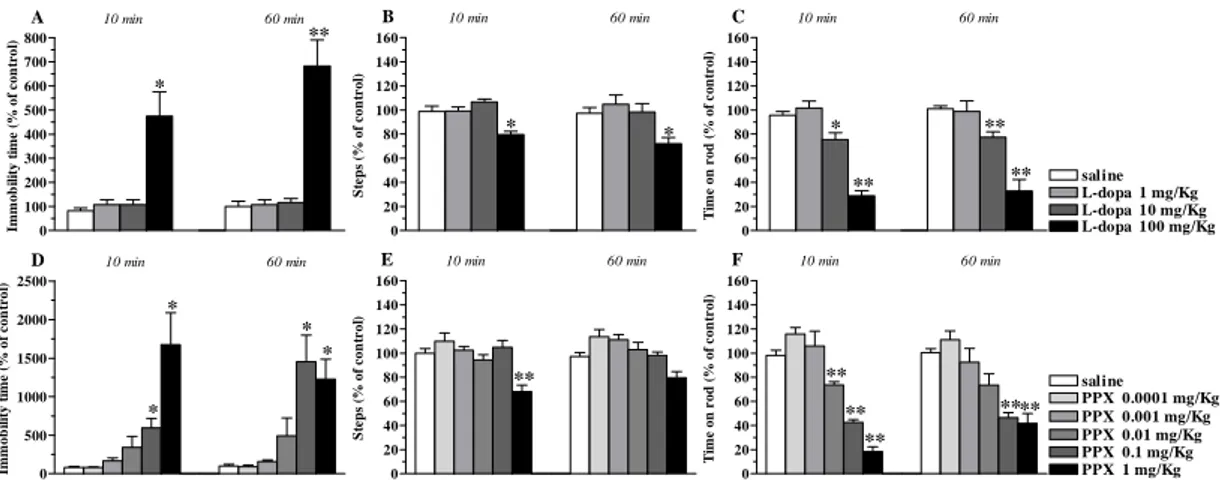
J-113397 caused long lasting inhibition of the immobility time in the bar test at 10 mg/Kg, lower doses being ineffective (Fig. 1A). J-113397 caused a dual regulation of stepping activity in the drag test (Fig. 1B) and rotarod performance (Fig. 1C). In both tests, facilitation was observed at 0.3 and 1 mg/Kg and reduction at 10 mg/Kg.
0 100 200 300 400 500 600 700 * A ** 10 min 60 min Im m o b il ity ti m e (% o f c o n tr o l) 0 20 40 60 80 100 120 140 160 ** ** ** B 10 min 60 min S tep s ( % o f co n tro l) 0 20 40 60 80 100 120 140 160 180 saline J-113397 0.1 mg/Kg J-113397 0.3 mg/Kg J-113397 1 mg/Kg J-113397 10 mg/Kg * * * C 10 min 60 min T im e on r o d ( % of co n tr o l)
Figure 1. 113397 dually modulated motor activity in C57BL/6J mice. Systemic administration of J-113397 (0.1-10 mg/Kg, i.p.) affected motor performance in the bar (A), drag (B) and rotarod (C) test. All tests were performed before (control session) and after (10 and 60 min) drug injection. Data are means ± SEM of 6 determinations per group and were expressed as percentage of the control session. *p<0.05,
Trap-101
Trap-101 did not affect the immobility time at any of the doses tested (Fig. 2A). However, Trap-101 dually modulated motor activity in the drag and rotarod test, increasing the number of steps (Fig. 2B) and time on rod (Fig. 2C) at 10 mg/Kg and reducing them at 30 mg/Kg. administration. These effects were observed only at 10 min after drug administration.
UFP-101
UFP-101 increased the immobility time at 30 nmol (Fig. 2D) and caused dual responses in the drag (Fig. 2E) and rotarod test (Fig. 2F), namely facilitation at 1 and 3 nmol and marked inhibition at 30 nmol. Differently from non peptide antagonists, the effects of UFP-101 were detected also at 60 min after injection.
0 40 80 120 160 200 240 280 320 360 400 A 10 min 60 min Im m o b ilit y t im e ( % o f c o n tr o l) 0 20 40 60 80 100 120 140 160 B * * 10 min 60 min S teps ( % of con trol ) 0 20 40 60 80 100 120 140 160 Trap-101 0.1 mg/Kg saline Trap-101 1 mg/Kg Trap-101 30 mg/Kg C Trap-101 10 mg/Kg * * 10 min 60 min T im e on r o d ( % of co n tr o l) 0 1000 2000 3000 4000 5000 6000 D ** ** 10 min 60 min Im m o b ilit y t im e ( % o f c o n tr o l) 0 20 40 60 80 100 120 140 160 180 200 E ** ** * ** ** ** St e p s ( % o f c o nt r o l) 10 min 60 min 0 20 40 60 80 100 120 140 160 UFP-101 0.1 nmol saline UFP-101 1 nmol UFP-101 10 nmol F UFP-101 3 nmol UFP-101 30 nmol ** ** ** ** **** 10 min 60 min T im e o n r o d ( % o f c o n tr o l)
Figure 2. Trap-101 and UFP-101 dually modulated motor activity in C57BL/6J mice. Systemic administration of Trap-101 (0.1-30 mg/Kg, i.p.), or i.c.v. injection of UFP-101 (0.1-30 nmol) affected motor performance in the bar (A, D), drag (B, E) and rotarod (C, F) test. All tests were performed before (control session) and after (10 and 60 min) drug injection. Data are means ± SEM of 6 determinations per group and were expressed as percentage of the control session. *p<0.05, **p<0.01 different from saline (RM ANOVA followed by contrast analysis and the sequentially rejective Bonferrroni’s test).
DA receptor subtypes differentially modulate motor actions of NOP receptor antagonists
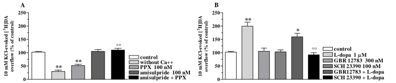
We previously reported that NOP receptor antagonists elevate striatal DA release in rats, suggesting that endogenous N/OFQ tonically inhibits nigro-striatal DA transmission (Marti et al., 2004a). We therefore employed selective DA receptor antagonists to unravel the contribution of endogenous DA to motor actions of NOP


![Figure 14. L-dopa increased spontaneous tritium efflux from synaptosomes. L-dopa (1-100 µM) elevated spontaneous tritium efflux from a preparation of striatal synaptosomes in superfusion pre-loaded with [ 3 H]-DA (A)](https://thumb-eu.123doks.com/thumbv2/123dokorg/4700146.44757/49.892.152.764.255.442/increased-spontaneous-synaptosomes-elevated-spontaneous-preparation-synaptosomes-superfusion.webp)
![Thiazolo[5,4-d]thiazole-based organic sensitizers with improved spectral properties for application in greenhouse-integrated dye-sensitized solar cells](data:image/gif;base64,R0lGODlhAQABAIAAAP///wAAACH5BAEAAAAALAAAAAABAAEAAAICRAEAOw==)