1
Università degli Studi di Catania
Scuola Superiore di Catania
International PhD
in
STEM CELLS
XXIV cycle
THE CELL CYCLE GENES REGULATION OF IPS CELLS AND
THE ROLE OF SWI/SNF CHROMATIN REMODELING ENZYMES
DURING THEM DIFFERENTIATION
Maurizio Rossi
Coordinator of PhD (Tutor): Prof. Daniele Condorelli
Co-Tutor: Dr. Luigi Vitelli
2
INDEXABSTRACT... 6
1. INTRODUCTION……….. 9
1.1. Analysis of the cell cycle in ES cells……… 9
1.2 Induced pluripotent stem cells ……….……. 11
1.2.1 Mouse induced pluripotent stem cells ……..……… 11
1.3 ES and iPS cell cycle.……….………... 15
1.4 Cell-cycle control differs in differentiated cells and ES and iPS cells . 19 1.5 The cyclin E family ……….. 21
1.6 The family of transcription factors E2F ……… 23
1.7 The regulation of activity by the E2F proteins "pocket"……… 25
1.8 Rb-mediated inhibition of E2F ……….. 29
1.9 Active transcriptional repression by Rb ……… 31
1.10 The chromatin remodeling: the role of multiprotein complex SWI / SNF containing BRM and BRG-1………... 33
1.11 SWI/SNF-Brg1 Regulates Self-Renewal and Occupies Core Pluripotency-Related Genes in Embryonic Stem Cells ………... 40
1.12 Regulation of cyclin E1 gene by the complex repressor CERC2…….. 41
1.13 Differentiation of the cES and iPS ……….... 44
1.14 Retinoic acid as an inducer of differentiation ……… 46
1.15 Metastable states of pluripotency ………...……… 47
3
2. MATERIALS AND METHODS ……… 50
2.1 ES and iPS Cell Culture ………. 50
2.1.1 Lines of Mouse Embryonic Stem Cells CGR8 and iPS..……… 50
2.2 MICE ………... 52
2.3 Cell Culture ………... 52
2.4 Retroviral production and ips Cell Induction……… 53
2.5 Chemical treatment of Ips And cES with Retinoic Acid……….. 55
2.6 Embryonic bodies induction (ascorbic acid treatment)………. 56
2.7 EPICS induction with L-Prolyn ………..………. 58
2.8 The cell counting and sampling ……… 59
2.9 Analysis of nuclear protein in cES and iPS ……… 60
2.10 Colorimetric assay for quantitative protein analysis ……… 61
2.11 Acrylamide Electrophoresis with SDS (SDS PAGE)……… 62
2.12 cES and iPS gene expression analysis through Western Blotting……… 63
2.13 Used Antibodies..……….….……….. 64
2.14 Immunoprecipitation with Dyneabeads ……… 65
2.15 EMSA (Electrophoretic Mobility Shift Assay)………... 67
2.15.1 Marking End in 5 '(CERM)………... 67
2.15.2 Annealing and Acrylamide Gel Electrophoresis On (CERM)……… 68
2.15.3 Marking End In 5’ DHFR Promoter ……… 69
2.15.4 Annealing And Acrylamide Gel Electrophoresis On (DHFR) ………… 70
2.16 Real Time PCR ……… 72 2.16.1 RNA extraction ……… 72 2.16.2 RNA Precipitation ……… 72 2.16.3 Washing of RNA………... 73 2.16.4 RNA Quantization..……….….……… 73 2.16.5 Retrotranscription ………... 73
4
3 THESIS RESEARCH OBJECTIVE... 75
4 RESULTS (1 st Part)……… 79
4.1 Exploiting experimental procedures for culturing and differentiating ips cells to
cardiomyocytes and neurons……… .79
4.1.2 In vitro differentiation of embryonic bodies generated by ips: effect of ascorbic acid
(aa)……… 80
4.1.3 Copperative effect of seeding density and the ascorbic acid effect on the differentiation of EB derived ips……… 82 4.1.4 Induction of ips-epics cells by treatment with l-prolyn……… 84 4.1.5 Ttreatment of ips with retinoic acid (commitment toward neural
differentiation)……… 85
4.2 RESULTS (2 st Part)……… 87 4.2.1 iPS Treatment with GMEM medium without LIF for inducing differentiation
……… 87
4.2.2 Protein expression analysis of the cell cycle genes during treatment of ips cells with
retinoic acid……… 89
4.2.3 Expression analysis of the genes specifying SWI/SNF Chromatin Remodeling
Complexes in iPS cells……… 91
4.2.4 Effect of treatment of ips with retinoic acid on differentiative markers (real time
pcr analysys) ……… 93
4.2.5 Interaction of the complex SWI / SNF (BRM AND BRG-1) with HDAC1 during proliferation and differentiation of ips: BRG-1 and HDAC1 interact in ips induced to differentiate with ra: reciprocal immunoprecipitation and western blot analysis… 94
5
4.2.6 The complex interaction with E2F/Rb with chromatin remodeling enzymes in iPS treated with Retinoic Acid. ……… 97 4.2.7 Id2 interacts with Rb in ES and iPS treated with LIF……… 99 4.2.8 HDAC1 co-immunoprecipitates with E2F ……… 101 4.2.9 mSin3A forms a complex with HDAC1 in iPS induced to differentiate withouth LIF
and treated with RA: reciprocal immunoprecipitation and Western blot analysis
……… 103
4.2.10 BRG-1 interacts with Cyclin E during the differentiation induced by Retinoic
Acid………. 105
4.2.11 Rb interacts with HDAC1 in iPS treated with RA………. 107 4.2.12 Prelimiar Results on the study of the genes that regulate the cell cycle during differentiation of iPS induced in EPISC by L-Prolyn ……… 109 4.2.13 Prelimiar Results on the study of the genes that regulate the cell cycle during differentiation of iPS by Ascorbic Acid ……… 111 4.2.14 Characterization of Complex Repressor CERM / CERC by EMSA In cES And iPS Treated With LIF And With Retinoic Acid. ……… 113 4.2.15 E2F/Rb complex interactions with some genes that control the cell cycle in iPS cells treated with LIF and RA ……… 117 4.2.16 CRE-DNA binding complexes in RA and EPA+RA treated ES cells. … 119 5 DISCUSSION……… 121 6 FIGURE……… 134 7 BIBLIOGRAPHY……… 163
6
ABSTRACT
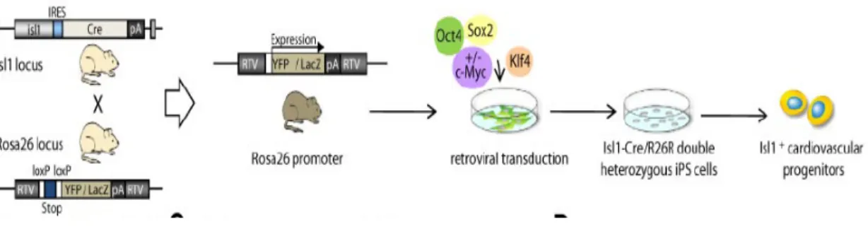
The pluripotent cell state can be achieved by ectopic expression of some transcription factors that can reprogram somatic cells to achieve pluripotency. (Takahashi, K., and Yamanaka, S.; 2006; Takahashi, K., and Yamanaka, S.; 2007 ). One of the purpose of these studies is to identify which genes are modulated during the early stages of differentiation of iPS .We analyzed the expression of some proteins that regulate the cell cycle by Western blot: cyclin A, cyclin E, CDK2, the family members Rb (Rb105, p130 and p107), some subunits of the complex SWI / SNF (BRM and BRG-1) histone deacetylases ,mSin3A, and E2F2 and E2F4. The results show that the expression of cyclin A and cyclin E proteins are present in high quantities in undifferentiated iPS and their expression increase at 1h and then decreases at 3h, 6h, 24h in iPS induced to differentiate with retinoic acid (RA). In this studies I have been found that the level of CYC E in iPS is less than cES. As a further control, were carried out analysis of RealTime-PCR The data of immunoprecipitation shows that the interaction of BRG-1-HDAC-1 (data not shown) is strongly stimulated in the presence of RA 5uM for 1h and little without RA. The undifferentiated ESCs express this protein very weakly. The results indicate the presence of complex Rb/Id2 in ES and iPS (undifferentiated) samples.
Another objectives of my thesis was to study the regulation of gene expression during the cyclic E1 "self-renewal" of mouse iPS. This result confirms that during self-replication of the CES a multiprotein complex binds to the promoter sequence of CERM cyclin E1 is
7
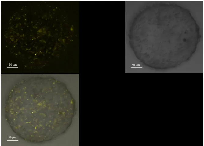
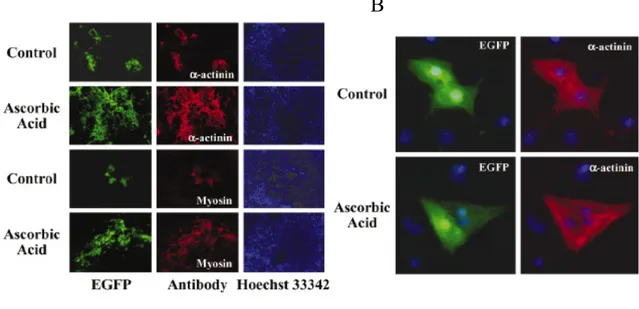
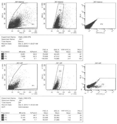
expressed constitutively and without cell cycle periodicity in the iPS. Brefly in IPS treated with LIF and RA the CERC2 complex is made by P130-E2F4-CYCE-Rb proteins suìimilarly to what observed for the ESC cells. I have obtained differentiated cardiomyocytes from Embryonic Bodies Furtermore the FACS analysis has shown that the number of cardiomyocytes was about 8% of the total cells in the Embryonic bodies. My studies on the differentiative effect of ascorbic acid show that iPS cells behave as ES cells. In fact, ascorbic acid at a concentration of 50 µg/ml final has a stimulant effect on the differentiation of EB. The results with the treatment of iPS withAscorbic Acid indicate that this chemical induce the differentiation in cardiomyocytes .In fact I have shown that the level of CYC A, CDK2 and Oct 4 are decreasing following this treatment.
These results obtained has been very interesting and revealed the importance of the expression of cyclin E and of the Rb/E2F pathway during the early stages of differentiation in both ES cells into iPS. In pluripotent iPS and ES cells induced to differentiate, the length of the G1 phase of cell cycle is greatly increases, and at the molecular level, the complex cyclin E/Cdk2 kinase activity increased from the constitutive kinase activity in undifferentiated cells to a dependent activity as observed during the early stages of differentiation in the cell cycle.. In addition, the regulated activities of the cyclin E/Cdk2 complex results in the activation of the pathway Rb/E2F which in turn requires a cell cycle dependent transcriptional control of target genes for transcription factor E2F.
Overall, the results obtained in these studies supports the model that the cell cycle of ES and iPS cells abbreviated with a short G1 phase, is functionally linked to pluripotency. In conclusion the iPS cell have a similar cell cycle regulation than ES cells.
8
ACKNOWLEDGMENTSI would like to first thank my advisor and dissertation mentor Dr. Luigi Vitelli. His constant help was key in the writing and success of this PhD thesis. Special thanks to Prof.Rodolfo Negri for his help on the thesis grading. Moreover I thank a lot to Dr. Agostino Girolamo and Prof.A. Carè for the help in the stem cell culture.
Dedication
I dedicate this work to my Mother, friends and colleagues who have encouraged me to work towards my goals and never give up.
9
1 INTRODUCTION
1.1 ANALYSIS OF THE CELL CYCLE IN ES CELLS.
Study of the pluripotency of embryonic stem (ES) cells aims to investigate the mechanisms of activation of proliferative signals, and transcription of the "checkpoint" that control the cell cycle. The level of proliferation of ES cells is unusually high. This reflects the structure of the cell cycle that lacks gap phases (G) complete and basically consists of stages of DNA replication (S phase) alternating with chromosome segregation (M phase) (Burdon T.,et. Al. 2002). In detail the phases G1, S and G2 / M represent 20, 70 and 10%, respectively, of the total cycle. Based on the proportion of cells that are in S phase and the length of the cell cycle (11-12 hours), it was estimated that the ES cells is about 5.5-6 hours in S phase and for about 1.5 hours in G1 phase (Savatier et al., 1994 and 1995). For this reason, the cell cycle regulation in ESC is fundamentally different from that of other somatic cells. The division by the ESC is led unusually high activity of the complexes cdk2/ciclina A and E, which appears to be constitutived. The only kinase activity that shows periodicity in the cell cycle is associated with cyclin B, which is highest in mitosis (Stead et al., 2002).
Numerous experiments have demonstrated the role of E2F TF target gene in the control of cell cycle genes regulated particularly when cells leave the G0 phase and enter in the cycle. To study the role of E2F TF binding sites in the regulation of cell cycle, Le Cam et al.,(1999) have investigated the promoter of cyclin E1 in differentiated cells. In this study on quiescent mouse fibroblasts they have discovered that the activity of cyclin E1 gene
10
promoter is controlled by a DNA sequence defined as "cyclin E repressor module (CERM). The CERM is located downstream of the transcription start site and consists of a variant of the canonical E2F sites flanked by a sequence AT rich. Both sequences are necessary to bind a complex of high molecular weight E2F called cyclin E repressor complex 1 (CERC1) that prevents the transcription of cyclin E1. When the fibroblasts begin to proliferate, the complex CERC1 dissociates from CERM sequence and cyclin E1 transcription is induced. Similar studies have shown that even in quiescent K562cells , CERM regulates the repression of CCNE1 gene in the period between the end of mitosis and late G1 phase through its association with a high molecular weight E2F complex called CERC2 (Polanowska et al ., 2001). The repression of cyclin E1 gene is not sufficient to block the kinase activity of the complex: it requires the involvement of protein Cip / Kip, which inhibit specifically the catalytic subunit of CDK2. More recently, Dalton and White et al (Stead, 2002) have investigated by Chip analysis the Cyclin E promoter in ES cell during differentiation. Briefly they have found the p107 is recruited on E2F4 binding sites in the Cyclin E promoter during the changes in Cyclin E1 transcription during differentiation.
11
1.2 INDUCED PLURIPOTENT STEM CELLSPrevious work had already indicated that individual transcription factors, when overexpressed or deleted, could induce cell fate changes in somatic cells. For example, overexpression of the transcription factor MyoD induced the conversion of fibroblasts into myogenic cells (Davis, Weintraub et al. 1987). Mouse B cells were shown to reprogram into progenitors that can give rise to the hematopoietic lineage when Pax5 was removed (Nutt, Heavey et al. 1999). In ESCs, the activation of transcription factors can lead to the differentiation into a certain lineage. The indication that transcription factor overexpression can induce cell fate changes started attempts to reprogram cell types to other lineages including the induction of pluripotency from differentiated cells.
1.2.1 MOUSE INDUCED PLURIPOTENT STEM CELLS
Several transcription factors and genes are important for maintaining the pluripotency and ES cell phenotype in early embryos and ES cells. In 2006, Takahashi et al. selected 24 genes as candidates for factors that induce pluripotency in somatic cells base done the hypothesis that these factors play a role in the maintenance of ES cell identity (Takahashi and Yamanaka 2006). A βgal-neomycin cassette was inserted into the mouse Fbx15 gene by homologous recombination. Fbx15 was used as a marker for pluripotency. The 24 candidates were all together introduced into mouse embryonic fibroblasts (MEFs) from Fbx15βneo/βneo embryos by retroviral transduction. This resulted in neomycin resistant
12
colonies with morphology similar to ES cells. Critical candidates to induce the pluripotency were determined by excluding individual factors from the pool of transduced genes. Ten genes were identified of which the individual absence resulted in poor or no colony formation. Transduction with the combination of these 10 factors produced more ES cell-like colonies that all 24 genes together did. When individual factors were excluded from the 10-pool transduction into MEFs, only a few or no colonies were formed when the factors OCT4, SOX2, KLF4 or c-MYC were not present. Takahashi et al. showed that these four factors together could produce a similar number of colonies to the ones observed with the pool of 10 genes. This demonstrated that induced pluripotent stem cells (iPS cells) could be derived from MEFs by the transduction with the transcription factors OCT4, SOX2, c-MYC and KLF4. Global gene-expression profiles of ES cells, iPS cells and Fbx15βneo/βneo MEFs revealed that iPS cells are closely related to ES cells but not to fibroblasts. It is for these reasons that in this thesis we have studied the cell cycle genes regulation of ips cells and the role of SWI/SNF chromatin remodeling enzymes during them differentiation. Takahashi et al. also showed the pluripotency of iPS cells by the differentiation into all three germ layers during teratoma formation after a subcutaneous injection of iPS cells into nude mice. Using tail tip fibroblasts instead of MEFs from Fbx15βneo/βneo mice led to the same result. However, no chimeric mice were obtained. However, when endogenous Nanog expression was used for the selection of iPS cells instead of Fbx15, chimeric mice could be obtained (Maherali, Sridharan et al. 2007; Okita, Ichisaka et al. 2007). Using this selection stategy, iPS cells were generated that have a lot of characteristics similar to ES cells including feeder-independent growth while
13
maintaining the ES-like morphology in contrast to Fbx15 selected iPS cells (figure 4) (Takahashi and Yamanaka 2006). Maherali et al. investigated the epigenetic changes of reprogrammed fibroblasts and found that pluripotency genes were demethylated in iPS cells whereas this was only partially the case in Fbx-15 selected iPS cells. The obtained homogeneous cell lines were derived from a selection of 3 weeks postinfection whereas the heterogeneous lines were selected 1 week postinfection suggesting that delayed selection strategy increases the chance of a pure population of iPS cells (Maherali, Sridharan et al. 2007). The detection of global hypomethylation in female iPS cells suggests a similar epigenetic state to that of female ES cells. X-inactivation is regulated by two noncoding transcripts Xist and its antisense transcript Tsix. Undifferentiated cells have two active X chromosomes that both express Tsix to repress Xist expression. Upon differentiation Xist becomes upregulated on one of the X chromosomes followed by this chromosomes inactivation and Tsix expression disappears. The Nanog-GFP MEFs were shown to have one Xist RNA-coated inactive Xchromosome, whereas no Xist could be detected in iPS cells where Tsix was present in high levels indicating that the four transcription factors are able to induce X-reactivation and erase the chromatin modifications specific for the inactivated chromosome (Maherali, Sridharan et al. 2007). Furthermore, it was also reported by Maherali et al. that X-inactivation was random in differentiated iPS cells. 94.4 % genes in Nanog-selected iPS cells had a methylation pattern identical to that of ESC’s,whereas only 0.7% had a more MEF-like pattern. This suggests that reprogramming can reverse the epigenetic memory of a somatic cell to that of ESCs (Maherali, Sridharan et al. 2007). However, Okita et al. reported the development of neck tumors in chimeric mice
14
derived from the Nanog-iPS cell line that died soon after birth (Okita, Ichisaka et al. 2007). In these tumors the retroviral expression of c-MYC is reactivated whereas the expression of the transgenes remained low in normal tissues.
15
1.3 ES AND IPS CELL CYCLECell cycle regulation is another unique feature that distinguishes ES cells from differentiated cells. ES cells transit through cell cycle much faster than differentiated cells mainly due to a shortened G1 phase. In mouse embryonic fibroblasts (MEFs), the G1 phase lasts 15–20 h and temporally accounts for more than 80% of the cell cycle. However, in both mouse and human ES cells, G1 lasts 2–4 h and temporally accounts for only 15–20% of the cell cycle. This unique cell cycle pattern is further characterized by hyperphosphorylated RB protein, constitutively high activity of cyclin E and A-associated kinases, and a lack of expression of major CDK inhibitors (Stead et al., 2002). Upon differentiation, the ES cell-cycle pattern quickly switches to a MEF-like pattern (Savatier et al., 1996). The role of a shortened G1 phase in maintaining pluripotency is not clear, though the exclusivity of this unique cell cycle among cells that are pluripotent suggests it is important. Another difference between ES cells and somatic cells is the activity of telomerase, where ES cells, as well as many adult stem cells, show a much higher telomerase activity. Similar to ES cells, iPS cells exhibit a cell cycle with a shortened G1 phase (Maherali et al., 2007) and elevated telomerase activity (Takahashi and Yamanaka, 2006; Takahashi et al., 2007; Yu et al., 2007). The study of processes responsible for the undifferentiated state of ESC and iPS aims to investigate the mechanisms of activation of proliferative signals, and transcription of the "checkpoint" that control the cell cycle.
The level of proliferative CES is unusually high. This reflects the structure of the cell cycle that fails to complete phases G and basically consists of stages of DNA replication (S phase), alternating with chromosome segregation (M phase). In detail the stages G1, S and
16
G2 / M represent 20, 70 and 10% respectively of the total cycle. On the basis of the proportion of cells that are in S phase and the length of the cell cycle (11-12 hours), it was estimated that the ESC is about 5.5-6 hours in S phase and for about 1.5 hours in G1 phase (Savatier et al., 1994 and 1995). The high proliferative rate of the ESC seems to be independent of the route of regulation of Rb but related to the activity of some of the specific Cdk transition G1 / S that does not show periodicity in the cell cycle as the complex CDK2 / cyclin A1 and E1.
The only kinase showing periodicity in the cell cycle is associated with cyclin B, which is highest in mitosis (Stead et al., 2002). In addition, the kinase activity associated with Cdk4 is not detectable, cyclin D1 is present in low quantity and cyclin D2 is not expressed. This indicates that the mitogenic signals, particularly those transduced by receptor tyrosine kinases, are not coupled to the machinery of the cell cycle (Burdon et al., 2002) because it does not determine the expression of cyclin D.
Although present in limited quantities, the complex containing cyclin D1 and cdk4 can prevent the inhibitory action of p27 on cyclin complex E1/Cdk2 but do not contribute to constitutive phosphorylation of Rb (Burdon et al., 2002). Moreover, the absence of early G1 phase cyclin-dependent D shortens the G1 phase.
The complex CDK2-ciclina E1 constitutively active throughout the cell cycle promotes the transition from M directly to the late G1 phase in an inactive state while maintaining the pocket proteins Rb and p107.
As a consequence, E2F transcription factors are not subject to repression by pocket proteins and their genes are transcribed independently of the position of the loop. The repression of
17
cyclin E1 gene is not sufficient to block the kinase activity of the complex and it is necessary protein Cip / Kip that specifically inhibit the catalytic subunit of CDK2. In fact, the absence of inhibitors of CDKs, such as p21Cip1, p27Kip1 and p16INK4a as well as non-sensitive to the complex of cyclin D3-Cdk6, p16INK4a (Stead et al. 2002; Faast et al. 2004), seem to contribute to the absence of a checkpoint between the phases G1 / S in mouse ES cells.
Recently studies have shown that transcription factors Oct4 and Nanog play important roles in maintaining the undifferentiated state of mouse ES cells (Zhang W.W. et al., 2011). Oct4 (Pou5f1), belongs to the POU (Pit-Oct-Unc) transcription factor family. Oct4-null embryo dies at embryonic day 3.5 and the blastocyst are composed mainly of trophectodermal cells without the ICM. It has been proposed that, Oct4 controls pluripotency of ES cells in a dose-dependent manner (Niwa et al., 2000). A twofold induction of Oct4 led to ES cell differentiation into primitive endoderm and mesoderm. Loss of Oct4, on the other hand, triggers differentiation into trophectoderm lineages. These observations indicate that the appropriate level of Oct4 is critical for the maintenance of ES cells. Two independent studies confirme the essential roles of Nanog in ES cell maintenance and embryonic development (Chambers et al., 2003; Mitsui et al., 2003). Hence, Nanog is required for the ICM formation and primitive ectoderm development (Mitsui et al., 2003) because Nanog-null embryo fails in the formation of primitive ectoderm, and dies at embryonic day 4.5. Interestingly, over-expression of Nanog allows ES cells to bypass its dependence on the LIF and BMP signaling pathway (Chambers et al., 2003; Mitsui et al., 2003). These results
18
demonstrate the indispensable roles of Nanog in early embryonic development and ES cell maintenance.
Cyclin-dependent kinase 1 (Cdk1) along with cyclin B are involved in cell cycle regulation in eukaryotic cells. Deletion of Cdk1 leads to early embryonic lethality, suggesting its critical role in early embryonic development (Santamaria et al., 2007). Repression of Cdk1 resulted in the differentiation of trophoblast stem cells into giant cells. Furthermore, inhibition of Cdk1 caused rapid apoptosis of the ES cells (Ullah et al., 2008). These findings suggest that Cdk1 could be involved in the maintenance of the unique undifferentiated state of early stem cells. It was recently shown that Cdk1 is a member of the Oct4 interactome in mouse ES cells (Wang et al., 2006). Interestingly, many members of the Oct4 interactome network are necessary for the maintenance of self-renewal and pluripotency (Wang et al., 2006).
19
1.4 CELL-CYCLE CONTROL DIFFERS IN DIFFERENTIATED CELLS AND ES AND IPS CELLS
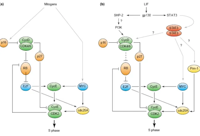
The pluripotent stem cells undergo unlimited self-renewal and differentiation in all types of body cells. Stem cells specifically regulate the structure of their cell cycle and this allows them to proliferate rapidly, including the expression of cell cycle regulators in the G1 phase of the cycle, and then quickly transition into S phase. In addition, stem cells because of the role of embryonic development (ES) must maintain genomic integrity and prevent the acquisition of mutations that would be transmitted to many cell lines. ES cells also express high levels of DNA repair proteins. Similarly, ES cells, induced pluripotent stem (iPS), are ready to proliferate and show a lack of cell cycle arrest G1 / S, extreme sensitivity to DNA damage, and a high level of expression of the repair genes DNA. The mechanisms that regulate the cell cycle in ES cells and genomic integrity iPS cells are similar, although not identical (Momcilovic O. et al 2011). Proliferation of differentiated mammalian cells is controlled primarily by regulating the progression through the G1 phase and entry into S phase. The (RB) retinoblastoma protein and its relatives P107 and P130 are responsible for controlling the G1 / S (Fig. 2). Phosphorylation regulates the activity of RB: hypophosphorylated (G1-specific) RB inhibits the expression of genes that are required for entry into S phase sequestering the E2F family of transcription factors. During the progression through the G1, RB is phosphorylated sequentially by cyclin complexes and cyclin dependent kinase (CDK). Phosphorylation by cyclin and cyclin D/CDK4 D/CDK6 induces a partial release of E2F, which is sufficient to activate the transcription of cyclin E and cdc25A genes. The cdc25A phosphatase removes phosphates from CDK2 inhibitors,
20
and the resulting cyclin E/CDK2 complex then completes the RB phosphorylation, leading to full release of E2F and activation of target genes and entry into S phase (Harbour, JW and Dean, DC 2000;, Bartek, J. and Lukas, J. 2001). A second initiative involves the participation of the protooncogenes c-myc, which directly stimulates the transcription of genes encoding cyclin E and cdc25A to generate cyclin E/CDK2 kinase complex (Bartek, J. and Lukas, J. 2001) (Fig. 2). The tumor suppressors p16INK4a and p27Kip1 inhibit the cyclin and cyclin D/CDK4 D/CDK6 E/CDK2 and cyclin, respectively. They are activated in response to various growth inhibitory signals, such as contact inhibition and senescence and terminal differentiation (Sherr, CJ and Roberts, JM,. 1999). Embryonic stem cells have a short G1 phase of about 1.5 hours during which hypophosphorylated RB is virtually undetectable (Savatier P, et.al 1994) Thus, RB is likely to be rephosphorylated immediately after mitosis in embryonic stem cells, the contrary to differentiated cell types. In addition to RB, the ES cells expressing P107 (Robanus Maandag E-1998), but not p130.
21
1.5 THE CYCLIN E FAMILYTwo types of cyclin E have been recently described: cyclin E1 and cyclin E2, which are closely homologous. These show almost identical patterns of expression during embryonic development and in a similar way to adult tissues. This suggests that the two cyclins E control overlapping phases in the progression from G1 to S. Alternatively, the co-expression of cyclins E may represent a mechanism of redundancy to ensure the normal entry into S phase (Geng et al., 2001).
Unlike cyclins D, which are induced by external factors, the expression of cyclins E is controlled by endogenous mechanisms and is highest in the transition G1 / S. Furthermore, these proteins may drive the progression into S phase even in the absence of Rb, or at least for one round of cell division when is suppressed the transactivation capacity of E2F. This suggests that cyclin E promotes S phase in different ways and at least one of the critical events controlled by cyclin E is independent of the complex E2F/Rb (Lukas et al., 1997). Other specific events of S phase may be regulated by the complex cyclin E/cdk2. These include the phosphorylation of proteins involved in DNA replication, activation of the phosphatase Cdc25A, the regulation of transcriptional programs through transcription factors such as ID2, ID3, BAF155 and SWI / SNF, the modulation of events of splicing and transcription genes for histones (Moroy and Geisen, 2004). However, mice double knockout cyclin E1-/ - E2-/ - are viable, suggesting that E-type cyclins are not absolutely required for embryonic development in mice. On the other hand the E type cyclin are
22
essential for the proper development of extraembryonic tissues, such as the placenta (Geng et al., 2003).
23
1.6. THE FAMILY OF TRANSCRIPTION FACTORS E2F
The family of E2F transcription factors is essential for temporal control of gene expression during the cell cycle in mammals, exerting both positive and negative effects. Indeed, the E2F binding sites are found in various promoters of genes whose products are required for the biosynthesis of nucleotides, such as dihydrofolate reductase (DHRF) and thymidine kinase (TK), for DNA replication (DNA polymerase α and Cdc6), for cell cycle progression (cyclin E, cyclin D1, c-myc, c-myb, and cdc2) (Wells et al., 2000). The activity of E2F is regulated mainly by association with different proteins.
The E2F proteins form heterodimers with DP family members (DP1 and DP2). The functional specificity of the complex E2F/DP is determined by E2F subunits, however, heterodimerization with DP proteins dramatically increases the DNA binding, transactivation and regulation by the Rb family members. Additionally, DP proteins are associated with all E2F in the cell (Zheng et al., 1999) (figure 1).
The family of E2F transcription factors can be divided into three distinct subgroups based on sequence homology and functional characteristics (Dyson, 1998). Studies of "knockout" gene made it possible to identify the distinct roles of E2F family members in the development and physiology of the rat.
The domains for DNA binding and dimerization are conserved in all E2F, E2F6 but lack the C-terminal sequences responsible for transactivation and protein binding "pocket". The activity of this protein is thus independent of control by the Rb family members. E2F6 can repress transcription by binding the protein and the binding protein YY1 Ring1 (RYBP), a
24
component of the Polycomb complex (PCG) (Trimarchi and Lees, 2002).In conclusion, every member of the same subgroup have different biological functions that are the result of differences in their expression, activity and / or regulation in vivo.
25
1.7. THE REGULATION OF ACTIVITY BY THE E2F PROTEINS "POCKET"
The activity of E2F is negatively regulated by protein "pocket". The Rb gene was the first tumor suppressor to be identified. Alterations in the Rb gene are caused by deletions or missense mutations that result in a truncated, nonfunctional, protein, or its complete absence. These changes are associated with different types of human cancer, including familial retinoblastoma, osteosarcoma, lung cancer, cervical cancer, prostate cancer, breast cancer and some forms of leukemia. P130 and p107 mutations, however, are rarely associated with tumors. The different biological properties probably reflect differences in the way of regulating E2F (Giovanni A et al. 2000).
The various members of the protein "pocket" is associated with E2F at different stages of the cell cycle: p130/E2F complexes are mostly found in quiescent or differentiated cells, the complex p107/E2F are prevalent in cells in S phase and complexes Rb / E2F are present both in resting cells and in actively dividing cells (Dyson, 1998).The activity of Rb and other family members is regulated by phosphorylation and is expressed in a cascade control of E2F, in which each member of the family of E2F has a definite role for cell cycle progression (Fig. 1).
In this model, E2F4 and E2F5 is translocated into the nucleus after binding to DP, p107 and p130. The phosphorylation of Rb, p107 and p130, by the cdk of the G1 phase, leading to the destruction of the repressor complexes containing E2F4 and E2F5. The result is a de-repression of several genes involved in controlling cell growth, including E2F1 and E2F2
26
genes. This de-repression allows the accumulation of E2F1 and E2F2 (and probably E2F3) which function as transcriptional activators of genes important for S phase, including cyclin E and cdk genes. These generate additional kinase activity, which completes the phosphorylation of Rb and facilitates the accumulation of E2F and the beginning of DNA replication. Finally, E2F1 and E2F2 activate cdk inhibitors (p18 and p19), the function of which is important for cell cycle progression. DP1 and DP2 phosphorylation, by cyclin complex A/cdk2 causes loss of activity of E2F1, E2F2 and E2F3 at the end of S phase (De Gregori et al., 1997).
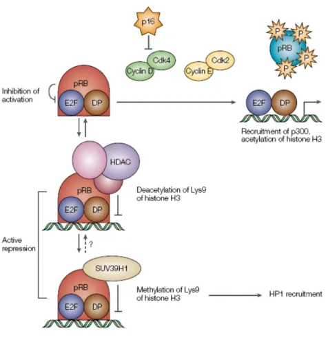
The binding domain of E2F to Rb and various family members is located at the C-terminal 18 amino acids and includes the transactivation domain. This explains how the Rb family members inhibit the transcription of E2F target genes. In fact E2F/DP the complex, bound to DNA, promotes the transcription of target genes through the transactivation domain. The complex with Rb linked to that site is unable to recruit the transcriptional machinery. Alternatively, Rb may recruit different proteins to the promoter, which affect chromatin structure, and actively repress transcription.These include the histone deacetylases (HDACs), two components of the ATPase complex SWI / SNF (BRM and BRG-1) and histone methyltransferases (MTAs, such as SUV39H1) (Trimarchi and Lees, 2002).
The histone deacetylases remove acetyl groups from octamer histone tails, facilitating the condensation of nucleosomes in chromatin. Gene expression is inhibited as well, since it blocked access to the promoter of transcription factors (Harbour and Dean, 2000). In addition, recent studies show that E2F1 can be acetylated and that this increases the binding of the complex E2F/DP DNA (Martinez-Balbas et al., 2000). Therefore inhibition of E2F
27
activity by the overall Rb / HDAC deacetylation can occur even for the same E2F, thereby reducing their ability to bind to DNA.
The complex SWI / SNF chromatin remodeling participates in adjusting the structure of nucleosomes and their position on the promoter. In particular, cooperate with Rb to block the activity of E2F transcription (Trouche et al., 1997). Recent evidence shows that Rb can recruit HDAC and SWI / SNF together in a single complex, in particular the complex HDAC-Rb-SWI/SNF repressor acts at the checkpoint of the cell cycle in G1 and S phase in the complex Rb-SWI/SNF ( Zhang et al., 2000).
The formation of complexes containing the SUV39H1 histone tails allows the modification of the adjacent, this means that the effects of gene silencing to be propagated throughout the locus (Trimarchi and Lees, 2002).
In conclusion, the response mediated by the E2F transcriptional activation can be both a repression, depending on the context in which the promoter bind E2F. The complexity of these mechanisms, due to the formation of a variety of multiprotein complexes, suggests a multiplicity of functions. It is not yet clear whether the individual E2F have distinct roles in controlling cell growth or, alternatively, different complexes containing Rb family members, may direct transcription factors to different E2F target genes or to the same genes but at different times (De Gregori et al., 1997).
The difficulty of this analysis is due to the fact that most of the cells contains all of the E2F and all members of the Rb family. However, in vivo studies using chromatin immunoprecipitation (Wells et al., 2000), show that most of the target genes of E2F is bound by specific members of the E2F family. In addition, the DNA binding by the E2F
28
proteins and "pocket" is a promoter-specific and depends on the phase of the cell cycle. One hypothesis is that members of the Rb family, linked to the promoter, can be used both for the binding of repressor proteins in resting cells for the binding of activator protein in cells in S phase In any case, the target genes of E2F are not regulated by a single complex, static, probably, every cell has the potential to form different transcription complexes (Wells et al., 2000).
29
1.8 RB-MEDIATED INHIBITION OF E2FRb can repress transcription by at least two different mechanisms. First, it can bind transcription factors such as E2F and block their ability to activate transcription (Flemington et al. 1993; Helin et al. 1993). Second, the Rb–E2F repressor complex that forms at promoters can actively repress transcription (Bremner et al. 1995; Sellers et al. 1995; Weintraub et al. 1995). The balance between these two activities in vivo is still in question. In this section, we address the potential mechanisms through which Rb might inactivate E2F. Because Rb binds E2F within its transactivation domain (Flemington et al. 1993; Helin et al. 1993), it was assumed initially that it physically blocks E2F transactivation. This idea is supported by in vitro studies in which Rb inhibited transcriptional activation by E2F1 in the apparent absence of other corepressors (Ross et al. 1999). However, Rb may also inhibit E2F by recruiting chromatin remodeling enzymes, including the HDACs mentioned above. The HDACs are a family of at least seven different enzymes that remove acetyl groups from the tails of histone octamers, which appears to facilitate condensation of nucleosomes into chromatin. This, in turn, inhibits gene expression by blocking access of transcription factors to the promoter (Kingston and Narlikar 1999; Kornberg and Lorch 1999; Wolffe and Hayes 1999). In contrast to experiments in vitro, transfection assays in cultured cells have suggested that interaction with HDAC is required for the inhibition of E2F1 by Rb (Brehm et al. 1998; Magnaghi et al. 1998). Other studies have only shown a partial requirement for HDAC activity in the
30
Rb-mediated inhibition of E2F activity (Luo et al. 1998; Lai et al. 1999a). E2F1 has been shown to interact with the histone acetyl transferases p300/CBP and p/CAF (Trouche et al. 1996). Thus, it is possible that Rb-mediated recruitment of HDAC to E2F acts to offset this histone acetyltransferase (HAT) activity (Fig. 1). It has also been shown recently that E2F1 can be acetylated, which increases the binding of the E2F/DP complex to DNA (Martinez-Balbas et al. 2000). Therefore, recruitment of HDAC to E2F via Rb may inhibit E2F activity by deacetylation of the protein, thereby inhibiting its binding to DNA. Rb can also interact with BRG1 and BRM—the two ATPase components of the human SWI/SNF chromatin remodeling complex, which is discussed in more detail below (Dunaief et al. 1994; Singh et al. 1995). Some results have shown that overexpression of BRG1/BRM can facilitate Rb-mediated inhibition of E2F1transcriptional activity (Trouche et al. 1997); however, other studies have found that E2F activity is inhibited efficiently in cells that are BRG1/BRM deficient (Weintraub et al. 1992; Zhu et al. 1993; Zhang et al. 2000). Thus the relative importance in vivo of these two potential mechanisms for inhibiting E2F transactivation—direct binding and masking of the E2F transactivation domain versus Rb-mediated recruitment of chromatin remodeling enzymes to inhibit E2F—is still unclear.
31
1.9 ACTIVE TRANSCRIPTIONAL REPRESSION BY RB
Binding of Rb and other pocket proteins to E2F does not simply inhibit E2F activity. The resulting Rb–E2F complex binds to promoters and actively represses transcription by blocking the activity of surrounding enhancers on the promoter (Weintraub et al. 1992; Hsiao et al. 1994; Johnson et al. 1994; Adnane et al. 1995; Bremner et al. 1995; Neuman et al. 1995; Sellers et al. 1995; Weintraub et al. 1995; Chow et al. 1996; Ferreira et al. 1998; Meloni et al. 1999). Whereas Rb requires sequences in the pocket and in the carboxy-terminal region to bind and inhibit E2F, the pocket alone is sufficient for active repression when Rb is tethered directly to a promoter (e.g., through a Gal4 DNA binding domain; Bremner et al. 1995; Sellers et al. 1995; Weintraub et al. 1995). This is explained by the recent finding that active repression by Rb is attributable, at least in part, to the recruitment of pocketbinding corepressors. Perhaps the best studied of these corepressors are the chromatin remodeling enzymes. Modification of chromatin structure is an important mechanism for regulating gene transcription (Felsenfeld 1992; Kingston and Narlikar 1999). One manner in which chromatin structure can be altered is by acetylation of histones. HATs have been shown to act as transcriptional coactivators that alter chromatin structure, thereby allowing transcription factors access to the promoter. In contrast, HDACs have been associated with transcriptional inhibition and are found in corepressor complexes (Alland et al. 1997; Grunstein 1997; Hassig and Schreiber 1997; Hassig et al. 1997; Heinzel et al. 1997; Laherty et al. 1997). Three of the seven known HDACs (HDAC1–
32
HDAC3) interact with Rb, and Rb can bind simultaneously to HDAC and E2F, allowing recruitment of an HDAC–Rb–E2F repressor complex at promoters of cell cycle genes (Brehm et al. 1998; Luo et al. 1998; Magnaghi et al. 1998; Lai et al. 1999a; Chen and Wang 2000; Dahiya et al. 2000). In two recent studies, Rb mutants that have amino acid substitutions in the LXCXE-binding site showed reduced binding to HDAC1 and HDAC2, but not HDAC3 (Chen and Wang 2000; Dahiya et al. 2000). The impaired interaction with HDAC1 and HDAC2 had no effect on the ability of the Rb mutants to inhibit transcriptional activation by E2F, but these mutants were unable to actively repress some genes and unable to maintain growth arrest. In addition, it has been shown that recruitment of an HDAC–Rb–E2F complex can actively repress transcription and regulate histone acetylation at the promoter (Luo et al. 1998).
33
1.10 CHROMATIN REMODELING: THE ROLE OF MULTIPROTEIN COMPLEX SWI / SNF CONTAINING BRM AND BRG-1
In the course of evolution the complex SWI / SNF has been stored for promoting events of chromatin remodeling and it is important for activation and for transcriptional repression of genes involved in the regulation of cell proliferation (Muchardt C. and Yaniv, M., 2001) . The chromatin-remodeling complexes are multiprotein complexes. The genes that encode complex SWI / SNF were initially characterized in Saccharomyces cerevisiae. The SWI genes or "faulthy mathing type switching” and SNF or" Sucrose Non-Fermenting "were defined as transcriptional activators involved in the regulation of a group of inducible genes (CWM Roberts and Orkin SH, 2004). In this organic complex SWI / SNF is composed by 11 proteins assembled into a complex of 2MDa. This complex acts transcriptional activity, affecting the structure of chromatin in the vicinity of the promoters of target genes (M. Carlson and BC Laurent, 1994). In mammals have been identified proteins homologous to many members of the complex SWI / SNF in yeast.
In Drosophila SWI2/SNF2 homologue is known as Brahma or brm (Tamkun JW et al., 1992). The gene encoding this protein was initially identified as a suppressor of Polycomb mutations. The protein resembles the brm complex SWI2/SNF2 throughout its length and contains a protein similar to yeast, a bromodomain type helicase and a domain with an ATP-binding site. As in yeast, brm is associated with a large complex that also contains SNR-1, the Drosophila homologue of SNF5. Even in mammalian cells have identified two
34
proteins closely related to SWI2/SNF2. These proteins, known in humans and mice hbrm/hSNF2alfa and BRG-1/hSNF2 Beta as mbrm and mBRG-1 (190 kDa) were both associated with the so-called including complex mammalian SWI / SNF containing at least eight proteins including homolog of SNF5 (hSNF5/IN1), BAF 155 and BAF 170, two homologous subunits of yeast SW13 e BAF a homologue of the 60 subunits of yeast SWP73 (Muchardt C. and Yaniv, M., 1993).
The multiprotein complex in mammalian SWI / SNF complex is composed of subunits that contain the BRG-1 or brm-1 activity with adenosine triphosphatase (ATPase) as central to hydrolyze ATP. BRG-1 and mbrm-1 are homologues of yeast SWI2/SNF2 ATP ase. There are two types of ATPases can be grouped into different subfamilies according to whether they contain a bromodominio (the SWI2/SNF2 subfamily), two pairs of cromodomini (the ISWI subfamily). All the members of different subfamilies seem to be part of a multi-subunit complex that can remodel nucleosomes by ATP-hydrolysis and increase or decrease the effects of repressive chromatin (SIf. S. et al., 2001). Within this complex, the two related protein BRG-1 and brm-1 are mutually exclusive indicating that co-exist at least two versions of the complex SWI / SNF in mammalian cells (Roberts, C. W. M. and Orkin H., 2004). Similar to their counterparts in yeast, the complex SWI / SNF purified in mammalian differentiated cells possess chromatin remodeling activity. The remodeling of chromatin is a process that participates in cell cycle control by acting directly on the structure of DNA. Chromatin consists of DNA wrapped in strings of nucleosomes (octamers of histone proteins, namely of two molecules of each histone H2A, H2B, H3 and
35
H4). Thanks to the nucleosomes, the chromatin structure in order to achieve higher order levels of organization that increase the compaction ratio.
The adjustment of the compaction ratio of nucleosomes structures is critical to the control of gene expression and is regulated by two mechanisms. The first mechanism is based on the modification of amino-terminal histone tails. The most studied covalent modification involves acetylation. This post-translational modification is the addition of acetyl groups mediated by histone acetyl transferase (HAT) on the lysine residues present N-terminal histone tails. The acetylation of histones cause relaxation of chromatin, facilitating access to transcription factors and the de-acetylation mediated by histone deacetylase makes the chromatin less accessible to transcription factors. The acetylation seems therefore more involved in the phenomena of transcriptional activation, in contrast to the deacetylation which causes repression of transcription. Recently, methylation has also played an important role in transcriptional repression. In fact, the histone methyltransferase SUV 23H1 seem involved in the phenomena transcriptional repression. The addition of a methyl group on lysine residues 9 Histone H3, by SUV39H1 creates a binding site with high affinity for the HP1 protein (heterochromatin protein 1) making the area of DNA, which was bound, transcriptionally inactive. The second mechanism depends on the complex SWI / SNF uses the energy released from ATP hydrolysis to move nucleosomes along the DNA helix facilitating the access of transcription factors. The complex SWI / SNF interacts with the chromatin is able to form stable areas of accessibility in the distribution of ordered nucleosomes to allow transcription factors access to their target sites.
36
It was also suggested a possible direct interaction of the complex SWI / SNF with RNA polymerase II holoenzyme to locate the start point of transcription (Owen-Hughes T., Workman JM 1996). The alteration of the ATP-dependent nucleosome is targeted to the histones made recognizable by modification of the enzyme complexes. The most common changes involve covalent bond formation by acetylation, phosphorylation, methylation and ubiquitination of histone molecules and / or non-covalently in the action of enzymes that remodel chromatin using the energy released by the hydrolysis of ATP ( Roberts C.W.M. S. and Orkin. H., 2004). In some cases it is then that the complex SWI / SNF and HAT may cooperate for the transcriptional regulation (Roberts C.W.M. and Orkin S.H., 2004). Moreover, the complex SWI / SNF can remodel chromatin causing alterations of the position and shape of the nucleosomes in an energy-dependent hydrolysis of ATP generated. This affects the stability of the octamer of nucleosome causing slippage along the same DNA strand or the transfer of the octamer in regions adjacent to those of transcriptionally active DNA, increasing the accessibility to the transcriptional machinery. Recently, it has also been shown that the complex SWI / SNF are involved in regulating gene expression, also important because they can recruit transcription factors that make the structure even more complex (W. Wang et al., 1996). In differentiated cells is well documented interaction between pRb and BRG-1 or brm-1, which can only occur when pRb is in its hypophosphorylated form (Roberts C.W.M. and Orkin S.H., 2004). This suggests that SWI2/SNF2 proteins associated with Rb have an important role in tumor suppression. THE BRG-1 subunit of the SWI2/SNF2 multiprotein complex is involved in controlling cell proliferation and is a likely candidate in the role of a tumor suppressor.
37
The mutant BRG-1 - / - is lethal at an early stage blastocysts, and heterozygous for the mutation in BRG-1 + / - induce rhabdoid tumor, an aggressive form of cancer in children that affects various types of tissues. The reintegration of the gene BRG-1 is able to stop the rapid proliferation of cancer cells (CWM Roberts and Orkin SH, 2004). It is believed that the arrest of the growth of cancer cells for the inclusion of BRG-1 is in part due to decreased expression of target genes of the E2F family transcription factors such as cyclin E, but even more by the increased mRNA transcription of the cyclin-dependent kinase inhibitors (CDI) as p21 and p15 (H. Kang et al., 2004). In differentiated mammalian cells the regulation of cell proliferation activities attributed to BRG-1 seems to depend on its ability to interact with the tumor suppressor pRb, thereby suppressing the expression of target genes dependent on E2F. pRb can bind both BRG-1 and brm. The BRG-1 complex / brm appears to regulate the phosphorylation of pRb through modulation of cyclin-dependent kinase inhibitor p21.
It has been shown that the complex HDAC-Rb-SWI/SNF repress the target genes of E2F such as cyclin E, cyclin A and cdc2 blocking the cycle at G1 phase. The activation of cyclin D/CDK4-6 complex in differentiated cells in response to mitogenic stimuli cause the phosphorylation of Rb. This allows the release of HDAC and the S-phase transition. The complex Rb-SWI/SNF is so able to activate the expression of cyclin E, whereas the expression of cyclin A is still repressed. Cyclin E forms a complex with CDK2 that can phosphorylate Rb and cause the release of the complex SWI / SNF, the activation of the expression of cyclin A and entry into G2 phase. The association ordered in time of the
38
BRG-1 with complexes HDAC and pRb allows the sequential activation of cyclin E and cyclin A by inducing progression of the cell cycle.
All the purified complexes contain different subunit except the complexes SWI2/SNF2 where BRG-1 and brm are subunite highly conserved. The similarities between the subunits are also found in the case of the human histone deacetylase complex remodeling of nucleosomes (NuRD) containing Mi-2 as ATP ase center. Nurd contains closely related subunits, which include HDAC-1 and -2 proteins associated to retinoblastoma (RBA) Rb and p46, and p48 itself (Sif, S. et al 2001), As mentioned above, if acetylation is a type of chromatin modification that triggers the gene expression, histone deacetylation results in the repression of gene expression. It was in fact shown that brm and BRG-1 are able to work on recruiting other chromatin complexes such as HDAC-1 and 2 and their counterparts co-repressors of yeast Sin3, and mSIn3A mSin3B. It has been proposed that the repression of specific promoters may be permitted by the recruitment of the complex HDAC/mSIn3A BRG-1/brm-1. This complex also contains Rb HDAC/mSin3A AP46, Rb AP48 and mSin3A associated proteins such as SAP18, and SAP30, whose function is unknown (Sif, S. et al., 2001).
By studying the interactions of individual fractions purified from the complex SWI / SNF that brm-1 is capable of co-precipitated with HDAC-1, HDAC2, Sin3A, and Rb AP48 and BRG-1 co-precipitate with small amounts of HDAC-2 and mSIn3A AP48 Rb but not with HDAC-1. This different composition of protein complexes associated with brm and BRG-1 may explain the different functional characteristics of these proteins.
39
In fact it seems that BRG-1 has a greater ability to hydrolyze ATP and remodel the chromatin and then working both in the conformation of the nucleosome and altering its position. BRM chromatin remodeling can only change the order in the position of nucleosomes (Sif S . et al., 2001). The expression of BRG-1 is ubiquitous at all stages of development from the earliest pre-implantation embryo, whereas the expression of brm is absent in these stages and increases at later stages in the embryo at the stage of blastula . At this stage the cells leave the cell cycle to be targeted to cell differentiation.
40
1.11 SWI/SNF-BRG1 REGULATES SELF-RENEWAL AND OCCUPIES CORE
PLURIPOTENCY-RELATED GENES IN EMBRYONIC STEM CELLS
The SWI/SNF-Brg1 chromatin remodeling protein plays critical roles in cell-cycle control and differentiation through regulation of gene expression. Loss of Brg1 in mice results in early embryonic lethality, and recent studies have implicated a role for Brg1 in somatic stem cell self-renewal and differentiation. However, little is known about Brg1 function in preimplantation embryos and embryonic stem (ES) cells. Il gruppo di Kidder B. L. , 2009, report that Brg1 is required for ES cell self-renewal and pluripotency. RNA interference-mediated knockdown of Brg1 in blastocysts caused aberrant expression of Oct4 and Nanog. In ES cells, knockdown of Brg1 resulted in phenotypic changes indicative of differentiation, downregulation of self-renewal and pluripotency genes (e.g., Oct4,
Sox2, Sall4, Rest), and upregulation of differentiation genes. Using genome-wide promoter
analysis (chromatin immunoprecipitation) they found that Brg1 occupied the promoters of key pluripotency-related genes, including Oct4, Sox2, Nanog, Sall4, Rest, and Polycomb
group (PcG) proteins. Moreover, Brg1 co-occupied a subset of Oct4, Sox2, Nanog, and
PcG protein target genes. These results demonstrate an important role for Brg1 in regulating self-renewal and pluripotency in ES cells.
41
1.12 REGULATION OF CYCLIN E1 GENE BY THE COMPLEX REPRESSOR
CERC2
In somatic cells the regulation of G1 / S is governed by sequential activation of pRb and cyclin complexes / Cdks. Precisely pRb forms a complex with E2F, recruiting HDAC and SWI / SNF, able to repress the transcription of genes involved in G1 / S cell cycle such as the gene encoding the cyclin E1 CCNE1. When, in response to appropriate stimuli proliferative, pRb is phosphorylated and inactivated by the complex cyclin / Cdks, the cells complete the G1 phase and progress into S phase. The phosphorylation of Rb by cyclin complex D1/Cdk4-6 promotes the release of HDACs from the domain "pocket" of pRb. The removal of HDAC leads to increased acetylation of a specific nucleosome located at the site of initiation of transcription of cyclin E1 and its subsequent activation. The activation of the promoter of cyclin E1, which occurs during the G1 / S decreases in S phase and disappeared during the period between the end of mitosis and the late G phase. The expression of cyclin E1 reflects transient activation of gene promoter CCNE1 (Cyclin E1 gene) that occurs during the G1 / S and then fall in the S phase and disappear during the period between the end of mitosis and late G phase.In quiescent mouse fibroblasts has been shown that the promoter activity of cyclin E1 gene is controlled by a DNA sequence defined as "cyclin E repressor module" (CERM), located downstream of the transcription start site and consists of a variant of the canonical E2F sites and a sequence rich in AT. Both sequences are necessary to bind a complex of high molecular weight E2F called "cyclin E repressor complex 1" (CERC1) that prevents the transcription of cyclin E1. When
42
fibroblasts begin to proliferate, the complex dissociates from the sequence CERC1 CERM and is induced transcription of cyclin E1.CERC1, even if it contains E2F4/DP1 pocket and the protein p130, E2F complexes differs from canonical in that it has additional components as observed through a glycerol gradient sedimentation slowed (Le Cam et al., 1999). Similar studies have shown that, even in proliferating K562 cells, the repression of the gene regulates CERM CCNE1 in the period between the end of mitosis and late G1 phase through its association with a high molecular weight E2F complex (Polanowska et al. 2001). Further studies have shown that this complex possesses histone deacetylase activity and was named CERC2.EMSA experiments performed with a DNA probe that spans the promoter region of the murine cyclin E1 gene (CERM probe) and nuclear extracts prepared from K562 cells blocked in G1, show that the complex CERC2 reacts with antibodies directed against p107, E2F4 , DP1, cyclin E1 and CDK2 but not with antibodies directed against Rb, p130 and E2F family members such as E2F1, E2F2, E2F3, E2F5 and cyclin A1. Ultimately CERC2 is a complex of high molecular weight containing G1-specific E2F4/DP1, p107, cyclic E1 and CDK2 (Polanowska et al., 2001). The repressive role of the CERC2 gene promoter of cyclin E1 requires conformational changes in chromatin structure.To investigate whether the complex contains CERC2 among its members and the HDAC complex SWI / SNF, other experiments were carried out EMSA and inhibitor TSA, which have shown that histone deacetylase activity exists in the K562 and BRG -1 and Brm are not associated with CERC2. In particular, the repression of the gene CCNE1 correlates with deacetylation of histones H3, H4, and methylation of arginine and lysine K9 of histone H4 R3 of the single nucleosome that includes CERM (Nielsen et al., 2001,).The
43
transcriptional activation of the gene CCNE1 during late G1 phase is determined by the reduction of methylation in the same region. Methylation of histone lysine methyl transferase is produced by lysine methylation of arginine SUVAR39H1 while the protein is left to the "R-methyltransferase" (PRMT). In mammals, nine proteins were identified PRMTs divided into two groups for their ability to catalyze one or two methylation reactions, both symmetric and asymmetric. In mammals the activity of PRMT1 and PRMT4 responsible for the formation of asymmetric dimethylarginine, is associated with gene activation (Bauer UM et al., 2002), while PRMT5 catalyzes the symmetrical dimethyl associated with transcriptional repression. It has recently been shown that arginine methyl transferase PRMT5 is part of the complex transcriptional repression of cyclin E1 promoter of the gene and its methyl-transferase activities sull'istone H4 is a prerequisite for the blockade of transcription and the consequent inhibition of cell proliferation (Fabbrizio E. et al., 2002).
44
1.13. DIFFERENTIATION OF THE CES AND IPS.
The removal of LIF from the culture medium of ESC is sufficient to induce differentiation programmed to form all types of cells which carry out embryonic development. This is possible in vitro through the formation of specific cellular structures called embryoid bodies (EB) for their similarity to post-implantation embryonic tissues. These cells contain the precursors of all three germ layers and mimic gastrulation and organogenesis early (Savatier et al., 1995). Alternatively, differentiation can be induced experimentally by adding the appropriate chemicals (such as retinoic acid in the case of this study) that address the ESC differentiation toward a specific system. For the incredible potential for differentiation in vitro, the ESCs and iPS are only one cell type to develop strategies for an efficient gene therapy. In addition, CES is an ideal system to study the mechanisms involved in the processes of early embryonic development and cell differentiation and tissue.From the molecular point of view, during the early stages of development, when cells must be targeted to specific lines, the differentiation of ESCs and iPS is associated with a rearrangement of the expression and gene silencing, processes that are intimately connected with the alteration of chromatin structure (Lee et al., 2004). In fact, the dynamic changes of chromatin structure facilitate or inhibit the access of transcription factors to DNA. At least two mechanisms can be used to alter or remodel the structure of chromatin: one involving multiprotein complexes SWI / SNF, which use the energy of ATP to change the position and / or conformation of nucleosomes, and the other involves the modification both
45
covalent DNA (cytosine methylation) and histone tails (phosphorylation, methylation and acetylation). In cells in active proliferation, the genes are regulated by the cyclic recruitment of HDAC and HAT but remain nell'eucromatina; the induction of differentiation leads to DNA methylation or histone H3, which triggers the repositioning of genes and nell'eterocromatina through their HDAC silencing (Fig. 3) (Ferreira et al., 2001).These processes were examined during in vitro differentiation of ESCs and iPS : histone acetylation, a marker dell'eucromatina, declines rapidly after induction of differentiation, and then be partially restored on the contrary, methylation of histone lisina9 H3, a marker of heterochromatin, increases following induction of differentiation. This is consistent with the model that the differentiation of stem cells is accompanied by a restriction of a series of genes that are expressed. The global deacetylation of histones is required for the differentiation of ESCs: the histone acetyltransferase and deacetylase transmit signals to initiate differentiation of the appropriate epigenetic modifications, as well as the elimination of the existing structure of chromatin and the establishment of a new pattern of global change is both gene-specific during the differentiation in vitro (Lee et al., 2004).Taken together, these results suggest that the structure of chromatin at the promoter of a gene key to differentiation is programmed early in development.
46
1.14 RETINOIC ACID AS AN INDUCER OF DIFFERENTIATION
Retinoic acid (RA), a derivative of vitamin A (retinol), has an important role as an endogenous factor during embryogenesis, morphogenesis and cell proliferation. It is known that RA has two distinct roles during the early stages of cell differentiation: it inhibits myogenesis and cardiogenesi, while simultaneously promotes neurogenesis and adipogenesis (zur Nieden et al., 2004). The RA may also regulate growth and differentiation of a wide variety of pre-malignant and malignant cells, both in vivo and in culture.
In CES, the RA can induce differentiation into cells ectoderm, mesoderm and endoderm, in a time-dependent and concentration. Under certain conditions, ESCs differentiate into extraembryonic cells, similar to the parietal endoderm (Chen and Gudas, 1996).The extensive self-replication of CES depends on the balance between signals that promote the proliferation and differentiation. In CES, the addition of RA induces differentiation in part by blocking the signals transduced by LIF (Tighe and Gudas, 2004).The main mechanism by which RA regulates the differentiation, is expressed through the action of nuclear receptors for RA (RAR and RXR), expressed in ESCs and during embryonic development. In the presence of different isomers of RA, RAR and RXR receptors form heterodimers that activate the transcription of specific genes for differentiation, including the homeotic genes. The RA may also indirectly regulate the expression of many genes involved in pre-and postnatal development (Chen and Gudas, 1996).
47
1.15 METASTABLE STATES OF PLURIPOTENCY
The term “metastability” has been previously used to describe transient changes within ICM like ES cell populations resulting from oscillations in Nanog or Stella gene expression (Chambers et al., 2007; Hayashi et al., 2007). The goup of Jacob Hanna (Hanna J et al 2009) apply this term to describe the interconversion between two distinct pluripotent states in NOD and 129 mouse strains. Them results suggest that the ICM and EpiSC pluripotent states may be in a “metastable” equilibrium dictated by the genetic background where exogenous factors can convert one state into another. Thus, one may consider the two states of pluripotency, the ICM/ES cell-like state and the epiblast/EpiSC cell-like state, as two different levels of pluripotency. Exogenous factors such as c-Myc and Klf4 in combination with Oct4 and Sox2 can induce the ICM-ES like state from somatic cells. However, the stability of the ES cell state is determined by the genetic background: while ICM-ES cells or iPS cells derived from a “permissive” genetic background such as 129 or C57BL/6 are stable once established in the presence of Lif, the ES cell like state of iPS cells or of ICM derived pluripotent cells of the ”non-permissive” NOD background remains unstable with the maintenance of the pluripotent state depending on the continuous expression of the exogenous factors in addition to Lif/Stat3 signaling. Inactivation of the transcription factors or removal of the inhibitors causes the ES like NOD cells to assume an EpiSC-like state, characterized by reduced pluripotency. Inter-conversion between these states can be controlled by the absence or presence of the same factors. Several lines of evidence support the notion that the conversions between the different pluripotency states are due to cells being inefficiently induced to successfully convert from one state to another, rather than
48
due to selection for rare pre-existing cells constantly present in heterogeneous stem cell populations. First, evidence for direct reprogramming of EpiSCs into iPS cells is supported by the observations that EpiSC cells do not convert spontaneously into ES like cells and that all derived Epi-iPS cell lines carried integrated viral transgenes. Second, the EpiSC to ES cell conversion requires multiple passages in defined media and continuous transgene induction, which is similar to generating iPS cells from somatic cells (Jaenisch and Young, 2008)). Third, it is unlikely that NOD ES cultures carry already rare EpiSC-like cells since the NOD iPS or ES lines were passaged routinely by trypsinization, which does not allow propagation of the EpiSC cells. Finally, the NOD EpiSC-like iPS cell line carried an identical Sox2 integration as its parental Dox dependent NOD iPS line indicating a clonal relation. An important question remains why only a small fraction of the NOD ES cells convert into an EpiSC state. One possibility is that after removal of the exogenous stimuli, the EpiSC state becomes one of several epigenetic states that can be acquired by the NOD ES cells upon differentiation.
49
1.16 CONTROL OF EMBRYONIC STEM CELL METASTABILITY BY L-PROLINE CATABOLISM
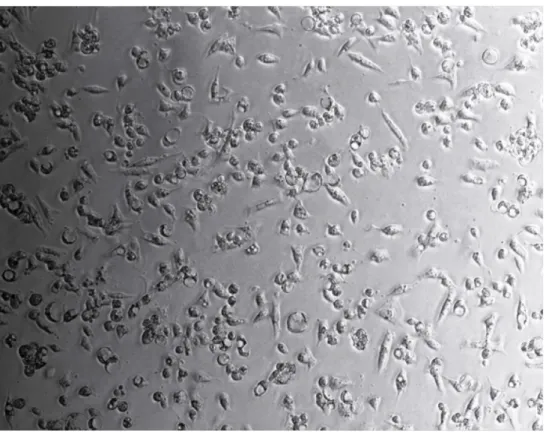
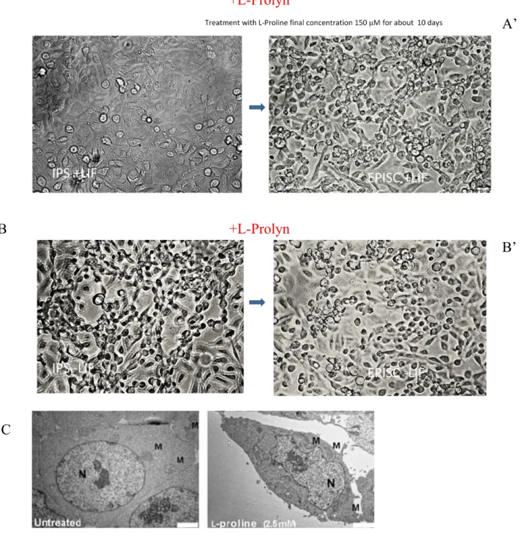
The molecular mechanisms controlling mouse Induced Pluripotent Stem Cells (iPS) metastability, i.e. their capacity to fluctuate between different states of pluripotency, are not fully resolved. The Casalino L., group (2011) has for first induced EPISC from CES using as inducer a fisiological esposition at L-Prolyn. I developed and used this platform, for induce the iPS to differentiate in EPISC. L-Pro, force iPS toward a novel epiblast stem cell (EpiSC)-like state, in a dose- and time-dependent manner. Unlike EpiSCs, L-Pro-induced cells (PiCs) contribute to chimeric embryos and rely on leukemia inhibitor factor (LIF) to self-renew. Furthermore, PiCs revert to ESCs or differentiate randomly upon removal of either L-Pro or LIF, respectively. Remarkably, PiC generation depends on both L-Pro metabolism (uptake and oxidation) and Fgf5 induction, and is strongly counteracted by antioxidants, mainly L-ascorbic acid (vitamin C, Vc). ESCs↔PiCs phenotypic transition thus represents a previously undefined dynamic equilibrium between pluripotent states, which can be unbalanced either toward an EpiSC-like or an iPS phenotype by L-Pro Vc treatments, respectively. All together, our data provide evidence that iPS metastability can be regulated at a metabolic level just as the CES.
50
2. MATERIALS AND METHODS
2.1. ES AND IPS CELL CULTURE
The cell cultures used in this study are:
- Mouse embryonic stem cell lines (ES CGR8) - Mouse Induced Pluripotent Stem Cell (iPS #202)
2.1.1 LINES OF MOUSE EMBRYONIC STEM CELLS CGR8 AND IPS.
A cell line used in the experiments of this study, is called CGR8 (Exponentially growing germ-line feeder-independent embryonic stem cell line Competent) and is derived directly from the blastocyst inner cell mass of a mouse. These are embryonic stem cells (ESCs) and in vitro retain the capacity to differentiate into all mature somatic phenotypes when induced by appropriate signals. The CGR8 can be grown indefinitely in culture in an undifferentiated state in the presence of leukemia inhibitory factor (LIF) (Savatier et al., 1995). Another cell line used in this study is iPS #202 .The CGR8 and iPS #202 grow in the culture medium GMEM ("Glasgow's Modified Eagle's Medium", BioMed), containing sodium bicarbonate (3.7 g / l) and complemented with fetal calf serum (FCS, 10%),