___________________________
Corresponding author: Prof. Nenad Bukvic, MD, PhD, Specialist of Medical Genetics, University
Hospital Policlinics of Bari, Medical Genetics Unit, Piazza Giulio Cesare, 11, 70125 Bari, Italy. e-mail: [email protected]; [email protected]. Tel: +39 080 5593621. Fax +39 080 5593618
UDC 575 DOI: 10.2298/GENSR1602753B
Original scientific paper
FAMILLY WITH TWO DIFFERENT CASES OF POST- AND PRE-NATAL L1 SYNDROME; WHEN HYDROCEPHALY BECOME “MULTIDISCIPLINARY
HEADACHE”
Nenad BUKVIC1, Francesca BOARETTO2, Giuseppe LOVERRO3, Francesco C.SUSCA4, Rosaura LOVAGLIO4, Margherita PATRUNO4, Dragoslav BUKVIC5, Srdjan STARCEVIC6,
Giovanni VAZZA2, Maria Luisa MOSTACIUOLLO2, Nicoletta RESTA4 1University Hospital Policlinics of Bari, Medical Genetics, Bari, Italy
2University of Padua, Department of Biology-Laboratory of Human Genetics, Padua, Italy 3University of Bari, Department of Gynecology and Obstetrics, Bari, Italy
4University of Bari, DIMO-Laboratory of Medical Genetics, Bari, Italy 5Niksic General Hospital - Department of Gynecology, Niksic, Montenegro 6Clinic for Ortopedic Surgery and Traumatology, MMA(VMA), Belgrade, Serbia
Bukvić N., F. Boaretto, G. Loverro, F. C.Susca, R. Lovaglio, M. Patruno, D. Bukvic, S.Starcevic, G. Vazza, M. L. Mostaciuollo, N. Resta (2016): Familly with two different
cases of post- and pre-natal L1 syndrome; When hydrocephaly become “multidisciplinary headache”.- Genetika, Vol 48, No.2, 753 -760.
In middle 90’s the scientific community classified as CRASH syndrome a clinical situation characterized by Corpus callosum hypoplasia, Retardation, Adducted thumbs, Spastic paraplegia, and Hydrocephalus. This pathology is also known as L1 syndrome and includes a spectrum of related neurological disorders with an X-linked recessive mode of inheritance associated to mutations in the human L1 Cell Adhesion Molecule gene (L1CAM;OMIM 308840). Here we report regarding a couple pass through our Genetic Counseling, during clinical diagnostic procedure in ~3 years old son, due to presence of multiple malformations such as hydrocephalus, agenesis of corpus callosum, tetraparesys, axial hypotony, cognitive and motor incompetency. The proband was the first male child of a healthy, non-consanguineous Italian couple with no family history of brain abnormalities, recurrent miscarriages, other birth defects and/or genetic illnesses. During Genetic Counseling, a diagnostic hypothesis of L1 syndrome was made, with in deep explanation of genetic testing possibilities. On the basis of our protocol, we fixed another appointment 2 weeks later, but unfortunately the family never showed up.
Approximately one year later, the Department of Gynecology and Obstetrics requested Genetic Counseling for a 33 years old woman, secondigravida (22° gestation week), with abnormal ultrasound findings showing severe fetal ventriculomegaly. The family history was unremarkable with no consanguinity; she was one of two sisters and had a healthy brother. Surprisingly the woman was the mother of our proband. On the basis of a diagnostic hypothesis of L1 syndrome (includes a spectrum of related neurological disorders with an X-linked recessive mode of inheritance), we performed molecular analysis on the proband’s DNA, mother’s DNA and fetal DNA. The mutational screening revealed the presence of a non sense c.2701C>T (p.Arg901*) mutation in the exon 20 of the L1CAM gene in all the tested DNA samples. This observation underline a necessity to consider L1-syndrome as potential part of differential diagnosis in all cases of hydrocephaly, same as undoubting of importance of genetic counseling with subsequent, extension of the molecular analysis to the relatives in order to provide an effective and reliable counseling and risk assessment to these families. Finally, the development of new analytical tools [primarily next generation sequencing (NGS)] and development of an assay/gene panel can be used to specifically target a L1 Syndrome for fast, reliable and high quality results in tricky clinical situation like this one.
Keywords: Hydrocephaly, L1 syndrome, L1CAM gene, pre- and post-natal
diagnosis
INTRODUCTION
In middle 90’s scientific community referred as CRASH syndrome, for clinical situation characterized by corpus callosum hypoplasia, retardation, adducted thumbs, spastic paraplegia, and hydrocephalus (FRANSEN et al., 1995; KENWRICK et al., 1996), even though this pathology is known as well as L1 syndrome. In fact, this syndrome represents a spectrum of related neurological disorders with an X-linked recessive mode of inheritance due to mutations in the human L1 cell adhesion molecule gene (L1CAM;OMIM 308840) which include: X-linked hydrocephalus with stenosis of the aqueduct of Sylvius (HSAS; OMIM 307000); mental retardation, adducted thumbs, shuffling gait, and aphasia (MASA) syndrome (OMIM 303350); spastic paraplegia type 1 (SP1; OMIM 312900); and agenesis of the corpus callosum (ACC; OMIM 304100) (CHIDSEY et al., 2014). Mutations in L1CAM have also been reported to be associated with Hirschsprung disease (JACKSON et al., 2009). The incidence is 1 in 30,000 male births (SCHRANDER-STUMPEL and FRINS, 1998). The clinical symptoms are highly variable, both within and between families (BURTON, 1979; HALLIDAY et al., 1986; FINCKH et al., 2000) and the mild ventricular enlargement is consistent with long survival whereas onset of fetal hydrocephalus during pregnancy (in utero) results in stillbirth or early infant mortality. The degrees of mental retardation and spasticity are also highly inconsistent. In almost 50% of observed cases, adducted thumbs are present but cannot be relied on as a diagnostic sign (HALLIDAY et al., 1986; KENWRICK
et al., 1996). Since 1949, when BICKERS and ADAMS (1949) described the first family with hydrocephalus and aqueduct stenosis in seven male relatives it was first thought that the hydrocephalus is caused by congenital stenosis of the aqueduct of Sylvius. STEVENS et al., (1993) bring the observation that it may rather be the result than the cause of hydrocephalus giving a motivation that aqueduct stenosis has also been reported in patients with hydrocephalus of other origin. The identification of the disease gene (L1CAM) and the mutation analyses have contributed to more accurate diagnostic procedures and to a better understanding of disease spectrum and pathogenesis.
Clinical Report
The couple was passed through our Genetic Counseling, during clinical diagnostic procedure in 3 years old son, due to presence of multiple malformations [hydrocephalus, agenesis of corpus callosum, tetraparesys, axial hypotony, cognitive and motor incompetency] requested after hospitalization. The proband was the first male child of a healthy, non consanguineous Italian couple with no known family history of brain abnormalities, recurrent miscarriages, other birth defects and/or genetic illnesses.
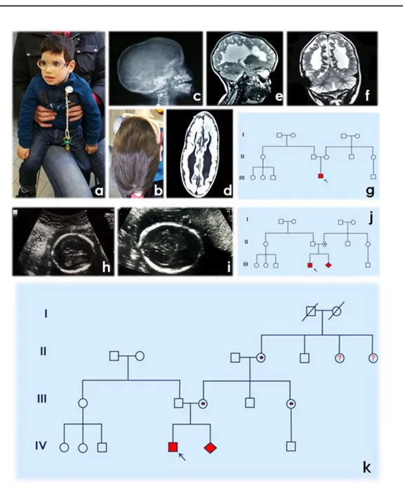
The proband was delivered at 36 weeks + 3days gestation by programed cesarean section after routine US control, performed in 33° g.w. when triventricular hydrocephalus was established. Until this moment, the pregnancy was completely uneventful and no positive US signs have been observed. The birth weight of 3.380 kg (90thcentile), length of 51 cm (90thcentile), and head circumference of 42 cm (>100th centile - macrocefalia). APGAR scores were 6/10 at 1’/5’, respectively. Post-natal Computed Tomography was performed at 2nd day of age and results were consistent with a primary diagnosis of hydrocephalus with aqueductal (Sylvius) stenosis massive triventricular dilation, absence of corpus callosum. Due to this findings, a ventriculo-peritoneal shunt was placed at 4-day of age. Subsequently (at 9 months of age), magnetic resonance image (MRI) evidenced normal dimension of IV ventricle which was in axes and metallic artifact on the right side corresponding to external derivation of shunt with introduction of catheter. In supratentorial region, a multiple malformations have been seen (absence of corpus callosum, colpocephalyc dilatation of lateral ventricles with irregular configuration of walls, scarce representation of white matter and cortical atrophy particularly in parieto-occipital region (Figure 1).
The proband was evaluated in the medical genetics unit at 2 years and 9 months of age. His weight was 12 kg (10th centile), hight was 87 cm (10thcentile). On physical examination, marked scaphocephaly was present, no adducted thumbs without contractures were noted (Figure 1). The proband had no reported seizures. In medical documentation, presented during Genetic Counseling we had found:
Routine hemato-chemical laboratory examination (hepatic, renal, hormonal, martial, ecc.) were normal. ORL examination within normal limits. Ophthalmological examination: OO +1.75 sf. Nistagmus was present in all positions. Normal male karyotype (46, XY) was observed; aCGH whole genome chromosomal microarray analysis was pursued confirm no microdeletions/microduplications in this patient.
On the basis of medical documentation, anamnesis, clinical signs during Genetic Counseling a diagnostic hypotheses of L1 syndrome has been made, with subsequent explanation regarding genetic tests possibilities and consequences of eventually positive results of molecular test for the patient, his mother and for the mother’s family. On the basis of our practice we fixed another appointment 2 weeks later, when collection of blood and signs of informed consensus should be performed, but unfortunately family has not shown up further.
Approximately one year later, the Department of Gynecology and Obstetrics requested Genetic Counseling for a 33 years old woman, secondigravida (22° gestation week), with abnormal ultrasound findings showing severe fetal ventriculomegaly. The family history was unremarkable with no consanguinity; she was one of two sisters and had a healthy brother. Surprisingly the woman was the mother of our proband, who was without any genetic testing.
Figure 1. a,b. Proband/patient phenotype; c. RTG – laterolateral radiography; d. MR - horizontal section; e. MR - mid-sagital (medial) section; f. MR - frontal (coronal) section; g. Pedigree after 1° Genetic Counseling; h,i. II pregnancy - ecography 22w+3d – ventriculomegaly; j. Pedigree after 2° Genetic Counseling and Genetic Tests results during II pregnancy; k. Pedigree after 3° Genetic Counseling and Genetic Test.
On the basis of the previous hypothesis of L1 syndrome, which become more probable after this clinical finding (see pedigree) we performed molecular analysis on proband’s DNA, mother’s DNA and fetal DNA, considering that no characterization was done at the moment of the first Genetic Counseling. The whole coding sequence of the L1CAM gene was analyzed by direct sequencing in an ABI 3730 DNA Analyzer (Applied Biosystems, Foster City, CA; conditions available on request). The alignment of the results with the sequences provided for L1CAM GeneBank accession number NM_000425 (NCBI, http://www.ncbi.nlm.nih.gov/) was carried out using the software SeqManII (DNASTAR. Madison, WI). This mutational screening revealed the presence in all the tested samples of the single nucleotide substitution c.2701C>T causing a stop codon at amino acid position 901 (p.Arg901*) in the exon 20 of the gene (Figure 2). The same mutation has been already reported in three different L1 patients (MAC FARLANE et al., 1997;
KANEMURA et al., 2006; VOS and HOFSTRA, 2010) and, as in our case, is generally associated to a
severe phenotype. Conversely to our proband, adducted thumbs were diagnosed in all cases. From a functional point of view the substitution detected results in a loss of function of L1 protein due to a truncation within its 3rd Fn III domain. Genotype-phenotype correlation studies suggest that the type of mutation have an important effect on the severity of the L1 syndrome (BERTOLIN et al., 2010). In general children with a loss of L1 protein were found more likely to die before the age of 3. On the basis of these results post-test Genetic Counseling has been performed with written report in which same genetic test for mother and subsequently for sister was recommended. Even these two cases (mother and sister of proband’s mother) carried the same mutation in heterozygosis (Figure 1 and 2) and due to this observations other family members (grandaunts and cousins belonging to the branch of the maternal grandmother) should be analyzed.
Figure 2(a) Family tree. Filled squares indicate affected males; arrow indicates proband; *indicates individuals whose DNA was available for the genetic analysis. (b) Frames designate the detected variation c.2701C>T(p.Arg901*)(boxed in black)in the forward sequence ofthe L1CAM gene exon 20. In the top: Refseq sequence (NM_000425). Proband (III-1) and fetal DNA are hemizygous for the variation. All the women (II-2, II-5 and I-2) are heterozygous.
DISCUSSION
As previously reported by VOS and HOFSTRA (2010), to date more than 200 different pathogenic L1CAM mutations have been reported distributed throughout the whole gene, with
large clinical variability between and sometimes within families
(http://www.l1cammutationdatabase.info/).
Since there are no predictive bio-markers for L1-syndrome, molecular analysis of the L1CAM gene is the only diagnostic tool for affected individuals and for the identification of healthy female carriers. In the same paper the authors (VOS and HOFSTRA, 2010) refer that in patients with three or more clinical characteristics but no affected relatives, the detection rate is around 58%. In the present study we identified L1CAM mutations in apparently sporadic cases with at least three disease features (hydrocephalus, agenesis of corpus callosum, tetraparesys, axial hypotony, cognitive and motor incompetency) and subsequently by the analysis of probands' relatives we evidenced several female carriers (see pedigree). This observation underline a necessity to consider L1-syndrome as potential part of differential diagnosis in all cases of hydrocephaly, same as undoubting of importance of pre- and post-test genetic counseling (BUKVIC and MARGAGLIONE, 2013; BUKVIC and ELLING, 2015; BUKVIC et al., 2015) with subsequent, if necessary, extension of the molecular analysis to the relatives in order to provide an effective and reliable counseling and risk assessment to these families.
Finally, the development of new analytical tools [primarily next generation sequencing (NGS)] and development of an assay/gene panel can be used to specifically target a L1 Syndrome for fast, reliable and high quality results in tricky clinical situation like this one.
ACKNOWLEDGMENT
The authors are extremely grateful to the patient and his family for participation in the study, and to Dr. Celestina Antonella Donato, Dr. Rosana Bagnulo and Dr. Annunziata De Luisi for their technical assistance. The authors would also like to thank Dr. Francesca Mercadante and Dr. Fabiana Cortellessa, for their assistance in MGC ambulatory and finally to Dr. Cristiano Simone and Dr. Alessandro Stella for critical review and comments on the manuscript.
Received February 17th, 2016 Accepted June 16th, 2016 REFERENCES
BERTOLIN C., F. BOARETTO, G. BARBON, L. SALVIATI, E. LAPI, M.T. DIVIZIA, L.GARAVELLI, G.OCCHI, G.VAZZA, M.L. MOSTACCIUO LO (2010): Novel mutations in the L1CAM gene support the complexity of L1 syndrome. J Neurol Sci., 294(1-2):124-126.
BICKERS D.S., R.D. ADAMS (1949): Hereditary stenosis of the aqueductof sylvius as a cause of congenital hydrocephalus. Brain, 72:246–262. PMID: 18136715
BUKVIC N., M.MARGAGLIONE (2013): Genetic counselling in post-genomic era-to be or not to be. World J. Med. Genet., 3(3): 9-13.
BUKVIC N., J. ELLING (2015): Genetics in the Art and Art in Genetics. Gene, 555: 14-22.
BUKVIC N., D. VARVARA, C. ROSSI, M. FELICIA FAIENZA, F. C. SUSCA, N. RESTA (2015): From clinical suspect to molecular confirmation of Noonan syndrome; contribution of “best practice” genetic counseling and new technical possibilities. Genetika, 47(3), 877-884.
BURTON B.K. (1979): Recurrence risk for congenital hydrocephalus. Clin. Genet., 16:47–53.
CHIDSEY B.A., E.E. BALDWIN, R. TOYDEMIR, L. AHLES, H. HANSON, D.A. STEVENSON (2014): L1CAM whole gene deletion in a child withL1 syndrome. Am. J. Med. Genet. Part. A 164:1555–1558.
FINCKH U., J. SCHRÖDER, B. RESSLER, A. VESKE, A. GAL (2000): Spectrum and detection rate of L1CAM mutations in isolatedand familial cases with clinically suspected L1 disease. Am. J. Med. Genet., 92:40–46.
FRANSEN E., V. LEMMON, G. VAN CAMP, L. VITS, P. COUCKE, P.J. WILLEMS (1995): CRASH syndrome: clinical spectrum of corpuscallosum hypoplasia, retardation, adducted thumbs, spasticparaparesis and hydrocephalus due to mutations in onesingle gene, L1. Eur. J. Hum. Genet., 3:273–284.
HALLIDAY J., C.W.CHOW, D. WALLACE, D.M. DANKS (1986): X-linked hydrocephalus: a survey of a 20 year period in Victoria, Australia. J. Med. Genet., 23:23–31.
JACKSON S.R., Y.S.GUNER, R.WOO, L.M.RANDOLPH, H.FORD, C.E. SHIN (2009): L1CAM mutation in association with X-linked hydrocephalus and Hirschsprung’s disease. Pediatric Surg. Int., 25:823–825.
KANEMURA Y., N.OKAMOTO, H.SAKAMOTO, T.SHOFUDA, H.KAMIGUCHI, M.YAMASAKI (2006): Molecular mechanisms and neuroimaging criteria for severe L1 syndrome with X-linked hydrocephalus. J. Neurosurg., 105 (5 Suppl): 403-12.
KENWRICK S., M.JOUET, D.DONNAI (1996): X-linked hydrocephalus and MASA syndrome. J. Med. Genet., 33:59–65. MACFARLANE J.R., J.-S. DU, M.E. PEPYS, S. RAMSDEN, D. DONNAI, R. CHARLTON, C. GARRETT, J. TOLMIE, J.R.W. YATES, C.
BERRY, D. GOUDIE, A. MONCLA, P. LUNT, S. HODGSON, M. JOUET, S. KENWRICK (1997): Nine novel L1 CAM mutations in families with X-linked hydrocephalus. Hum. Mutat., 9(6): 512–518.
SCHRANDER-STUMPEL C., J.P. FRYNS (1998): Congenital hydrocephalus: nosologyand guidelines for clinical approach and genetic counselling. Eur. J. Pediatr., 157:355–362.
STEVENSON R.E., J.C. HALL, R.M. GOODMAN (1993): Human malformationsand related anomalies.. Oxford Mon. Med. Gen., 27:68–110.
DVA RAZLIČITA SLUČAJA POST- I PRE-NATALNOG SINDROMA; KADA HIDROCEFALUS POSTAJE “MULTIDISCIPLINARNA GLAVOBOLJA” Nenad BUKVIC1, Francesca BOARETTO2, Giuseppe LOVERRO3, Francesco C. SUSCA4, Rosaura LOVAGLIO4, Margherita PATRUNO4, Dragoslav BUKVIC5, Srdjan STARCEVIC6,
Giovanni VAZZA2, Maria Luisa MOSTACIUOLLO2, Nicoletta RESTA4 1University Hospital Policlinics of Bari, Medical Genetics, Bari, Italy
2University of Padua, Department of Biology-Laboratory of Human Genetics, Padua, Italy 3University of Bari, Department of Gynecology and Obstetrics, Bari, Italy
4University of Bari, DIMO-Laboratory of Medical Genetics, Bari, Italy 5Niksic General Hospital - Department of gynecology, Niksic, Montenegro 6Clinic for Ortopedic Surgery and Traumatology, MMA(VMA), Belgrade, Serbia
Izvod
Sredinom 90-tih u naučnoj literaturi se koristi termin CRASH sindrom kao akronim za Corpus callosum hypoplasia, Retardation, Adducted thumbs, Spastic paraplegia, and Hydrocephalus. Ovo patološko stanje je takodje poznato pod nazivom L1 sindrom, koji obuhvata spektar neuroloških oboljenja sa X vezanim tipom nasljedjivanja, uzrokovan prisustvom mutacija unutar L1CAM (L1 Cell Adhesion Molecule) gena (OMIM 308840). Proband je prvo muško dijete (hidrocephalus, aplazije corpus callosum, tetraparesys, hypotonia, te kognitivne i mentalne deficijencije.zdravog) ne konsanguinog Italijanskog bračnog para, bez anamnestički pozitivne istorije za oboljenja CNS, ponavljanih spontanih pobačaja, congenitalnih anomalija ili genetskih oboljenja. Na osnovu medicinske dokumentacije, anamnestičkih podataka i objektivnog nalaza u toku obrade u Genetskom Savjetovalištu u okviru diferencijalne dijagnoze se postavlja hipoteza L1 sindroma, uz detaljno objašnjenje mogućnosti te strategije genetičkog testiranja. Na osnovu proceduralnog protokola, predložen je datum sljedećeg susreta (za 15 dana), medjutim bračni par se nije pojavio te tako nije bilo moguće uzeti material potreban za izvodjenje genetskih testova. Godinu dana poslije, odjeljenje za Ginekologiju i Akušerstvo zahtjeva Genetsko savjetovanje za 33 godine staru drugorotku (22° sedmica trudnoće) sa patološkim ultrazvučnim nalazom koji pokazuje veliko uvećanje moždanih komora fetusa (ventriculomegalia). Na naše iznenadjenje pacijentica je majka probanda. Na osnovu već postavljenje dijagnostičke hipoteze za L1 sindrom, koja sa ovom trudnoćom postaje daleko izvjesnija, a kako molekularna karakterizacija prethodno nije bila uradjena, ista se izvodi istovremeno na DNA probanda, DNA majke i na fetalnoj DNA dobijenoj nakon amniocenteze. Molekularnom analizom se utvrdjuje prisustvo mutacije: non sense c.2701C>T (p.Arg901*), ekson 20, L1CAM gena u svim testiranim DNA uzorcima što daje molekularnu potvrdu kliničke dijagnoze. Ova observacija ukazuje na važnost da L1-sindrom bude uvršten kao dio diferencijalne dijagnoze u svim slučajevima hidrocefalusa, kao i na nepobitnu važnost Genetskog savjetovališta, molekularne analize te izvodjenja iste unutar familije kako bi se omogućio efektivni i realan “counseling” uz kalkulaciju realnog rizika. Na kraju, ali ne manje značajna je činjenica da uz razvoj novih tehnoloških dostignuća i analitičkih mogućnosti, [next generation sequencing (NGS)] te razvoja eseja/genskih panela koji uključuju L1 sindrom bila bi moguća brža, kvalitetnija analiza sa sigurnijim i sveobuhvantijim rezultatima u kliničkim situacijama kao što je ova (22° sedmica trudnoće, bez prethodne karakterizacije probanda, itd.)
Primljeno 17. II 2016 Odobreno 16. VI.2016.