Lesion Load May Predict Long-Term
Cognitive Dysfunction in Multiple Sclerosis
Patients
Francesco Patti1☯*, Manuela De Stefano2☯, Luigi Lavorgna2, Silvia Messina1, Clara
Grazia Chisari1, Domenico Ippolito2, Roberta Lanzillo3, Veria Vacchiano3, Sabrina Realmuto4, Paola Valentino5, Gabriella Coniglio6, Maria Buccafusca7,
Damiano Paolicelli8, Alessandro D’Ambrosio2, Patrizia Montella2, Vincenzo Brescia Morra3, Giovanni Savettieri4, Bruno Alfano9, Antonio Gallo2, Isabella Simone8,
Rosa Viterbo8, Mario Zappia1, Simona Bonavita2,10, Gioacchino Tedeschi2
1 Department G.F. Ingrassia, Section of Neurosciences, University of Catania, Catania, Italy, 2 Department of Medical, Surgical, Neurological, Metabolic and Aging Sciences, Second University of Naples, Naples, Italy, 3 Department of Neurological Sciences, University‘Federico II,’ Naples, Italy, 4 Department of Experimental Biomedicine and Clinical Neurosciences-University of Palermo, Palermo, Italy, 5 Department of Medical Sciences, Institute of Neurology, University“Magna Graecia”, Catanzaro, Italy, 6 Department of Neurology, “Madonna delle Grazie” Hospital, Matera, Italy, 7 Department of Neurosciences, Psychiatry and
Anaesthesiology, University of Messina, Messina, Italy, 8 Department“Scienze Mediche di Base, Neuroscienze e Organi di Senso”, University of Bari, Bari, Italy, 9 Biostructure and Bioimaging Institute, National Research Council, Naples, Italy, 10 Neurological Institute for Diagnosis and Care“Hermitage Capodimonte”, Naples, Italy
☯ These authors contributed equally to this work. *[email protected]
Abstract
Background
Magnetic Resonance Imaging (MRI) techniques provided evidences into the understanding of cognitive impairment (CIm) in Multiple Sclerosis (MS).
Objectives
To investigate the role of white matter (WM) and gray matter (GM) in predicting long-term CIm in a cohort of MS patients.
Methods
303 out of 597 patients participating in a previous multicenter clinical-MRI study were en-rolled (49.4% were lost at follow-up). The following MRI parameters, expressed as fraction (f) of intracranial volume, were evaluated: cerebrospinal fluid (CSF-f), WM-f, GM-f and ab-normal WM (AWM-f), a measure of lesion load. Nine years later, cognitive status was as-sessed in 241 patients using the Symbol Digit Modalities Test (SDMT), the Semantically Related Word List Test (SRWL), the Modified Card Sorting Test (MCST), and the Paced Au-ditory Serial Addition Test (PASAT). In particular, being SRWL a memory test, both immedi-ate recall and delayed recall were evaluimmedi-ated. MCST scoring was calculimmedi-ated based on the number of categories, number of perseverative and non-perseverative errors.
OPEN ACCESS
Citation: Patti F, De Stefano M, Lavorgna L, Messina S, Chisari CG, Ippolito D, et al. (2015) Lesion Load May Predict Long-Term Cognitive Dysfunction in Multiple Sclerosis Patients. PLoS ONE 10(3): e0120754. doi:10.1371/journal.pone.0120754 Academic Editor: Glenn Wylie, Kessler Foundation Research Center, UNITED STATES
Received: November 14, 2014 Accepted: January 26, 2015 Published: March 27, 2015
Copyright: © 2015 Patti et al. This is an open access article distributed under the terms of theCreative Commons Attribution License, which permits unrestricted use, distribution, and reproduction in any medium, provided the original author and source are credited.
Data Availability Statement: All relevant data are within the paper.
Funding: These authors have no support or funding to report.
Competing Interests: The authors of this manuscript have the following competing interests: Dr. Patti has received honoraria for scientific lectures from Biogen Idec; Dr. De Stefano report no disclosure; Dr. Lavorgna has received travel payment from Merck-Serono, Novartis and Biogen Idec; Dr. Messina has received travel payment from Novartis, Biogen Idec, Bayer Schering, Merck Serono; report no disclosure;
Results
AWM-f was predictive of an impaired performance 9 years ahead in SDMT (OR 1.49, CI 1.12–1.97 p = 0.006), PASAT (OR 1.43, CI 1.14–1.80 p = 0.002), SRWL-immediate recall (OR 1.72 CI 1.35–2.20 p<0.001), SRWL-delayed recall (OR 1.61 CI 1.28–2.03 p<0.001), MCST-category (OR 1.52, CI 1.2–1.9 p<0.001), MCST-perseverative error(OR 1.51 CI 1.2–1.9 p = 0.001), MCST-non perseverative error (OR 1.26 CI 1.02–1.55 p = 0.032).
Conclusion
In our large MS cohort, focal WM damage appeared to be the most relevant predictor of the long-term cognitive outcome.
Introduction
Cognitive Impairment (CIm) has been recognized as an important feature of Multiple Sclerosis (MS), affecting up to 65% patients. CIm, such as memory impairment, reduced information processing speed, attention deficit, impaired executive function, can occur from the early stage of the disease and tends to worsen over time. The prevailing pattern of CIm in MS is repre-sented by attention, processing speed, memory, executive function and visuo-spatial deficits, while language abilities are typically unaffected [1]. In the last years, novel Magnetic Resonance Imaging (MRI) techniques have provided further evidences into the understanding of CIm in MS, highlighting the involvement of both white matter (WM) and gray matter (GM) damage in the development of disability [2,3].T1-, T2-lesion load (LL) and brain atrophy measures may predict the onset of CIm after several years [4,5]. Conversely, other studies showed a clear discrepancy between LL and severity of CIm in MS [6].WM abnormalities were weakly corre-lated with CIm, suggesting that WM abnormalities alone cannot fully explain the extent of clin-ical symptoms and CIm in MS [7,8].In the present study, WM, and GM atrophy and WM LL were obtained through a fully automated, operator-independent, multiparametric segmenta-tion method from a large MS populasegmenta-tion [9,10]. By using this approach, we recently showed that baseline (BL) GM atrophy and EDSS were the best long-term (9 years follow-up) predic-tors of clinical disease progression in relapsing remitting (RR) MS patients[11].Considering these findings, the aim of the present study was to investigate the role of WM and GM damage in predicting long term (9 years follow-up) CIm in a large multicenter cohort of MS patients.
Materials and Methods
Ethics statement
The study was previously approved by the Ethics Committee (EC) of the Second University of Naples and then by all local EC of each participating center: EC“Federico II” Naples, EC Uni-versity Hospital Policlinico Palermo, EC UniUni-versity Hospital Policlinico Catanzaro, EC Univer-sity Hospital Policlinico Messina, EC UniverUniver-sity Hospital Policlinico Bari, National Research Council, Naples. A written informed consent was obtained from every patient before study initiation.
Drs Chisari, Ippolito, Vacchiano, Valentino, Buccafusca, Paolicelli, D’Ambrosio, Montella, Viterbo, Realmuto and Prof. Alfano report no disclosure; Dr Lanzillo has received travel payment from Merck-Serono, Novartis, Biogen Idec, Schering-Plough and Teva; Dr Coniglio has received funding for a trip from Novartis; Dr. Bresciamorra has received travel payment from Novartis, Biogen Idec, Schering-Plough and Teva. Prof. Savettieri has received travel payment from Teva and honoraria for scientific lectures from Biogen Idec; Dr. Gallo has received travel payment from Biogen Idec and Schering-Plough and honoraria for scientific lectures from Biogen Idec; Prof. Simone has received honoraria for educational lectures from Biogen Idec, Bayer, Sanofi Aventis-Genzyme and Teva; Prof. Zappia has received funding for a trip from Schering-Plough; Prof. Bonavita has received travel payment from Merck-Serono, Novartis, Biogen Idec and Teva and honoraria for scientific lectures from Novartis; Prof. Tedeschi has received travel payment from Merck-Serono, Novartis, Biogen Idec and Teva and honoraria for scientific lectures from Novartis, Teva and Biogen Idec. Francesco Patti declares, on behalf of all authors, all potential competing interests do not alter their adherence to PLOS ONE policies on sharing data and materials.
Patients
In line with our recent paper, of the initial cohort of 597 MS patients participating to the previous multicenter clinical/MRI research, 303 subjects were enrolled in this 9-year follow-up (FU) study (49.4% lost at FU). The reasons for drop-out were: missing contact information (50.7%), unavail-ability (24.1%), refusal (20.1%), death (3.4%), and other major medical illnesses (1.7%). There were no significant differences in BL demographic or clinical data between patients lost at FU and those participating in the initial cross-sectional study, except for a minor percentage of RR patients in the dropped-out population (65% RR patients in the dropped out group, 72.4% in the FU group; p<0.02) [11]. Out of 303 subjects, 241 underwent the Neuropsychological (NPS) bat-tery. Furthermore, not all patients performed the whole NPS battery; a slight partial incomplete-ness was obtained for some test because of the challenging duration of the NPS examination. At BL and at FU, disability was measured by the EDSS and fatigue was evaluated by the Fatigue Se-verity Scale (FSS).ΔEDSS and ΔFSS represent the variation of these parameters during FU. Nine years after, 42 out of 241(17.4%) RRMS patients converted to Secondary-Progressive (SP) MS (RR! SP), while the remaining 199 (82.6%) did not change their disease phenotype (RR! RR).
MRI imaging
At BL, the enrolled patients underwent the MRI protocol described in the previous cross-sec-tional study [10]. In brief, conventional spin echo sequences were acquired to obtain
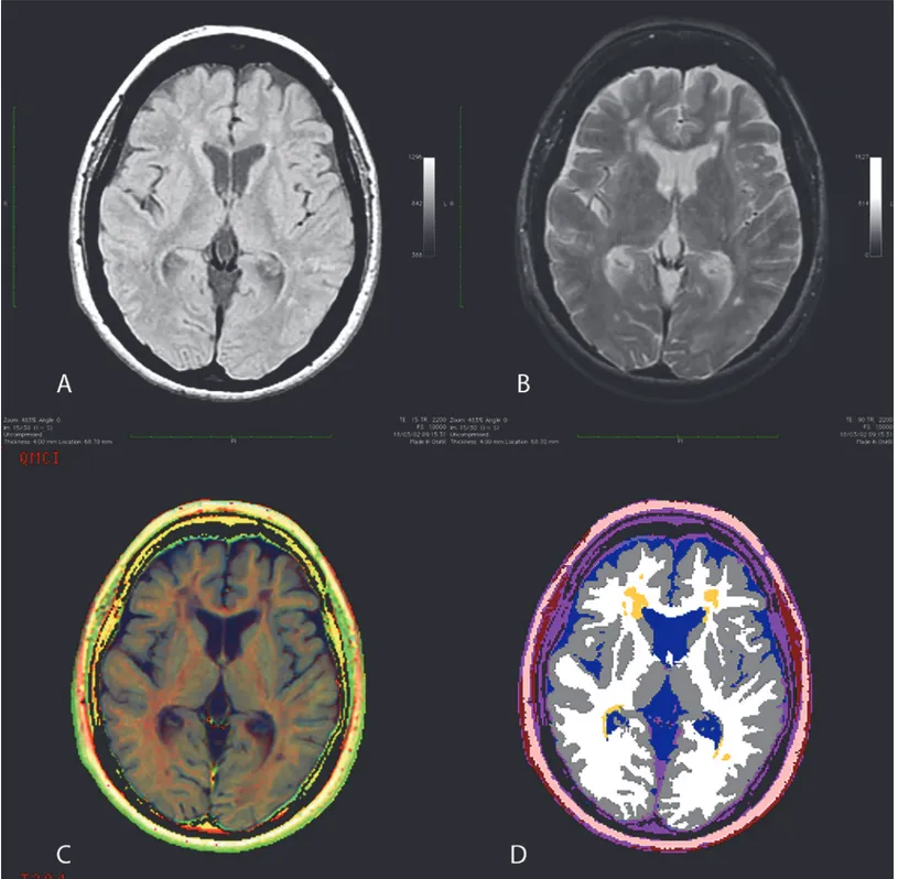
T1-weighted (TR/TE 600/15 msec, two averages) and dual echo (TR/TE2300/15–90 msec, one average) images, with 90°flip angle and 256 × 192 matrix size. All the studies were segmented using a multispectral, fully automated method, based on relaxometric characterization of brain tissues [9]. The program furnishes complete sets of multifeature images [R1 (= 1/T1), R2 (= 1/ T2), proton density (N(H))-based] and segmented images of the following intracranial tissues: cerebrospinal fluid (CSF), WM, GM, abnormal WM. AWM is a WM LL measure as deter-mined by the R1, R2, and N(H) information and morphological characteristics. The relaxo-metric method used provides GM segmentation not influenced by WM lesions [12]. For each study, a couple of interactive interslice movies of both multifeature and segmented images were produced, and two neuroimaging experts reviewed them (for a maximum of 2 minutes) to detect motion artifacts and segmentation errors due to the imperfect separation of nasal mu-cosa and vitreous humor from brain tissue. To normalize for head size variability, the volumes were expressed as fraction (f) of intracranial volume. AWM-f is measure of LL. The reduction of WM-f and GM-f indicate respectively WM and GM volume reduction. The increase of CSF-f indicates global brain volume reduction.Fig. 1shows a transverse slice of a patient with MS and depicts the major steps of the multiparametric method.
Neuropsychological evaluation (NPS)
At the 9-year FU visit, all patients underwent the same NPS battery, standardized among the participating centers. Tests were administered during daytime, in a quiet room. A brief neuro-psychological battery was administered to explore the following cognitive domains.
Sustained attention. Symbol Digit Modalities Test (SDMT, number of correct pairings). Patients are presented a series of nine symbols. Every symbol is paired with a single digit, labeled 1–9. In the test page a pseudo-randomized sequence of symbols is presented. Patients have to write the correct digit associated to every symbols as quick as possible in 90 seconds [13]
Verbal memory. Semantically related word list test (SRWL):the test consisted of five con-secutive immediate free-recall trials (I-R), followed by a 15-min delayed recall trial (D-R) and recognition of 16 words from 4 categories: animals, transportation, vegetables and furniture [14].
Executive functions, conceptual reasoning. Modified Card Sorting Test (MCST) was adapted to improve Wisconsin Card Sort Test (WCST), simplifying the task, removing the am-biguity in interpreting responses and providing measure of perseveration. The cards were
Fig 1. Axial image of a Multiple Sclerosis patient. (A) Proton density-weighted image; (B) T2-weighted image; (C) corresponding multiparametric image; (D) segmented image. In the segmented image, white matter is represented by white, gray matter by gray, cerebrospinal fluid by blue, and lesions by yellow. doi:10.1371/journal.pone.0120754.g001
always presented to each participant in the same order according to the sequence provided by the numbers at the back of the cards. Errors were scored as perseverative if the sorting response was the same category (color, form, number) as the previously incorrect response, or if the sort-ing response did not change after the patient was told that the rules had changed. An error was scored as not perseverative if the patient followed a sorting response to neither color, form or number (“other” response) with a second “other” response. MCST scoring is based on the number of categories completed and the number of perseverative and non-perseverative errors [15].
Information speed processing, working memory. Three-second version of Paced Audi-tory Serial Addition Test (PASAT) is a test measuring sustained attention and information pro-cessing speed. This test is included in the Brief Repeatable Battery of Neuropsychological test (BRB). A series of single digit numbers from a type record are presented to the subject at the rate of one every 3 seconds. The subject is asked to add every digit to the one immediately pro-ceeding. The test is composed by 61 digits, the maximum score is 60 [16].
PASAT and SDMT were corrected using the available normative data for the Italian popula-tion. We considered as cut off point a corrected score below the 5th percentile of the normative data [17]. SRWL raw scores were corrected for sex, age and education; cut-off points were cal-culated using the available normative data [14]. MCST raw scores were corrected for age; a cut off was calculated for every measure using the available internal tolerance limit according to the MCST-Roma version [18].
Higher values indicate better performances in all tests but MCST perseverative and non per-severative error; in these items higher values indicate worse performance. An overall Cognitive Index (CI) grading score was calculated for each patient. Considering the number of standard deviations (SD) below mean of normative values, cognitive tests were graded as follows:0, at or above mean value; 1, below mean but at or above 1 SD below mean; 2,< 1SD below mean but at or above 2SD below mean; 3,<2SD below mean but at or above 3SD below mean; 4, <3SD below mean. These grades were summed to obtain an overall CIm for each patient [19]. CIm was defined as the failure on at least two tests involving at least two different domain (verbal memory, attention/information processing speed and executive functions).
Statistical Analysis
Age, disease duration, NPS scores and MRI volume data are presented as means and SD (see
Table 1). Individual variables were checked for skewness and presence of outliers and the mean MRI parameters of all subjects were adjusted for age, gender and education using a linear re-gression model. Statistical analysis was performed using STATA 12.0, and a p value< 0.05 was considered statistically significant. Chi square test was used to compare NPS test score and CIm between RR! RR and RR!SP.
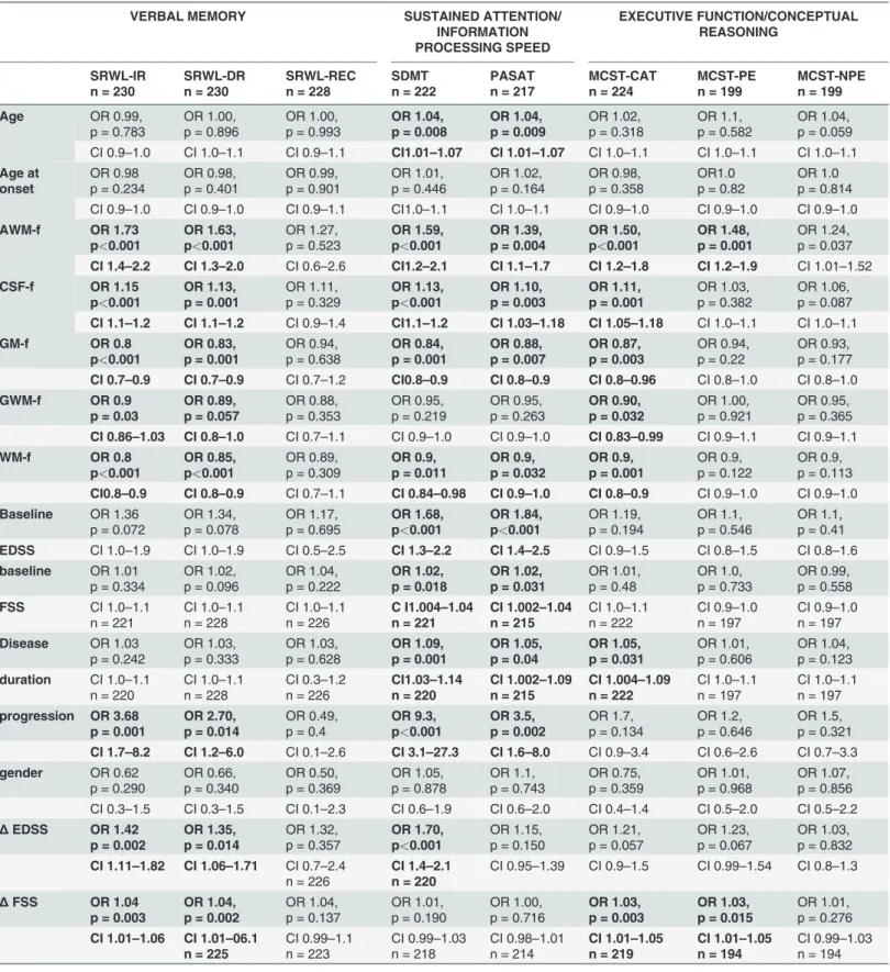
Several univariable logistic regression were fit considering binomial“impaired yes/no” vari-able for each NPS as dependent and age, age at onset, CSF-f, AWM-f, GM-f, GWM-f, WM-f, BLEDSS,ΔEDSS, BLFSS, ΔFSS, disease duration, progression to SPMS and gender as indepen-dent variables. Variables correlating with outcomes (p< 0.1) in univariable analysis were used as independent variables in logistic stepwise regression, considering p = 0.10 as the critical value for entering or excluding variables in the model. A negative binomial regression analysis was performed to evaluate the correlation with the severity of CIm index score.
Results
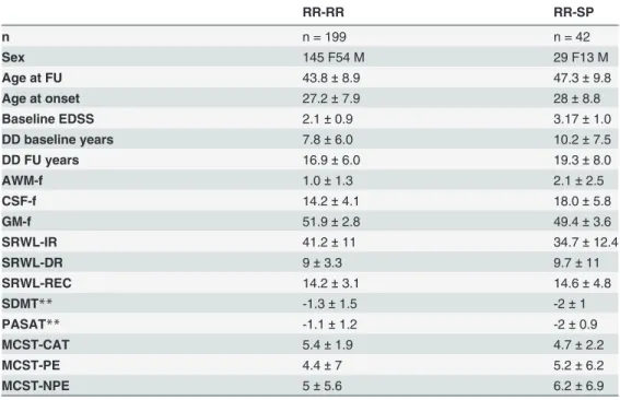
Demographic, clinical, cognitive and MRI data are summarized inTable 1. In Tables2and3
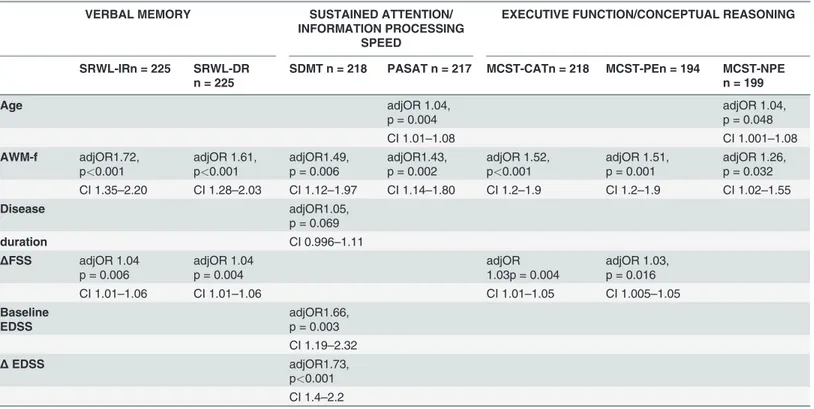
evaluated. Nineteen patients were excluded from the analysis because their SDMT scores were not available. If compared to the RR!SP, RR!RR group showed higher SDMT scores (p<0.001). The logistic regression step-wise model regarding SDMT showed a significant posi-tive correlation with AWM-f (OR 1.49, CI 1.12–1.97 p = 0.006), EDSS at BL (OR 1.066 CI 1.19–2.32 p = 0.003), ΔEDSS (OR 1.66 CI 1.19–2.32 p = 0.003), ΔFSS (OR1.66 CI 1.4–2.2 p<0.001). This means that for a 1% increase in AWM-f there was a 49% increased odds to be cognitively impaired at this test. Moreover, the EDSS at BL and the variations in EDSS and FSS scores were related to higher impaired scores at SDMT (seeTable 3).
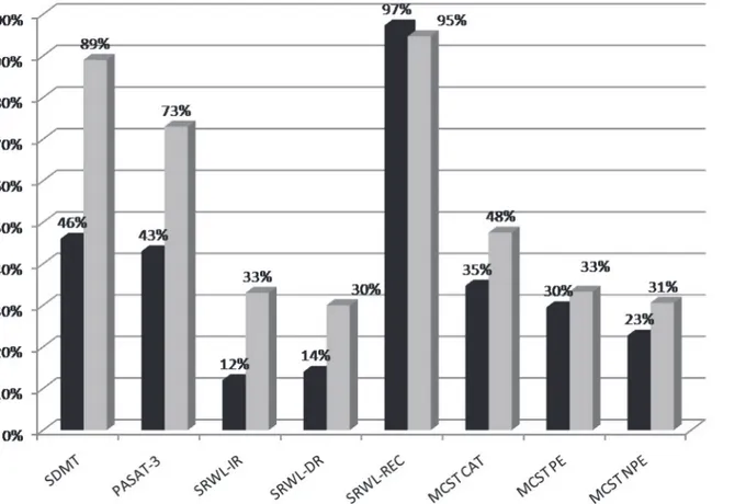
PASAT scores of 217 MS patients were evaluated. Twenty-four patients were excluded from the analysis because their PASAT scores were not available. Seventy-three percent of RR!SP and 43% of RR!RR patients’ scores were under cutoff (p = 0.002). PASAT showed a positive correlation with AWM-f (OR 1.43, CI 1.14–1.80 p = 0.002) and age (OR 1.04, CI 1.01–1.08 p = 0.004). In other words, for a 1% increase of AWM-f there was a 43% increased odds of hav-ing impaired PASAT scores. One year increase in age was related to 4% increased odds of im-paired score at this test (seeTable 3).
SRWL scores of 230 MS patients were evaluated. Eleven patients were excluded from the analysis because their SRWL scores were not available. Thirty-three per cent of RR!SP pa-tients and 12% of RR!RR papa-tients showed SRWL-immediate recall (IR) under cut off (p = 0.001). SRWL-IR score showed a positive correlation with AWM-f (OR 1.72 CI 1.35–2.20 p<0.001) and ΔFSS (OR 1.04 CI 1.01–1.06 p = 0.006) such that a 1% increase of AWM-f was related to 72% higher odds to be impaired at this test.ΔFSS was related to higher odds of im-paired performances at this test. The SRWL-delayed recall (DR) was statistically different be-tween 2 groups, with 30% RR!SP patients showing SRWL scores under cut off if compared to
Table 1. Demographic, clinical characteristics,*adjusted baseline MRI volume (as percentage of whole brain volume) and**neuropsychological test scores of MS patients (mean ± SD).
RR-RR RR-SP n n = 199 n = 42 Sex 145 F54 M 29 F13 M Age at FU 43.8± 8.9 47.3± 9.8 Age at onset 27.2± 7.9 28± 8.8 Baseline EDSS 2.1± 0.9 3.17± 1.0 DD baseline years 7.8± 6.0 10.2± 7.5 DD FU years 16.9± 6.0 19.3± 8.0 AWM-f 1.0± 1.3 2.1± 2.5 CSF-f 14.2± 4.1 18.0± 5.8 GM-f 51.9± 2.8 49.4± 3.6 SRWL-IR 41.2± 11 34.7± 12.4 SRWL-DR 9± 3.3 9.7± 11 SRWL-REC 14.2± 3.1 14.6± 4.8 SDMT** -1.3± 1.5 -2± 1 PASAT** -1.1± 1.2 -2± 0.9 MCST-CAT 5.4± 1.9 4.7± 2.2 MCST-PE 4.4± 7 5.2± 6.2 MCST-NPE 5± 5.6 6.2± 6.9
*Adjusted for age, gender and education. ** z score.
Table 2. Univariable analysis verbal memory, sustained attention/information processing speed, executive function/conceptual reasoning.
VERBAL MEMORY SUSTAINED ATTENTION/
INFORMATION PROCESSING SPEED EXECUTIVE FUNCTION/CONCEPTUAL REASONING SRWL-IR n = 230 SRWL-DR n = 230 SRWL-REC n = 228 SDMT n = 222 PASAT n = 217 MCST-CAT n = 224 MCST-PE n = 199 MCST-NPE n = 199 Age OR 0.99, p = 0.783 OR 1.00, p = 0.896 OR 1.00, p = 0.993 OR 1.04, p = 0.008 OR 1.04, p = 0.009 OR 1.02, p = 0.318 OR 1.1, p = 0.582 OR 1.04, p = 0.059 CI 0.9–1.0 CI 1.0–1.1 CI 0.9–1.1 CI1.01–1.07 CI 1.01–1.07 CI 1.0–1.1 CI 1.0–1.1 CI 1.0–1.1 Age at onset OR 0.98 p = 0.234 OR 0.98, p = 0.401 OR 0.99, p = 0.901 OR 1.01, p = 0.446 OR 1.02, p = 0.164 OR 0.98, p = 0.358 OR1.0 p = 0.82 OR 1.0 p = 0.814 CI 0.9–1.0 CI 0.9–1.0 CI 0.9–1.1 CI1.0–1.1 CI 1.0–1.1 CI 0.9–1.0 CI 0.9–1.0 CI 0.9–1.0 AWM-f OR 1.73 p<0.001 OR 1.63, p<0.001 OR 1.27, p = 0.523 OR 1.59, p<0.001 OR 1.39, p = 0.004 OR 1.50, p<0.001 OR 1.48, p = 0.001 OR 1.24, p = 0.037 CI 1.4–2.2 CI 1.3–2.0 CI 0.6–2.6 CI1.2–2.1 CI 1.1–1.7 CI 1.2–1.8 CI 1.2–1.9 CI 1.01–1.52 CSF-f OR 1.15 p<0.001 OR 1.13, p = 0.001 OR 1.11, p = 0.329 OR 1.13, p<0.001 OR 1.10, p = 0.003 OR 1.11, p = 0.001 OR 1.03, p = 0.382 OR 1.06, p = 0.087 CI 1.1–1.2 CI 1.1–1.2 CI 0.9–1.4 CI1.1–1.2 CI 1.03–1.18 CI 1.05–1.18 CI 1.0–1.1 CI 1.0–1.1 GM-f OR 0.8 p<0.001 OR 0.83, p = 0.001 OR 0.94, p = 0.638 OR 0.84, p = 0.001 OR 0.88, p = 0.007 OR 0.87, p = 0.003 OR 0.94, p = 0.22 OR 0.93, p = 0.177 CI 0.7–0.9 CI 0.7–0.9 CI 0.7–1.2 CI0.8–0.9 CI 0.8–0.9 CI 0.8–0.96 CI 0.8–1.0 CI 0.8–1.0 GWM-f OR 0.9 p = 0.03 OR 0.89, p = 0.057 OR 0.88, p = 0.353 OR 0.95, p = 0.219 OR 0.95, p = 0.263 OR 0.90, p = 0.032 OR 1.00, p = 0.921 OR 0.95, p = 0.365 CI 0.86–1.03 CI 0.8–1.0 CI 0.7–1.1 CI 0.9–1.0 CI 0.9–1.0 CI 0.83–0.99 CI 0.9–1.1 CI 0.9–1.1 WM-f OR 0.8 p<0.001 OR 0.85, p<0.001 OR 0.89, p = 0.309 OR 0.9, p = 0.011 OR 0.9, p = 0.032 OR 0.9, p = 0.001 OR 0.9, p = 0.122 OR 0.9, p = 0.113 CI0.8–0.9 CI 0.8–0.9 CI 0.7–1.1 CI 0.84–0.98 CI 0.9–1.0 CI 0.8–0.9 CI 0.9–1.0 CI 0.9–1.0 Baseline OR 1.36 p = 0.072 OR 1.34, p = 0.078 OR 1.17, p = 0.695 OR 1.68, p<0.001 OR 1.84, p<0.001 OR 1.19, p = 0.194 OR 1.1, p = 0.546 OR 1.1, p = 0.41 EDSS CI 1.0–1.9 CI 1.0–1.9 CI 0.5–2.5 CI 1.3–2.2 CI 1.4–2.5 CI 0.9–1.5 CI 0.8–1.5 CI 0.8–1.6 baseline OR 1.01 p = 0.334 OR 1.02, p = 0.096 OR 1.04, p = 0.222 OR 1.02, p = 0.018 OR 1.02, p = 0.031 OR 1.01, p = 0.48 OR 1.0, p = 0.733 OR 0.99, p = 0.558 FSS CI 1.0–1.1 n = 221 CI 1.0–1.1 n = 228 CI 1.0–1.1 n = 226 C I1.004–1.04 n = 221 CI 1.002–1.04 n = 215 CI 1.0–1.1 n = 222 CI 0.9–1.0 n = 197 CI 0.9–1.0 n = 197 Disease OR 1.03 p = 0.242 OR 1.03, p = 0.333 OR 1.03, p = 0.628 OR 1.09, p = 0.001 OR 1.05, p = 0.04 OR 1.05, p = 0.031 OR 1.01, p = 0.606 OR 1.04, p = 0.123 duration CI 1.0–1.1 n = 220 CI 1.0–1.1 n = 228 CI 0.3–1.2 n = 226 CI1.03–1.14 n = 220 CI 1.002–1.09 n = 215 CI 1.004–1.09 n = 222 CI 1.0–1.1 n = 197 CI 1.0–1.1 n = 197 progression OR 3.68 p = 0.001 OR 2.70, p = 0.014 OR 0.49, p = 0.4 OR 9.3, p<0.001 OR 3.5, p = 0.002 OR 1.7, p = 0.134 OR 1.2, p = 0.646 OR 1.5, p = 0.321 CI 1.7–8.2 CI 1.2–6.0 CI 0.1–2.6 CI 3.1–27.3 CI 1.6–8.0 CI 0.9–3.4 CI 0.6–2.6 CI 0.7–3.3 gender OR 0.62 p = 0.290 OR 0.66, p = 0.340 OR 0.50, p = 0.369 OR 1.05, p = 0.878 OR 1.1, p = 0.743 OR 0.75, p = 0.359 OR 1.01, p = 0.968 OR 1.07, p = 0.856 CI 0.3–1.5 CI 0.3–1.5 CI 0.1–2.3 CI 0.6–1.9 CI 0.6–2.0 CI 0.4–1.4 CI 0.5–2.0 CI 0.5–2.2 Δ EDSS OR 1.42 p = 0.002 OR 1.35, p = 0.014 OR 1.32, p = 0.357 OR 1.70, p<0.001 OR 1.15, p = 0.150 OR 1.21, p = 0.057 OR 1.23, p = 0.067 OR 1.03, p = 0.832 CI 1.11–1.82 CI 1.06–1.71 CI 0.7–2.4 n = 226 CI 1.4–2.1 n = 220 CI 0.95–1.39 CI 0.9–1.5 CI 0.99–1.54 CI 0.8–1.3 Δ FSS OR 1.04 p = 0.003 OR 1.04, p = 0.002 OR 1.04, p = 0.137 OR 1.01, p = 0.190 OR 1.00, p = 0.716 OR 1.03, p = 0.003 OR 1.03, p = 0.015 OR 1.01, p = 0.276 CI 1.01–1.06 CI 1.01–06.1 n = 225 CI 0.99–1.1 n = 223 CI 0.99–1.03 n = 218 CI 0.98–1.01 n = 214 CI 1.01–1.05 n = 219 CI 1.01–1.05 n = 194 CI 0.99–1.03 n = 194 doi:10.1371/journal.pone.0120754.t002
14% RR!RR scores (p = 0.012). A multivariable analysis showed a positive correlation be-tween SRWL-DR scores, AWM-f (OR 1.61 CI 1.28–2.03 p<0.001) and ΔFSS (OR 1.04 CI 1.01– 1.06 p = 0.004).In other words, for a 1% increase of AWM-f there was a 61% increased odds to have SRWL-DR score under cut-off andΔFSS was related to higher odds of impaired perfor-mances at this test (seeTable 3).
MCST scores of 224 patients were evaluated. Seventeen patients were excluded from the analysis because their scores were not available. The MCST-category (CAT) scores of 49% of RR!SP and 34% of RR!RR patients were under cut off (p = 0.08). The multivariable analysis showed a positive correlation between MCST-CAT, AWM-f (OR 1.52, CI 1.2–1.9 p<0.001) andΔFSS (OR 1.03 CI 1.01–1.05 p = 0.004). In other words, a higher AWM-f was related to a 52% increased odds of MCST-CAT impairment andΔFSS was related to higher odds of im-paired performances at this test. Out of 199 RR!RR patients only 30% and 24% showed per-severative (MCSTpe) and non perper-severative errors (MCSTnpe) respectively. The multivariable logistic regression step-wise model showed a positive correlation between AWM-f, MCSTpe (OR 1.51 CI 1.2–1.9 p = 0.001) and ΔFSS (OR 1.03 CI 1.005–1.05 p = 0.016), such that a higher AWM-f was related to a 51% increased odds to be cognitively impaired at this test andΔFSS was related to a higher odds of impaired performances at this test. Regarding MCSTnpe, a posi-tive correlation was found with age (OR 1.04 CI 1.001–1.08 p = 0.048) and AWM-f (OR 1.26 CI 1.02–1.55 p = 0.032). This means that AWM-f was related to a 26% increased odds to be cognitively impaired at MCSTnpe and one year increase in age was related to a 4% increased odds of impaired performances at this test (seeTable 3).
We evaluated cognitive performances of 227 patients. Out of them, 69 (28.6%) were cogni-tively impaired and 158 (69.6%) cognicogni-tively preserved. In particular the prevalence of CIm was 27.1% in RR!RR and 45% in RR!SP group (p = 0.025). As expected, cognitively impaired pa-tients showed a significantly higher overall CIm than cognitively preserved papa-tients (19.0±3.8
Table 3. Multivariate analysis verbal memory, sustained attention/information processing speed, executive function/conceptual reasoning.
VERBAL MEMORY SUSTAINED ATTENTION/
INFORMATION PROCESSING SPEED
EXECUTIVE FUNCTION/CONCEPTUAL REASONING
SRWL-IRn = 225 SRWL-DR n = 225
SDMT n = 218 PASAT n = 217 MCST-CATn = 218 MCST-PEn = 194 MCST-NPE n = 199 Age adjOR 1.04, p = 0.004 adjOR 1.04, p = 0.048 CI 1.01–1.08 CI 1.001–1.08 AWM-f adjOR1.72, p<0.001 adjOR 1.61, p<0.001 adjOR1.49, p = 0.006 adjOR1.43, p = 0.002 adjOR 1.52, p<0.001 adjOR 1.51, p = 0.001 adjOR 1.26, p = 0.032 CI 1.35–2.20 CI 1.28–2.03 CI 1.12–1.97 CI 1.14–1.80 CI 1.2–1.9 CI 1.2–1.9 CI 1.02–1.55 Disease adjOR1.05, p = 0.069 duration CI 0.996–1.11 ΔFSS adjOR 1.04 p = 0.006 adjOR 1.04 p = 0.004 adjOR 1.03p = 0.004 adjOR 1.03, p = 0.016 CI 1.01–1.06 CI 1.01–1.06 CI 1.01–1.05 CI 1.005–1.05 Baseline EDSS adjOR1.66, p = 0.003 CI 1.19–2.32 Δ EDSS adjOR1.73, p<0.001 CI 1.4–2.2 doi:10.1371/journal.pone.0120754.t003
and 10.8±3.9, p<0.001). The multivariable logistic regression step-wise model regarding CIm showed a positive correlation with AWM-f (OR 1.63, CI 1.3–2.1, p>0.001) and disease dura-tion (OR 1.04, CI 1.0–1.1, p = 0.06). A negative binomial regression analysis showed AWM-f was the only significant parameter showing an Incidence Rate Ratio of 1.09 (CI 1.06–1.12, p>0.001). In other words for a 1% increase of AWM-f there was a 9% increased overall CIm index score. Considering EDSS at baseline and FU visit we found median EDSS was 2.0 at BL and 3.0 at the end of FU (p< 0.001). Logistic regression showed a positive correlation between ΔEDSS and CIm (OR 1.29 CI 95% 1.05–1.57 p = 0.013): this means that one point increase in EDSS was related to 29% odds to be cognitively impaired. Considering FSS at baseline and FU, we found median FSS was 26 at BL and 37 at FU (p<0.001). Logistic regression showed a posi-tive correlation betweenΔFSS and CIm (OR 1.03 CI 95% 1.01–1.05 p = 0.004), such that one point increase of FSS was related to 3% odds to be cognitively impaired. Moreover, the multi-variable analysis showed a positive correlation betweenΔEDSS and SDMT (OR 1.73 p<0.001), and betweenΔFSS, and SRWL-IR (OR 1.04 p = 0.006), SRWL-DR (OR 1.04 p = 0.004), MCSTCAT (OR 1.03 p = 0.004) and MCST-pe (OR 1.03 p = 0.01).
Discussion
In this study we evaluated the role of WM, GM and LL (i.e. AWM-f) volumes in predicting the long-term occurrence of CIm in a large group of MS patients.
In the last years, it has been showed that CIm is more related to brain atrophy than to LL in mildly disabled MS patients [4,8]. On the other hand, WM lesions seem to play an important role in the development of CIm as well [20,21]. The contribution of WM damage to CIm is also confirmed by studies reporting a clear association between cognitive functioning and cor-tico–cortical and cortico–subcortical WM tracts damage [21,22]. In particular, the association between the WM damage and the impairment of information processing speed, generally stud-ied by PASAT and SDMT, is frequently demonstrated [23]. A 5 years-follow up study showed that brain atrophy is a good predictor of cognitive functioning in RRMS patients, although also T1-hypointense lesions showed a good predictive value [5]. This is in line with findings sug-gesting that atrophy of cortical and sub-cortical deep GM could be associated with WM lesion burden [24]. However, the pathophysiologic process remains poorly understood [25].
Using a fully automated segmentation method, we found that AWM-f, indicating LL, was the best predictor of CIm in MS patients. In particular, AWM-f was predictive of an impaired SDMT performance. The lower SDMT scores in RR!SP patients compared to RR!RR (pro-portion of RRMS and SPMS patients with impaired performance at each test represented in
Fig. 2) and the positive correlation with EDSS at BL, underline the influence of accrual of dis-ability and WM damage on cognitive performance. Since an interaction between motor disabil-ity and cognition has been demonstrated, especially on progressive patients, we cannot exclude that motor impairment may affect in part CIm [26]. However, our patients have a relatively mild disability, being baseline EDSS below 4 for both RRMS and SPMS (2.1 ± 0.9 and 3.17 ± 1 respectively). Our results showed higher PASAT scores in RR!RR if compared to RR!SP pa-tients. In the multivariable analysis, age and AWM-f were the only variables predictive of lower PASAT scores, confirming previous findings showing that lower age was related to better per-formances at this test [27].Taken together, these results underline the importance of WM tract integrity in the rapid transfer of information between the cortex and deep GM, suggesting the involvement of WM in processing speed deficits.
Regarding MCST, we found a positive correlation with AWM-f. This is in line with other studies, showing that although MCST has a lower sensitivity than other executive function tests in MS, it is strongly correlated with brain atrophy and LL [28].
RR!SP showed lower SRWL scores when compared to RR!RR group. Moreover, we found a positive correlation between AWM-f and the incidence of being classified as impaired on the SRWL.
Our results could be apparently in contrast with other studies reporting that patients with pro-gressive MS show deficits in information processing speed, attention, working memory, executive function, and verbal episodic memory, whereas CIm of RRMS patients is limited to information processing speed and working memory [29]. The association between verbal memory and GM pathology is frequently reported. Using a different battery (BRB) and a different MRI technique, Amato et al [30] showed a clear association between GM atrophy and verbal memory, suggesting the involvement of cortical regions in the neuropathological process at the early stage of the dis-ease; however, given the cross sectional nature of the study, a direct comparison with the results presented herein is not possible. Memory deficits of MS patients were initially thought to be due only to impaired retrieval. More recent explanations postulate that verbal memory impairment is the consequence of an inadequate acquisition and retrieval, both secondary to information pro-cessing insufficiency [31], although it is conceivable the impairment of memory and information processing speed may result from the same pathological process. In line with this perspective, a robust correlation between CIm and LL is reported in a number of studies conducted with mildly disabled patients or at the early stage of the disease [32–34] Using high field MRI, a correlation between Normal Appearing WM, SDMT and CVLT-II [35] scores has been demonstrated,
Fig 2. Proportion of RRMS and SPMS patients with impaired performance at each test. Legend: RRMS = relapsing-remitting multiple sclerosis, SPMS = secondary progressive multiple sclerosis, SDMT = symbol digit modality test, PASAT-3 = 3.0 inter-stimulus interval of Paced Auditory Serial Addition Test, SRWL = semantically related word list test, IR = immediate recall, DR = delayed recall, REC = recognition, MCST CAT = modified card sorting test categories, MCST-PE = MCST perseverative errors, MCST-NPE = MCST non-perseverative errors.
highlighting the crucial role of WM tract integrity in verbal learning, ensuring rapid transfer of information between cortex and deep GM. In our study, we support the role of a fully automated, operator-independent, multiparametric segmentation method to measure AWM-f, as marker of LL and both WM and GM volumes [10].MRI was performed 9 years before the NPS, which al-lows us to suppose that AWM-f could be considered an early predictor of CIm, strengthening the concept of a possible involvement of WM in the development of CIm. It is conceivable that CIm in our patients is, at least in part, caused by central neural pathways-disconnection. A “discon-nection model” could interpret the involvement of multiple cognitive domains in this pathology as a series of disconnection syndromes affecting different cognitive networks. We propose that disruption of cortical WM tracts leads to reduced connectivity between cortico-cortical and cor-tico-subcortical cognitive processing regions, resulting in deficits in specific cognitive domains. On the other hand, this kind of model does not exclude GM pathology [3,30] which may play a more important role as the disease progress [36]. Recently, a 13-year follow-up study showed that GM magnetization transfer ratio (MTR) was the only MRI predictor of global CIm, support-ing the notion that GM plays a major role in the long-term development of CIm [37]. However, we cannot rule out that GM damage is secondary to WM damage, emphasizing the role of WM as an early marker of CIm.
Our study has the appeal of focusing on the long term predicting value of MRI parameters in a real life setting of MS management, being our cohort not in the early phase of the disease and having a relatively low disability.
However, our project has several limitations. First, though we had the great advantage to use the same (mobile) scanner in each participating center so that all patients shared the same protocol on the same scanner, we used a 1.0 Tesla MRI scanner which did not allow us to per-form more advanced MRI measurements (i.e. WM tractography). Second, we did not have a baseline cognitive evaluation, therefore we could not assess the cognitive profile of patients at such time-point. Third we did not perform a FU MRI to assess the longitudinal change in brain volume. On the other hand, the use of an accurate and automatic segmentation method gave us the advantage to simultaneously assess LL, WM and GM volumes in a large population of patients and in a time-saving fashion. Finally, we did not use a universally accepted method to calculate the overall CIm, thus possibly underestimating the real burden of CIm. Since there is a lack of a standardized classification criteria, we used two different strategies to have an esti-mate of global CIm: a composite index score based on a graded system [19] and a domain spe-cific CIm. Furthermore, since the number of test may influence cognitive outcome, we decided to use a brief NPS evaluation, investigating the most commonly affected cognitive domains in MS, giving us the opportunity to test a large number of patients [38].Although we used a brief NPS battery, not all patients completed tests. However, given the small number of missing data for every test (19 were excluded from SDMT analysis, 24 from PASAT analysis, 11 from SRWL analysis, 17 from MCST) we do not believe this may affect our results.
In conclusion, our findings suggest that WMLL, a reliable and relatively easy to acquire MRI parameter, may have a role in the pathology of CIm in MS patients and could be consid-ered as an early predictor of future cognitive decline. Further longitudinal studies are needed to better clarify the relation between WMLL and GM damage. In particular, we suggest that the use of automated segmentation procedures might be useful for planning future studies focused on selecting the best parameters for monitoring cognitive decline in MS patients.
Acknowledgments
We thank Dr Valentina Panetta for providing general advice and technical help in statistical analysis. The authors thank Serono Foundation International for covering the expenses of the
truck rental. The authors are particularly grateful to Prof Ugo Nocentini, who provided general advice and shared his data about MCST.
Author Contributions
Conceived and designed the experiments: FP LL SB GT MZ GS IS RL PV VB. Performed the experiments: CGC DI VV SR GC MB DP AD PM BA AG RV. Analyzed the data: MD SM FP LL. Contributed reagents/materials/analysis tools: CGC DI VV SR. Wrote the paper: FP MD LL SM GT.
References
1. Patti F. Cognitive impairment in multiple sclerosis. Mult Scler. 2009; 15(1):2–8. doi:10.1177/ 1352458508096684PMID:18805842
2. Pirko I, Lucchinetti CF, Sriram S, Bakshi R. Gray matter involvement in multiple sclerosis. Neurology. 2007; 68(9):634–42. PMID:17325269
3. Morgen K, Sammer G, Courtney SM, Wolters T, Melchior H, Blecker CR, et al. Evidence for a direct as-sociation between cortical atrophy and cognitive impairment in relapsing-remitting MS. NeuroImage. 2006; 30(3):891–8. PMID:16360321
4. Deloire MS, Ruet A, Hamel D, Bonnet M, Dousset V, Brochet B. MRI predictors of cognitive outcome in early multiple sclerosis. Neurology. 2011; 76(13):1161–7. doi:10.1212/WNL.0b013e318212a8be PMID:21444901
5. Summers M, Fisniku L, Anderson V, Miller D, Cipolotti L, Ron M. Cognitive impairment in relapsing-re-mitting multiple sclerosis can be predicted by imaging performed several years earlier. Mult Scler. 2008; 14(2):197–204. PMID:17986503
6. Fulton JC, Grossman RI, Udupa J, Mannon LJ, Grossman M, Wei L, et al. MR lesion load and cognitive function in patients with relapsing-remitting multiple sclerosis. AJNR Am J Neuroradiol. 1999; 20 (10):1951–5. PMID:10588124
7. Rovaris M, Comi G, Filippi M. MRI markers of destructive pathology in multiple sclerosis-related cogni-tive dysfunction. J Neurol Sci. 2006; 245(1–2):111–6. PMID:16674978
8. Zivadinov R, Sepcic J, Nasuelli D, De Masi R, Bragadin LM, Tommasi MA, et al. A longitudinal study of brain atrophy and cognitive disturbances in the early phase of relapsing-remitting multiple sclerosis. J Neurol Neurosurg Psychiatry. 2001; 70(6):773–80. PMID:11385012
9. Alfano B, Brunetti A, Larobina M, Quarantelli M, Tedeschi E, Ciarmiello A, et al. Automated segmenta-tion and measurement of global white matter lesion volume in patients with multiple sclerosis. Journal of magnetic resonance imaging: JMRI. 2000; 12(6):799–807. PMID:11105017
10. Tedeschi G, Lavorgna L, Russo P, Prinster A, Dinacci D, Savettieri G, et al. Brain atrophy and lesion load in a large population of patients with multiple sclerosis. Neurology. 2005; 65(2):280–5. PMID: 16043800
11. Lavorgna L, Bonavita S, Ippolito D, Lanzillo R, Salemi G, Patti F, et al. Clinical and magnetic resonance imaging predictors of disease progression in multiple sclerosis: a nine-year follow-up study. Mult Scler. 2013.
12. Prinster A, Quarantelli M, Orefice G, Lanzillo R, Brunetti A, Mollica C, et al. Grey matter loss in relaps-ing-remitting multiple sclerosis: a voxel-based morphometry study. Neuroimage. 2006; 29(3):859–67. PMID:16203159
13. Smith A. Symbol digit modalities test1991.
14. Mauri M. Archivio di Psicologia, Neurologia e Psichiatria. 1997; 58(5–6):621–45.
15. Nelson HE. A modified card sorting test sensitive to frontal lobe defects. Cortex. 1976; 12(4):313–24. PMID:1009768
16. Gronwall DM. Paced auditory serial-addition task: a measure of recovery from concussion. Percept Mot Skills. 1977; 44(2):367–73. PMID:866038
17. Amato MP, Portaccio E, Goretti B, Zipoli V, Ricchiuti L, De Caro MF, et al. The Rao's Brief Repeatable Battery and Stroop Test: normative values with age, education and gender corrections in an Italian pop-ulation. Mult Scler. 2006; 12(6):787–93. PMID:17263008
18. Nocentini U, Di Vincenzo S, Panella P, Pasqualetti P, Caltagirone C. La valutazione delle funzioni ese-cutive nella pratica neuropsicologica: dal Modified Card Sorting Test al Modified Card Sorting Test— Roma version. Dati di standardizzazione. NUOVA RIVISTA DI NEUROLOGIA. 2002.
19. Camp SJ, Stevenson VL, Thompson AJ, Miller DH, Borras C, Auriacombe S, et al. Cognitive function in primary progressive and transitional progressive multiple sclerosis: a controlled study with MRI corre-lates. Brain. 1999; 122 (Pt 7):1341–8. PMID:10388799
20. Benedict RH, Weinstock-Guttman B, Fishman I, Sharma J, Tjoa CW, Bakshi R. Prediction of neuropsy-chological impairment in multiple sclerosis: comparison of conventional magnetic resonance imaging measures of atrophy and lesion burden. Arch Neurol. 2004; 61(2):226–30. PMID:14967771
21. Benedict RH, Bruce JM, Dwyer MG, Abdelrahman N, Hussein S, Weinstock-Guttman B, et al. Neocorti-cal atrophy, third ventricular width, and cognitive dysfunction in multiple sclerosis. Arch Neurol. 2006; 63(9):1301–6. PMID:16966509
22. Amato MP, Portaccio E, Goretti B, Zipoli V, Battaglini M, Bartolozzi ML, et al. Association of neocortical volume changes with cognitive deterioration in relapsing-remitting multiple sclerosis. Arch Neurol. 2007; 64(8):1157–61. PMID:17698706
23. Papadopoulou A, Muller-Lenke N, Naegelin Y, Kalt G, Bendfeldt K, Kuster P, et al. Contribution of corti-cal and white matter lesions to cognitive impairment in multiple sclerosis. Mult Scler. 2013.
24. Bendfeldt K, Blumhagen JO, Egger H, Loetscher P, Denier N, Kuster P, et al. Spatiotemporal distribu-tion pattern of white matter lesion volumes and their associadistribu-tion with regional grey matter volume re-ductions in relapsing-remitting multiple sclerosis. Hum Brain Mapp. 2010; 31(10):1542–55. doi:10. 1002/hbm.20951PMID:20108225
25. Tozer DJ, Chard DT, Bodini B, Ciccarelli O, Miller DH, Thompson AJ, et al. Linking white matter tracts to associated cortical grey matter: a tract extension methodology. Neuroimage. 2012; 59(4):3094–102. doi:10.1016/j.neuroimage.2011.10.088PMID:22100664
26. Leone C, Patti F, Feys P. Measuring the cost of cognitive-motor dual tasking during walking in multiple sclerosis. Mult Scler. 2014.
27. Wills S, Leathem J. The effects of test anxiety, age, intelligence level, and arithmetic ability on Paced Auditory Serial Addition Test performance. Appl Neuropsychol. 2004; 11(4):180–7. PMID:15673489 28. Parmenter BA, Zivadinov R, Kerenyi L, Gavett R, Weinstock-Guttman B, Dwyer MG, et al. Validity of
the Wisconsin Card Sorting and Delis-Kaplan Executive Function System (DKEFS) Sorting Tests in multiple sclerosis. J Clin Exp Neuropsychol. 2007; 29(2):215–23. PMID:17365256
29. Ruet A, Deloire M, Charre-Morin J, Hamel D, Brochet B. Cognitive impairment differs between primary progressive and relapsing-remitting MS. Neurology. 2013; 80(16):1501–8. doi:10.1212/WNL. 0b013e31828cf82fPMID:23516324
30. Amato MP, Bartolozzi ML, Zipoli V, Portaccio E, Mortilla M, Guidi L, et al. Neocortical volume decrease in relapsing-remitting MS patients with mild cognitive impairment. Neurology. 2004; 63(1):89–93. PMID:15249616
31. Guimaraes J, Sa MJ. Cognitive dysfunction in multiple sclerosis. Front Neurol. 2012; 3:74. doi:10. 3389/fneur.2012.00074PMID:22654782
32. Amato MP, Portaccio E, Stromillo ML, Goretti B, Zipoli V, Siracusa G, et al. Cognitive assessment and quantitative magnetic resonance metrics can help to identify benign multiple sclerosis. Neurology. 2008; 71(9):632–8. doi:10.1212/01.wnl.0000324621.58447.00PMID:18725589
33. Bastianello S, Giugni E, Amato MP, Tola MR, Trojano M, Galletti S, et al. Changes in magnetic reso-nance imaging disease measures over 3 years in mildly disabled patients with relapsing-remitting multi-ple sclerosis receiving interferon beta-1a in the COGnitive Impairment in MUltimulti-ple Sclerosis
(COGIMUS) study. BMC neurology. 2011; 11(1):125.
34. Patti F, Amato MP, Trojano M, Bastianello S, Tola MR, Goretti B, et al. Cognitive impairment and its re-lation with disease measures in mildly disabled patients with relapsing-remitting multiple sclerosis: baseline results from the Cognitive Impairment in Multiple Sclerosis (COGIMUS) study. Mult Scler. 2009; 15(7):779–88. doi:10.1177/1352458509105544PMID:19542262
35. Bomboi G, Ikonomidou VN, Pellegrini S, Stern SK, Gallo A, Auh S, et al. Quality and quantity of diffuse and focal white matter disease and cognitive disability of patients with multiple sclerosis. J Neuroimag-ing. 2011; 21(2):e57–63. doi:10.1111/j.1552-6569.2010.00488.xPMID:20626570
36. Tiberio M, Chard DT, Altmann DR, Davies G, Griffin CM, Rashid W, et al. Gray and white matter volume changes in early RRMS: a 2-year longitudinal study. Neurology. 2005; 64(6):1001–7. PMID:15781816 37. Filippi M, Preziosa P, Copetti M, Riccitelli G, Horsfield MA, Martinelli V, et al. Gray matter damage
pre-dicts the accumulation of disability 13 years later in MS. Neurology. 2013; 81(20):1759–67. doi:10. 1212/01.wnl.0000435551.90824.d0PMID:24122185
38. Fischer M, Kunkel A, Bublak P, Faiss JH, Hoffmann F, Sailer M, et al. How reliable is the classification of cognitive impairment across different criteria in early and late stages of multiple sclerosis? J Neurol Sci. 2014; 343(1–2):91–9.
