CLINICAL RESEARCH ARTICLE
Irisin serum levels are positively correlated with bone mineral
status in a population of healthy children
Graziana Colaianni1, Maria F. Faienza2, Lorenzo Sanesi1,5, Giacomina Brunetti3, Patrizia Pignataro1, Luciana Lippo1, Sara Bortolotti1, Giuseppina Storlino1, Laura Piacente2, Gabriele D’Amato4, Silvia Colucci3and Maria Grano1
BACKGROUND: Irisin is a myokine secreted by skeletal muscle during physical activity. Irisin treatment increased cortical bone mineral density (BMD) in young healthy mice and restored bone and muscle mass loss in a mouse model of disuse-induced osteoporosis and muscular atrophy. In humans, Irisin was positively correlated with BMD in young athletes. Considering that the bone mass reached during childhood is one of the most important determinants of lifelong skeletal health, we sought to determine if Irisin levels were correlated with bone mineral status in children.
METHODS: Irisin and bone metabolic markers were quantified in sera and bone mineral status was evaluated by quantitative ultrasound in a population of 34 healthy children (9.82 ± 3.2 years).
RESULTS: We found that Irisin levels were positively correlated with the amplitude-dependent speed of sound Z-score (r= 0.305; p < 0.001), bone transmission time Z-score (r= 0.375; p < 0.001) and osteocalcin (r = 0.370; p < 0.001), and negatively with Dickkopf WNT Signaling Pathway Inhibitor 1 (r= −0.274; p < 0.001).
CONCLUSION: In a regression analysis model, Irisin was one of the determinants of bone mineral status to a greater extent than bone alkaline phosphatase and parathyroid hormone, indicating that Irisin might be considered as one of the bone formation markers during childhood.
Pediatric Research (2019) 85:484–488; https://doi.org/10.1038/s41390-019-0278-y
INTRODUCTION
The myokine Irisin is produced by skeletal muscle during physical activity. It was firstly described as a hormone-like molecule triggering the “browning response” in white adipose tissue, a trans-differentiation program through which white adipocytes acquire features of brown adipocytes, regulating thermogenesis and increasing energy expenditure.1 A few years later, we demonstrated that a weekly dose, 35 times lower than that required to activate the browning response, efficaciously increased cortical bone mineral density (BMD), suggesting that the skeleton is the primary target organ of Irisin action.2 Our hypothesis stemmed from the idea that, since physical activity is a powerful stimulus for bone formation, this exercise-induced myokine could have beneficial effects on the skeleton. We previously reported that treatment with recombinant Irisin (r-Irisin) increases cortical bone mass in young healthy mice2 and prevents bone loss in non-weight-bearing mice.3
Numerous studies in humans aimed to clarify the correlation between circulating levels of Irisin and bone parameters in both physiological and disease conditions. Considering its exercise-mimetic activity, resulting in it also being defined as the “sport hormone”, Irisin was found to be positively correlated to total body BMD and bone strength in young athletes.4 Interestingly, we recently found in soccer players that Irisin levels were
positively correlated with total body BMD and with BMD at different bone sites, suggesting a systemic effect of Irisin on bone, independently of site-specific mechanical loading.5
In healthy adults, Irisin serum levels were inversely correlated with sclerostin6 and in post-menopausal women Irisin was inversely correlated to vertebral fragility fractures.7,8 Moreover, we recently explored a possible correlation between Irisin and the bone status in a subpopulation of 96 children diagnosed with childhood type 1 diabetes mellitus (T1DM). In these patients, Irisin levels were positively correlated with bone mineral status and osteocalcin and negatively with hemoglobin A1c%, glucose, and years of diabetes.9 Early childhood and puberty are periods of rapid growth and bone mineral accrual, key determinants of lifelong skeletal health.10–12Although the correlation between Irisin levels and BMD has been extensively studied in adults, few studies have reported Irisin relationship with bone status during childhood. To this end, we aimed to correlate the serum levels of Irisin with anthropometric parameters and bone mineral status in healthy children.
MATERIALS AND METHODS Subjects
This study was conducted at the Giovanni XXIII Pediatric Hospital of Bari, Italy, and included 34 healthy Caucasian children
Received: 13 July 2018 Revised: 18 December 2018 Accepted: 1 January 2019 Published online: 15 January 2019
1
Department of Emergency and Organ Transplantation, School of Medicine-University of Bari, Bari, Italy;2
Paediatric Unit, Department of Biomedical Science and Human Oncology, University of Bari, Bari, Italy;3Department of Basic Medical Sciences, Neuroscience and Sense Organs, Section of Human Anatomy and Histology, School of Medicine-University of Bari, Bari, Italy and4
Neonatal Intensive Care Unit, Di Venere Hospital, Bari, Italy Correspondence: Maria Grano ([email protected])
5
Present address: PhD School in“Tissue and Organ Transplantation and Cellular Therapies”, Department of Emergency and Organ Transplantation, School of Medicine-University of Bari, Bari, Italy
(mean age 9.82 ± 3.252 years), enrolled among those referred to our hospital for minor surgery or electrocardiographic screening. All children underwent an adapted version of a validated questionnaire to assess the level of physical activity performed weekly.13 Exclusion criteria from the study were any kind of disease and the use of vitamin and mineral supplements, medications, or dietary restrictions. None of the children recruited in this study had a history of fracture. Written informed consent was obtained from the children’s parents. This study was approved by local ethical committee. All the procedures used were in accordance with the guidelines of the Declaration of Helsinki on Human Experimentation.
Anthropometric measurements
Body mass index (BMI) was calculated as the weight/height2 ratio. Height, weight, and BMI were converted to sex-specific and age-Z-score-standard deviation score based on reference data published by the Center for Disease Control and Preven-tion.14 Puberty was classified according to Tanner’s staging system.15
Biochemical measurements
Serum samples were assayed for glycemia, calcium, phosphorus, bone alkaline phosphatase (B-ALP), intact parathyroid hormone (PTH), 25(OH)-Vitamin D, osteocalcin, C-terminal telopeptide of type I collagen (CTX-I), Dickkopf-related protein 1 (DKK-1), and sclerostin as previously reported.16Irisin serum concentrations were detected using a competitive enzyme-linked immunosor-bent assay (ELISA) kit (AdipoGen, Liestal, Switzerland) with an intra-assay coefficient of variation ≤6.9%. This ELISA kit allows the largest range of measurement (0.001–5 µg/mL) and is the most sensitive (0.001 µg/mL). The ELISA kit includes a polyclonal antibody recognizing the native Irisin and r-Irisin under competition in Irisin-coated plates. We followed the manufacturer’s instructions for all the analyses. The colorimetric reaction was measured using a spectrophotometer (Eon, BioTek, Winooski, VT, USA) at the end of the assay.
Quantitative ultrasonography
Bone quality was evaluated by quantitative ultrasound (QUS) measurements, performed with a DBM Sonic 1200 bone profiler (Igea S.r.l., Carpi, MO, Italy) employing a sound frequency of 1.25 MHz, as previously reported.16 QUS is a radiation-free technique that evaluates bone mineral status in the peripheral skeleton by computing the amplitude-dependent speed of sound (Ad-SoS), which reflects the ultrasound velocity inside the bone, and the bone transmission time (BTT), reflecting the bone characteristics without soft tissue interference.17The International Society for Clinical Densitometry (ISCD) 2007 Position Develop-ment Conference (PDC) addressed clinical applications of QUS for the diagnosis of osteoporosis, fracture risk assessment, treatment monitoring, and quality assurance/quality control.18 Statistical analysis
All data were analyzed by SPSS statistics version 22. We analyzed correlations between Irisin and somatic features, and skeletal and metabolic parameters. We checked all variables using a Shapiro–Wilk normality test. Gender differences were analyzed by t test for normally distributed variables and the Mann–Whitney test in other cases. To perform comparisons, we used the Pearson's correlation for normally distributed variables and Spearman's correlation in other cases. Multiple linear regression analysis was used to investigate the determinants, adjusted for age, of bone mineral status measured as the BTT Z-score (model 1; Fig.3a) and Ad-SoS Z-score (model 2; Fig.3b). In all analyses, a p value of <0.05 was considered statistically significant.
RESULTS
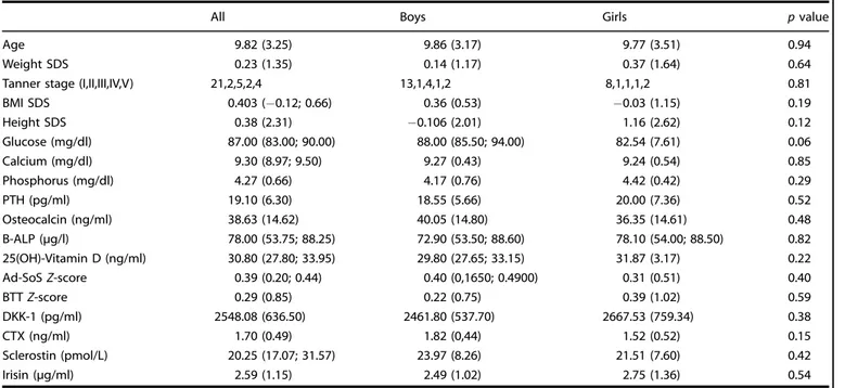
Table1shows the participant characteristics. Parametric variables are reported as mean and standard deviation, and non-parametric variables as median and interquartile range (IQR). No differences in gender distribution were observed between the groups. All children performed an average of 2 h per week of school sports. None of the children practiced sport at a competitive level.
Table 1. Demographic, anthropometric, and metabolic parameters
All Boys Girls pvalue
Age 9.82 (3.25) 9.86 (3.17) 9.77 (3.51) 0.94
Weight SDS 0.23 (1.35) 0.14 (1.17) 0.37 (1.64) 0.64
Tanner stage (I,II,III,IV,V) 21,2,5,2,4 13,1,4,1,2 8,1,1,1,2 0.81
BMI SDS 0.403 (−0.12; 0.66) 0.36 (0.53) −0.03 (1.15) 0.19 Height SDS 0.38 (2.31) −0.106 (2.01) 1.16 (2.62) 0.12 Glucose (mg/dl) 87.00 (83.00; 90.00) 88.00 (85.50; 94.00) 82.54 (7.61) 0.06 Calcium (mg/dl) 9.30 (8.97; 9.50) 9.27 (0.43) 9.24 (0.54) 0.85 Phosphorus (mg/dl) 4.27 (0.66) 4.17 (0.76) 4.42 (0.42) 0.29 PTH (pg/ml) 19.10 (6.30) 18.55 (5.66) 20.00 (7.36) 0.52 Osteocalcin (ng/ml) 38.63 (14.62) 40.05 (14.80) 36.35 (14.61) 0.48 B-ALP (µg/l) 78.00 (53.75; 88.25) 72.90 (53.50; 88.60) 78.10 (54.00; 88.50) 0.82 25(OH)-Vitamin D (ng/ml) 30.80 (27.80; 33.95) 29.80 (27.65; 33.15) 31.87 (3.17) 0.22 Ad-SoS Z-score 0.39 (0.20; 0.44) 0.40 (0,1650; 0.4900) 0.31 (0.51) 0.40 BTT Z-score 0.29 (0.85) 0.22 (0.75) 0.39 (1.02) 0.59 DKK-1 (pg/ml) 2548.08 (636.50) 2461.80 (537.70) 2667.53 (759.34) 0.38 CTX (ng/ml) 1.70 (0.49) 1.82 (0,44) 1.52 (0.52) 0.15 Sclerostin (pmol/L) 20.25 (17.07; 31.57) 23.97 (8.26) 21.51 (7.60) 0.42 Irisin (µg/ml) 2.59 (1.15) 2.49 (1.02) 2.75 (1.36) 0.54
BMIbody mass index, SDS standard deviation score, PTH parathyroid hormone, B-ALP bone alkaline phosphatase, Ad-SoS amplitude-dependent speed of sound
Z-score; BTT bone transmission time Z-score; DKK-1 Dickkopf-related protein 1, CTX C-terminal telopeptides of type I collagen
485
1234567
Therefore, the intensity of physical activity was considered comparable in the children and no assessment of the correlation of physical activity with Irisin and bone status was made.
Mean Irisin levels in boys did not differ significantly (p = 0.54) compared to girls (boys, Irisin levels, 2.49μg/ml; girls, Irisin levels, 2.75μg/ml). No differences were observed in Irisin levels between pre- and post-pubertal children (pre-pubertal, Irisin levels, 2.72μg/ml; post-pubertal, Irisin levels, 2.36μg/ml; p = 0.37), nor analyzed by gender.
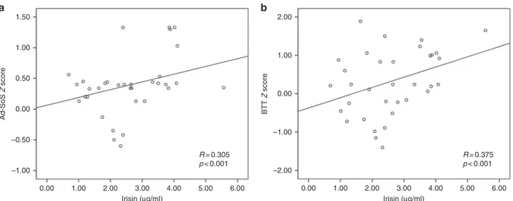
In all children, Irisin levels were positively correlated with the Ad-SoS Z-score (r= 0.305; p < 0.001) (Fig.1a) and BTT Z-score (r= 0.375; p < 0.001) (Fig. 1b), after adjustment for age. Further-more, Irisin levels were positively correlated with osteocalcin (r= 0.370, p < 0.001) (Fig.2a) and negatively with DKK-1 (r= 0.274, p < 0.001) (Fig.2b). Although to a lesser extent, Irisin levels were negatively correlated with glucose (r= −0.139; p = 0.011), and positively with the main standardized bone resorption marker CTX (r= 0.142; p = 0.03) (Fig. 2c). We also found a positive correlation between Irisin and sclerostin levels (r= 0.20; p= 0.001), but only in males, whereas there was a tendency toward an opposite correlation in females (Fig.2d).
To investigate major determinants of bone mineral status, two multiple linear regression analyses had been performed. In model 1, the dependent variable was the bone mineral status measured as the BTT Z-score, and in model 2, the dependent variable was the bone mineral status measured as the Ad-SoS Z-score. In model 1, Irisin had a greater positive influence (β value) on bone mineral status measured as the BTT Z-score than other independent variables, such as B-ALP and PTH (Fig.3a). In model 2, Irisin had a similar influence on bone mineral status, measured as the Ad-SoS Z-score, to other independent variables, such as osteocalcin and B-ALP (Fig.3b).
DISCUSSION
This study highlights for the first time a positive correlation between Irisin serum levels and bone mineral status in a population sample of children. The linkage between Irisin and bone tissue has emerged in 2014 when we showed that Irisin produced by skeletal muscle, excised from mice after exercise, increased osteoblast differentiation in vitro.19Thisfinding received increasing attention in the following years and stimulated the scientific community to investigate involvement of Irisin on bone metabolism in humans. Positive correlation between Irisin and
BMD were found in young athletes4 and in soccer players.5 Moreover, Irisin serum levels were negatively correlated with sclerostin5 and vertebral fragility fractures.6,7 All these studies were performed in adults or young adults. Only one study showed, by a linear regression analysis, that circulating Irisin was one of the determinants of BMD in a population sample of children aged 6–8 years.20
Much experimental evidence has suggested that bone mass in childhood is a predictor of pediatric fractures and that the amount of bone mass achieved by skeletal maturity is one of the most important contributors to peak bone mass, which in turn is the major determinant of osteoporotic fractures in the elderly.21 Here we showed that Irisin levels are positively correlated with bone mineral status in children,finding no differences accordingly to the pubertal stage, indicating that this myokine forms the bridge between healthy muscle and bone also during childhood. Notably, we found that Irisin was positively correlated with circulating osteocalcin, the osteoblast-derived hormone recently shown to enhance energy metabolism, skeletal muscle function, and brain development.22We and other authors have previously shown that a long-term treatment with r-Irisin increased osteocalcin messenger RNA expression in primary murine and rat osteoblasts.2,23To our knowledge, the present study is thefirst to report a positive correlation between Irisin and osteocalcin also in humans.
We also showed that Irisin was negatively correlated with DKK-1, one of the bone anabolic WNT pathway inhibitors,16 confirming the positive association between Irisin and bone status in these children. This result suggests that encouraging children to practice physical activity, thus enhancing Irisin production by skeletal muscle, might be one of the best non-pharmacologic strategies to preserve bone health. Moreover, this concept could also be applied to pediatric pathologic conditions, such as T1DM in children and adolescents, since we have previously shown that low BMD is associated with high circulating levels of DKK-1 in these pediatric patients.16
A previous study reported that serum levels of Irisin were inversely correlated with sclerostin in adults.6In this population of children, instead we found a positive correlation between Irisin and sclerostin. However, subdividing the children by gender, this association was observed only in males, with a tendency to an opposite correlation in females. Interestingly, a previous study showed that, in children, sclerostin was positively associated with bone age, reached a peak mean value, and then declined in late
1.50 a b 2.00 1.00 0.00 –1.00 –2.00 1.00 0.50 0.00 –0.50 Ad-SoS Z score BTT Z score –1.00 0.00 1.00 2.00 3.00 Irisin (µg/ml) Irisin (µg/ml) 4.00 5.00 R = 0.305 p < 0.001 R = 0.375 p < 0.001 6.00 0.00 1.00 2.00 3.00 4.00 5.00 6.00
Fig. 1 Irisin serum levels in children are positively correlated with bone mineral status evaluated by quantitative ultrasound (QUS) measurements in the peripheral skeleton by computing the amplitude-dependent speed of sound (Ad-SoS) (a) and the bone transmission time (BTT) (b)
puberty in both girls and boys.24 They also found that serum sclerostin levels were associated with cortical porosity, suggesting that changes in sclerostin production during growth may play a role in defining cortical structure.24 Albeit speculative, thesefindings could explain why we found a positive correlation between Irisin and sclerostin that, although opposite to that with DKK-1, it might be related to the achievement of peak bone mass, rather than to a negative effect on bone mass per se.
Our results also showed that Irisin is the strongest positive determinant of the BTT Z-score, more than B-ALP and PTH, which are the bone turnover markers most commonly used to evaluate
bone metabolism.25–28Although this study has several limitations, such as the small sample size, the use of a single group of subjects, the cross-sectional design that does not provide information about cause-and-effect relationships, and the use of QUS measurements, the results presented herein indicate a positive link between Irisin serum levels and bone quality in children. Regarding bone quality measurements in this population of children, QUS had several advantages over the more commonly used dual energy X-ray absorptiometry (DXA) scan, mainly because it is radiation-free. Although QUS feasibility has been ascertained in numerous studies, evidence of its validity has not
Model 1. Determinants of bone mineral status (BTT Z SCORE)
a b PTH (pg/ml) Osteocalcin (ng/ml) DKK-1 (pg/ml) CTX (ng/ml) Sclerostin (pmol/L) B-ALP (µg/l) Irisin (µg/ml) PTH (pg/ml) Osteocalcin (ng/ml) DKK-1 (pg/ml) CTX (ng/ml) Sclerostin (pmol/L) B-ALP (µg/l) Irisin (µg/ml) 0.216 –0.040 –0.369 0.127 0.009 0.220 0.335 <0.001 Beta p value 0.595 <0.001 0.111 0.924 <0.001 <0.001 0.071 0.278 –0.227 –0.090 0.010 0.243 0.230 0.250 Beta p value <0.001 <0.001 0.275 0.911 <0.001 <0.001 Model 2. Determinants of bone mineral status (Ad-SoS Z SCORE)
Fig. 3 Irisin has higher positive influence on bone mineral status measured as bone transmission time (BTT) Z-score than other independent variables, such as B-ALP and PTH. Multiple linear regression analysis was used to investigate the determinants of bone mineral status. In model 1, the dependent variable was the bone mineral status measured as BTT Z-score (a). In model 2, the dependent variable was the bone mineral status measured as Ad-SoS Z-score (b)
7000 a b c d 400,000 350,000 300,000 250,000 200,000 150,000 100,000 6000 5000 Osteocalcin (ng/ml) DKK-1 (pg/ml) 4000 3000 2000 1000 0.00 Glucose (mg/dl) CTX (ng/ml) B-ALP (µg/l) Sclerostin (pmol/L) –0.139 Sclerostin (pmol/L) 0.466 0.001 Male Female –0.063 0.516 R p R p 0.142 –0.098 0.20 0.011 R p value 0.03 n.s. 0.001 1.00 2.00 3.00 Irisin (µg/ml) 4.00 R = 0.370 p < 0.001 R = –0.274 p < 0.001 5.00 6.00 0.00 1.00 2.00 3.00 Irisin (µg/ml) 4.00 5.00 6.00
Fig. 2 Irisin serum levels in children are positively correlated with the “bone-building” protein osteocalcin (a) and negatively with Dickkopf-related protein 1 (DKK-1) (b), inhibitor of the bone anabolic WNT pathway. Irisin serum levels are negatively corDickkopf-related with glucose, and positively with C-terminal telopeptides of type I collagen (CTX), and sclerostin levels (c). Gender difference was observed in the correlation between Irisin and sclerostin (d)
been established for all kinds of populations, particularly because QUS-measured bone density might be different between the dominant and the non-dominant foot.29However, although DXA remains the gold standard for the diagnosis of osteoporosis, many studies have shown the effectiveness of QUS in screening and the early diagnosis of osteoporosis.30,31
Overall, these findings suggest that Irisin might be considered in future as one of the bone formation markers during childhood.
ACKNOWLEDGMENTS
This work was supported by Ministero dell’Istruzione, dell’Università e della Ricerca, PRIN 2015JSWLTN_003 (Progetto di Ricerca d’Interesse Nazionale, Grant 2015).
AUTHOR CONTRIBUTIONS
G.C., M.F.F., L.S., G.B., and M.G. designed research; L.S., P.P., L.L., S.B., G.S., L.P., and G.D. performed research; G.C., M.F.F., L.S., G.B., S.C., and M.G. analyzed, interpreted, and discussed the data; and G.C., M.F.F., and M.G. wrote the paper. All authors approved thefinal version of the manuscript to be published.
ADDITIONAL INFORMATION
Competing interests: The authors declare no competing interests.
Publisher’s note: Springer Nature remains neutral with regard to jurisdictional claims in published maps and institutional affiliations.
REFERENCES
1. Boström, P. et al. A PGC1-α-dependent myokine that drives brown-fat-like development of white fat and thermogenesis. Nature 481, 463–468 (2012).
2. Colaianni, G. et al. The myokine irisin increases cortical bone mass. Proc. Natl. Acad. Sci. USA 112, 12157–12162 (2015).
3. Colaianni, G. et al. Irisin prevents and restores bone loss and muscle atrophy in hind-limb suspended mice. Sci. Rep. 7, 2811 (2017).
4. Singhal, V. et al. Irisin levels are lower in young amenorrheic athletes compared with eumenorrheic athletes and non-athletes and are associated with bone density and strength estimates. PLoS ONE 9, e100218 (2014).
5. Colaianni, G. et al. Irisin levels correlate with bone mineral density in soccer players. J. Biol. Regul. Homeost. Agents 31(Suppl. 1), 21–28 (2017).
6. Klangjareonchai, T. et al. Circulating sclerostin and irisin are related and interact with gender to influence adiposity in adults with prediabetes. Int. J. Endocrinol. 2014, 261545 (2014).
7. Palermo, A. et al. Irisin is associated with osteoporotic fractures independently of bone mineral density, body composition or daily physical activity. Clin. Endocrinol. (Oxf.) 82, 615–619 (2015).
8. Anastasilakis, A. D. et al. Circulating irisin is associated with osteoporotic fractures in postmenopausal women with low bone mass but is not affected by either teriparatide or denosumab treatment for 3 months. Osteoporos. Int. 25, 1633–1642 (2014).
9. Faienza, M. F. et al. High irisin levels are associated with better glycemic control and bone health in children with type 1 diabetes. Diabetes Res. Clin. Pract. 141, 10–17 (2018).
10. Loomba-Albrecht, L. A. & Styne, D. M. Effect of puberty on body composition. Curr. Opin. Endocrinol. Diabetes Obes. 16, 10–15 (2009).
11. Sopher, A. B., Fennoy, I. & Oberfield, S. E. An update on childhood bone health: mineral accrual, assessment and treatment. Curr. Opin. Endocrinol. Diabetes Obes. 22, 35–40 (2015).
12. Golden, N. H. & Abrams, S. A. Optimizing bone health in children and adolescents. Pediatrics 134, e1229–e1243 (2014).
13. Mannocci A. et al. International Physical Activity Questionnaire for Adolescents (IPAQ A): reliability of an Italian version. Miner. Pediatr.https://doi.org/10.23736/
S0026-4946.16.04727-7(2018).
14. Ogden, C. L., Flegal, K. M., Carroll, M. D. & Johnson, C. L. Prevalence and trends in overweight among US children and adolescents, 1999–2000. JAMA 288, 1728–1732 (2002).
15. Tanner, J. M. & Whitehouse, R. H. Clinical longitudinal standards for height, weight, height velocity, weight velocity, and stages of puberty. Arch. Dis. Child 51, 170–179 (1976).
16. Faienza, M. F. et al. High sclerostin and Dickkopf-1 (DKK-1) serum levels in chil-dren and adolescents with type 1 diabetes mellitus. J. Clin. Endocrinol. Metab. 102, 1174–1181 (2017).
17. Baroncelli, G. I. et al., Phalangeal Quantitative Ultrasound Group. Cross-sectional reference data for phalangeal quantitative ultrasound from early childhood to young-adulthood according to gender, age, skeletal growth, and pubertal development. Bone 39, 159–173 (2006).
18. Krieg, M. A. et al. Quantitative ultrasound in the management of osteoporosis: the 2007 ISCD Official Positions. J. Clin. Densitom. 11, 163–187 (2008). 19. Colaianni, G. et al. Irisin enhances osteoblast differentiation in vitro. Int. J.
Endocrinol. 2014, 902186 (2014).
20. Soininen, S. et al. Body fat mass, lean body mass and associated biomarkers as determinants of bone mineral density in children 6-8years of age—The Physical Activity and Nutrition in Children (PANIC) study. Bone 108, 106–114 (2018). 21. Wren, T. A. et al. Longitudinal tracking of dual-energy X-ray absorptiometry bone
measures over 6 years in children and adolescents: persistence of low bone mass to maturity. J. Pediatr. 164, 1280–1285.e2 (2014).
22. Karsenty, G. Update on the biology of osteocalcin. Endocr. Pract. 23, 1270–1274 (2017).
23. Qiao, X. et al. Irisin promotes osteoblast proliferation and differentiation via activating the MAP kinase signaling pathways. Sci. Rep. 6, 18732 (2016). 24. Kirmani, S. et al. Sclerostin levels during growth in children. Osteoporos. Int. 23,
1123–1130 (2012).
25. Epelak, I. &Čvorišćec, D. Biochemical markers of bone remodeling. Biochem. Med. 19, 17–35 (2009).
26. Ventura, A. et al. Glucocorticoid-induced osteoporosis in children with 21-hydroxylase deficiency. Biomed. Res. Int. 2013, 250462 (2013).
27. Brunetti, G. et al. Impaired bone remodeling in children with osteogenesis imperfecta treated and untreated with bisphosphonates: the role of DKK1, RANKL, and TNF-α. Osteoporos. Int. 27, 2355–2365 (2016).
28. Brunetti, G. et al. Genotype–phenotype correlation in juvenile Paget disease: role of molecular alterations of the TNFRSF11B gene. Endocrine 42, 266–271 (2012). 29. Mergler, S., Lobker, B., Evenhuis, H. & Penning, C. Feasibility of quantitative
ultrasound measurement of the heel bone in people with intellectual disabilities. Res. Dev. Disabil. 31, 1283–1290 (2010).
30. Delvecchio, M. et al. Evaluation of impact of steroid replacement treatment on bone health in children with 21-hydroxylase deficiency. Endocrine 48, 995–1000 (2015).
31. Aceto, G. et al. Bone health in children and adolescents with steroid-sensitive nephrotic syndrome assessed by DXA and QUS. Pediatr. Nephrol. 29, 2147–2155 (2014).


