Lab Resource: Stem Cell Line
Generation and characterization of the human iPSC line IDISi001-A
isolated from blood cells of a CADASIL patient carrying a
NOTCH3 mutation
Héctor Fernández-Susavila
a, Cristina Mora
b, Marta Aramburu-Núñez
a, Rita Quintas-Rey
c, Susana Arias
a,
Manuel Collado
d, Esteban López-Arias
a, Tomás Sobrino
a, José Castillo
a, Patrizia Dell'Era
b, Francisco Campos
a,⁎
a
Clinical Neurosciences Research Laboratory, Department of Neurology, Health Research Institute of Santiago de Compostela (IDIS), Hospital Clínico Universitario, Santiago de Compostela, Spain
b
Cellular Fate Reprogramming Unit, Department of Molecular and Translational Medicine, University of Brescia, 25123 Brescia, Italy
c
Galician Public Foundation of Genomic Medicine, Genomics Medicine Group, University of Santiago de Compostela, USC, IDIS, Santiago de Compostela, Spain
dHealth Research Institute of Santiago de Compostela (IDIS), Hospital Clínico Universitario, Santiago de Compostela, Spain
a b s t r a c t
a r t i c l e i n f o
Article history:
Received 30 October 2017
Received in revised form 10 January 2018 Accepted 18 January 2018
Available online 31 January 2018
Cerebral autosomal dominant arteriopathy with subcortical infarcts and leukoencephalopathy (CADASIL) is the most common form of hereditary stroke disorder. It is caused by mutations in NOTCH3 that lead to progressive degeneration of the smooth muscle cells in blood vessels. There is currently no treatment for this disorder. We reprogrammed to pluripotency blood mononuclear cells isolated from a patient carrying a NOTCH3 mutation by using a commercially available non-integrating system. The success in the generation of this iPSC line (IDISi001-A) suggests that the NOTCH3 mutation did not limit cell reprogramming and offers an unprecedented opportunity for studying and modeling CADASIL pathology.
© 2018 Published by Elsevier B.V. This is an open access article under the CC BY-NC-ND license (http:// creativecommons.org/licenses/by-nc-nd/4.0/).
Resource table.
Unique stem cell line identifier
IDISi001-A Alternative name(s) of
stem cell line
N/A
Institution Health Research Institute of Santiago de Compostela (IDIS)
Contact information of distributor
Héctor Fernández Susavila; Francisco Campos Pérez
[email protected] [email protected]
Type of cell line iPSC
Origin Human
Additional origin info Age: 67 Sex: male
Ethnicity if known: Caucasian
Cell Source Blood
Clonality Clonal
Method of reprogramming
CytoTune™-iPS 2.0 Sendai Reprogramming Kit (Thermo Fisher Scientific). The episomal reprogramming vectors include the four Yamanaka factors OCT4, SOX2, KLF4, and c-Myc
Genetic Modification No Type of Modification N/A
Associated disease Cerebral autosomal dominant arteriopathy with subcortical infarcts and leukoencephalopathy (CADASIL)
Gene/locus NOTCH3 [c.3724CNT(p.Arg1242Cys)], germinal mutation/19p13.12
Method of modification Not applicable Name of transgene or resistance Not applicable Inducible/constitutive system Not applicable Date archived/stock date N/A Cell line repository/bank N/A
Ethical approval Written informed consent was obtained from the patient. The study was approved by the Scientific Ethics Committee for the Region of Santiago-Lugo (protocol number 2016/450) and the Commission on Guarantees for the Donation and Use of Human Cells and Tissues of Instituto de Salud Carlos III (protocol number 428-346-1)
Resource utility
To date, there is no effective treatment for cerebral autosomal dom-inant arteriopathy with subcortical infarcts and leukoencephalopathy (CADASIL). The research into CADASIL mechanisms, which has been limited mainly to the experiments in Notch3 mutant animal models,
⁎ Corresponding author at: Clinical Neurosciences Research Laboratory, Hospital Clínico Universitario, Travesa da Choupana s/n, 15706 Santiago de Compostela, Spain.
E-mail address:[email protected](F. Campos).
https://doi.org/10.1016/j.scr.2018.01.023
1873-5061/© 2018 Published by Elsevier B.V. This is an open access article under the CC BY-NC-ND license (http://creativecommons.org/licenses/by-nc-nd/4.0/).
Contents lists available atScienceDirect
Stem Cell Research
has not generated successful results for the clinical practice so far. We believe that the generation of human iPSC-derived muscle cells from CADASIL patients will facilitate gene-editing approaches for develop-mental biology studies as well as for disease modeling and drug screening.
Resource details
CADASIL is defined as cerebral autosomal dominant arteriopathy produced by the mutation of the NOTCH3 gene (Joutel et al., 2000). The main neurological symptoms of CADASIL include migraine with aura, psychiatric problems, ischemic episodes, and cognitive deficits. This disease causes degeneration of the smooth muscle due to the depo-sition of granular osmiophilic material within the basal lamina, progres-sive loss of smooth muscle cells, andfibrosis of the media layer of micro-arteries (Chabriat et al., 2009). Currently, the experimental modeling of CADASIL is restricted to the use of Notch3 knockout mice, which, how-ever, present important limitations in reproducing the phenotype of this disease (Ayata, 2010).
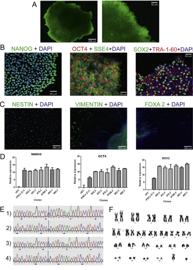
In this study, we report forfirst time the generation of a human iPSC line from a patient with CADASIL carrying a NOTCH3 ArgN Cys mutation at codon 1242 of exon 23. Human peripheral blood mononuclear cells (PBMCs) from this patient were reprogrammed into iPSCs (IDISi001-A) using the non-integrative CytoTune iPS 2.0 Reprograming System (Thermo Fisher Scientific) to deliver Yamanaka's factors OCT4, SOX2, KLF4, and c-Myc. iPSCs colony formation was observed daily. Colo-nies had characteristic round-shaped morphology with refracting borders and contained small round-shaped cells with a large nu-cleus/cytoplasm ratio. After 3 months, thefirst colonies were frozen. During the reprogramming process, the alkaline phosphatase test was used to select the potential pluripotent colonies (Fig. 1A). Once the cell line was established, the following standard tests were car-ried out: A) PCR analysis for the absence of mycoplasma (Supple-mental Fig. 1); B) PCR analysis to confirm the loss of the Sendai virus (SeV) used for the reprogramming (Supplemental Figs. 2 and 3); C) short tandem repeat (STR) analysis used to demonstrate that the generated iPSCs genetically matched donor's cells (Supplemen-tal Fig. 4); and D) PCR analysis of the NOTCH3 gene to confirm the presence of the mutation in the iPSCs (Supplemental Fig. 5). Subse-quently, the expression of the membrane and nuclear pluripotency markers (Tra-1-60, SSEA4, SOX-2, OCT-4, and NANOG, using the an-tibodies showed inTable 1) was evaluated by immunofluorescence analysis (Fig. 1B). Cells from the new iPSC line were spontaneously differentiated in vitro into the three germ layers by the induction of embryoid body (EB) formation (Fig. 1C). Expression levels of genes encoding transcription factors essential to maintain pluripo-tent embryonic stem cell phenotype (SOX2, OCT4, and NANOG) were evaluated by qPCR in different clones (#42.2, #53.3, #76.2, #78B.2, #83.3, #80.3) derived from the same cell line. These results were compared to those obtained with a human foreskinfibroblast (HFF) cell line, used as SeV-uninfected cell control, and to those in al-ready characterized iPSC positive control cells (iPSCs C+) (Fig. 1D). In addition, the presence of the mutation previously diagnosed in the patient in IDISi001-A cells was confirmed by forward and reverse sequencing of the corresponding NOTCH3 fragment (Fig. 1E, panels 1 and 2, respectively) and compared to the results of similar sequenc-ing of the same fragment from a healthy subject without NOTCH3 mutation (Fig. 1E; panels 3 and 4, respectively). Finally, the karyo-type of the new reprogrammed cell line showed a normal chromo-somal profile, with no abnormalities (Fig. 1F). In conclusion, we believe that the generation of a human iPSC line from a CADASIL pa-tient will facilitate studies of developmental biology and stimulate disease modeling for drug screening and therapeutic gene-editing approaches for this important cerebral arteriopathy (see Graphical abstract) (Table 2).
Materials and methods
Reprogramming of CADASIL-PBMCs
A peripheral blood sample was taken from a patient diagnosed with CADASIL, whose mutation was located at codon 1243 of exon 23 of NOTCH3 (genotype: Arg1242Cys/normal). This mutation had not been reported before. The mutation was analyzed by sequencing the entire coding region of the gene (new generation sequencing technique, SOLID 5500XL, Thermo Fisher Scientific).
To obtain PBMCs, the blood sample was mixed with PBS + 2% FBS and centrifuged in a SepMate-50 tube (StemCell Technologies) with Lymphoprep (StemCell Technologies) in a density gradient.
Before reprogramming, PBMCs were cultured for 3 days using Stempro-34 SFM (Gibco) with cytokines (StemCell Technologies). Reprogramming was performed using a CytoTune-iPS 2.0 Sendai Reprogramming Kit (Thermo Fisher Scientific). Three hundred thou-sand cells were infected at the following multiplicity of infection (MOI) values: KOS = 5; c-MYC = 5; KLF4 = 3.
Cell culture
Cells were cultured in mTeSR1 medium (StemCell Technologies). On thefirst and second passage, cells were maintained in the wells of a 12-well plate. On the third passage, they were transferred into 12-wells of a 6-well plate, and from passage four and thereafter, they were cultured in 35-mm Petri dishes. All wells and dishes were coated with Matrigel™ matrix (Corning). For thefirst and second passages, colonies were se-lected and picked with the help of an Origio Stripper (Origio) with Strip-per tips (Origio) of 175μm in diameter. Later passages were performed mechanically, using cell lifters.
PCR and qPCR
Total RNA from undifferentiated IDISi001-A cell line, human fibro-blasts (considered as negative control), and a previously established iPSC line was isolated using a Quick-RNA™ MiniPrep kit (Zymo Re-search) and quantified with Nanodrop (Thermo Fisher Scientific). For RT-PCR, 1μg of cDNA was generated using a iScriptTM cDNA Synthesis Kit (BIORAD) following the manufacturer's instructions. PCR to analyze the presence/absence of SeV was performed using a Ready Mix REDTaq PCR Reaction Mix (Sigma-Aldrich). For qPCR, iTaq Universal SYBR Green Supermix (BIORAD) was used. In order to check for the expression of pluripotency factors, qPCR for NANOG, OCT4, and SOX2 was performed.
Table 1shows all primer sequences used for PCR and qPCR. All primers (they came lyophilized but were diluted to afinal concentration of 100μM with nuclease-free H2O) were designed in-house and ordered
from Sigma-Aldrich. Alkaline phosphatase
For the alkaline phosphatase (AP) test, an AP Live Stain Kit (Thermo Fisher Scientific) was used. This test was performed always after 3–5 days of cell culture based on plate confluence and according to the manufacturer's instructions. Colonies were not affected by the pro-cess and kept growing normally after the AP test.
Immunocytochemistry
Immunocytochemistry tests for the pluripotency nuclear markers SSEA4 and TRA1–60 and for the pluripotency membrane markers SOX2 and OCT4 were performed using a Pluripotent Stem Cell 4-Marker Immuno Kit (Thermo Fisher Scientific) according to the manu-facturer's instructions.
To analyze NANOG (Thermo Fisher Scientific) expression, a standard immunocytochemistry protocol from our lab was
employed. Nuclei were stained with 4 ′,6-diamidino-2-phenylindole (DAPI).
Karyotyping
Cells were blocked in their metaphase by incubating them with Colcemid (Gibco) for 3 h. Then, the cells were detached by adding
trypsin-EDTA for 8–10 min and washed 2–3 times with DMEM +5% PBS. After that, they were centrifuged and exposed to a hypotonic 0.075 M KCL solution for 10 min at 37 °C. Cells were thenfixed by the triple exposure to methanol/glacial acetic acid mixture (3:1). To check the karyotypes, Giemsa staining was performed. A total of 29 meta-phases were analyzed, all of which were normal without any kind of chromosomal anomalies.
Analysis of the mutation
The NOTCH3 fragment containing the mutation was amplified by PCR. The samples were sequenced by New Generation Sequencing using a SOLID 5500XL genetic analyzer.
In vitro embryoid body formation and differentiation
Once a Petri dish reached approximately 85% confluence, colonies were washed carefully with DMEM-F12 and after that, Embryoid Body Medium with 20% FBS (EBM20%) was added. This medium contained DMEM-F12 with Glutamax, 20% FBS, non-essential aminoacids 1× (Gibco), P/S 1×, and 50 mMβ-mercaptoethanol (Gibco).
Colonies were cut making a grid pattern with the aid of a sterile 200-μL pipette and then, they were detached from the plate either mechanically or enzymatically (Dispase solution, StemCell Technologies). Once detached, clusters were transferred to a well of an ultra-low 6-well attachment plate (Corning) with EBM20% plus 10μM ROCK inhibitor (StemCell Techonologies). EBs already formed at day 1. EBs were maintained for 7 days with medium replacement every other day. On day 7, EBs were transferred into a chamber slide (Sarstedt) previously coated with Matrigel™, 6–7 EBs per well, to allow their attachment and differentiation. EBs inside the chamber slides were maintained for 14 days in EBM20% (plus ROCK inhibitor just thefirst plating day), before performing immuno-cytochemistry experiments.
For the characterization of the three germ layers, the endoderm was identified using an anti-FOXA2 antibody (Thermofisher; 1:100 dilu-tion), the mesoderm was identified using an anti-vimentin antibody (Bioss Antibodies; 1:100 dilution), whereas the ectoderm was identified using an anti-nestin antibody (Abcam; 1:200 dilution).
Acknowledgements
This study has been partially supported by grants from Instituto de Salud Carlos III (PI17/0054), Spanish Research Network on
Table 1
Characterization and validation.
Classification Test Result Data
Morphology Photography Normal Not shown but available from the authors upon
request Phenotype Immunocytochemistry OCT4, NANOG, SOX2, SSEA4, TRA-1-60 Fig. 1, panel B
RT-qPCR Relative gene expression: SOX2, OCT4, NANOG
Fig. 1, panel D
Genotype Karyotype (G-Banding) 46XY, Resolution 400 Fig. 1, panel F
Identity Microsatellite PCR (mPCR) OR Short tandem repeat analysis Fig. 4, supplementaryfile
short tandem repeat analysis Not performed N/A
Mutation analysis (IF APPLICABLE)
Sequencing Heterozygous Diagnostic available from the authors upon
request Southern blot OR whole genome
sequencing
Substitution Fig. 1, panel E
Microbiology and virology Mycoplasma Negative Fig. 1, supplementary
Differentiation potential Embryoid body formation FOXA-2, vimentin, nestin Fig. 1, panel C Donor screening (OPTIONAL) HIV 1 + 2 Hepatitis B, Hepatitis C Not performed Not available Genotype additional info
(OPTIONAL)
Blood group genotyping Not performed Not available
HLA tissue typing Not performed Not available
Table 2 Reagents details.
Antibodies used for immunocytochemistry/flow-cytometry
Antibody Dilution Company Cat # and RRID
Pluripotency markers Rabbit anti-OCT4 1:100 Thermo Fisher, A24867
Mouse anti-SSEA4 1:100 Thermo Fisher, A24866
Rat anti-SOX2 1:100 Thermo Fisher, A24759
Mouse anti-TRA-1-60 1:100 Thermo Fisher,24868
Rabbit anti-NANOG 1:200 Thermo Fisher, PA-1-097X
Differentiation markers Rabbit anti-FOXA2 1:100 Thermo Fisher, 720061
Rabbit anti-vimentin 1:100 BIOSS Antibodies, bs-0756R
Rabbit anti-nestin 1:100 Abcam, ab93157
Secondary antibodies Alexa Fluor 488 Goat anti-mouse IgG3 1:250 Thermo Fisher, A24877
Alexa Fluor 594 Donkey anti-rabbit 1:250 Thermo Fisher, A24870
Alexa Fluor 488 Donkey anti-rat 1:250 Thermo Fisher, A24876
Alexa Fluor 594 Goat anti-mouse IgM 1:250 Thermo Fisher,A24872
DyLight 488 anti-rabbit IgG 1:200 Vector Laboratories, DI-1488
Biotinylated anti-mouse IgG 1:200 Vector Laboratories, BA-2001
Primers
Target Forward/Reverse primer (5′-3′)
Episomal plasmids (qPCR) SeV plasmid GGATCACTAGGTGATATCGAGC/ACCAGACAAGAGTTTAAGAGATATGTATC
Pluripotency markers (qPCR) NANOG AGGAAGACAAGGTCCCGGTCAA/TCTGGAACCAGGTCTTCACCTGT
OCT4 GGGTTTTTGGGATTAAGTTCTTCA/GCCCCCACCCTTTGTGTT
SOX2 CAAAAATGGCCATGCAGGTT/AGTTGGGATCGAACAAAAGCTATT
Cerebrovascular Diseases RETICS-INVICTUS (RD12/0014), the Ministry of Economy and Competitiveness of Spain (SAF2014-56336-R), Xunta de Galicia (Consellería Educación GRC2014/027), and the European Union program FEDER. Furthermore, F. Campos (CP14/00154) and T. Sobrino (CP12/03121 & CPII17/00027) are recipients of research con-tracts from the Miguel Servet Program of Instituto de Salud Carlos III. The funders had no role in the study design, data collection and analysis, decision to publish, or preparation of the manuscript.
Appendix A. Supplementary data
Supplementary data to this article can be found online athttps://doi. org/10.1016/j.scr.2018.01.023.
References
Ayata, C., 2010.CADASIL: experimental insights from animal models. Stroke 41 (10 Suppl), S129–134.
Chabriat, H., et al., 2009.Cadasil. Lancet Neurol. 8 (7), 643–653.
Joutel, A., et al., 2000.The ectodomain of the Notch3 receptor accumulates within the cerebrovasculature of CADASIL patients. J. Clin. Invest. 105 (5), 597–605.