UNIVERSITÀ DEGLI STUDI DI SALERNO
Dipartimento di Farmacia
Dottorato di ricerca
in Scienze Farmaceutiche
Ciclo XIII NS – 2012-2015
Coordinatore: Chiar.mo Prof. Gianluca Sbardella
Analysis and evaluation of potential nutraceutical
milk and dairy products
derived from the Centrale del latte di Salerno
settore scientifico disciplinare di afferenza
: CHIM/08
Dottorando Tutore
Dott. Giacomo Pepe Chiar.mo Prof.
Pietro Campiglia
Co-tutore Chiar.mo Prof.
Index
Chapter I:
Bovine milk, monolithic columns and comprehensive HPLC ... - 1 -
1.1. Introduction ... - 2 -
1.2. Lipids ... - 3 -
1.2.1. Fatty acids ... - 3 -
1.2.1.1. Saturated fatty acids ... - 3 -
1.2.1.2 Unsaturated fatty acids ... - 3 -
1.2.1.3. Conjugated linoleic acid (CLA) ... - 4 -
1.2.1.4. Trans vaccenic acid ... - 4 -
1.2.2. Phospholipids and glycosphingolipids... - 5 -
1.3. Minerals, vitamins and antioxidants ... - 5 -
1.3.1. Calcium ... - 5 - 1.3.2. Selenium ... - 6 - 1.3.3. Iodine ... - 6 - 1.3.4. Magnesium ... - 6 - 1.3.5. Zinc ... - 7 - 1.3.6. Vitamin E ... - 7 - 1.3.7. Vitamin A ... - 7 - 1.3.8. Folate ... - 8 - 1.3.9. Riboflavin ... - 8 - 1.3.10. Vitamin B12 ... - 8 - 1.4. Proteins ... - 9 - 1.4.1. Whey Proteins ... - 9 - 1.4.1.1. 𝛼-Lactoalbumin... - 10 - 1.4.1.2. 𝛽-Lactoglobulin ... - 11 -
Index 1.4.1.4. Lactoferricin ... - 11 - 1.4.1.5. Immunoglobulins ... - 12 - 1.4.1.6. Lactoperoxidase ... - 13 - 1.4.2. Caseins ... - 13 - 1.4.2.1. 𝛼- and 𝛽-Casomorphins ... - 14 - 1.4.2.2. Caseinphosphopeptides... - 14 - 1.5. Aim of work ... - 15 -
1.6. Monolithic stationary phases ... - 15 -
1.6.1. Preparation techniques for organic monolithic columns ... - 18 -
1.6.1.1. Activation of capillary... - 19 -
1.6.1.2. Thermally and radiation polymerization ... - 21 -
1.6.2. Monomers ... - 23 -
1.6.3. Porogens ... - 25 -
1.6.4. Crosslinking monomer ... - 25 -
1.7. Comprehensive HPLC ... - 28 -
1.7.1. Optimization of the LC × LC method ... - 35 -
1.7.1.1. Optimization of the first dimension and mobile phase incompatibility ... 35
-1.7.1.2. Peak focusing at the head of the secondary column ... - 37 -
1.7.1.3. Optimization of the second dimension analysis time ... - 37 -
1.8. References ... - 39 -
Chapter II: Susceptibility to denaturation of caseins in milk samples for improving protein conformational study and their identification ... - 53 -
Abstract ... - 54 -
Keywords ... - 54 -
Abbreviations ... - 54 -
2.1. Introduction ... - 55 -
Index
2.2. Materials and methods ... - 58 -
2.2.1. Reagents and standards ... - 58 -
2.2.2. Sampling and sample preparation ... - 58 -
2.2.3. HPLC separation ... - 59 -
2.2.4. Far-UV CD ... - 60 -
2.2.5. Identification of caseins by MALDI-TOF-MS ... - 60 -
2.3. Results and discussion... - 61 -
2.3.1. HPLC separation of purified casein fractions ... - 61 -
2.3.2. Far-UV CD data of caseins ... - 63 -
2.3.3. Identification of casein fraction variants ... - 66 -
2.4. Conclusions ... - 67 -
2.5. References ... - 68 -
Chapter III: Analysis of bovine milk caseins on organic monolithic columns: An integrated capillary liquid chromatography–high resolution mass spectrometry approach for the study of time-dependent casein degradation - 72 - Abstract ... - 73 -
Keywords ... - 74 -
Abbreviations ... - 74 -
3.1. Introduction ... - 75 -
3.1.1. Aim of work ... - 77 -
3.2. Materials and methods ... - 77 -
3.2.1. Materials ... - 77 -
3.2.2. Sample preparation ... - 78 -
3.2.3. Preparation of the organic monolithic columns ... - 78 -
3.2.4. Capillary HPLC–UV: chromatographic conditions... - 79 -
3.2.4.1. Analysis of standards ... - 81 -
3.2.4.2. Analysis of casein fractions by Cap-HPLC–UV ... - 82 - 3.2.4.3. Analysis of casein fractions in high resolution
Index
(HR-Cap-HPLC–UV) ... - 82 -
3.2.5. Mass spectrometry analysis: direct infusion ... - 83 -
3.2.6. Analytical conditions in Capillary-HPLC–high-resolution-MS ... - 84 -
3.3. Results and discussion... - 85 -
3.3.1. Preparation of the organic monolithic capillary columns ... - 85 -
3.3.2. Analysis of casein standards present in bovine milk through Cap-HPLC–UV ... - 86 -
3.3.3. High Resolution Mass Spectrometry of casein standards... - 89 -
3.3.4. Analysis of casein fraction in bovine milk ... - 95 -
3.3.5. Cap-LC–HRMS analysis of week IV casein fractions ... - 100 -
3.4. Conclusions ... - 109 -
3.5. References ... - 110 -
Chapter IV: Evaluation of two sub-2 μm stationary phases, core-shell and totally porous monodisperse, in the second dimension of on-line comprehensive two dimensional liquid chromatography, a case study: separation of milk peptides after expiration date ... - 117 -
Abstract ... - 118 -
Keywords ... - 118 -
Abbreviations ... - 118 -
4.1. Introduction ... - 120 -
4.1.1. Aim of work ... - 122 -
4.2. Materials and methods ... - 123 -
4.2.1. Chemicals ... - 123 -
4.2.2. Sampling and sample preparation ... - 123 -
4.2.3. Peptides isolation ... - 124 -
4.2.4. Columns ... - 124 -
4.2.4.1. Organic monolithic column ... - 124 -
Index
4.2.5. Evaluation of column perfomances ... - 125 -
4.2.5.1. Instrumentation ... - 125 -
4.2.5.2. Chromatographic conditions: kinetic evaluation of monolithic column ... - 126 -
4.2.5.3. Efficiency evaluation of sub-2 μm columns... - 126 -
4.2.6. Analysis of soluble fractions ... - 127 -
4.2.6.1. Analysis of whey proteins by Cap-HPLC-UV ... - 127 -
4.2.6.2. Analysis of whey proteins by Cap-LC-MS ... - 128 -
4.2.7. 1D and LC × UHPLC analysis of peptides ... - 128 -
4.2.7.1. Instrumentation ... - 128 -
4.2.7.2. Chromatographic conditions: comparative high efficiency monodimensional (1D) analysis ... - 129 -
4.2.7.3. Chromatographic conditions: LC × UHPLC (set-up 1 and 2) ... - 129 -
4.2.8. Identification of peptides ... - 130 -
4.3. Results and discussion... - 131 -
4.3.1. Analysis of whey proteins present in bovine milk ... - 131 -
4.3.1.1. Kinetic and thermodynamic evaluation of monolithic column ... - 131 -
4.3.1.2. Analysis of whey proteins in Cap-LC-UV and Cap-LC-HRMS .... - 133 -
4.3.1.3. Cap-LC-UV analysis of whey proteins extracted from semi-skim milk after expiration date. ... - 138 -
4.3.2. Analysis of peptides ... - 139 -
4.3.2.1. Kinetic and thermodynamic evaluation of 2D columns ... - 139 -
4.3.2.2. Preliminary search of chromatographic condition in both dimensions ... - 143 -
4.3.2.3. Application of the LC × UHPLC approach to the separation of peptides of soluble fraction of milk ... - 143 -
4.3.2.4. Performance evaluation and comparison of set-ups #1 and #2 .... - 144 -
4.3.3. Identification of peptides by 1D high efficiency monodimensional- MS analysis ... - 153 -
Index
4.5. References ... - 157 -
Chapter V:
Figure captions
Chapter I:
Figure 1.1. Comparison between monolithic (left) and packed
stationary phases (right)... 16 -Figure 1.2. SEM-image of the porous structure of a typical
monolithic silica column (A), enlarged view of the entrance to a macropore or throughpore (B) and mesoporous structure of the
skeleton (C)... 16 -Figure 1.3. Comparison of the efficiency of particle-packed and
monolithic columns... 18 -Figure 1.4. Activation of inner wall surface of fused silica capillary.. 19 -Figure 1.5. Reaction scheme of silanating “Tentacle type”... 20 -Figure 1.6. Reaction scheme of derivatization “Grafting-on-to ... 21 -Figure 1.7. Decomposition reaction of azobisisobutyronitrile... 21 -Figure 1.8. Comparison between monolithic polimer obtained by
radiation (left) and thermally (right) polymerization...
Figure 1.9. Comparison between monolithic columns obtained by
radiation (left) and thermally (right) polymerization. 1: Uracil; 2: Benzaldehyde; 3: Nitrobenzene; 4: Ethylbenzene; 5: Butylbenzene….
22
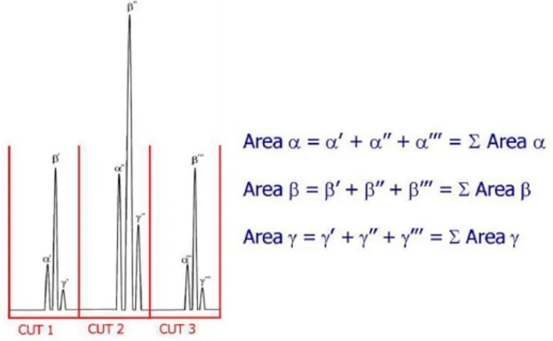
23 -Figure 1.10. Non-modulated peak α (a) and peak α modulated 5
times (b) ... 30 -Figure 1.11. Effects of modulation on 3 co-eluting peaks... 30 -Figure 1.12. Quantitative procedure in LC × LC... 31
-Figure captions
Figure 1.13. Peak intensity colour-scale for comprehensive HPLC
analyses... 32 -Figure 1.14. Data elaboration in comprehensive two-dimensional
HPLC: (A) 1D non-modulated peak; (B) modulated 2D peaks; (C) fractions of modulated peaks, analyzed in 2 dimensions; (D) bidimensional data visualization (color); (E) bidimensional data
visualization (grayscale); (F) three-dimensional visualization... 32 -Figure 1.15. 10-port two-position switching valve... 33
-Chapter II:
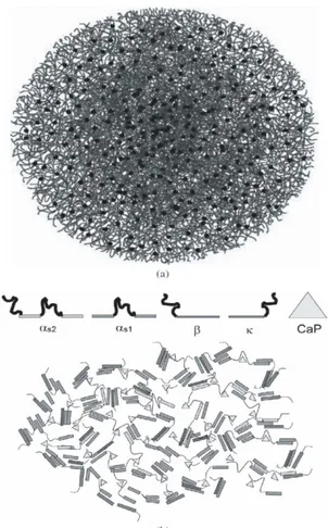
Figure 2.1. (a) The nanocluster model for casein micelles. Casein
monomers are thread-like, while the dark circles represent calcium phosphate nanoclusters. (b) The model of Horne – dual bonding model of casein micelles using casein monomers as indicated. Protein-protein interactions occur between hydrophobic regions (rectangular bars) while the protein hydrophilic regions (loops) bind
to calcium phosphate clusters (triangles)... 57 -Figure 2.2. HPLC chromatograms of casein fractions dissolved in:
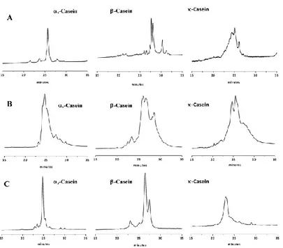
(A) water; (B) solution of 8 M urea and water/acetonitrile (70:30); (C) solution of 0.3% β-mercaptoethanol, water/acetonitrile (70:30); and (D) solution of 8M urea, 165mM Tris-HCl, 44mM sodium citrate and 0.3% β-mercaptoethanol. I, κ-casein; II, αS2-casein; III, αS1
-casein; and IV, β-casein. Peaks 1–4, κ-casein in different states of glycosylation; peaks 5 and 6: αS2- and αS1-casein, respectively; and
-Figure captions
Chapter III:
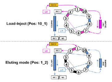
Figure 3.1. Sketch of 10-valve port used for load-trapping purposes.. 83 -Figure 3.2. Chromatograms of casein standards on (A) a 27-cm long
Protein-Cap-RP-Lauryl-γ-Monolithic column (270 mm × 0.250 mm, L. × I.D.); (B) on an ACE C4 column packed with 3 μm particles (150 mm × 0.300 mm, L. × I.D.); and (C) on a Jupiter C4 column packed with 5 μm particles (150 mm × 0.300 mm, L. × I.D.). Gradient time: 30 min. Commercially available casein standards were dissolved in water. UV detection at 214 nm. See Section 3.2.4.1
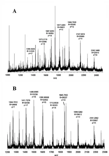
for full experimental details... 88 -Figure 3.3. Mass spectra of αS-CN (A) and β-CN (B) obtained
through direct infusion of a 4.0 μM sample solution in MeCN/iso-propanol/H2O/CF3COOH 45/45/9.9/0.1 (v/v/v/v). Instrument: LTQ
Orbitrap MS ESI (+) detector. R = 60,000, mass range: 1000–2500; maximum injection time: 100 ms; 1 microscan...
Figure 3.4. Expansion of mass spectrum of αS-CN obtained through
direct infusion of a 4.0 µM sample solution in MeCN/iso-propanol/ H2O/CF3COOH 45/45/9.9/0.1 (v/v/v/v). Instrument: LTQ Orbitrap
MS ESI (+) detector. R = 60,000, mass range: 1000-2500; maximum injection time: 100ms; 1 microscan...
Figure 3.5. Expansion of mass spectrum of β-CN obtained through
direct infusion of a 4.0 µM sample solution in MeCN/iso-propanol/ H2O/CF3COOH 45/45/9.9/0.1 (v/v/v/v). Instrument: LTQ Orbitrap
MS ESI (+) detector. R = 60,000, mass range: 1000-2500; maximum injection time: 100ms; 1 microscan...
Figure 3.6. Deconvoluted spectra of αs-CN (A) and β-CN (B)
obtained using the Xtract software. Monoisotopic masses are
90
91
-Figure captions
reported. Experimental conditions as in Figure 3.3...
Figure 3.7. Deconvoluted spectra of αS-CN (A) and β-CN (B)
obtained using the Xtract software. Averaged molecular masses are reported, determined by matching the measured isotopic pattern to a calculated isotope pattern obtained from the elemental composition of an average hypothetical amino acid. Experimental conditions as in text (section 3.2.5.)...
92
93 -Figure 3.8. Chromatographic profile of casein fraction extracted
from basal whole, semiskim and skim milk and at weeks I–IV from expiry date. Column: Protein-Cap-RP-Lauryl-γ-Monolithic (27 cm × 0.250 mm, L. × I.D.). Elution: from 20% to 40% of mobile phase B with a 60-min gradient time; flow rate: 10 μL/min. Tcol = 60 °C. UV detection at 214 nm. See Section 3.2.4.2. for full experimental details………...
Figure 3.9. High-resolution (HR-Cap-LC) chromatogram of casein
fraction of basal skim milk (A) and of casein fraction extracted at week IV from skim milk (B) using a 50-cm long Protein-Cap-RP-Lauryl-γ-Monolithic column (0.250 mm I.D.). Elution: 120-min gradient from 15% to 45% of mobile phase (B); flow rate: 7 μL/min. Tcol = 60 °C. UV detection at 214 nm. Full experimental details in Section 3.2.4.3..……...
Figure 3.10. Total ion current chromatogram (Cap-LC–HRMS
analysis) of casein fraction extracted at week IV from skim milk (B) using a 50-cm long protein-Cap-RP-lauryl-γ-monolithic column (0.250 mm I.D.). Elution: 60-min gradient from 15% to 45% of mobile phase (B); flow rate: 7 μL/min. Tcol = 60 °C. UV detection at 214 nm. LTQ Orbitrap MS ESI (+) detector. R = 60,000, mass
99
-Figure captions
range: 1000–3000 amu. Full experimental details in Section 3.2.6…..
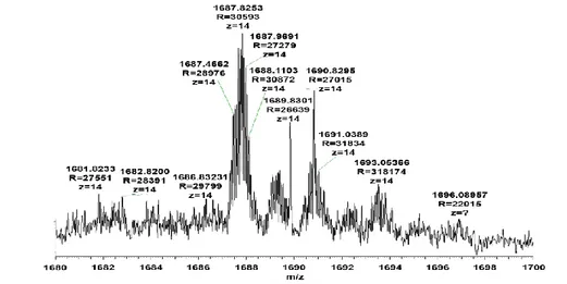
Figure 3.11. Mass spectra of peaks 1–4 in region 1 in TIC
chromatogram reported in Figure 3.10. Instrument: LTQ Orbitrap MS ESI (+) detector. R = 60,000, mass range: 1000–3000; maximum injection time: 100 ms for 1 microscan...
Figure 3.12. Mass spectrum of peak 6 of TIC chromatogram in
Figure 3.10. (see text). Instrument: LTQ Orbitrap MS ESI (+) detector. R = 60,000, mass range: 1000-3000; maximum injection time: 100 ms for 1 microscan...
Figure 3.13. Mass spectrum of peak 5 in region 2 in TIC
chromatogram reported in Figure 3.10. Instrument: LTQ Orbitrap MS ESI (+) detector. R = 60,000, mass range: 1000–3000; maximum injection time: 100 ms for 1 microscan...
101
-- 102 -
106
107 -Figure 3.14. Range of exact molecular masses of αS1-CN.
Monoisotopic exact molecular masses were obtained by
deconvolution of high resolution mass spectrum 5 in Figure 3.13. using Xtract software...
Figure 3.15. Range of exact molecular masses of αS2-CN.
Monoisotopic exact molecular masses were obtained by
deconvolution of high resolution mass spectrum 5 in Figure 3.13. using Xtract software...
Figure 3.16. Range of exact molecular masses of κ-CN.
Monoisotopic exact molecular masses were obtained by
deconvolution of high resolution mass spectrum 5 in Figure 3.13. (see text) using Xtract software……...
107
108
-Figure captions
Chapter IV:
Figure 4.1. Expansion of software window showing the 2D continuous shifted gradient...
Figure 4.2. Chromatographic characterization and efficiency test of
Protein-Cap-RP-Lauryl-γ-Monolithic column (250 × 0.250 mm L. × I.D.). (A): Φ: 3.00 μL/min, ΔPc: 817 psi; (B) Φ: 2.00 μL/min, ΔPc:
523 psi; Φ: 1.00 μL/min, ΔPc: 287 psi...
Figure 4.3. Experimental van Deemter for n-pentyl-benzene, column:
Protein-Cap-RP-Lauryl-γ-Monolithic column (250 × 0.250 mm, L. × I.D.). Mobile phase: ACN/H2O; 60/40, v/v; ƞ: 0.72 × 10⁻³ Pa s at 25
°C. Column oven: 25 °C; Detection: UV at 214 nm. Injection volume: 50 ƞL...
Figure 4.4. Chromatographic profile of whey fraction. Column:
Protein-Cap-RP-Lauryl-γ-Monolithic (250 × 0.250 mm, L. × I.D.); Pre-column: Protein-Cap-RP-Lauryl-γ-Monolithic (50 × 0.200 mm, L. × I.D.). Elution: from 10% to 35% of mobile phase B with a 65-min gradient time; flow rate: 10 μL/65-min. Tcol = 60 °C. UV detection at 214 nm. See Section 4.2.6.1. for full experimental details...
Figure 4.5. Total ion current of whey fraction using a 250 cm long
protein-Cap-RP-Lauryl-γ-Monolithic column (0.250 mm I.D.). Elution: 70-min gradient from 15% to 45% of mobile phase (B); flow rate: 10 μL/min. Tcol = 60 °C. Exactive Orbitrap MS-ESI (+) detector. R = 10,000, mass range: 500-4000 amu. Full experimental details in Section 4.2.6.2...
Figure 4.6. Mass spectrum of peaks A in TIC chromatogram of whey
protein. Monoisotopic exact molecular masses were obtained by deconvolution of high resolution mass spectrum in Figure 4.5. using
- 130 -
132
-- 133 --
134
-Figure captions
Xtract software. Averaged molecular masses are reported, determined by matching the measured isotopic pattern to a calculated isotope pattern obtained from the elemental composition
of an average hypothetical amino acid... 136 -Figure 4.7. Mass spectrum of peaks B in TIC chromatogram of whey
fraction. Deconvoluted spectra of BSA obtained using the Promass
software…... 136
-Figure 4.8. Mass spectrum of β-Lactoglobulin B. Monoisotopic exact
molecular masses were obtained by deconvolution of high resolution
mass spectrum in Figure 4.5. using Xtract software... 137 -Figure 4.9. Range of exact molecular masses of β-Lactoglobulin A.
Monoisotopic exact molecular masses were obtained by
deconvolution of high resolution mass spectrum in Figure 4.5. using Xtract software…...
Figure 4.10. Chromatographic profile of whey fraction extracted
from semi-skimed milk at weeks I-IV from expiry date. Column: Protein-Cap-RP-Lauryl-γ-Monolithic (25 cm × 0.250 mm, L. × I.D.). Elution: from 10% to 35% of mobile phase B with a 70-min gradient time; flow rate: 10 μL/min. Tcol = 60 °C. UV detection at 214 nm. See Section 4.2.6.1. for full experimental details...
137
138 -Figure 4.11. Experimental van Deemter (A) and Popple plot (B) for
ethylbenzene, columns: ( ) KinetexTM C18 50 × 3.0 mm, 1.7 μm, ( ) TitanTM C18 50 × 3.0 mm, 1.9 μm, ( ) KinetexTM 50 × 3.0 mm, 2.6 μm. Mobile phase: ACN/H2O; 60/40, v/v; (viscosity given as (ƞ):
0.59 × 10⁻³ Pa s at 35 °C). Column oven: 35 °C; Detection: UV at 214 nm, sampling rate: 80 points/s; resolution: 4.8 nm; no filter.
-Figure captions
Figure 4.12. Comparison of isocratic elution on the KinetexTM C18 50 × 3.0 mm, 1.7 μm (black line), TitanTM C18 50 × 3.0 mm, 1.7 μm (red dotted line). Column oven: 35 °C; Detection: UV at 214 nm, Sampling rate: 80 points/s; resolution: 4.8 nm; no filter. Injection volume: 0.5 μL. Sample: small molecule mixture. Results are expressed as retention factors (k) and theoretical plates per column
(N)... 142 -Figure 4.13. Comparison of a fast gradient elution on the KinetexTM
C18 50 × 3.0 mm, 1.7 μm (red dotted line), TitanTM C18 50 × 3.0 mm, 1.9 μm (black line). Instrument: Waters UPLC; Detector: PDA; Cell: 500 nL; UV at 214 nm; Tcol: 40 °C; Vinj: 6.0 μL; Mobile phase: (A) H2O/ACN 95/5 v/v + 0.1% v/v TFA; (B) ACN/H2O 95/5
v/v + 0.1% v/v TFA; Flow rate: 1.5 mL/min. Sample: peptides <
3,000 Da fraction... 142 -Figure 4.14. 2D LC × UHPLC plot of peptides < 3,000 Da fraction
after four week from expiration date. 1D column Discovery® 150 × 1.0 mm, 5 μm, flow rate 20 μL/min; column oven: 25 °C, injection volume 2 μL. 2
D column TitanTM C18 50 × 3.0 mm, 1.9 μm.; flow rate 2.5 mL/min. Modulation time 60 sec; column oven: 60
°C... - 149 - Figure 4.15. 2D LC × UHPLC plot of peptides < 3,000 Da fraction
after four week from expiration date. 1D column Discovery® 150 × 1.0 mm, 5 μm; flow rate 20 μL/min; column oven: 25 °C; injection volume 2 μL. 2
D column KinetexTM C18 50 × 3.0 mm, 1.7 μm; flow
rate 2.5 mL/min. Modulation time 60 sec; column oven: 60 °C... 149 -Figure 4.16. 2D LC × UHPLC plot of peptides < 3,000 Da fraction
Figure captions
1.0 mm, 5 μm, flow rate 20 μL/min; column oven: 25 °C, injection volume 2 μL. 2D column TitanTM C18 50 × 3.0 mm, 1.9 μm; flow rate 2.5 mL/min. 2D Modulation time 45 s; column oven: 60
°C... 150
-Figure 4.17. 2D LC × UHPLC plot of peptides < 3,000 Da fraction
after four week from expiration date. 1D column Discovery® 150 × 1.0 mm, μm; flow rate 20 μL/min; column oven: 25 °C; injection volume 2 μL. 2D column KinetexTM C18 50 × 3.0 mm, 1.7 μm; flow rate 2.5 mL/min. Modulation time 45 s; column oven: 60
°C... 150 -Figure 4.18. Three dimensional projection of 2D LC × UHPLC plot
of peptides < 3,000 Da fraction obtained using in second dimension a (A) TitanTM C18 and (B) KinetexTM C18 columns, respectively, with a modulation time of 45 s. The LC × LC data were visualized and elaborated in three dimensions using Chromsquare® ver. 1.5.01
software…... 151 -Figure 4.19. Chromatograms of high efficiency monodimensional
comparative analysis of the peptides belonging to < 3,000 Da fraction. TitanTM C18 2 × 100 × 2.1 mm, 1.9 μm, nc: 454 (A);
KinetexTM C18 100 + 50 × 2.1 mm, 1.7 μm, nc: 490 (B)... 152
-Figure 4.20. Total ion chromatograms of high efficiency
monodimensional comparative analysis of the peptides < 3,000 Da fraction. Columns top: TitanTM C18 2 × 100 × 2.1 mm, 1.9 μm;
-Table captions
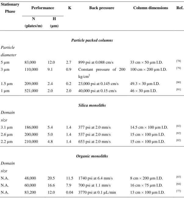
Chapter I:
Table 1.1. Representative performance data for a variety of packed and
monolithic columns... 27
-Chapter II:
Table 2.1. Precision (repeatability and reproducibility of analysis
method) of peak retention times of caseins from whole bovine milk samples of CdLdS dissolved in denaturing solution consisting of 8M urea, 165mM Tris-HCl, 44mM sodium citrate and 0.3%
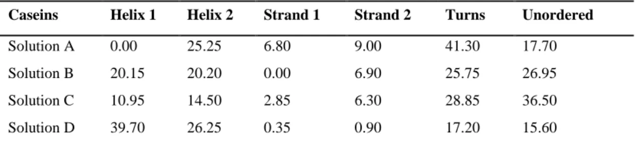
β-mercaptoethanol... 62 -Table 2.2. Relative amounts of secondary structures present in casein
different solutions... 64 -Table 2.3. MALDI-TOF-MS data of casein fraction variants... 67
-Chapter III:
Table 3.1. Exact measured monoisotopic molecular masses of peaks 1-4
reported in Figure 3.11. (see text). Monoisotopic exact molecular masses were obtained by deconvolution of high resolution mass spectrum 5 in
Figure 3.11. using Xtract software... 103 -Table 3.2. Summary of monoisotopic exact molecular masses obtained
by the deconvolution of the high resolution mass spectrum of peak 5 (see
-Table captions
Chapter IV:
Table 4.1. Peak capacity values for comparative monodimensional
analysis and for comprehensive LC × UHPLC set-ups #1 and #2, values
are corrected for both orthogonality and undersampling effect…... 145 -Table 4.2. Sequence and masses of identified milk peptides after four
-- 1 --
CHAPTER I
Chapter I: Bovine milk, monolithic columns and comprehensive HPLC
- 2 - 1.1. Introduction
Milk has been described as nature‟s most complete food. However, both the traditional and contemporary view of the role of milk has been remarkably expanded beyond the horizon of nutritional subsistence of infants. Milk is more than a source of nutrients for any neonate of mammalian species, as well as for growth of children and nourishment of adult humans. Recent studies have shown that milk furnishes a broad range of biologically active compounds protecting neonates and adults against pathogens and illnesses, such as immunoglobulins, antibacterial peptides, antimicrobial proteins, oligosaccharides and lipids, besides many other components at lower concentrations (called “minor” components, but with considerable potential benefits).
Among the many valuable constituents in milk, the high levels of calcium play an important role in the development, strength, and density of bones in children and in the prevention of osteoporosis in elderly people. Calcium is also beneficial in reducing cholesterol absorption, and in controlling body weight and blood pressure
[1]
.
Bovine milk is a source of lipids, proteins, amino acids, vitamins and minerals. It contains immunoglobulins, hormones, growth factors, cytokines, nucleotides, peptides, polyamines, enzymes and other bioactive peptides. The lipids in milk are emulsified in globules coated with membranes. The proteins are in colloidal dispersions as micelles. The casein micelles occur as colloidal complexes of protein and salts, primarily calcium. Lactose and minerals are mainly in solution. Bovine milk composition has a dynamic nature, and the composition varies with stage of lactation, age, breed, nutrition, energy balance and health status of the udder. Milk contains many different types of fatty acids. All these components make milk a nutrient rich food item [2].
Chapter I: Bovine milk, monolithic columns and comprehensive HPLC
- 3 - 1.2. Lipids
1.2.1.Fatty acids
Milk contains about 33 g total lipid (fat)/L. Triacylglycerols, which account for about 95 % of the lipid fraction, are composed of fatty acids of different length (4-24 C-atoms) and saturation. Each triacylglycerol molecule is built with a fatty acid combination giving the molecule a liquid form at body temperature. Other milk lipids are diacylglycerol (about 2% of the lipid fraction), cholesterol (less than 0.5%), phospholipids (about 1%), and free fatty acids (FFA) accounting to less than 0.5% of total milk lipids. Increased levels of FFA in milk might result in off-flavours in milk and dairy products, and the free volatile short-chain fatty acids contribute to the characteristic flavours of ripened cheese [3].
1.2.1.1. Saturated fatty acids
More than half of the milk fatty acids are saturated, accounting to about 19 g/L whole milk. The specific health effects of individual fatty acids have been extensively studied. Butyric acid (4:0) is a wellknown modulator of gene function, and may also play a role in cancer prevention. Caprylic and capric acids (8:0 and 10:0) may have antiviral activities, and caprylic acid has been reported to delay tumour growth. Lauric acid (12:0) may have antiviral and antibacterial functions, and might act as an anti caries and anti plaqueagent. Interestingly, Helicobacter
pylori can be eradicate by this fatty acid. Capric and lauric acid are, moreover
addressed for inhibition of COX-I and COX-II. Finally, stearic acid (18:0) does not seem to increase serum cholesterol concentration, and is not atherogenic [4].
1.2.1.2. Unsaturated fatty acids
Oleic acid is the single unsaturated fatty acid with the highest concentration in milk accounting to about 8 g/litre whole milk. Accordingly milk and milk products
Chapter I: Bovine milk, monolithic columns and comprehensive HPLC
- 4 -
contribute substantially to the dietary intake of oleic acid in many countries. Oleic acid is considered to be favourable for health, since diets with high amounts of this monounsaturated fatty acid reduce plasma cholesterol, LDL-cholesterol as well triacylglycerol concentrations. Moreover, replacement of saturated fatty acids with cis unsaturated fatty acids reduces risk for coronary artery disease. Several studies also indicate a cancer protective effect of oleic acid, but the data are not fully persuasive. Milk fat is rich in oleic acid (about 25%) containing also a very high ratio oleic acid/polyunsaturated fatty acids. A diet rich in milk fat therefore may be helpful to increase this ratio in the total dietary fatty acids [5].
1.2.1.3.Conjugated linoleic acid (CLA)
Bovine milk, milk products and bovine meat are the main dietary sources of the cis-9, trans-11 isomer of conjugated linoleic acid. In most cases this isomer is the most abundant CLA-isomer in bovine milk. Minor amounts of other geometrical and positional isomers of CLA also occur in milk, with different biological effects. Milk content of cis-9, trans-11 CLA vary considerably, but may constitute about 0,6% of the fat fraction. The biochemical role of CLA effects in metabolism added to its reported anti-proliferative and pro-apoptotic effect on various types of cancer cell, makes CLA potential therapeutic agent in nutritional cancer therapy [6].
1.2.1.4. Trans vaccenic acid
The main trans 18:1 isomer in milk fat is vaccenic acid, (18:1, trans-11, VA), but trans double bounds in position 4 to 16 is also observed in low concentrations in milk fat. The amount of VA in milk fat may vary, constituting 1.7%, or 4-6% of the total fatty acid content. Typically, the concentration of VA may be about 2-4% when the cows are on fresh pasture and about 1-2% on indoor feeding. Normally, naturally increase in cis-9, trans-11 CLA in milk also results in increased
Chapter I: Bovine milk, monolithic columns and comprehensive HPLC
- 5 -
concentration of VA. VA has a double role in metabolism as it is both a trans fatty acid and a precursor for cis-9, trans-11 CLA [7].
1.2.2. Phospholipids and glycosphingolipids
Phospholipids and glycosphingolipids accounts to about 1% of total milk lipids. These lipids contain relatively larger quantities of polyunsaturated fatty acids than the triacylglycerols. They have different functional roles, such as cations binding, emulsions stabilization, enzymatic activity regulation on the globule surface, cell-cell interactions, differentiation, proliferation, immune recognition, transmembrane signaling, hormones receptors and growth factors. Gangliosides are one of these components found in milk. Gangliosides (with more than one sialicacid moiety) are mainly found in nerve tissues, and they have been demonstrated to play important roles in neonatal brain development, receptor functions, allergies, for bacterial toxins [8].
1.3. Minerals, vitamins and antioxidants
Milk contains many minerals, vitamins and antioxidants. The antioxidants have a role in prevention of oxidation of the milk, and they may also have protective effects in the milk-producing cell and for the udder. Most important antioxidants in milk are the mineral selenium and the vitamins E and A. As there are many compounds that may have antioxidative function in milk, measurement of total antioxidative capacity of milk may be a useful tool [9].
1.3.1. Calcium
The calcium concentration in bovine milk is about 1 g/L. Thus, daily intake of milk and milk products has a central role in securing calcium intake. In human nutrition adequate calcium intake is essential. Getting enough calcium in the diet gives
Chapter I: Bovine milk, monolithic columns and comprehensive HPLC
- 6 -
healthy bones and teeth, and it may also help prevent hypertension, decrease the risk of colon, breast cancer and kidney stones, improving, at the same time, weight control [10].
1.3.2. Selenium
Selenium concentrations in body fluids and tissues are directly related to selenium intake. Selenium is important in human health; it has a role in the immune- and antioxidant system and in DNA synthesis and repair. Selenium protects against many types of cancer [11]. There are evidences about selenium role in asthma protection, since low selenium intake usually worsen the asthma symptoms. Selenium deficiency has even been linked to adverse mood states. Selenium is also a component of enzymes involved in metabolism of thyroid hormone [12]. Recommended daily intake of selenium is 55 µg, and the optimal selenium concentration in bovine milk may be discussed. If milk contains about 50-100 µg selenium/L, it would be a good selenium source.
1.3.3. Iodine
Iodine is an essential component of the thyroid hormones. These hormones control the regulation of body metabolic rate, temperature regulation, reproduction and growth. The recommended iodine intake is 150 µg/d for adults. Accordingly, a daily intake of 0.5 litres milk with an average content of 160 µg iodine/L meets about 50% of the requirement. However, it is important to underline the great seasonal variation in iodine content of milk.
1.3.4. Magnesium
Magnesium is ubiquitous in foods, and milk is a good source, containing about 100 mg/L milk. Recommended intake is 400 mg/day for men and 310 mg/day for women. Magnesium has many functions in the body, participating in more than
Chapter I: Bovine milk, monolithic columns and comprehensive HPLC
- 7 -
300 biochemical reactions. Magnesium deficiency has been linked to atherosclerosis as results of oxidative stress [10].
1.3.5. Zinc
Zinc is an essential part of several enzymes and metalloproteins. Zinc has several functions in the body, in DNA repair, cell growth and replication, gene expression, protein and lipid metabolism, immune function and hormone activity [10]. Milk is a good zinc source; containing about 4 mg/L. Recommended intake is 8 and 11 mg/day for adult female and male. The bioavailability of zinc is better from milk than from vegetable food, and inclusion of milk in the diet may improve total bioavailability of zinc [13].
1.3.6. Vitamin E
Vitamin E concentration in milk is about 0,6 mg/L, but may increase 3–4 folds by proper feeding regimes. Recommended intake is 15 mg/day [10]. Vitamin E is not a single compound; it includes tocoferols and tocotrienols. In whole milk, tocopherol is the major form of vitamin E (> 85%); gamma-tocopherol and alpha-tocotrienol are present to a lesser extent (about 4% each of the total tocoferols and tocotrienols fraction) [14]. Observational studies indicate that high dietary intake of vitamin E are associated with decreased risk for cancer and coronary heart disease, and that vitamin E can stimulate T-cells and increase the immune defence system. Milk seems to be a food item favouring absorption and transportation of vitamin E from ingested food into the chylomicrons [15].
1.3.7. Vitamin A
Milk is a good source of retinoids, containing 280 µg/L. Recommended daily intake is 700–900 µg/da. Vitamin A has a role in vision, proper growth,
Chapter I: Bovine milk, monolithic columns and comprehensive HPLC
- 8 -
reproduction, immunity, cell differentiation, in maintaining healthy bones as well as skin and mucosal membranes [10].
1.3.8. Folate
Bovine milk contains 50 µg folate/L. Studies indicate that 5-methyl-tetrahydrofolate is the major folate form in milk. Recommended intake of folate is 400 µg/day for adults. Many scientists believe that folate deficiency is the most prevalent of all vitamin deficiencies. It is generally accepted that folate supplementation (400 µg/day) before conception and during the first weeks of pregnancy reduces the risk of neural tube defects. A higher total folate intake was associated with a decreased risk of incident hypertension, particularly in younger women [16].
1.3.9. Riboflavin
Milk is a good source of riboflavin containing 1.83 mg of the vitamin per liter milk
[10]
. Daily recommended intake is 1.1 and 1.3 mg for women and men, respectively. Riboflavin is part of two important coenzymes participating in a numerous metabolic pathways in the cell. It has a role in the antioxidant performance of glutathione peroxidase and DNA repair via the ribonucleotid reductase pathway.
1.3.10. Vitamin B12
Milk is also a good source of vitamin B12, being 4.4 µg/L. The daily recommendation is 2.4 μg [10]. Vitamin B12 is found only in animal foods, and plays a central role in folate and homocysteine metabolism, by transferring methyl groups. Vitamin B12 deficiency may cause megaloblasticanaemia and breakdown of the myelin sheath.
Chapter I: Bovine milk, monolithic columns and comprehensive HPLC
- 9 - 1.4. Proteins
Bovine milk contains about 32 g protein/L. The milk proteins have a high biological value, and milk is therefore a good source for essential amino acids. Milk proteins can exert a wide range of physiological activities, including enhancement of immune function, defense against pathogenic bacteria, viruses and yeasts, and development of the gut and its functions [17,18]. Besides the biologically active proteins naturally occurring in milk, a variety of bioactive peptides are encrypted within the sequence of milk proteins that are released upon suitable hydrolysis of the precursor. A wide range of bioactivities has been reported for milk protein components.
Bovine milk possesses a protein system constituted by two major families of proteins: caseins (insoluble) and whey proteins (soluble). Caseins (𝛼S1, 𝛼S2, 𝛽, and
𝜅) are the predominant phosphoproteins in the milk of ruminants, accounting for about 80% of total protein, while the whey proteins, representing approximately 20% of milk protein fraction, include 𝛽-lactoglobulin, 𝛼-lactalbumin, immunoglobulins, bovine serum albumin, bovine lactoferrin, lactoperoxidase and other minor components. Different bioactivities have been associated with these proteins. In many cases, caseins and whey proteins act as precursors of bioactive peptides that are released, in the body, by enzymatic proteolysis during gastrointestinal digestion or during food processing. The biologically active peptides are of particular interest in food science and nutrition due to their physiological roles, including opioid-like features, as well as immunomodulant, antihypertensive, antimicrobial, antiviral and antioxidant activities [19,20].
1.4.1. Whey Proteins
The liquid part of milk, whey, has traditionally not received the same attention paid to source milk, probably because it is a by-product of cheese making, barely
Chapter I: Bovine milk, monolithic columns and comprehensive HPLC
- 10 -
considered an additive to animal feeding. Interestingly, Hippocrates already commended the health properties of whey in Ancient Greece. During the Middle Age, whey was considered not simply a medicine but also even as an aphrodisiac potion and a skin balm: it was in fact a regular component of salves and ointments to soothe burns, to inspire vitality and to cure various illnesses. Recent decades have witnessed an increased interest in whey protein products and their nutritional and active role upon human health has been clearly disclosed.
Whey proteins represent approximately 20% of milk protein fraction and include 𝛽-lactoglobulin, 𝛼-lactalbumin, immunoglobulins, bovine serum albumin, bovine lactoferrin, and lactoperoxidase, together with other minor components.The actual concentrations of whey proteins depend onthe type of whey (acid or sweet), the source of milk (bovine, caprine or ovine), the time of the year, the type of feed, the stage of lactation and the quality of processing.
1.4.1.1. 𝛼-Lactoalbumin
𝛼-Lactoalbumin (𝛼-LA) is, quantitatively, the second most important protein in whey, representing ca. 20% (w/w) of the total whey protein inventory, and is fully synthesized in the mammary gland [21]. It contains 123 amino acid residues with a molecular weight of 14,175 kDa and isoelectric point between 4.2 and 4.5. In aqueous solution, 𝛼-LA has a globular structure stabilized by four disulphide bonds, and, actually, three genetic variants (A, B, and C) have already been identified. This globular protein consists in a single polypeptide chain with eight cysteine residues, and it is physiologically important for its involvement in lactose synthesis [22].
Chapter I: Bovine milk, monolithic columns and comprehensive HPLC
- 11 -
1.4.1.2. 𝛽-Lactoglobulin
𝛽-Lactoglobulin (𝛽-Lg) is quantitatively a noncasein protein in bovine milk (58%
w/w). It is a small, soluble, and globular protein, but its quaternary structure is pH
dependant. At pH of 3.0 and above 8.0, 𝛽-Lg is a monomer molecule with a molecular weight of 18 kDa, while, at pH between 7.0 and 5.2, it is a stable dimer with molecular mass of about 36.7 kDa; at pH between 5.2 and 3.5 it is an octamer with molecular mass of 140 kDa. 𝛽- Lg is composed mainly of 𝛽-sheet motifs and consists of 162 aminoacid residues [23]. The high nutritional and functional value of 𝛽-Lg is widely recognized and has made this protein an ingredient of choice in the formulation of modern foods and beverages [24].
1.4.1.3. Bovine Serum Albumin
Bovine serum albumin (BSA) is not synthesized in the mammary gland, but appears in milk following passive leakage from the blood stream. It contains 582 aminoacid residues with a molecular weight of 66,267 kDa; it also possesses 17 intermolecular disulphide bridges and one thiol-group at residue 34 [22]. Because of its size, BSA can bind to free fatty acids and other lipids as well as flavour compounds a feature that is severely hampered upon denaturation. Its heat-induced gelation at pH 6.5 is initiated by an intermolecular thiol-disulphide interchange, similar to what happens with 𝛽-Lg [25].
1.4.1.4. Lactoferricin
Lactoferrin (LF-B), an iron-binding glycoprotein with a molecular weight of about 80 kDa (703 aminoacid), is mainly found in external secretions that include breast milk and saliva and in the secretory granules of neutrophils. In addition to its antimicrobial effects, it is well known to possess a variety of biological activities, like regulation of immune response, cells transcriptional activation and antiviral
Chapter I: Bovine milk, monolithic columns and comprehensive HPLC
- 12 -
properties [26-28]. The antimicrobial activity of bovine lactoferrin has been attributed to the bovine lactoferricin fragment (LfcinB), which, unlike the parental glycoprotein, displays no iron-binding capacity. In fact, the LfcinB is considered as the active domain responsible for antimicrobial activity of LF-B against a wide range of microorganisms. Lactoferricin is a cationic peptide produced by acid pepsin hydrolysis of mammalian lactoferrin [29] and consists of 25 aminoacid residues (FKCRRWQWRMKKLGAPSITCVRRAF), including two cysteine residues that create a disulfide bond linking the highly positively charged NH2
-terminal and the COOH--terminal regions of the peptide. LfcinB has a high content of asymmetrically clustered basic aminoacid residues, giving the peptide a net positive charge of 7.84 at pH 7.0 [30].
1.4.1.5. Immunoglobulins
Immunoglobulins (IG) constitute a complex group whose components are produced by B-lymphocytes; they significantly contribute to the whey protein content, exerting an important immunological function (especially in colostrums). These proteins are present in the serum and physiological fluids of all mammals, behaving as receptors, when attached to surfaces, as well as antibodies, when released in blood and lymph. IG are subject to postnatal transfer via colostrums, as the placenta does not permit passage of macromolecules [31]. In terms of quaternary structure, IG are either monomers or polymers of a four-chain molecule, consisting of two light polypeptide chains (with a molecular weight in the range 25,000 kDa) and two heavy chains (with molecular weight of 50,000-70,000 kDa). There are, however, three basic classes of IG: IGG, IGA and IGM, although IGG is often sub-divided into two subclasses: IGG1 and IGG2. Up to 80% (w/w) of all IG in milk or whey is represented by IGG [21] but, qualitatively, the family of IG found in bovine whey and colostrums includes IGA and secretory IGA, IGG1, IGG2 and IGG fragments, IGM, IGE, J-chain or components, and free secretory components.
Chapter I: Bovine milk, monolithic columns and comprehensive HPLC
- 13 -
1.4.1.6. Lactoperoxidase
Lactoperoxidase (LP) is present in a variety of animal secretions such as tears, saliva and milk. It is one of the most abundant enzymes in plain milk, representing 1% (w/w) of the total protein pool in whey [32]. The complete LP system (i.e. enzyme plus substrate) was originally characterized in milk by its activity, depending on many factors, e.g. animal species, breed and lactation cycle. Other members of the group of oxidoreductases include myeloperoxidase (present in neutrophils and monocytes), eosinophil peroxidase and thyroid peroxidase. Chemical sequencing unfolded a great degree of homology among them, which suggests a close evolutionary relationship for these enzymes. Peroxidases utilize hydrogen peroxide to oxidise thiocyanate to hypothiocyanate, and are active in a variety of anatomic locations [33].
1.4.2. Caseins
Caseins are currently the main source of milk derived biologically active peptides. Different peptides are released from the original sequence of the parent protein by enzymatic hydrolysis, during gastrointestinal digestion or cheese ripening. Casein-derived peptides are considered as highly prominent ingredients for health-promoting functional foods and pharmaceutical preparations. Casein has been considered a valuable amino acid supply source since ancient times. In the latter half of the 1970s, a variety of bioactive peptides has been isolated from a digestion of casein [34]. Caseins (𝛼S1, 𝛼S2, 𝛽, and 𝜅) are the predominant phosphoproteins in
Chapter I: Bovine milk, monolithic columns and comprehensive HPLC
- 14 -
1.4.2.1. 𝛼- and 𝛽-Casomorphins
𝛽-Casomorphins (𝛽-CMs) are a group of exogenous opioid-like peptides derived from the hydrolysis of 𝛽-casein and were first isolated from an enzymatic casein digest [35]. Their primary amino acid sequence is NH2
-Tyr-Pro-Phe-Pro-Gly-Pro-Ile-Pro-Asn-Ser-Leu-COOH, located in bovine 𝛽-casein at positions 60–70. It has been reported that 𝛽-CMs reach significant level in the stomach because they are fairly resistant to proteolysis due to their proline-rich sequence [36]. 𝛼-Casomorphins (exorphins) have been isolated from peptic hydrolysates of 𝛼-casein fractions. In general, their structures differ considerably from those of 𝛽-casomorphins. Active fractions were shown to be a mixture of two separate peptides derived from 𝛼1-casein fragments 90–95 and 90–96 [Arg90
-Tyr-Leu-Gly-Tyr-Leu95-(Glu96)]. The N-terminal arginine residue was also reported to be
essential for opioid activity. 𝛼- and 𝛽-Casomorphins may be produced by the enzymatic action of different proteases released from tumor cells [37]. Indeed, Hatzoglou et al. [38] have shown that five different casomorphins, 𝛼-casein fragments 90–95 and 90–96, 𝛽-Casomorphins 7 (BCM7) fragment 60–66, 𝛽- Casomorphins 5 (BCM5) fragment 60–64, and the morphiceptin, the amide of 𝛽-Casomorphins 4, exert antiproliferative action on T47D cells, blocking cells in G0/G1 phase.
1.4.2.2. Caseinphosphopeptides
Caseinphosphopeptides (CPPs) are a family of bioactive peptides derived from digestion of casein. Their name is due to their high content of phosphorylated sites, and they are characterized by the ability to bind and solubilize calcium [39]. This property is accounted for their anticancer activity against intestinal tumor HT-29 cells, by modulation of cell proliferation and apoptosis [40].
Chapter I: Bovine milk, monolithic columns and comprehensive HPLC
- 15 - 1.5. Aim of work
The main target of my PhD is aimed to the reutilization and valorization of potential nutraceutical milk and dairy products derived from the “Centrale del latte di Salerno”. The research was focused on the analysis of bio-macromolecules recovered in by-products and waste derived from milk industry, by means of powerful analytical tools such as newly designed liquid chromatography platforms (monolithic columns and comprehensive two-dimensional liquid chromatography). In this regard, several matrices were considered. In particular, commercial milk samples stored at room temperature after expiration date were analyzed, in order to study the degradation of the protein fraction and identify biologically active peptides, potentially useful for the production of innovative value-added products and, simultaneously, reduce the amount of food waste and its environmental impact. Collected results showed how these techniques are the best strategies to identify and characterize new bioactive molecules, capable of modulating basal human physiological processes, that can be included in functional food and nutraceuticals.
1.6. Monolithic stationary phases
Whereas one of the main objectives of the analytical chemistry is to speed up the analysis; in order to fulfill these objectives without using extremely high pressures, monolithic stationary phases have been developed (Figure 1.1.). The term „„monolith‟‟ refers to unibody structures, which consist of one single piece of porous material composed of interconnected repeating cells or channels [41]. The major advantage of monolithic supports is the fast mass transport between the monolithic support and the surrounding liquid [42,43].
Chapter I: Bovine milk, monolithic columns and comprehensive HPLC
- 16 -
Figure 1.1. Comparison between monolithic (left) and packed stationary phases (right).
The porous rod fills totally the cylindrical volume of the column and possesses interconnected skeletons (diameter about 1.5-2.0 μm) and interconnected flow paths (through-pores, 10-12 nm) through the skeletons (Figure 1.2.).
Figure 1.2. SEM-image of the porous structure of a typical monolithic silica column (A),
enlarged view of the entrance to a macropore or throughpore (B) and mesoporous structure of the skeleton (C).
A monolithic column with small-sized skeletons and large through-pores can reduce the diffusion path length and flow resistance compared to a particle-packed column.
It is evident that a support structure which has a large (through-pore size)/(skeleton size) ratio, unattainable with a particle-packed column, can provide both high
Chapter I: Bovine milk, monolithic columns and comprehensive HPLC
- 17 -
permeability and high column efficiency. A wide range of monolithic columns, either capillary-sized or larger-sized, have been reported.
Depending on the nature of the monolithic support we can distinguish two major types of monoliths: organic polymer-based monoliths and silica-based monoliths
[44]
.
Initially, monolithic columns were prepared from organic polymer-gel materials; high-speed separations of polypeptides and proteins were performed with these supports, but relatively low efficiency for small solutes compared with particles-packed columns was evidenced due to the presence of micropores in the polymer-gel structure. Moreover, at the presence of organic solvents, these early monoliths tend to swell and contract, thus causing dramatic effects for the efficiency of the separation and peak symmetry [45]. Monolithic silica columns, prepared by using a sol-gel process, have shown lower pressure drop and higher efficiency than particle-packed columns.
The advantage of this type of column consists of a high-speed separation of both small and large molecules This is allowed by a porosity up to 80%, which corresponds to 15% higher permeability than particle-packed columns.
Another advantage is the possibility to perform analyses with very high efficiency even at very high flow rates. This is due to the porous structure minimizing back pressures.
The efficiency of the separation of these columns does not significantly decrease when the flow rate is increased as it happens for the conventional particle-packed columns. This is clear looking at Figure 1.3., showing the van Deemter curve, obtained by plotting the height of the theoretical plate versus the linear velocity
[46-48]
. The efficiency of these columns is thus very similar to columns packed with 3 μm particles.
Chapter I: Bovine milk, monolithic columns and comprehensive HPLC
- 18 -
Figure 1.3. Comparison of the efficiency of particle-packed and monolithic columns.
Moreover, high flow rate (10 mL/min) are allowed with monoliths, leading to a reduced analysis time up to 70%. In the case of gradient analysis, the re-conditioning time between two analyses is also reduced.
However, the main drawback of using monolithic columns is their preparation, strongly limited by the length of the column. A straight monolithic column longer than 15 cm cannot be prepared without drawbacks [49,50]. Therefore, the monolithic silica columns (4-6 mm I.D.) for conventional HPLC are prepared in a mold capable of providing high-speed separation, leading to a reduction of the maximum number of theoretical plates The generation of a larger number of theoretical plates requires the use of a series of connected columns.
1.6.1. Preparation techniques for organic monolithic columns
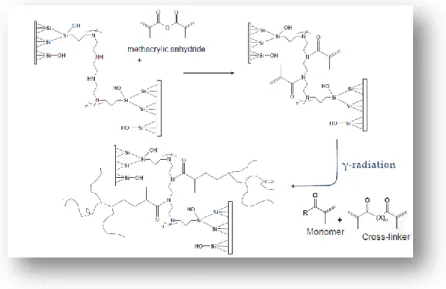
The preparation of organic monolithic stationary phases for chromatographic columns is accomplished through an “in situ” heat- or γ-rays-induced process. Polymerization mixtures consist of acrylic monomers, cross-linker and porogens. Before undertaking the polymerization process a step of pretreatment of the capillary must be necessarily carried out, in order to homogeneously distribute the
Chapter I: Bovine milk, monolithic columns and comprehensive HPLC
- 19 -
monolith within the capillary. The synthesis of the monolithic support includes a series of very important steps which are listed below:
Activation of the inner wall of the capillary through the step of “Etching” silanating “New Tentacle Type” and polymerization “Grafting on to”. Filling of the capillaries with the polymerization mixture formed by the
various components mixed in the right proportions. Polymerization by irradiation with γ-rays.
Capillaries drying for uncured monomers and porogens removal.
1.6.1.1. Activation of capillary
The pretreatment of the capillary provides three distinct phases:
“Etching”:
Transformation of siloxane groups present on the surface of the capillary in silanol groups (Figure 1.4.). Cifuentes et al. [51] proved that etching of columns with NaOH followed by leaching with HCl gives more reproducible surface treatment.
Chapter I: Bovine milk, monolithic columns and comprehensive HPLC
- 20 - Silanating step“Tentacle type”
The capillary surface is usually modified with a bi-functional silanizing reagent such as vinyl silane, acrylate silane or methacrylate silane. The most common reagent used is 3-(trimethoxysilyl)propyl methacrylate (Figure 1.5.). This step allows the immobilization of the monolithic polymer on the inner wall of the capillary.
Figure 1.5. Reaction scheme of silanating “Tentacle type”.
Polymerization phase “Grafting-on-to”
In this last phase of pre-treatment a solution of methacrylic anhydride is used in order to create a high number of unsaturated groups on which the polymerization process relies (Figure 1.6.).
Chapter I: Bovine milk, monolithic columns and comprehensive HPLC
- 21 -
Figure 1.6. Reaction scheme of derivatization “Grafting-on-to”.
1.6.1.2. Thermally and radiation polymerization
In case the polymerization is carried out through the use of heat (thermal polymerization), a compound (“photoinitiator” radical) is added to the polymerization mixture.
Azobisisobutyronitrile (AIBN) is quite popular. It decomposes while irradiated at 365 nm to afford free radicals [52-54]. Figure 1.7. shows the decomposition process involving AIBN, which, at high temperatures, releases N2 and generates two
radicals [55-57].
Chapter I: Bovine milk, monolithic columns and comprehensive HPLC
- 22 -
Thermally initiated free radical polymerization was the first method used for the preparation of rigid polymer-based monolith [58-60]. This process is very simple and its origin can be traced down to techniques typically applied in the preparation of porous beads by using suspension polymerization. This type of polymerization is generally treated in the literature as a “clone” of bulk polymerization in which each droplet of the dispersed phase containing monomer is an individual bulk reactor. Polymerizations initiated using high energy radiation such as γ-rays or electron beam belong to the group of “exotic” approaches to monoliths.
The major advantages of this method are no need for an initiator and thus absence of any functional groups at the chain ends, wide range of operating temperatures and wide choice of materials for containers, including stainless steel tubes.
The polymerization process triggered by γ rays allows a better distribution of the cross-linked structure for the entire length of the capillary, as is evident in Figure 1.8. in which a monolithic stationary phase obtained by a process of thermal polymerization is compared with a stationary phase obtained by polymerisation radiation. SEM images show that the monolith obtained by radical polymerization has a more homogeneous morphology.
Figure 1.8. Comparison between monolithic polimer obtained by radiation (left) and
Chapter I: Bovine milk, monolithic columns and comprehensive HPLC
- 23 -
This is why the photoinitiated monoliths exhibits about twice back pressure, thus indicating a difference in the porous structure (Figure 1.9.).
Figure 1.9. Comparison between monolithic columns obtained by radiation (left) and
thermally (right) polymerization. 1: Uracil; 2: Benzaldehyde; 3: Nitrobenzene; 4: Ethylbenzene; 5: Butylbenzene.
Although use of gamma rays can provide secondary compounds, this process remains more advantageous than thermal polymerization. Pore size control by irradiation rate, lower operating temperatures and resulting freedom in low boiling solvents choice represent the main advantages of the technique.
1.6.2. Monomers
Various functional monomers can be applied in preparation of monoliths. Glycidyl methacrylate with a highly reactive epoxy ring is the most frequently used in the synthesis of methacrylate ester-based monoliths [61]. However, the effects of other methacrylate monomers on separation properties of monolithic columns, such as
Chapter I: Bovine milk, monolithic columns and comprehensive HPLC
- 24 -
C6- [62], C8- [63], C16- [64], C4-, C12-, C18-, and isobornyl- methacrylate monomers
[65]
has been investigated for the preparation monolithic stationary phases with modulated hydrophobicities. The column prepared with C12 alkylmethacrylate functional monomer has shown the highest efficiency; however, the C18-methacrylate monolithic column provided better permeability [66]. The column based on the lauryl methacrylate (LMA) monomer provided the best separation of peptidic fragments containing less than 20 amino acids in tryptic digests of cytochrome C [67]. Butyl methacrylate (BMA) and 2-hydroxy ethyl methacrylate monomers were polymerized with 1,3-butanediol dimethacrylate cross-linker to produce hydrophobic monoliths for proteins chromatographic separation [68].
The styrene–divinylbenzene co-polymer monolithic stationary phases generally show lower polarity than their polymethacrylate counterparts. Their hydrophobicity can be tuned by adding a methacrylate to the polymerization mixture. Addition of methacrylic acid in the polymerization mixture significantly improves the surface area of prepared monoliths up to 261 m2/g in comparison to 0.1 m2/g for the material prepared in absence of methacrylic acid. In this way increased resolution for small aromatic compounds was attained [69]. Incorporation of a methacrylate monomer to the polymerization mixture for preparation of poly(divinylbenzene-alkyl methacrylate) monolithic columns improved peak symmetry with respect to poly-(styrene-co-divinylbenzene) columns [70,71] and decreased the retention of aromatic compounds, probably because of decreased π–π interactions in presence of long hydrophobic alkyl moieties in the monolithic structure [72].
Another approach tested for tuning the polarity of monolithic stationary phases was mixing two different monomers in the polymerization mixture. Partial substitution (25%) of BMA by LMA produced columns with enhanced retention of neutral analytes, while the replacement of larger percentages (75%) resulted in columns with lower retentivity [73].
Chapter I: Bovine milk, monolithic columns and comprehensive HPLC
- 25 -
In spite of numerous reports on variation of properties of monolithic columns by varying the chemistry of functional monomers in the polymerization mixture, to the best of our knowledge the effects of hydrophobicity and other properties of functional monomers on the separation of small compounds and large biopolymers on monolithic stationary phases have not been systematically compared so far.
1.6.3. Porogens
The choice of pore-forming solvent or porogen is a key tool that may be used for the control of porous properties without changing the chemical composition of the final polymer. In general, larger pores are obtained using more macroporogen due to an earlier onset of phase separation. The porogenic solvent controls the porous properties of the monolith through the solvation of the polymer chains in the reaction medium during the early stages of the polymerization [74,75]. Infact, the space originally occupied by porogens contributes to the formation of pores inside monolith phase. During the polymerization step the solvent molecules are incorporated within the polymer and then be removed by washing, thereby leaving a series of interconnected channels between them which will represent the “chromatographic bed”.
1.6.4. Crosslinking monomer
The porosity and the retentivity of the (poly)-methacrylate monolithic stationary phase can be changed by employing different alkyl dimethacrylate cross-linkers with various alkyl chain lengths between the two methacrylate units. Using 2-methyl-1,8-octanediol dimethacrylate as the cross-linker increased the proportion of mesopores in the final monolith, in comparison to the ethylene dimethacrylate (EDMA) cross-linker. The increased surface area in the mesopores improved the column efficiency [76].