Low expression of surface major histocompatibility com-plex (MHC) class I molecules and defects in antigen pro-cessing machinery make human neuroblastoma (NB) cells appropriate targets for MHC unrestricted immuno-therapeutic approaches. Human T-cell receptor (TCR) Vγ9Vδ2 lymphocytes exert MHC-unrestricted antitumor activity and are activated by phosphoantigens, whose expression in cancer cells is increased by aminobisphos-phonates. With this background, we have investigated the in vivo anti-NB activity of human Vγ9Vδ2 lympho-cytes and zoledronic acid (ZOL). SH-SY-5Y human NB cells were injected in the adrenal gland of immunodefi-cient mice. After 3 days, mice received ZOL or human Vγ9Vδ2 T cells or both agents by intravenous administra-tion once a week for 4 weeks. A significantly improved overall survival was observed in mice receiving Vγ9Vδ2 T cells in combination with ZOL. Inhibition of tumor cell proliferation, angiogenesis and lymphangiogenesis, and increased tumor cell apoptosis were detected. Vγ9Vδ2 T lymphocytes were attracted to NB-tumor masses of mice receiving ZOL where they actively modified tumor microenvironment by producing interferon-γ (IFN-γ), that in turn induced CXCL10 expression in NB cells. This study shows that human Vγ9Vδ2 T cells and ZOL in combina-tion inhibit NB growth in vivo and may provide the ratio-nale for a phase I clinical trial in patients with high-risk NB.
Received 31 October 2012; accepted 7 February 2013; advance online publication 12 March 2013. doi:10.1038/mt.2013.38
INTRODUCTION
Neuroblastoma (NB) is the most frequent extracranial solid tumor in children, with more than 50% of patients displaying metastatic disease at diagnosis.1 Bone marrow is the most common site of NB metastasis.2 Despite aggressive therapies, the outcome of children with metastatic NB at diagnosis still remains poor, with approxi-mately one third of them surviving at 5 years.3 Many efforts are
ongoing to develop new therapeutic strategies, and immunother-apy has attracted interest especially as adjuvant to standard front-line therapies.4 A better understanding of interactions between tumor cells, tumor microenvironment and the immune system is instrumental for the development of novel therapeutic approaches aiming at potentiating anti-NB immune response while dampen-ing tumor-driven immunosuppressive mechanisms. Indeed, dif-ferent preclinical and clinical strategies of antibody-mediated and cell-mediated NB immunotherapy have been developed in the last years.4
To design appropriate immunotherapeutic protocols for human NB, the multiple mechanisms of immune evasion adopted by tumor cells must be taken into account.5–9 Low expression of surface major histocompatibility complex (MHC) class I mol-ecules and defects in the antigen processing machinery of human NB cells allow the latter cells to escape the attack of tumor-specific T cells.5,10 On the other hand, downregulation of MHC class I mol-ecules makes NB cells appropriate targets for MHC unrestricted immunotherapeutic approaches making use of natural killer cells or T-cell receptor (TCR) gamma delta (γδ) T cells.11–13 Interferon-γ (IFN-γ) can upregulate MHC class I and some components of the antigen processing machinery in cancer cells including NB cells, exert antiangiogenic activity by inducing CXCL9 and CXCL10 expression, and re-program antitumor immune responses, thus modifying the tumor microenvironment.14
In the blood of healthy subjects, γδ T cells represent 1–5% of circulating T lymphocytes and display predominantly the Vγ9Vδ2 TCR.15 Vγ9Vδ2 T cells possess the unique capacity to recognize in a MHC-unrestricted way and be activated by natural nonpeptide phosphorylated intermediates of isoprenoid metabolism. These include exogenous prenyl pyrophosphates from bacteria and par-asitic protozoa as well as endogenous prenyl pyrophosphates, e.g., isopentenyl pyrophosphate (IPP), deriving from the mevalonate pathway that operates in human cells.16
Tumor cells that produce elevated concentrations of IPP can be recognized and killed by Vγ9Vδ2 T cells.16,17 Tumor cell produc-tion of IPP can be boosted by exposure to aminobisphosphonates,
The first two authors contributed equally and the last two authors contributed equally.
Correspondence: Ignazia Prigione, Laboratory of Oncology, Istituto Giannina Gaslini, Via G.Gaslini 5, 16148 Genoa, Italy.
E-mail: [email protected]
Mechanisms of the Antitumor Activity of Human
Vγ9Vδ2 T Cells in Combination With Zoledronic
Acid in a Preclinical Model of Neuroblastoma
Emma Di Carlo
1,2, Paola Bocca
3, Laura Emionite
4, Michele Cilli
4, Giuseppe Cipollone
5, Fabio Morandi
3,
Lizzia Raffaghello
3, Vito Pistoia
3and Ignazia Prigione
31Anatomic Pathology and Molecular Medicine, Department of Medicine and Sciences of Aging, “G. d’Annunzio” University, Chieti, Italy;
2Ce.S.I. Aging Research Center, “G. d’Annunzio” University Foundation, Chieti, Italy; 3Laboratory of Oncology, “Istituto Giannina Gaslini”, Genova, Italy; 4Animal Model Facility, IRCCS Azienda Ospedaliera Universitaria San Martino, IST, Istituto Nazionale per la Ricerca sul Cancro, Genova, Italy; 5Department of Biomedical Science, “G. d’Annunzio” University, Chieti, Italy
1034 1043
γδ T Cells and ZOL for Neuroblastoma Immunotherapy
Molecular Therapy 10.1038/mt.2013.38 12
March2013
21 531October2012
7February2013
a class of drugs that inhibit osteoclastic reabsorption.18,19 The lat-ter drugs specifically inhibit the IPP-processing enzyme pharne-syldiphosphate synthase in the mevalonate pathway leading to accumulation of IPP.20 Zoledronic acid (ZOL) is an aminobisphos-phonate that, besides its antiosteoclastic activity, exerts direct and indirect antitumor effects via inhibition of tumor growth and angiogenesis and induction of malignant cell apoptosis.21–25 ZOL, especially when combined with IL-2, has a strong capacity to acti-vate and expand human Vγ9Vδ2 T cells displaying an effector-memory immunophenotype.26–28
Vγ9Vδ2 T cells exert their antineoplastic activity by MHC unrestricted killing of tumor cells and antibody-dependent cel-lular cytotoxicity, as well as by activation of other immune effector mechanisms through cytokine release.29–31 The potent antitumor activity of Vγ9Vδ2 T cells against solid tumors,32 multiple myeloma and lymphomas33 supports their use in clinical trials, especially in combination with conventional chemotherapy.18,34 ZOL has been clinically approved for the treatment of patients with bone metas-tasis from solid tumors and multiple myeloma.24,35,36 So far, the combination of adoptively transferred Vγ9Vδ2 T cells and ZOL has been tested in a few preclinical models of adult human malig-nancies and in a recent phase I clinical trial.28,34,37
In this study, the antitumor activity of human Vγ9Vδ2 T cells and ZOL in combination has been investigated in an orthotopic mouse model of human NB38,39 and the mechanisms involved have been identified.
RESULTS
Lysis of NB cell lines by ZOL-expanded Vγ9Vδ2 T cells from normal donors and patients with NB
These experiments were carried out in preparation for the in
vivo studies reported in the following sections. TCR Vγ9Vδ2
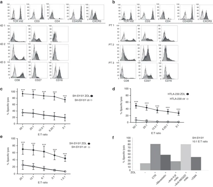
cell populations were expanded in vitro by ZOL stimulation of peripheral blood mononuclear cells from four normal donors and three untreated patients with stage 4 NB at diagnosis. Figure 1 shows the immunophenotype of Vγ9Vδ2 T cell–expansions derived from three normal donors (Figure 1a) and from three patients with NB (Figure 1b), respectively. A common CD3+ CD4− CD45R0+ CXCR3+ phenotype characterized both normal donor- and patient-derived Vγ9Vδ2 T cells (Figure 1a,b, upper histogram plots). Differently, variable patterns of expression of CD8, CD27, and CD16 markers were detected in each Vγ9Vδ2 T cell–expansion, either from normal donors (Figure 1a) or patients (Figure 1b).
Overnight pretreatment of SH-SY-5Y NB cells, subsequently used for in vivo experiments, and HTLA-230 NB cells with 10 μmol/l and 50 μmol/l ZOL, respectively, significantly increased their susceptibility to killing by Vγ9Vδ2 T cells expanded from normal donors, at all effector: target ratios tested (P < 0.001, Figure 1c,d). Similar results were observed when patient derived Vγ9Vδ2 T cells were used as effectors against ZOL sensitized SH-SY-5Y target cell line (P < 0.001, Figure 1e).
Patient (Figure 1f) and normal donor (data not shown) derived Vγ9Vδ2 T cells lysed ZOL sensitized SH-SY-5Y NB cells
via TCR-dependent and perforin-mediated mechanisms, as
dem-onstrated by treatment of effector cells with anti-Vγ9 blocking mAb or the vacuolar-type H+-ATPase inhibitor concanamycin A,
respectively. Blocking of NKG2D on Vγ9Vδ2 T cells was ineffec-tive (Figure 1f).
Finally, incubation of target cells with mevastatin, which pre-vents ZOL-induced IPP accumulation, during ZOL pretreatment dampened Vγ9Vδ2 T cell–mediated lysis (Figure 1f).
In vivo efficacy of combined administration of Vγ9Vδ2 T cells and ZOL
To investigate in vivo the anti-NB activity of Vγ9Vδ2 T cells in combination with ZOL we used an orthotopic mouse model that mimics closely the growth pattern of human NB.38,39 The SH-SY-5Y NB cell line was implanted in the adrenal gland of nude/nude athymic mice because this cell line was previously well characterized for its patterns of growth and spreading following orthotopic xenograft.39 Three days after tumor implantation, mice were weekly i.v. treated with 5 × 10⁶ Vγ9Vδ2 T cells, ZOL (150 μg/ Kg), or both, in combination, for 4 weeks.
A statistically significant improvement of survival was observed in mice receiving Vγ9Vδ2 T cells after ZOL pretreat-ment, in comparison with untreated mice (P = 0.024; median survival times: 42 and 48 days for untreated and treated mice, respectively) (Figure 2a). In contrast, survival of mice treated with Vγ9Vδ2 T cells (Figure 2b) or ZOL (Figure 2c) as single agents was not significantly different from that of the control group (P > 0.05; median survival times: 43 days for Vγ9Vδ2 T cells or ZOL treatment groups, 42 days for control group).
Morphological and immunohistochemical analyses disclose the mechanisms underlying the
in vivo anti-NB activity of Vγ9Vδ2 T cells and ZOL
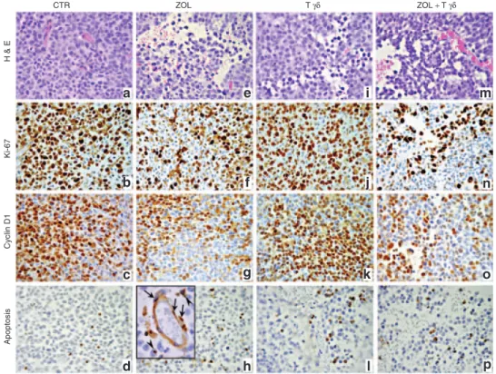
Tumors from nu/nu mice (n = 6) orthotopically implanted with human NB SH-SY-5Y cells, were composed of large primitive-appearing cells growing in solid sheets and sometimes arranged in Homer-Wright pseudorosettes. Such cells displayed large vesicu-lar nuclei and prominent nucleolus, scant cytoplasm and poorly defined cell borders (Figure 3a). These tumors showed high pro-liferative activity, as assessed by Ki-67 immunostaining (Figure 3b and Table 1), frequent cyclin D1 expression (Figure 3c) and a few apoptotic figures, as assessed by the TUNEL assay (Figure 3d and Table 1). Tumor growth was supported by a richly branched vascular network composed of microvessels of both murine and human origin as revealed, respectively, by anti-mouse-CD31 and anti-human-vWF immunostainings (Figure 4a,b and Table 1).
ZOL treatment (n = 6) resulted in frequent ischemic-hemor-rhagic foci of the tumor masses (Figure 3e), associated with sig-nificant (P < 0.05) impoverishment of human vWF+ endothelial microvessels (Figure 4e and Table 1). In contrast, counts of CD31+ murine microvessels did not differ significantly in ZOL treated mice compared with controls (Figure 4d and Table 1). ZOL treat-ment virtually abolished tumor cell expression of VEGF-A com-pared with controls (Figure 4f and Table 1), whereas tumor cell proliferation and Cyclin D1 expression appeared substantially unchanged (Figure 3f,g; Table 1) along with the frequency of apoptotic events (Figure 3h and Table 1) which may also involve endothelial cells (inset in Figure 3h).
Injection of human Vγ9Vδ2 T lymphocytes (n = 7) promoted very small collapses of the tumor tissues (Figure 3i) in association
with a significantly (P < 0.05) increased tumor cell apoptosis ver-sus controls only (Figure 3l and Table 1). Tumor cell prolifera-tion and cyclin D1 expression (Figure 3j,k), along with VEGF-A expression (Figure 4i), were basically unchanged in compari-son with control, and the micro-vascular network integrity was substantially unaltered in both murine and human components (Figure 4g,h and Table 1).
Tumors from mice treated with ZOL 16 hours before human Vγ9Vδ2 T lymphocyte infusion were harvested 6 hours after the administration of the latter cells (n = 9). This combined treat-ment induced a peculiar “worm-eaten” feature of the tumor tissue (Figure 3m) in association with significant inhibition of tumor cell proliferation (P < 0.05) and reduced cyclin D1 expression (Figure 3n,o and Table 1) compared with tumors
Figure 1 Immunophenotypic and cytotoxic properties of Vγ9Vδ2 T cells expanded in vitro by ZOL stimulation of PBMC from normal donors and patients with NB. PBMC from normal donors and patients with NB at diagnosis were cultured in the presence of ZOL and IL-2. Proliferating cells were expanded in IL-2 containing medium. Immunophenotype and cytotoxicity were tested after 10/12 day culture. (a,b): immunophenotypic profiles of Vγ9Vδ2 T cells expanded from (a) three normal donors (ND) and (b) three patients with NB (Pt). Upper histogram plots in (a) and (b) show markers, from a representative sample, expressed with the same pattern in each Vγ9Vδ2 T cell expansion. The other histogram plots show markers dif-ferently expressed in each Vγ9Vδ2 T cell expansion. Grey histograms represent isotype controls and black histograms are the specific stainings. (c,d) Vγ9Vδ2 T cells expanded from four normal donors were tested for cytotoxic activity against untreated and ZOL-sensitized SH-SY-5Y and HLA-230 human NB cells. Results are expressed as mean percentage of specific lysis ± SD from four independent experiments (***P < 0.001). (e) Vγ9Vδ2 T cells expanded from three patients with NB were tested for cytotoxic activity against untreated and ZOL-sensitized SH-SY-5Y NB cells. Results are expressed as mean percentage of specific lysis ± SD from three independent experiments (***P < 0.001). (f) Cytotoxic activity of Vγ9Vδ2 T cells expanded from one patient with NB against ZOL-sensitized SH-SY-5Y NB cells was tested, at 10:1 E:T ratio, after preincubation of effectors with medium (CTR) or with anti-TCR Vγ9 mAb, or anti-NKG2D mAb or concanamycin A (CMA) and after preincubation of target cells with Mevastatin. Results from one representative experiment of three performed are shown. NB, neuroblastoma; PBMC, peripheral blood mononuclear cells.
0 20 40 60 80 100 0 20 40 60 80 100 0 20 40 60 80 100 0 20 40 60 80 100 0 20 40 60 80 100 0 20 40 60 80 100 0 20 40 60 80 100 0 CD8 100 80 60 40 % Specific lysis 20 0 *** *** *** *** *** *** *** *** *** *** *** *** *** *** E:T ratio 50:1 25:1 12.5:1 6.25:1 3:1 100 80 60 40 % Specific lysis 20 0 % Specific lysis 0 10 20 30 40 50 60 70 80 90 100 E:T ratio ZOL CTR
+Mevastatin +Anti-V γ9 mAb +Anti-NKG2D mAb +CMA − + + + + + 50:1 25:1 12.5:1 6.25:1 3:1 100 80 60 40 % Specific lysis 20 0 E:T ratio 25:1 12:1 6:1 3:1 1.5:1 ND 1 TCR Vδ2 CD3 CD4 CD45R0 CXCR3 TCR Vδ2 CD3 CD4 CD45R0 CXCR3 ND 2 ND 3 CD27 CD16 SH-SY-5Y ZOL SH-SY-5Y ctr HTLA-230 ZOL HTLA-230 ctr SH-SY-5Y 10:1 E:T ratio SH-SY-5Y ZOL SH-SY-5Y ctr 20 40 60 80 100 0 20 40 60 80 100 0 20 40 60 80 100 0 20 40 60 80 100 0 20 40 60 80 100 0 20 40 60 80 100 0 20 40 60 80 100 0 20 40 60 80 100 0 20 40 60 80 100 0 CD8 PT 1 PT 2 PT 3 CD27 CD16 20 40 60 80 100 0 20 40 60 80 100 0 20 40 60 80 100 0 20 40 60 80 100 0 20 40 60 80 100 0 20 40 60 80 100 0 20 40 60 80 100 0 20 40 60 80 100 0 20 40 60 80 100 0 20 40 60 80 100 0 20 40 60 80 100 0 20 40 60 80 100 a b c d e f ***
from untreated or single agent treated mice. Tumor cell apop-tosis was significantly (P < 0.05) increased in mice treated with ZOL and human Vγ9Vδ2 T lymphocytes (Figure 3p and Table 1) compared with tumors from untreated, but not single agent treated, mice. Tumor masses from mice that received the com-bined treatment showed scarce to absent VEGF-A expression (Figure 4l) and a severely compromised micro-vascular net-work, because (i) murine microvessels were significantly (P <
0.05) reduced in comparison with untreated or single agent treated animals, and (ii) human microvessels were significantly (P < 0.05) reduced when compared with tumors from untreated or human Vγ9Vδ2 T lymphocyte treated mice (Figure 4j,k and
Table 1).
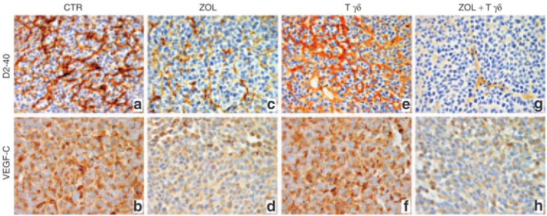
Immunohistochemistry for D2-40/podoplanin revealed that control tumors were endowed with a robust lymphatic microvas-culature of human origin and also showed a distinct expression
Figure 2 Survival of mice orthotopically engrafted with SH-SY-5Y cells treated with ZOL and human Vγ9Vδ2 T cells as single agents or in com-bination. Five-week-old female athymic (nude/nude) Balb/c mice were orthotopically xenografted in the left adrenal gland with human SH-SY-5Y NB cells. Three days after, mice were randomized into four groups receiving different treatments (one weekly treatment for 4 weeks). Pooled results from two different experiments are shown. The overall survival of mice receiving human Vγ9Vδ2 T cells and ZOL in combination was significantly higher than that of control mice (P = 0.024, Kaplan–Meier, log-rank test) (a). In contrast, the overall survival of mice treated with Vγ9Vδ2 T cells alone (b) or ZOL alone (c) did not differ significantly from that of control mice.
100 80 60 40 % Of sur vi va l 20 0 0 20
Days after tumor engraftment
40 60
100 Control (n = 16)
ZOL + γδ T cells (n = 15) Control (n γδ T cells (n = 15)= 16) Control (n ZOL (n = 15)= 16)
80 60 40 % Of sur vi va l 20 0 0 20 40 60 100 80 60 40 % Of sur vi va l 20 0 0 20 40 60 a b c
Figure 3 Histologic features and immunohistochemical analysis of proliferative activity and apoptosis of tumors developed in mice that received treatments with ZOL and/or human Vγ9Vδ2 T cells. H&E staining of tumors developed after orthotopic injection of SH-SY-5Y NB cells in untreated mice (a) and in mice treated with ZOL (e), Vγ9Vδ2 T cells (i) or ZOL + Vγ9Vδ2 T cells (m). Immunostaining with anti-Ki-67 and anti-Cyclin D1 Abs of tumors developed in untreated mice (b,c) and in mice treated with ZOL (f,g), Vγ9Vδ2 T cells (j,k) or ZOL + Vγ9Vδ2 T cells (n,o). TUNEL assay performed on tumors developed in untreated mice (d) and in mice treated with ZOL (h; inset panel: arrows, endothelial cells; arrowheads, neoplastic cells), Vγ9Vδ2 T cells (l) or ZOL + Vγ9Vδ2 T cells (p). (a–p: ×400; inset panel in h: ×1,000). Abs, antibodies; H&E, hematoxylin and eosin.
CTR Apoptosis Cyclin D1 Ki-67 H & E ZOL T γδ ZOL + T γδ a e i m b f j n c g k o d h l p
of VEGF-C (Figure 5a,b and Table 1). Similar features were observed in tumors from Vγ9Vδ2 T lymphocyte only treated mice (Figure 5e,f), whereas tumors from ZOL treated mice showed sig-nificant reduction of lymphatic vasculature (P < 0.05) and reduced VEGF-C expression (Figure 5c,d and Table 1) when compared with control tumors. Finally, in tumors from mice receiving the combined treatment VEGF-C expression was reduced similarly to that observed in tumors from animal receiving ZOL only, whereas
lymphatic vasculature was significantly decreased (P < 0.05) com-pared with untreated or single agent treated animals (Figure 5h,g and Table 1).
In summary, the most remarkable and significant anti-NB effects were obtained when ZOL and Vγ9Vδ2 T cells were admin-istered in combination. ZOL alone showed significant antiangio-genic activity, whereas Vγ9Vδ2 T cells administered alone affected significantly tumor cell apoptosis.
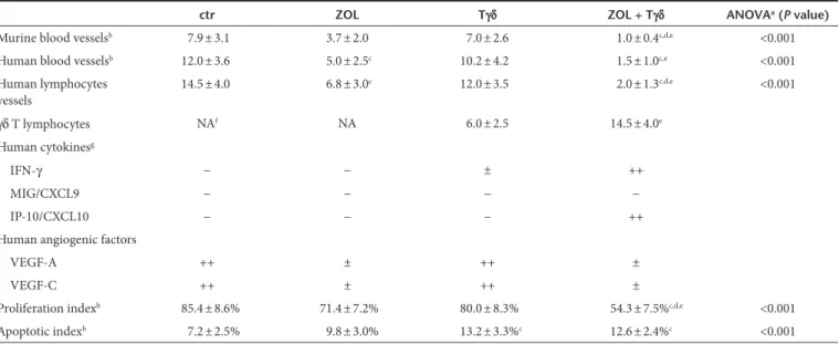
Table 1 Immunohistochemical analyses of tumors developed after SH-SY-5Y cell injection in the adrenal gland of athymic nu/nu mice untreated or treated with ZOL, human Vγ9Vδ2 T cells or both
ctr ZOL Tγδ ZOL + Tγδ ANOVAa (P value)
Murine blood vesselsb 7.9 ± 3.1 3.7 ± 2.0 7.0 ± 2.6 1.0 ± 0.4c,d,e <0.001
Human blood vesselsb 12.0 ± 3.6 5.0 ± 2.5c 10.2 ± 4.2 1.5 ± 1.0c,e <0.001
Human lymphocytes vessels 14.5 ± 4.0 6.8 ± 3.0 c 12.0 ± 3.5 2.0 ± 1.3c,d,e <0.001 γδ T lymphocytes NAf NA 6.0 ± 2.5 14.5 ± 4.0e Human cytokinesg IFN-γ − − ± ++ MIG/CXCL9 − − − − IP-10/CXCL10 − − − ++
Human angiogenic factors
VEGF-A ++ ± ++ ±
VEGF-C ++ ± ++ ±
Proliferation indexb 85.4 ± 8.6% 71.4 ± 7.2% 80.0 ± 8.3% 54.3 ± 7.5%c,d,e <0.001
Apoptotic indexb 7.2 ± 2.5% 9.8 ± 3.0% 13.2 ± 3.3%c 12.6 ± 2.4%c <0.001
aP < 0.001 One-way analysis of variance (ANOVA) for comparisons between four groups. bAssessment of cytokine and angiogenic factor expression and counts of
microvessels and γδ T cells were performed at ×400 in a 0.180 mm2 field. At least three samples (one sample/tumor growth area) and 8–10 randomly chosen fields/
sample were evaluated. Results are expressed as mean ± SD of CD31 (murine blood vessels), or vWF (human blood vessels), or D2-40/podoplanin (human lymphatic vessels) positive microvessels per field; or TCRγδ positive cells per field; or Ki-67 or TUNEL positive cells/number of total cells evaluated on formalin-fixed sections by immunohistochemistry. cP < 0.05 Tukey test compared with tumors from untreated (ctr) mice. dP < 0.05 Tukey test compared with tumors from ZOL only treated
mice. eP < 0.05 Tukey test compared with tumors from Tγδ lymphocyte only treated mice. fNot applicable. gThe expression of cytokines and angiogenic factors was
defined as absent (−), scarce (±), moderate (+), or frequent (++) on paraffin embedded (VEGF-A and VEGF-C) or cryostat (IFN-γ, MIG/CXCL9, IP-10/CXCL10) sections stained with the corresponding antibody.
Figure 4 Vascular network in tumors developed in mice that after orthotopic engraftment of SH-SY-5Y cells received treatments with ZOL and/or human Vγ9Vδ2 T cells. Immunostaining with anti-CD31, anti-vWF and anti-VEGF-A Abs of tumors developed after orthotopic injection of SH-SY-5Y NB cells in untreated mice (a, b and c, respectively) and in mice treated with ZOL (d–f), Vγ9Vδ2 T cells (g–i) or ZOL + Vγ9Vδ2 T cells (j–l). (a–l: ×400). Abs, antibodies.
CTR VEGF-A vWF CD31 ZOL T γδ ZOL + T γδ a b c d e f g h i j k l
Perturbation of tumor microenvironment by infiltrating Vγ9Vδ2 T lymphocytes in mice treated with Vγ9Vδ2 T cells and ZOL
Tumor localization of adoptively transferred human Vγ9Vδ2 T lymphocytes, alone or combined with ZOL, and their influ-ence on tumor microenvironment were investigated by immuno-histochemical analyses of tumor masses from NB orthotopically implanted mice. Mice were treated with ZOL 16 hours before receiving or not human Vγ9Vδ2 T lymphocytes, and were sac-rificed 6 hours after the completion of the third cycle of therapy.
By contrast with tumors developed in untreated (Figure 6a) or ZOL treated mice (Figure 6b), tumors developed in mice receiving human Vγ9Vδ2 T lymphocytes alone (Figure 6c), or in combina-tion with ZOL (Figure 6d), were infiltrated by human γδ T lym-phocytes. Such infiltration was significantly (P < 0.05) higher in tumors from mice receiving the combined treatment (Figure 6d and Table 1) and most of γδ T lymphocytes expressed the cyto-toxic molecule T-cell intracellular antigen-1 (TIA-1), as revealed by TCRγδ/TIA-1 double staining (inset in Figure 6d). In addi-tion, immunohistochemical examination of serial tissue sections showed that hIFN-γ production was distinct in tumor masses from mice receiving the combined treatment that appeared infiltrated by γδ T lymphocytes (Figure 7c and Table 1), whereas it was barely detectable in the tumors from mice receiving Vγ9Vδ2 T lympho-cytes alone (Figure 7b) and obviously absent in tumors from ZOL treated (Figure 7a) or control animals (Table 1). Analyses of IFN-γ inducible chemokine production revealed that CXCL10/IP10 was detectable in the cytoplasms of tumor cells from mice receiving the combined treatment (Figure 7f and Table 1), whereas it was absent in tumors from Vγ9Vδ2 T lymphocytes treated mice (Figure 7e) and in tumors from ZOL treated (Figure 7d) or control animals (data not shown). In contrast, expression the IFN-γ inducible anti-angiogenic chemokine CXCL9 was not detected in tumors from any group of mice (Table 1).
Analyses of murine tumor infiltrating leukocytes, performed with anti-F4/80 and anti-pan-natural killer cells, revealed a scarce to moderate macrophage infiltrate at the tumor border and few macrophages penetrating the innermost areas, whereas natural
Figure 5 Lymphatic vessel network and VEGF-C expression in tumors developed in mice that after orthotopic engraftment of SH-SY-5Y cells received treatments with ZOL and/or human Vγ9Vδ2 T cells. Immunostaining with anti-D2-40 and anti-VEGF-C Abs of tumors developed (a,b) in untreated mice and (c,d) in mice treated with ZOL, (e,f) Vγ9Vδ2 T cells or (g,h) ZOL + Vγ9Vδ2 T cells. (a,c,e,g: ×400; b,d,f,h: ×630). Photographs are representative of the staining detected in at least 6 out of 8 randomly chosen fields/tumor sample. Three samples were examined for each experi-mental group. Abs, antibodies.
CTR a c e g b d f h VEGF-C D2-40 ZOL T γδ ZOL + T γδ
Figure 6 Detection and characterization of human Tγδ lymphocytes in the tumor masses developed in mice that after orthotopic engraft-ment of SH-SY-5Y cells received human Vγ9Vδ2 T cells and/or ZOL. Immunostaining with anti-TCRγδ Ab of tumors developed in (a) untreated mice and (b) in mice treated with (c) ZOL, Vγ9Vδ2 T cells or ZOL + Vγ9Vδ2 T cells (d). Double immunostaining with anti-TCRγδ (red) and anti-TIA-1 (intracytoplasmic granular brown dots) Abs (inset in d). (a–d: ×400; inset in d: ×1,000). Abs, antibodies.
CTR
a b c d
T
γδ
ZOL T γδ ZOL + T γδ
Figure 7 Detection of IFN-γ and CXCL10 in the tumor masses devel-oped in mice that, after orthotopic engraftment of SH-SY-5Y cells, received ZOL, human Vγ9Vδ2 T cells or both. Immunostaining with anti-IFN-γ and anti-CXCL10 Abs in tumors from mice treated with (a,d) ZOL or (b,e) Vγ9Vδ2 T cells only and in tumors from mice treated with (c,f) ZOL + Vγ9Vδ2 T cells. (a–f: ×1,000). Abs, antibodies.
a b c d e f ZOL CXCL10 IFN-γ T γδ ZOL + T γδ
killer cells were completely absent (data not shown). No relevant differences emerged in the content of murine infiltrating leuko-cytes within tumors from the different treatment groups (data not shown).
These data demonstrate that (i) i.v. injected human Vγ9Vδ2 T lymphocytes are able to migrate into NB-tumor masses grown in orthotopically xenografted mice; (ii) the tumor localization of i.v. injected human Vγ9Vδ2 T lymphocytes is potentiated by ani-mal pretreatment with ZOL; and (iii) tumor localized Vγ9Vδ2 T cells can exert their functional programs and interfere with tumor microenvironment.
DISCUSSION
Few studies have addressed the preclinical anti-NB activity of human γδ T lymphocytes or ZOL. Treatment of immunodefi-cient mice carrying human metastatic NB with human γδ T cells from normal individuals was ineffective.40 Likewise, ZOL alone administered in a NB murine model of bone invasion inhib-ited osteoclast activity and tumor cell proliferation, but did not improve survival.21 Finally, it has been reported that ZOL sensi-tizes NB-derived tumor-initiating cells to cytolysis mediated by human Vγ9Vδ2 T cells.37
We asked whether the combination of human Vγ9Vδ2 T lymphocytes and ZOL could exert synergistic anti-NB effects in
vivo based upon the notion that ZOL may sensitize tumor cells to
Vγ9Vδ2 T cell–mediated lysis. Indeed, we showed that Vγ9Vδ2 T lymphocytes with an effector/central memory phenotype could
be expanded from peripheral blood of normal individuals and patients with NB upon incubation with ZOL, and that these effec-tor cells killed more efficiently NB cells that had been pretreated with ZOL. Recognition and lysis of NB cells by Vγ9Vδ2 T lym-phocytes were found to depend on engagement of TCR but not NKG2D, release of cytotoxic granules, and ZOL-induced IPP accumulation in tumor cells. Taken together, these results are con-sistent with previous reports in other tumor models.16,18
In vivo experiments were next carried out using an
ortho-topic human NB model in immunodeficient mice whereby malig-nant cells are implanted in the adrenal gland capsule.38,39 Mice received ZOL alone or Vγ9Vδ2 T cells alone or both, with ZOL administered 16 hours before Vγ9Vδ2 T lymphocytes to allow ZOL-mediated tumor cell sensitization. The survival of human NB carrying mice was significantly improved only by ZOL and Vγ9Vδ2 T cells in combination, pointing to the therapeutic syn-ergism of these agents. However, ZOL or Vγ9Vδ2 T cells admin-istered alone exerted significant effects on tumor angiogenesis or tumor cell apoptosis, respectively.
Tumor blood microvessels were lined by tumor-derived or host-derived endothelial cells, consistently with previous studies showing that human NB cells can transdifferentiate into endo-thelial cells carrying the same genetic abnormalities, e.g., MYCN amplification, through a phenomenon known as vascular mim-icry.41,42 ZOL alone significantly reduced the proportion of human blood microvessels, without any additional effect observed in mice treated with ZOL and Vγ9Vδ2 T cells. NB tumors from control
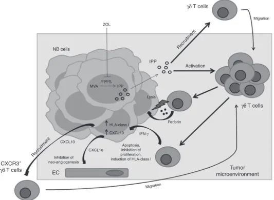
Figure 8 A model for the effects of combined treatment with ZOL and human Vγ9Vδ2 T cells on NB tumor microenvironment. ZOL-mediated inhibition of FPPS results in intracellular accumulation of upstream metabolites of the MVA pathway including IPP, which activates and attracts Vγ9Vδ2 T cells. The latter cells kill tumor cells through a perforin-dependent mechanism and release IFN-γ that may be involved in inhibition of NB cell pro-liferation and/or induction of apoptosis. Vγ9Vδ2 T cell-secreted IFN-γ upregulates HLA class I expression on NB cells thus increasing their immuno-genicity, and induces CXCL10 expression in tumor cells. CXCL10 inhibits neo-angiogenesis by binding to CXCR3 on endothelial cells and recruits a new wave of CXCR3+ Vγ9Vδ2 T cells to the tumor site. EC, endothelial cells; FPPS, farnesyl pyrophosphate synthase; IPP, isopentenyl pyrophosphate; MVA, mevalonate. NB cells IPP Activation Recr uitment Recr uitment Perforin Migration ZOL Lysis HLA-class I CXCL10 CXCL10 Inhibition of neo-angiogenesis Tumor microenvironment CXCL10 CXCR3+ γδ T cells EC IFN-γ γδ T cells γδ T cells Apoptosis, inhibition of proliferation, induction of HLA-class I Migration MVA FPPS IPP
mice showed a rich network of human lymphatic microvessels that was significantly inhibited by ZOL alone and even more by ZOL and Vγ9Vδ2 T cells in combination. Consistent with these findings, ZOL treatment reduced expression of VEGF-A and VEGF-C, that are crucial mediators of angiogenesis and lymphan-giogenesis, respectively.43,44
Although inhibition of VEGF-A expression is a well-known component of the antitumor activity of ZOL,36 the finding that the latter drug dampened VEGF-C expression in a mouse model of a human malignancy is novel and warrants further investigation. In this respect, inhibition of VEGF-C expression was reported in a rat model of oral wound healing as result of the impact of ZOL-mediated osteoclast suppression in the bone marrow microenvironment.45
Only ZOL and Vγ9Vδ2 T cells in combination decreased significantly mouse derived blood microvessels in the tumor microenvironment. The finding that ZOL alone failed to inhibit formation of the latter microvessels is so far unexplained and war-rants further investigation.
Vγ9Vδ2 T cells alone increased significantly tumor cell apop-totic index and this effect was not amplified by their combination with ZOL. Tumor cell proliferation was unaffected by the single agents whereas it was significantly inhibited by ZOL and Vγ9Vδ2 T cells.
Taken together, these results indicate that the synergistic therapeutic activity of the latter agents was attributable to de novo inhibition of tumor cell proliferation and mouse derived blood microvessel formation, as well as to augmented inhibition of human lymphatic microvessel network compared with ZOL alone.
Histological and immunohistochemical studies provided insight into the mechanisms underlying the synergism between ZOL and Vγ9Vδ2 T cells. Tumors from mice treated with the latter combination were found to contain significantly higher numbers of human Vγ9Vδ2 T cells than tumors from mice receiving Vγ9Vδ2 T cells alone. These infiltrating lymphocytes expressed the cytotoxic granule associated molecule TIA-1 and IFN-γ, whereas NB cells expressed at high level CXCL10, but not CXCL9.46 These results support the following scenario (Figure 8): (i) ZOL induces accumulation in tumor cells of IPP, which activates and attracts Vγ9Vδ2 T cells, as previously shown in a breast cancer model;19 (ii) the latter cells can kill NB cells through a perforin dependent mechanism and release IFN-γ, that in turn induces CXCL10 expression in tumor cells; (iii) IFN-γ may be involved in inhibition of NB cell proliferation and/or induction of apoptosis;14 iv) CXCL10 can inhibit neo-angiogenesis by binding to CXCR3 on endothelial cells and recruit a new wave of CXCR3+ Vγ9Vδ2 T cells to the tumor site. An implication of this model is that Vγ9Vδ2 T cell-derived IFN-γ can upregulate HLA class I expression on NB cells thus increasing their immunogenicity.5
This study may provide the rationale for the design of a phase I clinical trial based upon administration of in vitro expanded autologous Vγ9Vδ2 T cells together with ZOL in patients with metastatic NB and low tumor burden. Although caution is needed due to the potential interference of ZOL with actively growing bones, a phase I study combining ZOL and low-dose cyclophos-phamide in patients with NB has recently been published with encouraging results.47
The therapeutic protocol used in our study may be improved as follows in the perspective of the clinical application: (i) Vγ9Vδ2 T cells can be infused more frequently than weekly (e.g., every other day) to allow a continuous trafficking of these cells to the tumor site and administered for longer time frames34 (ii) bisphos-phonates including ZOL have high tropism for bone matrix lead-ing to low plasma concentrations.48 It is therefore conceivable that the therapeutic activity of ZOL can be increased by targeting the drug to the tumor site in nanoparticles coated with tumor-specific molecules, for example anti-GD2 antibody to NB cells.49
MATERIALS AND METHODS
Cell lines. The human NB cell lines SH-SY-5Y39 and HTLA-230 (kindly
pro-vided by Professor Emil Bogenmann) were cultured in Dulbecco’s modi-fied Eagle’s medium (EuroClone, Milan, Italy) supplemented with 10% fetal bovine serum (Invitrogen, Carlsbad, CA), HEPES buffer, l-glutamine, penicillin-streptomycin, and nonessential aminoacids (EuroClone). In vitro Vγ9Vδ2 T cell–expansion. Peripheral blood mononuclear cells were isolated from blood samples of normal donors or untreated patients with stage 4 NB at diagnosis by Ficoll (Sigma-Aldrich, St Louis, MO) density centrifugation. Samples were taken after permission in accordance with informed consent and the study was approved by the local Ethical Committee. Cells were resuspended in RPMI 1640 supple-mented with l-glutamine, penicillin-streptomycin, nonessential aminoac-ids (EuroClone) and 5% pooled human AB serum (complete medium). To expand TCR Vγ9Vδ2 cells, peripheral blood mononuclear cells were resuspended in complete medium and cultured in the presence of ZOL (5 μmol/l, Enzo Life Sciences; Vinci-Biochem, Italy) and 50 U/ml of recom-binant (r) hIL2 (PROLEUKIN, Novartis FarmaS.p.A., Origgio, Italy) in 96-well round bottom microtiter plates (BD Falcon, BD Biosciences, NJ). Proliferating T cells were maintained in IL-2 containing medium. The immunophenotype of proliferating T cells was checked using anti-TCR Vδ and CD3 monoclonal antibodies. Cultures containing higher than 90% TCR Vδ2+ cells were used for further experiments, at least 2 weeks after
ZOL stimulation.
Flow cytometry. The following monoclonal antibodies were used: CD3 PE-Cy7, CD16 FITC, (Beckman-Coulter, Marseille, France), anti-TCR Vδ2PE, CD45R0 APC, CD27 PE, CXCR3 FITC (BD Biosciences, San Jose, CA), MultiMix Triple Colour Reagent Anti Human CD3/CD4/CD8 (Dako, Glostrup, Denmark). Cells were resuspended in Dulbecco’s phosphate-buffered saline (Sigma-Aldrich) with 1% fetal bovine serum (staining solution) and stained with specific monoclonal antibodies for 30 minutes in the dark at 4 °C. Cells were then washed once with staining solution and analyzed by flow cytometry. Isotype mached fluochrome-conjugated murine Ig were used as negative controls. Flow cytometric analyses were performed by FACS Calibur cytometer (BD Biosciences) and data were analyzed by CellQuest (BD Biosciences) or Kaluza (Beckman-Coulter) softwares. Results were expressed as percent of positive cells.
Cytotoxicity assays. Cytotoxicity assays were performed using the 4 hours
51Cr-release test.50 Vγ9Vδ2 T cells in vitro expanded by ZOL, from
nor-mal donors and patients with NB, were used as effectors. Human NB cell lines, either untreated or treated overnight with ZOL (10–50 μmol/l) were used as targets. Effectors cells were seeded in duplicate at different concen-tration in 96-well V-bottom microtiter plates (BD Falcon) together with 5 × 10³ 51Cr-labeled target cells in a final volume of 200 μl. In some
experi-ments, effector cells were incubated with anti-TCR Vγ9 (clone 7A5; Pierce Endogen, Rockford, IL) or anti-NKG2D blocking monoclonal antibod-ies (R&D Systems, Minneapolis, MN) before being added to 51Cr-labeled
target cells. To inhibit perforin-mediated cytotoxicity, Vγ9Vδ2 T cells were incubated with concanamycin A (100 ng/ml; Sigma), an inhibitor
of vacuolar-type H+-ATPase, for 2 hours at 37 °C before being added to 51Cr-labeled target cells. To inhibit IPP-mediated target cell recognition
by Vγ9Vδ2 T cells, Mevastatin (MEV, 25 μ; Sigma) was added to target cells 6 hours before and during ZOL pretreatment. Concanamycin A or MEV were re-added during cytotoxicity assay. Results were expressed as percentage of specific lysis.
In vivo studies. Five-week-old female athymic (nude/nude) Balb/c mice (Harlan Laboratories S.r.l., S. Pietro al Natisone, Italy) were anesthetized with ketamine (Imalgene 1000; Merial Italia SpA, Milan, Italy) subjected to laparotomy and orthotopically xenografted in the left adrenal gland with human SH-SY-5Y NB cells (1.2 × 10⁶ cells/mouse suspended in 10 μl
serum-free culture medium). Three days after cell inoculation, mice were randomized into four groups: (i) untreated mice, receiving normal saline, (ii) mice treated with ZOL (Zometa from Novartis, Basel, Switzerland; 150 μg/Kg), (iii) mice treated with Vγ9Vδ2 T cells (5 × 10⁶/mouse in 100 μl
serum-free culture medium), and (iv) mice treated with Vγ9Vδ2 T cells (5 × 10⁶/mouse) 16 hours after Zometa (150 μg/Kg) injection. All
treat-ments were performed by intravenous administration once a week for 4 weeks. Mice were inspected at least twice weekly for tumor development and sacrificed by cervical dislocation when signs of poor health or abdomi-nal dilatation were observed.
This protocol was reviewed and approved by Ethical Committee of National Cancer Institute (Genova, Italy) and by Italian Ministry of Health.
Morphological and immunohistochemical analysis. Histological and immunohistochemical studies were performed on tumor masses explanted from mice 6 hours after the third cycle of therapy.
NB tumor masses derived from untreated and treated mice were fixed in 4% neutral buffered formalin or frozen in OCT compound. Formalin fixed tissues were, then, embedded in paraffin, sectioned at 4 μm, and stained with hematoxylin and eosin (Sigma).
For immunohistochemistry on the formalin-fixed, paraffin-embedded samples, sections were incubated for 30 minutes with mouse anti-human Ki-67, mouse anti-human von Willebrand Factor (vWF), mouse anti-human D2-40/podoplanin (Dako), rat anti-mouse CD31 (Dianova, Hamburg, Germany), rabbit anti-human cyclin D1/Bcl-1 (Thermo Scientific, Fremont, CA), rabbit human VEGF-A (Santa Cruz, Santa Cruz, CA), rabbit anti-human VEGF-C (Zymed, San Francisco, CA), rat anti-mouse F4/80 (Caltag Laboratories, Burlingame, CA), and rat anti-mouse CD49b (Pan-NK cells, clone DX5; Pharmingen, San Diego, CA) antibodies.
For immunohistochemistry on the frozen samples, cryostat sections were fixed in acetone for 10 minutes. After washing in PBS/Tween-20, sections were stained with mouse anti-human TCRγδ (clone B1) (BD, Franklin Lakes, NJ), goat human IFN-γ (Santa Cruz), rabbit anti-human MIG/CXCL9 (PeproTech, Rocky Hill, NJ), or mouse anti-anti-human IP10/CXCL10 (Abcam, Cambridge, UK) antibodies. Immune complexes were detected using the Bond Polymer Refine Detection Kit according to the manufacturer’s protocol (Leica, Wetzlar, Germany), then sections were counterstained with hematoxylin and eosin.
TCRγδ/T-cell intracellular antigen-1 (mouse anti-human TIA-1) (Abcam, Cambridge, UK) double staining was performed using the EnVision G/2 Doublestain System, Rabbit/Mouse (Dako), according to the manufacturer’s protocol.
DNA fragmentation associated with apoptosis was detected in 4 µm tissue sections by TUNEL staining with the ApopTag Plus Peroxidase In Situ Apoptosis Kit (Millipore, Billerica, MA) according to the manufacturer’s protocol.
Statistical analysis
In vivo and in vitro studies: In vivo experiments were performed at least twice with similar results. Survival curves were constructed using the Kaplan–Meier method and compared by log-rank test. In vitro data were
from three independent experiments, results were expressed as mean ± SD, statistical significance was determined by two-way analysis of variance. All analyses were performed using Graph-Pad Prism 5.0 software (Graph-Pad Software, San Diego, CA). P values of <0.05 were considered statistically significant.
Immunohistochemical studies: Variables were reported as mean ± SD.
Between-group differences in vessel or cell count were assessed by one-way analysis of variance. The difference between each pair of means was evaluated using the Tukey pairwise multiple comparisons test. The SPSS software, version 11.0 (SPSS, Chicago, IL) was used, with P < 0.05 as the significance cut-off.
ACKNOWLEDGMENTS
This work has been supported by grants from Ricerca Corrente, Ministero della Salute (contributo per la ricerca intramurale) and Associazione Italiana Ricerca sul Cancro (A.I.R.C.) Milano, Italy (IG n. 13134 to E. Di Carlo) and Fondazione Cassa di Risparmio della Provincia di Chieti to E.D.C. The work was done in Genova and Chieti, Italy, Europe. The authors declared no conflict of interest.
REFERENCES
1. Maris, JM, Hogarty, MD, Bagatell, R and Cohn, SL (2007). Neuroblastoma. Lancet
369: 2106–2120.
2. DuBois, SG, Kalika, Y, Lukens, JN, Brodeur, GM, Seeger, RC, Atkinson, JB et al. (1999). Metastatic sites in stage IV and IVS neuroblastoma correlate with age, tumor biology, and survival. J Pediatr Hematol Oncol 21: 181–189.
3. Matthay, KK, Reynolds, CP, Seeger, RC, Shimada, H, Adkins, ES, Haas-Kogan, D et
al. (2009). Long-term results for children with high-risk neuroblastoma treated on a
randomized trial of myeloablative therapy followed by 13-cis-retinoic acid: a children’s oncology group study. J Clin Oncol 27: 1007–1013.
4. Seeger, RC (2011). Immunology and immunotherapy of neuroblastoma. Semin Cancer
Biol 21: 229–237.
5. Prigione, I, Corrias, MV, Airoldi, I, Raffaghello, L, Morandi, F, Bocca, P et al. (2004). Immunogenicity of human neuroblastoma. Ann N Y Acad Sci 1028: 69–80. 6. Morandi, F, Levreri, I, Bocca, P, Galleni, B, Raffaghello, L, Ferrone, S et al. (2007).
Human neuroblastoma cells trigger an immunosuppressive program in monocytes by stimulating soluble HLA-G release. Cancer Res 67: 6433–6441.
7. Castriconi, R, Dondero, A, Augugliaro, R, Cantoni, C, Carnemolla, B, Sementa, AR et
al. (2004). Identification of 4Ig-B7-H3 as a neuroblastoma-associated molecule that
exerts a protective role from an NK cell-mediated lysis. Proc Natl Acad Sci USA 101: 12640–12645.
8. Raffaghello, L, Prigione, I, Airoldi, I, Camoriano, M, Morandi, F, Bocca, P et al. (2005). Mechanisms of immune evasion of human neuroblastoma. Cancer Lett 228: 155–161. 9. Soldati, R, Berger, E, Zenclussen, AC, Jorch, G, Lode, HN, Salatino, M et al. (2012).
Neuroblastoma triggers an immunoevasive program involving galectin-1-dependent modulation of T cell and dendritic cell compartments. Int J Cancer 131: 1131–1141. 10. Raffaghello, L, Prigione, I, Bocca, P, Morandi, F, Camoriano, M, Gambini, C et al.
(2005). Multiple defects of the antigen-processing machinery components in human neuroblastoma: immunotherapeutic implications. Oncogene 24: 4634–4644. 11. Foreman, NK, Rill, DR, Coustan-Smith, E, Douglass, EC and Brenner, MK (1993).
Mechanisms of selective killing of neuroblastoma cells by natural killer cells and lymphokine activated killer cells. Potential for residual disease eradication. Br J Cancer
67: 933–938.
12. Schilbach, KE, Geiselhart, A, Wessels, JT, Niethammer, D and Handgretinger, R (2000). Human gammadelta T lymphocytes exert natural and IL-2-induced cytotoxicity to neuroblastoma cells. J Immunother 23: 536–548.
13. Schilbach, K, Frommer, K, Meier, S, Handgretinger, R and Eyrich, M (2008). Immune response of human propagated gammadelta-T-cells to neuroblastoma recommend the Vdelta1+ subset for gammadelta-T-cell-based immunotherapy. J Immunother 31: 896–905.
14. Pistoia, V, Bianchi, G, Borgonovo, G and Raffaghello, L (2011). Cytokines in neuroblastoma: from pathogenesis to treatment. Immunotherapy 3: 895–907. 15. De Rosa, SC, Andrus, JP, Perfetto, SP, Mantovani, JJ, Herzenberg, LA, Herzenberg, LA et
al. (2004). Ontogeny of gamma delta T cells in humans. J Immunol 172: 1637–1645.
16. Morita, CT, Jin, C, Sarikonda, G and Wang, H (2007). Nonpeptide antigens, presentation mechanisms, and immunological memory of human Vgamma2Vdelta2 T cells: discriminating friend from foe through the recognition of prenyl pyrophosphate antigens. Immunol Rev 215: 59–76.
17. Gober, HJ, Kistowska, M, Angman, L, Jenö, P, Mori, L and De Libero, G (2003). Human T cell receptor gammadelta cells recognize endogenous mevalonate metabolites in tumor cells. J Exp Med 197: 163–168.
18. Mattarollo, SR, Kenna, T, Nieda, M and Nicol, AJ (2007). Chemotherapy and zoledronate sensitize solid tumour cells to Vgamma9Vdelta2 T cell cytotoxicity. Cancer
Immunol Immunother 56: 1285–1297.
19. Benzaïd, I, Mönkkönen, H, Stresing, V, Bonnelye, E, Green, J, Mönkkönen, J et al. (2011). High phosphoantigen levels in bisphosphonate-treated human breast tumors promote Vgamma9Vdelta2 T-cell chemotaxis and cytotoxicity in vivo. Cancer Res 71: 4562–4572.
20. Luckman, SP, Hughes, DE, Coxon, FP, Graham, R, Russell, G and Rogers, MJ (1998). Nitrogen-containing bisphosphonates inhibit the mevalonate pathway and prevent post-translational prenylation of GTP-binding proteins, including Ras. J Bone Miner Res
21. Peng, H, Sohara, Y, Moats, RA, Nelson, MD Jr, Groshen, SG, Ye, W et al. (2007). The activity of zoledronic Acid on neuroblastoma bone metastasis involves inhibition of osteoclasts and tumor cell survival and proliferation. Cancer Res 67: 9346–9355. 22. Bäckman, U, Svensson, A, Christofferson, RH and Azarbayjani, F (2008). The
bisphosphonate, zoledronic acid reduces experimental neuroblastoma growth by interfering with tumor angiogenesis. Anticancer Res 28(3A): 1551–1557. 23. Coscia, M, Quaglino, E, Iezzi, M, Curcio, C, Pantaleoni, F, Riganti, C et al. (2010).
Zoledronic acid repolarizes tumour-associated macrophages and inhibits mammary carcinogenesis by targeting the mevalonate pathway. J Cell Mol Med 14: 2803–2815. 24. Green, JR and Guenther, A (2011). The backbone of progress–preclinical studies and
innovations with zoledronic acid. Crit Rev Oncol Hematol 77 Suppl 1: S3–S12. 25. Gnant, M and Clézardin, P (2012). Direct and indirect anticancer activity of
bisphosphonates: a brief review of published literature. Cancer Treat Rev 38: 407–415. 26. Clézardin, P and Massaia, M (2010). Nitrogen-containing bisphosphonates and
cancer immunotherapy. Curr Pharm Des 16: 3007–2014.
27. Kondo, M, Sakuta, K, Noguchi, A, Ariyoshi, N, Sato, K, Sato, S et al. (2008). Zoledronate facilitates large-scale ex vivo expansion of functional gammadelta T cells from cancer patients for use in adoptive immunotherapy. Cytotherapy 10: 842–856.
28. Sato, K, Kimura, S, Segawa, H, Yokota, A, Matsumoto, S, Kuroda, J et al. (2005). Cytotoxic effects of gammadelta T cells expanded ex vivo by a third generation bisphosphonate for cancer immunotherapy. Int J Cancer 116: 94–99.
29. Bonneville, M and Scotet, E (2006). Human Vgamma9Vdelta2 T cells: promising new leads for immunotherapy of infections and tumors. Curr Opin Immunol 18: 539–546. 30. Bonneville, M and Scotet, E (2006). Human Vgamma9Vdelta2 T cells: promising new
leads for immunotherapy of infections and tumors. Curr Opin Immunol 18: 539–546. 31. Nussbaumer, O, Gruenbacher, G, Gander, H and Thurnher, M (2011). DC-like
cell-dependent activation of human natural killer cells by the bisphosphonate zoledronic acid is regulated by ?d T lymphocytes. Blood 118: 2743–2751.
32. Bouet-Toussaint, F, Cabillic, F, Toutirais, O, Le Gallo, M, Thomas de la Pintière, C, Daniel, P et al. (2008). Vgamma9Vdelta2 T cell-mediated recognition of human solid tumors. Potential for immunotherapy of hepatocellular and colorectal carcinomas.
Cancer Immunol Immunother 57: 531–539.
33. Castella, B, Vitale, C, Coscia, M and Massaia, M (2011). V?9Vd2 T cell-based immunotherapy in hematological malignancies: from bench to bedside. Cell Mol Life
Sci 68: 2419–2432.
34. Nicol, AJ, Tokuyama, H, Mattarollo, SR, Hagi, T, Suzuki, K, Yokokawa, K et al. (2011). Clinical evaluation of autologous gamma delta T cell-based immunotherapy for metastatic solid tumours. Br J Cancer 105: 778–786.
35. Morgan, G and Lipton, A (2010). Antitumor effects and anticancer applications of bisphosphonates. Semin Oncol 37 Suppl 2: S30–S40.
36. Clézardin, P (2011). Bisphosphonates’ antitumor activity: an unravelled side of a multifaceted drug class. Bone 48: 71–79.
37. Nishio, N, Fujita, M, Tanaka, Y, Maki, H, Zhang, R, Hirosawa, T et al. (2012). Zoledronate sensitizes neuroblastoma-derived tumor-initiating cells to cytolysis mediated by human ?d T cells. J Immunother 35: 598–606.
38. Engler, S, Thiel, C, Förster, K, David, K, Bredehorst, R and Juhl, H (2001). A novel metastatic animal model reflecting the clinical appearance of human neuroblastoma: growth arrest of orthotopic tumors by natural, cytotoxic human immunoglobulin M antibodies. Cancer Res 61: 2968–2973.
39. Khanna, C, Jaboin, JJ, Drakos, E, Tsokos, M and Thiele, CJ (2002). Biologically relevant orthotopic neuroblastoma xenograft models: primary adrenal tumor growth and spontaneous distant metastasis. In Vivo 16: 77–85.
40. Otto, M, Barfield, RC, Martin, WJ, Iyengar, R, Leung, W, Leimig, T et al. (2005). Combination immunotherapy with clinical-scale enriched human gammadelta T cells, hu14.18 antibody, and the immunocytokine Fc-IL7 in disseminated neuroblastoma.
Clin Cancer Res 11: 8486–8491.
41. Pezzolo, A, Parodi, F, Corrias, MV, Cinti, R, Gambini, C and Pistoia, V (2007). Tumor origin of endothelial cells in human neuroblastoma. J Clin Oncol 25: 376–383. 42. Hendrix, MJ, Seftor, EA, Hess, AR and Seftor, RE (2003). Vasculogenic mimicry and
tumour-cell plasticity: lessons from melanoma. Nat Rev Cancer 3: 411–421. 43. Carmeliet, P and Jain, RK (2011). Molecular mechanisms and clinical applications of
angiogenesis. Nature 473: 298–307.
44. Tammela, T and Alitalo, K (2010). Lymphangiogenesis: Molecular mechanisms and future promise. Cell 140: 460–476.
45. Yamashita, J, Koi, K, Yang, DY and McCauley, LK (2011). Effect of zoledronate on oral wound healing in rats. Clin Cancer Res 17: 1405–1414.
46. Groom, JR and Luster, AD (2011). CXCR3 ligands: redundant, collaborative and antagonistic functions. Immunol Cell Biol 89: 207–215.
47. Russell, HV, Groshen, SG, Ara, T, DeClerck, YA, Hawkins, R, Jackson, HA et al. (2011). A phase I study of zoledronic acid and low-dose cyclophosphamide in recurrent/ refractory neuroblastoma: a new approaches to neuroblastoma therapy (NANT) study.
Pediatr Blood Cancer 57: 275–282.
48. Chen, T, Berenson, J, Vescio, R, Swift, R, Gilchick, A, Goodin, S et al. (2002). Pharmacokinetics and pharmacodynamics of zoledronic acid in cancer patients with bone metastases. J Clin Pharmacol 42: 1228–1236.
49. Di Paolo, D, Loi, M, Pastorino, F, Brignole, C, Marimpietri, D, Becherini, P et al. (2009). Liposome-mediated therapy of neuroblastoma. Meth Enzymol 465: 225–249. 50. Morandi, F, Chiesa, S, Bocca, P, Millo, E, Salis, A, Solari, M et al. (2006). Tumor
mRNA-transfected dendritic cells stimulate the generation of CTL that recognize neuroblastoma-associated antigens and kill tumor cells: immunotherapeutic implications. Neoplasia 8: 833–842.