SCUOLA NORMALE SUPERIORE
Pisa
CLASSE DI SCIENZE MATEMATICHE, FISICHE E NATURALI CORSO DI PERFEZIONAMENTO IN NEUROBIOLOGIA
Triennio 2005-2007
Tesi di Perfezionamento
THE ANTIDEPRESSANT FLUOXETINE RESTORES PLASTICITY IN THE ADULT VISUAL CORTEX
CANDIDATO
José Fernando MAYA VETENCOURT
RELATORE Prof. Lamberto MAFFEI
INDEX
INTRODUCTION Page 01
Mood disorders and antidepressant treatment Page 01
Depression and amine neurotransmitters Page 01
Antidepressant drugs (ADs). Historical perspective Page 03 Neuronal circuitries affected in depression Page 06
Hypothesis for the etiology of depression Page 07
Antidepressant drugs (ADs) and neuronal plasticity Page 10
Increased BDNF expression induced by ADs Page 11
ADs and synaptic plasticity Page 13
Critical periods for experience-dependent plasticity Page 15
Avian song learning Page 16
Language in human beings Page 17
Critical period for visual system development Page 18
Determinant factors of the critical period for visual cortical plasticity Page 21
Glutamatergic NMDA receptors Page 22
Neurotrophic factors Page 23
Activity of the CRE-CREB system Page 24
Extracellular matrix molecules Page 25
Visual evoked potentials (VEPs) and visual acuity Page 30
Functional development of the rat primary visual cortex Page 33
Visual deprivation and amblyopia in humans Page 34
Aim of the thesis and experimental design Page 37
EXPERIMENTAL PROCEDURES Page 39
Animal treatment and fluoxetine administration Page 39
Surgical procedures Page 39
In vivo electrophysiology Page 40
Behavioral assessment of visual acuity Page 42
In vivo brain microdialysis Page 44
Histology Page 46
High performance liquid chromatography (HPLC) Page 46
LTP recording Page 47
Western blot Page 47
Enzyme linked immunosorbent assay (ELISA) Page 48 Cortical infusion of the benzodiazepine diazepam (Dz) Page 49 Cortical infusion of mercaptopropionic acid (MPA) Page 49
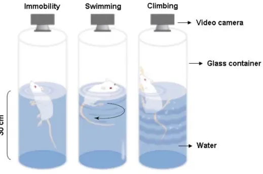
Rat forced swim test Page 50
RESULTS
Chronic fluoxetine administration restores OD plasticity
in the adult visual cortex Page 51
Chronic fluoxetine administration promotes the recovery of
normal visual functions in adult amblyopic rats Page 52
Chronic treatment with fluoxetine
causes a reduction of intracortical inhibition Page 54
Chronic treatment with fluoxetine reactivates long-term potentiation
of neural transmission in the adult visual cortex Page 56
Modulation of GAD65/67 expression induced by
chronic fluoxetine administration Page 57
Chronic fluoxetine treatment increases BDNF expression
in the adult visual cortex Page 58
Cortical diazepam administration prevents the effects induced by
chronic treatment with fluoxetine in visual cortical plasticity Page 60
Antidepressant effects induced by chronic treatment with fluoxetine Page 61
DISCUSSION Page 65
Chronic fluoxetine administration restores OD plasticity in the adult
Chronic treatment with fluoxetine promotes the recovery of vision
in adult amblyopic rats Page 67
Shift in the intracortical inhibitory-excitatory balanced mediated by an increased serotonergic transmission following chronic
antidepressant treatment Page 68
GAD65/67 expression following chronic treatment with fluoxetine Page 70
BDNF protein expression in the adult visual cortex
Following antidepressant treatment Page 71
BDNF and GAD65/67 interaction in the adult rat visual cortex Page 73
Long-term potentiation of neural transmission in the adult rat
visual cortex following antidepressant treatment Page 74
Antidepressant-like behavioural response induced by
chronic fluoxetine administration Page 75
Potential clinical application for chronic antidepressant treatment
in amblyopia and other neurological disorders Page 76
REFERENCES Page 78
INTRODUCTION
Mood disorders and antidepressant treatments
Depression and amine neurotransmitters
Mood disorders are among the most prevalent forms of mental illness. These are afflictions whereby the prevailing emotional mood is distorted or inappropriate to the circumstances, with a lifetime incidence of 10–25% in women and 5–12% in men (Blazer, 2000). Such pathologies are recurrent, life threatening due to the risk of suicide, and a major cause of morbidity worldwide. The most severe of these afflictions are major depression and manic depression; the latter also called “bipolar disorder” because such patients experience alternating episodes of depression and euphoria (Miklowitz & Johnson, 2006).
Depression has been described for several millennia, nevertheless, a distinction between a disturbance of cognitive faculties (a thought disorder) and a disturbance of emotions (a mood disorder), was recently introduced during early development of modern classification of mental illnesses in the 20th century. The term melancholia, which means “black bile” in greek, was first used by Hippocrates around 400 B.C. to refer to an alteration of mood, which was thought to depend on the balance of four humors, blood, phlegm, yellow and black bile. An excess of black bile was believed to cause depression (see Kandel et al., 2000). Most symptoms of this pathology were recognized in ancient times, as were the contributions of innate predispositions and external factors in causing the illness. Clinically, depression is defined by a set of standard criteria which include: depressed mood, low self esteem, feelings of hopelessness, worthlessness and guilt, decreased ability to concentrate and think, altered appetite, insomnia or hypersomnia, low energy, increased agitation, decreased interest in pleasurable stimuli, e.g., sex, food, social interactions, and recurrent thoughts of death and suicide. A diagnosis of depression is made when a certain number of the above symptoms are reported for longer than a 2 week period of time, and when symptoms disrupt normal social and occupational functioning (reviewed in Nestler et al., 2002).
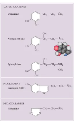
There are four effective treatments for depression and bipolar disorders: electroconvulsive therapy (ECT), antidepressant drugs, lithium, and anticonvulsants. Of all the treatments, ECT has been used for the longest period of time, over 50 years. Although the therapeutic mechanism of ECT is not fully understood, it is thought to be related to alterations in the sensitivity of aminergic receptors (Nestler, 1998). Amine transmitters regulate many brain functions and are also active in the peripheral nervous system. Because biogenic amines are implicated in a wide range of behaviors, ranging from central homeostatic functions to cognitive phenomena such as attention, it is not surprising that defects in biogenic amines function are implicated in most psychiatric disorders. There are five well-established amine neurotransmitters, the three catecholamines: dopamine, norepinephrine (noradrenaline), and epinephrine (adrenaline), as well as histamine and serotonin (Figure 1).
Figure 1. Amine neurotransmitters. The catecholamines, so named because they all share the catechol
moiety (i.e., a hydroxylated benzene ring), make up a distinctive subgroup within the biogenic amines. Serotonin and histamine contain an indole ring and an imidazole ring, respectively. Insert, space-filling model for norepinephrine. Taken from Purves et al., 2004.
All catecholamines are derived from a common precursor, the amino acid tyrosine. Dopamine is synthesized in the brain stem and hypothalamus. Dopaminergic neurons in the substantia nigra and ventral tegmental area provide a major ascending pathway that terminates in the striatum, the prefrontal and temporal cortex as well as the limbic system. Hypothalamic dopaminergic neurons instead, provide descending pathways to autonomic areas of the brain stem and the spinal cord (see Kandel et al., 2000). Dopamine is believed to be involved in motivation, reward and reinforcement, and plays also a poorly understood role in some sympathetic ganglia (Feldman et al., 1997).
Norepinephrine is used as a neurotransmitter in the locus coeruleus, a brainstem nucleus that projects diffusely to a variety of forebrain targets and influences sleep and wakefulness, attention and feeding behavior (reviewed in Berridge & Waterhouse, 2003). The most prominent noradrenergic neurons are sympathetic ganglion cells, which employ norepinephrine as the major peripheral transmitter. Epinephrine-containing neurons in the central nervous system, instead, are primarily in the lateral tegmental system and in the medulla and project to the hypothalamus and thalamus (Feldman et al., 1997). Serotonin (5-HT), in contrast, is synthesized from the amino acid tryptophan and is found in groups of neurons in the raphe regions of the pons and upper brain stem, which have widespread projections to the forebrain with an important role in sleep, wakefulness and behavioral arousal (Abrams et al., 2004).
Antidepressant drugs (ADs). Historical perspective.
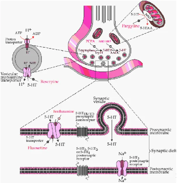
The functionality of amine synapses is critically important in psychopharmacology, with drugs affecting the synthesis, receptor binding, or catabolism of these neurotransmitters being among the most important agents of the modern pharmacology. Following their synthesis in the cytoplasm of presynaptic terminals, all the amine transmitters are packaged in synaptic vesicles and released into the synaptic cleft by means of exocytosis when the neurons fires an action potential. The neurotransmitter then interacts with pre- and post-synaptic receptors, and such activity is limited by active reuptake of the released molecule into presynaptic terminals as well as into glial cells (Feldman et al., 1997) (Figure 2). Inside the presynaptic terminals the
transmitters are packaged again into vesicles or catabolized primarily by the mitochondrial enzymes monoamine oxidase (MAO).
Figure 2. Serotonin synapse. Schematic diagram illustrating the synthesis, metabolism, neurotransmitter
release and pre-synaptic reuptake. Pre and postsynaptic receptors and sites of action of some antidepressant drugs are shown. Note that fluoxetine selectively inhibits the presynaptic reuptake of 5-HT, which enhances the postsynaptic serotonergic transmission. Taken from Feldman et al., 1997.
Drugs effective in treating depression act primarily on the serotonergic and noradrenergic systems of the brain (reviewed in Wong et al., 1995). The first drug used to ameliorate mood disorders, in particular schizophrenia, was reserpine. It was developed in the 1950s and initially used as treatment for hypertension because it blocks the noradrenergic transmission causing a reduction in the ability of the sympathetic division of the visceral motor system to cause constriction of blood vessels (Purves et al., 2004). Soon after, it was clear that a major side effect in hypertensive patients treated with reserpine was depression. It was then found that reserpine decreases serotonin and noradrenaline levels by inhibiting the uptake of neurotransmitters into synaptic vesicles, in the presynaptic terminal (see Figure 2), thereby keeping the transmitter into the cytoplasm where it undergoes degradation by the mitochondrial enzymes monoamine oxidase (Pletscher et al., 1956). This finding was of importance because it suggested the possibility that alterations in monoaminergic transmission might be involved in disorders of mood.
The monoamine hypothesis of depression, as first described in the 1960s, stated that depression was caused by a deficiency in the serotonergic and noradrenergic systems at functionally important receptor sites in the brain (Bunney et al., 1965; Schildkraut, 1965; Coppen, 1967). Indeed, the first drugs identified as antidepressants were discovered by chance when medications developed for other illnesses were found to elevate mood of psychiatric patients. Particularly, isoniazid, a drug initially used as treatment of tuberculosis, was found to ameliorate mood of patients receiving it. Later studies performed in depressed patients, who did not have tuberculosis, demonstrated an antidepressant effect of the drug. Isoniazid increases the concentration of serotonin and noradrenaline by interfering with the degradation of these neurotransmitters by the enzyme MAO, thus increasing its total content in the axonic terminals of neurons (see Figure 2). Further support to this notion came with the discovery that imipramine, an experimental antihistamine with a tricyclic structure, induced antidepressant effects. Imipramine blocks the reuptake of both serotonin and noradrenaline to the presynaptic nerve endings, thereby prolonging the action of these transmitters in the synapse (reviewed in Castren, 2005). These findings revolutionized the recognition and treatment of mood disorders shedding light into some of the molecular mechanisms involved in
depression. Because it became clear that both isoniazid and imipramine increase the extracellular levels of two important neurotransmitters in the brain: serotonin and noradrenaline; an increase in the total concentration of these neurotransmitters after treatment with these two drugs was the starting point for the proposal of the monoamine hypothesis of depression.
Neuronal circuitries affected in depression
Abnormalities in many brain areas are thought to mediate the diverse symptoms of depression. This notion is supported by human brain imaging studies which have demonstrated changes in blood flow in several brain regions, including areas of prefrontal and cingulate cortex, hippocampus, striatum, amygdala and thalamus. Similarly, anatomic studies of brains of depressed patients obtained at autopsy report abnormalities in many of these same brain regions (for review see Nestler et al., 2002). Knowledge of the function of such brain areas under normal conditions suggest the aspects of depression to which they may contribute. Neocortex and hippocampus may mediate cognitive aspects of depression, such as memory impairments and feeling of worthlessness, hopelessness, guilt and recurrent thoughts of suicide. The ventral striatum or nucleus accumbens, and the amygdale, are important in emotional memory and could as a result mediate the decreased drive and reward for pleasurable activities, anxiety, and reduced motivation that predominate in many patients (reviewed in Nestler et al., 2006).
A prominent mechanism by which the brain reacts to acute and chronic stress is the activation of the hypothalamic-pituitary-adrenal (HPA) axis. Under such conditions, neurons in the paraventricular nucleus (PVN) of the hypothalamus secrete corticotrophin releasing factor (CRF), which stimulates the synthesis and release of adrenocorticotropin (ACTH) from the pituitary gland. ACTH then stimulates the synthesis and release of glucocorticoids (cortisol in humans, corticosterone in rodents) from the adrenal cortex. Glucocorticoids exert profound effects on general metabolism and dramatically affects behavior via direct actions on numerous brain regions (reviewed by Nestler et al., 2002). The activity of the HPA axis is controlled by different brain pathways, including an inhibitory action exerted by the hippocampus and an excitatory action mediated by the
amygdala. Levels of glucocorticoids that are seen under normal physiological conditions seem to enhance hippocampal inhibition of HPA activity. They may also enhance general hippocampal functions and promote certain cognitive abilities. However, sustained elevations of glucocorticoids seen under conditions of stress or depression may damage hippocampal neurons particularly CA3 pyramidal cells (Sapolsky, 2000). Stress and the resulting increase in cortisol also reduce neurogenesis in the adult hippocampal dentate gyrus (Fuchs & Gould, 2000), which in turn reduces the inhibitory hippocampal action on the HPA axis; further increasing circulating glucocorticoid levels and subsequent hippocampal damage. Such positive feedback process with pathological consequences has been implicated in the onset of depression. Abnormal, excessive activation of the HPA axis is observed in approximately 50% of depressed patients and these abnormalities are corrected by antidepressant treatment (Arborelius et al., 1999; Holsboer, 2001).
Hypothesis for the etiology of depression
The pathologic effects of stress on hippocampus have contributed to a hypothesis for depression that proposes a role for neurotrophic factors in the etiology of this pathology and its treatment (Duman et al., 1997). Neurotrophic factors were first characterized by regulating neuronal growth and differentiation during development, but it has become clear that they are also potent regulators of plasticity and survival of adult neurons. The neurotrophic hypothesis of depression states that a deficiency in neurotrophic support may contribute to hippocampal pathology during the development of the illness, and that reversal of this deficiency induced by antidepressant treatments ameliorate the symptoms. Work in this hypothesis has focused on the brain derived neurotrophic factor (BDNF) (Reviewed in D’Sa & Duman, 2002), one of the most prevalent neurotrophins in the adult brain. Acute and chronic stress decrease levels of BDNF expression in the dentate gyrus and pyramidal cell layer of hippocampus in rodents. This reduction appears to be mediated partly via stress-induced glucocorticoids and partly by stress-induced decrease in serotonergic transmission (Smith et al., 1995). Conversely, chronic administration of virtually all classes of antidepressant treatments
increases BDNF expression in these regions (Nibuya et al., 1995), and prevent the stress-induced decrease in the neurotrophin levels. These findings raise the possibility that antidepressant induced upregulation of BDNF could repair some stress-induced damage to the hippocampus and protect vulnerable neurons from further damage. Furthermore, an enhancement of long-term potentiation and other forms of synaptic plasticity induced by BDNF in the hippocampus has been reported (Korte et al., 1996; Kang et al., 1997), which suggests that BDNF induced by antidepressants may promote hippocampal function. Importantly, these findings could also explain why the clinical antidepressant response show a time delay of several weeks: it would require sufficient time for levels of BDNF to gradually raise and exert its neurotrophic effects.
The neurotrophic hypothesis predicts that agents that promote BDNF expression and signaling might be clinically effective antidepressants, so different approaches focusing on understanding BDNF gene expression following antidepressant treatment have been developed. There is evidence for the transcription factor cAMP-response-element binding protein (CREB) regulating BDNF expression. For instance, BDNF expression is induced in vitro and in vivo by CREB (Tao et al., 1998; Conti et al., 2002) and all major classes of antidepressants increase levels of CREB expression and function in several brain regions including the hippocampus (Nibuya et al., 1996; Thome et al., 2000). An increase in CREB activity by microinjections of a viral vector encoding CREB into the hippocampal dentate gyrus exerts antidepressant-like effects, as evidenced in the forced swim test and learned helplessness test (Chen et al., 2001). In contrast, levels of CREB are reduced in the temporal cortex of depressed patients (Dowlatshahi et al., 1998). While these effects could be mediated by numerous target genes under control of CREB, it does illustrate novel strategies by which influencing hippocampal function in the context of depression, and makes likely a role for BDNF on it. Indeed, it has been demonstrated that single bilateral infusions of BDNF into the dentate gyrus of hippocampus or the overexpression of its primary receptor TrkB produce an antidepressant effect in behavioral models of depression (Shirayama et al., 2002; Koponen et al., 2005).
Importantly, experimental evidence arguing against the possibility that an impairment of BDNF-TrkB signaling may underlie the pathophysiology of depression
has been recently found. Mice lacking the tyrosine kinase receptor TrkB in the forebrain and transgenic mice overexpressing the dominant-negative T1 form of TrkB, for instance, show no depressive behavior (Zorner et al., 2003; Saarelainen et al., 2003). In contrast, in both BDNF knockouts and TrkB.T1 transgenic mice antidepressant treatment no longer induces a reduction of immobility in behavioral models of depression (Saarelainen et al., 2003), indicating that BDNF-TrkB signaling is necessary to mediate the behavioral response induced by antidepressants but may not be involved in the etiology of depression (reviewed in Martinowich et al., 2007).
Most of the clinical studies have focused on the hippocampus as the site involved in the generation and treatment of depression (Reviewed by Campbell & Macqueen, 2004). However, while the hippocampus is undoubtedly involved in the illness, it is unlikely that it accounts completely for these phenomena. The pathology in the hippocampus explains symptoms like alterations in learning and memory seen in depression but it does not represent the wide spectrum of symptoms. Brain imaging and autopsy studies have provided evidence for abnormalities in several brain areas of depressed individuals well beyond the hippocampus. The role of subcortical structures like the hypothalamus and amygdala in the regulation of motivation, sleep, appetite, energy levels, circadian rhythms, and responses to pleasurable stimuli, prominently affected in depressed patients, has also been recognized (reviewed in Nestler et al., 2002). Given the pervasive symptoms of depression, it is likely that the pathophysiology of this disorder and the mechanisms by which currently available treatments reverse symptoms, involve diverse neural circuits in numerous brain regions.
Recent experimental findings support an alternative hypothesis for depression which proposes a role for neurogenesis in its etiology. Magnetic resonance imaging studies have shown a reduction of hippocampal volume in depressed patients, event that has been correlated to a reduction of the number of hippocampal neurons (Videbech & Ravnkilde, 2004; Campbell et al., 2004). In addition, all clinically effective antidepressant drugs increase neurogenesis in the adult hippocampus (Malberg et al., 2000). Even though these findings indicate adult hippocampal neurogenesis as a candidate mechanism for the etiology of depression and as a substrate of antidepressant action, there is evidence arguing against it. For instance, at structural level it is highly
unlikely that changes in adult hippocampal neurogenesis account for the reduction of hippocampal volume observed in depressed patients. Stereological analysis of hippocampal volume in irradiated mice indeed, a strategy that impairs adult hippocampal neurogenesis, revealed no a significant reduction of volume in the hippocampus (Santarelli et al., 2003). In addition the fact that ablation of neurogenesis does not elicit a depression-like behavioral phenotype undermines a role for neurogenesis in the etiology of depression (Airan et al., 2007; Santarelli et al., 2003).
In contrast to the neurotrophic or neurogenic view of depression recent observations support an alternative hypothesis for such neurological disorder, which suggests that an impaired activity-dependent neuronal signaling may underlie this pathology and that antidepressant drugs might exert a therapeutic effect by enhancing information processing in the affected neuronal networks through activity-dependent mechanisms of synaptic plasticity (reviewed by Castren, 2005). This alternative idea, known as the network hypothesis, predicts that depression arise from an impaired synaptic plasticity in the CNS rather than from an alteration in the balance of signaling molecules. Evidence supporting this notion comes from the finding that disruption of adult hippocampal neurogenesis impairs the antidepressant-like behavioral response induced by antidepressants drugs (Santarelli et al., 2003). According to this idea, antidepressants may promote mechanisms of neuronal plasticity through an initial effect on monoamine metabolism, leading to an improved processing of information in neuronal networks involved in mood regulation (reviewed by Castren, 2005). Interestingly, differentiation of newly generated neurons takes several weeks (van Praag et al., 2002) which is a time course that correlates with the delayed onset of the therapeutic effects induced by antidepressant drugs. These findings suggest a role for neuronal plasticity in the action of ADs.
ADs and neuronal plasticity
Despite years of study, the biological basis of depression and the precise mechanisms of antidepressant efficacy remains unclear. Early research on depression focused on changes in neurotransmitter concentrations and its receptors. The results of
these studies, however, were in disagreement with clinical observations of the therapeutic action of ADs. Although the effects of antidepressants on monoamine metabolism occur soon after administration, it typically takes several weeks of continued treatment for the clinical antidepressant response to appear (Nestler et al, 1998). Further research made clear that long-term antidepressant treatment produces adaptive changes in monoamine receptors and intracellular signal transduction pathways associated (Sulser et al., 1978), which centered the attention on the effects of long-term antidepressant treatments on neurotrophic factors and coupled receptors as well as on intracellular signaling molecules (Duman et al., 1997; Manji et al., 2001; Coyle & Duman, 2003).
Increased BDNF expression induced by ADs
Research on the therapeutic effects induced by chronic administration of antidepressants has been developed focusing on the regulation of key signaling pathways involved in cellular survival, neurogenesis and neuronal plasticity in the adult brain. The fact that chronic antidepressant treatment increases the expression of BDNF and its primary receptor TrkB, in the frontal cortex and hippocampus of adult rats, was early demonstrated by Nibuya et al. in 1995 using in situ hybridization and northern blot analysis. Because neurotrophins were known to promote growth and development of immature neurons as well as to enhance the survival and function in neural cells in the adult (for review see Lindvall et al, 1994), a possible role for BDNF in mediating the therapeutic effects induced by antidepressants was suggested. That neurogenesis in the adult rat hippocampus is increased by chronic antidepressant treatment was elegantly shown by Malberg et al. (2000). In this study, different types of ADs were systemically given to adult animals and the acute and chronic effects on hippocampal neurogenesis were assessed by immunohistochemistry using the thymidine analogue bromodeoxyuridine (BrdU) as a marker for dividing cells. The authors found an increased number of BrdU labelled cells in the dentate gyrus of the adult rat hippocampus after chronic antidepressant administration but not after acute treatment. Moreover, an enhanced proliferation of hippocampal cells and differentiation into mature neurons was shown by combining BrdU labelling with specific markers for neuronal and glial cells.
These findings not only suggested a common molecular event induced by different ADs but highlighted a mechanism that is consistent with the time course for the therapeutic actions of antidepressants.
It has also been observed that adult neurogenesis and BDNF-induced TrkB signaling are critical molecular events in mediating the therapeutic effects of ADs. In a very elegant set of experiments Santarelli et al. (2003) showed that the antidepressant-like behavioral response to ADs is blocked if hippocampal neurogenesis is disrupted through a restricted x-irradiation in the mouse brain. These data provided a correlation between behavioral responses induced by antidepressants and the induction of hippocampal neurogenesis and suggest that generation of neural cells in the adult hippocampus may be necessary for the therapeutic action induced by ADs. On the other hand, single bilateral infusion of BDNF into the dentate gyrus of hippocampus was shown to induce an antidepressant effect in two behavioral models of depression: the learn helplessness and forced swim test (Shirayama et al., 2002). Furthermore, the authors evaluated whether a decreased phosphorylation of the primary BDNF receptor TrkB, induced by co-administration of BDNF with a broad spectrum tyrosine kinase inhibitor (K252a) into the hippocampus, impaired the antidepressant-like behavioral response induced by the neurotrophin in the learn helplessness test. K252a cortical administration completely blocked the behavioral effects induced by cortical BDNF administration. Taken together, these data suggest a correlation between BDNF-TrkB signaling and adult hippocampal neurogenesis, which may account for the behavioral effects induced by antidepressants. Experimental evidence in support to this notion comes from the finding that cortical administration of exogenous BDNF in adult animals not only increases CREB phosphorylation, but enhances long-term potentiation (LTP) of neural transmission in the adult hippocampus (Ying et al., 2002).
One important mechanism at the basis of the therapeutic action of ADs is the phosphorylation of the transcription factor cAMP-response element binding protein (CREB), which is known to promote BDNF expression. Direct evidence for the regulation of gene transcription via the cAMP-mediated second messenger pathway implicated in the actions induced by antidepressants was obtained in vivo using transgenic mice with a CRE-LacZ reporter gene construct (Thome et al., 2000). In these
mice, stimulation of the CRE site leads to an increase expression of the LacZ gene product, β-galactosidase, making it possible to evaluate the influence of antidepressant administration on CRE-mediated gene transcription. Levels of β-galactosidase assessed by fluorescence immunohistochemistry revealed an increased CRE-mediated gene transcription in limbic structures and cerebral cortex, induced by chronic treatment with three distinct classes of antidepressants. Furthermore, immunohistochemical analysis for p-CREB revealed that chronic antidepressant treatment increased phosphorylation of CREB, result that was consistent with the enhanced CRE-mediated gene expression. These findings, together with those of Nakagawa et al. (2002) in which the phosphorylation of CREB was shown to be required for hippocampal neurogenesis induced by the antidepressant rolipram, indicate a critical role for CREB phosphorylation in mediating the therapeutic effects of antidepressant drugs.
ADs and synaptic plasticity
Recent studies in animal models suggest that antidepressant treatments enhance synaptic connectivity in the brain. Particularly, an increased dendritic spine synapse formation in the adult rat hippocampus and frontal cortex was shown to be induced by chronic administration of the selective serotonin reuptake inhibitor (SSRI) fluoxetine (Hajszan et al., 2005). In depressed patients, brain imaging and neuropathological studies point toward a reduced neuronal activity and synaptic connectivity in the brain as evidenced by a decreased hippocampal and prefrontal cortex volume (reviewed by Castren, 2004) and antidepressant treatment seems to prevent or restore the structural and functional deficits observed in depression (Czeh et al., 2001).
The role of ADs in the remodeling and strengthening of specific neural synapses in the adult brain has also been addressed in rodents. Sairanen et al. (2007) evaluated the effect of chronic antidepressant treatment on the expression of three plasticity associated marker proteins: the polysialylated form of the nerve cell adhesion molecule (PSA-NCAM), the phosphorylated form of CREB (p-CREB) and the growth-associated protein 43 (GAP-43). PSA-NCAM is a cell surface protein involved in the regulation of axon growth (Cremer et al., 1997) and has been associated with the differentiation of newly
generated neurons in the rodent hippocampus (Seki & Arai, 1993), cell migration (Yoshida et al., 1999), synaptogenesis (Dityatev et al., 2004) and long-term potentiation of neural transmission (Muller et al., 1996; Cremer et al., 1998), while the phosphorylated form of CREB is known to be a permissive factor for neuronal plasticity. The GAP-43 protein is critically involved in synaptogenesis, axonal sprouting and the regulation of the cytoskeletal organization in the nerve ending (Benowitz & Routtenberg, 1997). Thus, Sairanen et al. (2007) assessed the expression of these proteins using immunohistochemistry to study mechanisms of synaptic plasticity induced by chronic treatment with the antidepressant imipramine on different areas in the forebrain of adult rats. Increased levels of PSA-NCAM, p-CREB and GAP-43 were found to be induced by chronic but not acute imipramine treatment in the hippocampus, medial prefrontal cortex and piriform cortex of adult animals, indicating an effect of antidepressants on the remodeling of neuronal networks.
Recent experimental evidence also suggests that a disturbance of brain plasticity is involved in animal models of depression and that chronic antidepressant treatment may counteract these alterations. In a set of elegant experiments adult rats were exposed to chronic mild stress conditions for three weeks to examine later long-term synaptic plasticity in the hippocampal CA1 region by whole-cell patch clamp (Holderbach et al., 2007). The authors found that chronic mild stress facilitated long-term depression (LTD) and had no effect on long-term potentiation (LTP), while chronic treatment of the SSRI fluvoxamine during the stress paradigm prevented the induction of LTD and increased the extent of LTP induction. Moreover, LTP was shown to be enhanced in non-stressed animals treated with fluvoxamine compared to stressed and non-stressed rats, which suggested an effect of antidepressants on synaptic plasticity of normal subjects.
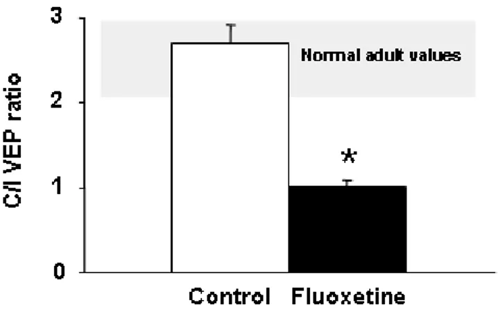
More recently, Normann et al. (2007) assessed this notion by using visual evoked potentials (VEPs) as a model to study cortical responses and its plasticity in the human visual system. Since repeated presentation of visual stimuli is known to produce a form of learning which subsequently improves the perception of these stimuli, the authors were able to assessed synaptic transmission in depressed patients and healthy individuals after chronic treatment with the SSRI sertraline. The neuronal correlates of perceptual learning in the visual system have been studied in awake mice (Frenkel et al., 2006), in which the
enhancement of evoked responses after repetitive presentation of visual stimuli is thought to reflect an increased long-term synaptic plasticity, as similarly suggested for the human visual system (Teyler et al., 2005). Normann et al. (2007) showed that chronic antidepressant administration in healthy human subjects increases the amplitude of early components (P1 and N1) of visual evoked potentials (VEPs) in response to repeated presentation of visual stimuli, whereas the polarity of such modulation was inverted in depressed patients. Taken together, these data suggest a role for neuronal plasticity in the action of ADs. How changes in plasticity are translated into the therapeutic effects induced by ADs is, however, currently unknown.
Critical periods for experience-dependent plasticity
The rich diversity of human personalities, abilities, and behavior is undoubtedly generated by the uniqueness of individual human brains. These fascinating neurobiological differences among human beings derive from both genetic and environmental influences. The first steps in the construction of neural circuitries, i.e., the establishment of distinct brain regions, the generation of neurons, the formation of major axon tracts, the guidance of growing axons to appropriate targets, and the initiation of synaptogenesis, rely largely on the tight interaction between genes and environment. Once the basic patterns of neural connections in the brain are established, however, patterns of neuronal activity (including those that are elicited by experience) modify the synaptic circuitry of the developing brain. Neuronal activity generated by interactions with the external world in postnatal life thus provides a mechanism by which the environment can influence brain structure and function.
The neural mechanisms that result from genetic and environmental interactions and their developmental consequences are sufficient to create some remarkably sophisticated innate behaviors. For most animals, the behavioral repertoire, including foraging, fighting and mating strategies, largely relies on patterns of connectivity established by intrinsic developmental mechanisms. However, the nervous system clearly adapts to and is influenced by the particular circumstances of an individual’s environment. These environmental factors are especially influential in early life, during
temporal windows called “critical periods”. Psychologists and ethologists have long recognized this notion, that early postnatal life is a period of special sensitivity to environmental influences on animal’s behavior. It is thought to be a critical period during which a given behavior is especially susceptible to, an indeed requires, specific environmental influences to develop normally (Purves et al., 2004). Once this period ends, such behavior is largely unaffected by subsequent experience and the failure to be exposed to appropriate stimuli during the critical period causes alterations that are irreversible on late developmental stages.
Avian song learning
The existence of critical periods has been demonstrated in several species (reviewed by Berardi et al., 2000) which include, for instance, song in birds and language in humans. Particularly well characterized is the sensitive period for learning courtship songs by oscine songbirds such as canaries and finches (reviewed by Doupe & Kuhl, 1999). In these species, the quality of early sensory exposure is the major determinant of subsequent perceptual and behavioral capabilities. The developmental periods for learning these and other behaviors are restricted during postnatal life, suggesting that the nervous system changes in some manner to become refractory to further experience.
Thus, avian song learning illustrates the interactions between intrinsic and environmental factors in this developmental process. Many birds sing to attract mates, but oscine songbirds are special in that their courtship songs are dependent on auditory and vocal experience. The sensitive period for song learning comprises an initial stage of sensory acquisition, when the juvenile bird listens to and memorizes the song of a nearby adult male tutor (usually of its own species), and a subsequent stage of vocal learning, when the young bird matches its own song to the now memorized tutor model via auditory feedback. This sensory motor learning stage ends with the onset of sexual maturity, when songs become acoustically stable. Even constant exposure to other songs after sensory acquisition at the end of the sensitive period does not affect this memory: the songs heard during sensory acquisition, but not later, are those that the bird vocally mimics (Purves et al., 2004).
On the other hand, additional features of song learning indicate an intrinsic predisposition for this specialized form of vocal learning. First, juveniles often need to hear the tutor song only 10 or 20 times to then vocally mimic it many months later. Second, when presented with a variety of songs played from tape recordings that include their own and other species’ songs, juvenile birds preferentially copy the song of their own species, even with no external reinforcement (reviewed in Doupe & Kuhl, 1999). These observations show that juveniles are not really “naive,” but are innately biased to learn the songs of their own species over those of others. Therefore, intrinsic factors make the nervous system of oscine birds especially sensitive to songs that are species typical. It is likely that similar biases influence human language learning.
Language in human beings
In contrast to canaries and finches, human beings require large postnatal experience to produce and decode speech sounds that are the basis of language. The various forms of early language exposure, including the “baby talk” that parents and other adults often use to communicate with children as they begin to acquire language may actually serve to emphasize important perceptual distinctions that facilitate proper language production and comprehension (Purves et al., 2004). Importantly, for this linguistic experience to be effective it must occur in early life. The requirement for perceiving and practicing language during a critical period is apparent in studies of language acquisition in congenitally deaf children. Whereas most babies begin producing speech like sounds at about 7 months (babbling), congenitally deaf infants show obvious deficits in their early vocalizations, and such individuals fail to develop language if not provided with an alternative form of symbolic expression. If, however, these deaf children are exposed to sign language at an early age, they begin to “babble” with their hands just as a hearing infant babbles audibly (Petitto & Marenhette, 1991). This suggests that, regardless of the modality, early experience shapes language behavior.
Examples of pathological situations in which normal children were never exposed to a significant amount of language point toward the same notion. One well-documented case is that of a girl who was raised by deranged parents until the age of 13 under
conditions of almost total language deprivation. Despite intense subsequent training, she never learned more than a rudimentary level of communication (see Kandel et al., 2002). This and other examples of so-called “feral children” starkly define the importance of early experience for language development as well as other aspects of social communication and personality. In contrast to the devastating effects of deprivation on children, adults retain their ability to speak and comprehend language even after long periods without exposure to human communication. The normal acquisition of human speech is subject to a critical period: the process is sensitive to experience or deprivation during a restricted period early in life and is relatively refractory to similar experience or deprivations in adulthood.
Critical period for visual system development
Although critical periods for language and other distinctively human behaviors are in some ways the most compelling examples of this phenomenon, it is difficult to study the underlying changes in the human brain. A much clearer understanding of how changes in connectivity might contribute to critical periods has come from studies of the developing visual system in experimental animals with highly developed visual abilities, particularly cats and monkeys. In an extraordinarily influential series of experiments in the 1960s, David Hubel and Torsten Wiesel found that depriving animals of normal visual experience during a restricted period of early postnatal life irreversibly alters neuronal connections and functions in the visual cortex. These observations provided the first evidence that the brain translates the effects of early experience, i.e., patterns of neural activity, into more or less permanently altered wiring of neural circuitries.
Electrophysiological recordings from the primary visual cortex of adult cats evidenced that the two eyes differentially activated cortical neurons and cells with similar preference for one eye were grouped together into ocular dominance columns (Hubel & Wiesel, 1963). It became clear that sensory inputs from the two eyes are first integrated in the primary visual cortex, where most afferents from the lateral geniculate nucleus of the thalamus terminate. In most mammals the afferent terminals form an alternating series of eye-specific domains in cortical layer IV called ocular dominance columns,
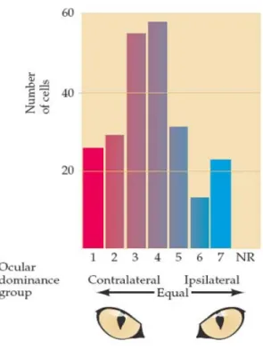
which are represented as stripes of cortical neurons that are driven only by stimulation of one eye or the other. All these units, together with orientation columns, are functionally organized into hypercolumns which process visual stimuli from discrete regions of the visual field. Thus, using electrophysiological recordings Hubel & Wiesel (1963) found that ocular dominance distribution across cortical layers of the visual cortex is roughly Gaussian in adult normal cats (Figure 3).
Figure 3. Ocular dominance distribution of single cell recordings in the visual cortex of adult cats. Cells in
group 1 and 7 are activated only by the contralateral or by the ipsilateral eye, respectively. Group 4 cells are driven by both eyes equally while 2/3 and 5/6 driven mainly by contra and ipsi eye, respectively. Taken from Purves et al., 2004.
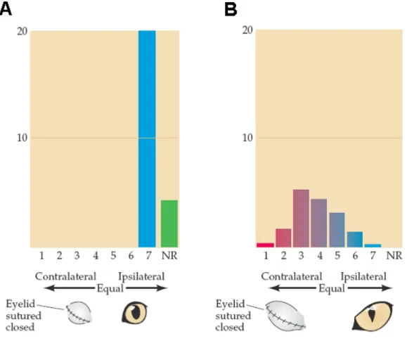
The researchers then asked whether this normal distribution of ocular dominance could be altered by visual experience. Using single cell recordings, they observed that occluding one eye early in development (a treatment referred to as monocular deprivation) led to the reduction in the number of cortical cells responding to that eye, in parallel with a robust increment in the number of neurons activated by the open eye (Wiesel & Hubel, 1963; Hubel & Wiesel, 1970) (Figure 4A). In contrast, monocular deprivation has no effect on ocular dominance distribution of adult animals (Figure 4B).
Figure 4. Effect of early closure of one eye on the ocular dominance distribution in the visual cortex. (A)
Following closure of one eye from 1 week after birth until 2.5 months, no cells could be activated by the deprived contralateral eye. Some cells could not be activated by either eye (NR). (B) A much longer period of monocular deprivation in an adult cat had little effect on ocular dominance distribution, though the overall cortical activity was diminished. Taken from Purves et al., 2004.
Recordings from the retina and lateral geniculate layers related to the deprived eye indicated that these more peripheral stations in the visual pathway worked quite normally. Thus, the absence of cortical cells that responded to stimulation of the closed eye was not a result of retinal degeneration or a loss of retinal connections to the thalamus. Rather, the deprived eye had been functionally disconnected from the visual cortex. Consequently, such animals are behaviorally blind in the deprived eye.
Because the same treatment is completely ineffective in the adult animal (Figure 4B), this early temporal window characterized by enhanced plasticity in response to experience is a typical example of a critical period. The shift in the ocular dominance distribution in response to monocular deprivation has been reported to occur in all mammals tested (reviewed by Berardi et al., 2000), accompanied by other dramatic effects such as poor development of visual acuity for the deprived eye, a condition known as amblyopia (reviewed in Odom, 1983) which is a permanent pathology in the adult. Even if the formerly deprived eye is subsequently left open indefinitely, little or no recovery of visual acuity occurs. Similar experiments in the monkey have shown that the same phenomenon occurs in primates, although the critical period is longer than in cats, up to six months (Horton & Hocking, 1999).
Determinant factors of the critical period for visual cortical plasticity
A further question for understanding how experience modulates neural circuits during critical periods is how patterns of activity are transduced to modify neural connections and to make these changes permanent. The ability of the central nervous system to process external stimuli into long term changes rely on activity dependent neural mechanisms that induce structural and functional modifications which underlie, for instance, processes of learning and memory. Extensive research during the last years shed light on determinant aspects of critical periods for experience-dependent plasticity. The mechanisms that modulate neural circuits functioning and connectivity rely on signals generated by the synaptic activity associated with sensory experience or motor performance, the basic neural processes by which experience is represented. A number of different signaling molecules, including NMDA receptors, neurotrophic factors, the
activity of the CRE-CREB system, extracellular matrix molecules and the GABAergic inhibitory transmission have been recognized as important regulators of visual cortical plasticity through changes that occur with correlated neural activity (reviewed by Berardi et al., 2003).
Glutamatergic NMDA receptors
Experimental evidence for the role of NMDA receptors in the regulation of visual cortical plasticity first came from the finding that blockade of such receptors inhibits the effects of monocular deprivation (Bear et al., 1990). The use of NMDA receptors antagonists and antisense oligonucleotides to reduce the expression of the NR1 subunit, without affecting visually driven activity, confirmed this notion (Roberts et al., 1998; Daw et al., 1999), demonstrating the NMDA receptor involvement in visual cortical plasticity. Additionally, the expression of NMDA receptors is developmentally regulated and modified by electrical activity. Their subunit composition varies in the visual cortex, from an increased expression of NMDA receptors containing the 2B subunit to a high presence of receptors containing the subunit 2A, with a time course that nearly parallels that of the functional development of the visual cortex. The expression of the NR2A subunit also correlates with the progressive diminishment of NMDA mediated currents (Flint et al., 1997). Dark rearing, which delays the critical period closure and impairs functional properties of the visual cortex, delays both the developmental shortening of NMDA receptor currents and expression of the NR2A subunit, suggesting a possible role for the 2B-to-2A switch in the closure of the critical period (Philpot et al., 2001). Nevertheless, experimental evidence against a causal role of NMDA receptors in determining the closure of the critical period comes from the finding that transgenic mice engineered to maintain prolonged NMDA responses by deletion of the NR2A subunit, show an unaltered sensitivity to MD which was restricted to the typical critical period and normally delayed by dark rearing from birth (Fagiolini et al., 2003). Moreover, western blot analysis confirmed a late postnatal onset of NR2A protein expression in wild type animals (P18) well in advance of the critical period (P28). Taken together, these observations indicate that the late onset and experience-dependent profile of NR2A
subunits, known to determine LTP occurrence in the hippocampus, is not relevant for critical period expression in the visual cortex (reviewed by Hensch, 2005).
Neurotrophic factors
Experimental evidence that link neurotrophins and neuronal plasticity, particularly in the neocortex and hippocampus, came out in the 1990s. One of the first evidences regarding neurotrophic factors and visual cortical plasticity came from the work of Maffei et al., in 1992, who demonstrated that the physiological shift in ocular dominance distribution could be prevented by infusion of the nerve growth factor (NGF) into the cortex during the critical period. Later, Cabelli et al., in 1995, evidenced that infusion of BDNF and the neurotrophin 4/5 (NT4/5) during the critical period, but not NGF, prevented the formation of ocular dominance columns in the cat visual cortex; finding of importance not only because it showed that neurotrophins modulate the patterning of projections within the visual cortex, but also because it suggested that thalamic axons compete for limiting amounts of neurotrophic factors.
Although plausible, such experiments, did not address the role of neurotrophins in activity dependent plasticity, since the effects observed still could be explain by a growth promoting action of BDNF and NT4/5 on thalamic axons within layer IV of the cortex, independent of neuronal activity. The requirement of neuronal activity for the occurrence of BDNF effects was evidenced by McAllister et al., in 1996, who demonstrated that inhibition of spontaneous electrical activity, glutamatergic synaptic transmission or inhibition of activation of L-type Ca2+ channels, prevented the BDNF induced dendritic growth in the visual cortex.
In addition, the requirement of electrical activity for the occurrence of the effects induced by NGF on ocular dominance plasticity has also been demonstrated. It was tested in animals subjected to complete monocular blockade of retinal discharges through intravitreal tetrodoxin (TTX) injections, at the peak of the critical period, while NGF was concurrently delivered into the visual cortex by osmotic minipumps. Analysis of single cell recordings revealed that while infusion of NGF is effective in preventing the ocular
dominance shift in young monocularly deprived rats, no rescue can be observed in TTX injected animals intracortically infused with NGF (Caleo et al., 1999).
Striking evidence for the role of BDNF in regulating the critical period for visual cortical plasticity came from studies performed in transgenic mice overexpressing BDNF in the visual cortex, with no alterations in the normal cellular pattern of expression or release and an effectively restricted expression of the neurotrophin in excitatory neocortical neurons (Huang et al., 1999). In these animals BDNF overexpression accelerates both the development of visual acuity and the time course of ocular dominance, thus supporting a crucial role for neurotrophins in visual cortical development and plasticity. The reciprocal regulation between neurotrophins and electrical activity seems to provide a mechanism by which active neuronal connections are selectively strengthened. As mentioned before, neurotrophins seem to require the presence of electrical activity to exert their actions (Sala et al., 1998; Caleo et al., 1999). Indeed, it has been demonstrated that both weak synaptic plasticity and localized BDNF application, which by themselves do not alter synaptic efficacy, induces long-term potentiation of synaptic transmission (Kovalchuk et al., 2002), finding that suggest a synergistic effect of neurotrophins and electrical activity in promoting synaptic plasticity. Accordingly, although BDNF promotes the phosphorylation of the transcription factor cAMP-response-element binding protein (CREB), it evokes only weak CREB mediated gene transcription unless it is coupled with electrical activity (Hu et al., 1999).
Activity of the CRE-CREB system
The initial molecular events of neural plasticity, which are changes in synaptic efficacy that do not require protein synthesis, are followed by long-lasting changes in neural circuitries that depend on gene expression and subsequent synthesis of proteins (reviewed in Berardi et al., 2003). The intracellular signaling pathways underlying the integration of electrical activity and neurotrophin transmission, for instance, involve the activation of three different protein kinases: cAMP-dependent protein kinase (PKA), extracellular-signal-regulated kinase (ERK1/2) and Ca2+/calmodulin-dependent protein kinase II (CaMKII), whose activity is necessary for the shift of ocular dominance after
monocular deprivation in young animals (Taha et al., 2002; Di Cristo et al., 2001; Beaver et al., 2001). Each kinase is activated by specific patterns of extracellular signals and is tightly woven within a network of mutual interactions. Activation of such intracellular pathways leads to changes in gene expression mediated by an up-regulation of transcription factors like egr1/zif68 and CREB, and subsequent protein synthesis, as demonstrated for ocular dominance plasticity in the visual cortex (Mower et al., 2002). Activated kinases must translocate to the nucleus where they phophorylate CREB, which then initiates the expression of genes under control of the cAMP-response element (CRE) promoter. Then, the consequent production of transcripts essential for establishment and maintenance of plasticity, does take place (reviewed by Silva et al., 1998).
A critical role for the CRE-CREB system in regulating the expression of genes involve in the physiological plasticity events during postnatal neocortical development, using transgenic mice carrying a CRE-lacZ reporter, has been demonstrated as well (Pham et al., 1999). It was found that calcium- and cAMP-regulated signaling pathways in visual cortical neurons are activated by monocular deprivation in young animals, events that precede the plastic modification typically observed after manipulations of visual stimuli early in development. Indeed, immunofluorescence analysis of visual cortical sections of CRE-lacZ transgenic mice monocularly deprived during the critical period, demonstrated an increased number of lacZ-positive cells in the visual cortex contralateral to the deprived eye. Moreover, the induction of these molecular events was dramatically down-regulated following the end of the critical period for visual cortical plasticity (Pham et al., 1999). These results show that CREB mediated gene expression is involved in the occurrence of visual cortical plasticity during development and suggest that a reduction in the activity of the CRE-CREB system may be implicated in the decline of plasticity observed during late postnatal development.
Extracellular matrix molecules
A correlation between the extracellular environment and adult visual cortical plasticity has also been demonstrated. There is evidence indicating that removal of important components present in the extracellular environment of the central nervous
system is necessary for experience-dependent plasticity to take place. The extracellular protease tissue plasminogen activator (tPA), for instance, is induced by electrical activity as an immediate early gene (Qian et al., 1993) and its proteolytic activity in the visual cortex is increased during monocular deprivation (Mataga et al., 2002). Initial evidence for the role of tPA in visual cortical plasticity came from the work of Mataga et al., in 1996, with the finding that its pharmacological inhibition attenuates the ocular dominance shift induced by monocular deprivation in young animals. This finding was then confirmed through pharmacological studies of the effects of monocular deprivation on tPA-knockout mice. Ocular dominance plasticity in these mice is impaired and can be rescued by exogenous tPA administration (Mataga et al., 2002). tPA has a wide spectrum of possible molecular targets, including extracellular matrix proteins (Wu et al., 2000), growth factors (Yuan et al., 2002), membrane receptors and cell adhesion molecules (Endo et al., 1999) which suggest an important role for this protease in visual cortical plasticity.
Additional data confirming the inhibitory action of the extracellular environment on visual cortical plasticity came from the work of Pizzorusso et al., in 2002, which focused on a class of glycoproteins that are major components of the extracellular matrix, the chondroitin-sulphate proteoglycans (CSPGs). Such molecules are composed of a core protein and CSPG glycosaminoglycan chains, which show a wide expression in the central nervous system, where they give a structural support to the extracellular environment acting mainly as physical barriers (Faissner & Steindler, 1995). CSPGs typically condense in lattice-like structures designated as perineuronal nets (PNNs) during postnatal development (Hockfield et al., 1990; Bruckner et al., 2000), and completely ensheath inhibitory interneurons assuming a perisynaptic localization at sites of synaptic contacts (Zaremba et al., 1989; reviewed by Celio et al., 1998). In a set of very elegant experiments, Pizzorusso et al. (2002) showed that the developmental maturation of the extracellular matrix was inhibitory for experience-dependent plasticity in the visual cortex. The authors used immunohistochemical analysis for Wisteria Floribunda Agglutinin (WFA), which binds to CSPG glycosaminoglycan chains, to demonstrate that the formation of adult-like PNNs around inhibitory interneurons coincided with the end of the critical period. Then, they showed that rearing animals in
complete darkness from birth, a strategy that is known to prolong the critical period for ocular dominance plasticity (see Berardi et al., 2000), inhibits the developmental maturation of PNNs. Soon after, the authors analyzed the effects of CSPGs degradation
in vivo with the enzyme chondroitin ABC (chABC) on visual cortical plasticity. This
treatment caused a degradation of PNNs in the adult visual cortex and reactivated plasticity for ocular dominance in monocularly deprived adult rats, demonstrating that developmental maturation of PNNs contribute to the progressive reduction of plasticity that occurs in the visual cortex at the end of the critical period.
That degradation of extracellular matrix components is a useful strategy to promote structural and functional recovery from visual deficits in the adult visual cortex, was recently demonstrated by Pizzorusso et al. (2006). In this case, long-term monocularly deprived animals were subjected to reverse suture in parallel to intracortical injections of the enzyme chABC which digest glycosaminoglycan chains of CSPGs, and single-unit recordings were then performed. The electrophysiological analysis showed a complete recovery of ocular dominance distribution in the visual cortex contralateral to the long-term deprived eye in chABC treated animals. Moreover, full recovery of both visual acuity and receptive field size of the long-term deprived eye was shown to be induced by chABC treatment. Thus, the authors demonstrated that intracortical infusion of chABC, coupled to reverse lid-suturing, favored the recovery of vision in adult rats with normal visual functions permanently impaired after long-term monocular deprivation.
A critical role for myelin-associated proteins in adult visual cortical plasticity, which include ligands for the Nogo receptor (NgR) has also been recognized. In particular, some proteins that are components of the myelin sheath including Nogo and the myelin basic protein (MBP) have been found to be chemo-repulsive for growing axons. Indeed, such molecules display an important function during development of the central nervous system in mediating axon growth and guidance (Purves et al., 2004). In addition, axon growth inhibition after injury of the adult nervous system appears to reflect the activity of inhibitory signals produced by glia and other cells at the lesion site through NgR signaling.
As the vast majority of visual cortical neurons express the NgR, McGee et al. (2005) evaluated whether the NgR-mediated myelin inhibition of neurite outgrowth contributes to the closure of the critical period for ocular dominance plasticity in rodents. Initially, the authors demonstrated that maturation of intracortical myelination correlates with the end of the critical period. Afterwards, they used transgenic mice lacking the NgR to investigate the involvement of NgR-signaling in restricting OD plasticity in the adulthood. Analysis of adult NgR transgenic mice, well after the end of the critical period (P120), showed that OD plasticity persisted into adulthood since an OD shift of visual cortical neurons was elicited by MD. Moreover, it was demonstrated that transgenic mice lacking main ligand of the NgR: Nogo, showed a similar susceptibility to MD in the adulthood thus confirming that NgR-dependent mechanisms participate directly in restricting visual cortex experience-dependent plasticity (McGee et al., 2005). Intracortical GABAergic inhibition
It has become clear that inhibition has an important role in sculpting patterns of electrical activity. This action contributes to the detection of imbalance of activity between the afferents to single cortical neurons. A failure in the timing of arrival of synaptic inputs on a post-synaptic neuron has been correlated with a failure in plasticity. The fact that manifestation of visual cortical plasticity requires inhibitory transmission was first shown by Hensch et al. (1998), using transgenic mice lacking the 65-Kda isoform of the GABA-synthesizing enzyme GAD (GAD65). Experience dependent plasticity in young animals in response to monocular deprivation, is impaired in these transgenic mice. Normal plasticity in these animals is rescued by enhancement of GABAergic transmission, in the visual cortex, by means of benzodiazepines administration. Thus, if the intracortical inhibition is reduced in young animals, the critical period onset is delayed, suggesting that there is an inhibitory threshold to be surpassed before the critical period can start. In contrast, if the intracortical inhibition is precociously enhanced by diazepam administration (Fagiolini & Hensch, 2000) the critical period starts earlier.
It has also been demonstrated that the time course and closure of the critical period for visual cortical plasticity is mediated by the maturation of intracortical inhibition, which involves BDNF expression. The development of intracortical inhibition, indeed, is accelerated in BDNF overexpressing mice (Huang et al., 1999) which suggest that BDNF controls the time course of the critical period by accelerating the maturation of the GABA-mediated inhibition. It is known that intracortical inhibition provides strong control over activity-dependent synaptic plasticity (Artola & Singer, 1987; Kirkwood & Bear, 1994) and matures slowly in comparison to excitation. It has been suggested that such a developmental mismatch between inhibition and excitation provides a temporal window for the critical period, when the organization of neuronal circuitries can be strongly influenced by sensory experience. Thus, the maturation of intracortical inhibitory circuitries sets the threshold for both the start and the end of the critical period. Consistent with this notion, dark rearing which prolongs the closure of the critical period also delay the development of intracortical inhibition (Fagiolini et al., 1994; Benevento et al., 1992).
The use of in vitro models of neuronal plasticity, term potentiation and long-term depression (LTP/LTD) of neural transmission, has also shed light on the molecular mechanisms underlying activity-dependent modifications of synaptic plasticity that depend on intracortical inhibition. In particular, reliable LTP of synaptic responses in layer II/III can be elicited by theta burst stimulation of layer IV in visual cortical slices of both young and adult animals. In contrast, LTP in layer II/III after electrical stimulation from the white matter (WM-LTP) can be obtained only in slices from the visual cortex of young rats but not in those of adult animals. Kirkwood & Bear (1994) first suggested that the maturation of inhibitory circuitries in layer IV is one of the mechanisms responsible for the closure of the critical period for the occurrence of WM-LTP. Indeed, theta burst stimulation from the white matter, in the adult rat, failed to induce LTP unless a GABAA receptor antagonist was applied to visual cortical slices. Notably, susceptibility to WM-LTP roughly coincides with the critical period for ocular dominance plasticity and, as the critical period, can be prolonged by dark rearing (Kirkwood et al., 1995). In support to this hypothesis, BDNF overexpressing mice, which show an accelerated maturation of intracortical inhibition, displayed an accelerated developmental decline of WM-LTP in
the visual cortex. The magnitude of LTP in wild-type mice undergoes a sharp decline between the fourth and fifth postnatal weeks, whereas in transgenic mice such decline in LTP occurred one week earlier (Huang et al., 1999). Moreover, it has also been demonstrated that WM-LTP can be rescued by blocking intracortical inhibition in slices derived from BDNF transgenic mice.
As to the diversity of intracortical inhibitory networks involved in the occurrence of plasticity in the visual cortex, the use of transgenic knockin animals with mutations in particular α-subunits of GABAA receptors, has given the opportunity to analyze whether specific GABA circuitries underlie plasticity in the visual system (Fagiolini et al., 2004). Because specific GABAA receptor-mediated currents can be enhanced by certain benzodizepines, whose sensitivity is determined by particular α-subunit composition, the triggering of ocular dominance plasticity through cortical administration of the benzodiazepine diazepam in parallel to monocular deprivation in young animals with point mutations in the α-1 α-2 and α-3 subunits that contribute to the benzodiazepine binding site, showed that only α1-containing inhibitory GABAergic circuitries regulate the expression of the critical period for visual cortical plasticity. In particular, point mutations of a histidine (H) to an arginin (R) in different subunits: 1(H101R), α-2(H101R) and α-3(H101R), which renders individual GABAA receptors insensitive to diazepam, were used. The authors initially showed that wild-type mice (in which normally no plasticity occurs after brief monocular deprivation soon after eye opening) that were cortically treated with the benzodiazepine agonist zoldipem, showed a marked ocular dominance shift in favor of the non deprived eye. Afterwards, they went on to examined ocular dominance plasticity in α-1(H101R) animals and found that no shift of ocular dominance occurred after cortical infusion of diazepam in parallel to brief monocular deprivation. In contrast, α-2(H101R) animals displayed an ocular dominance shift of visual cortical neurons similar to that of control animals after benzodiazepine administration (Fagiolini et al., 2004).
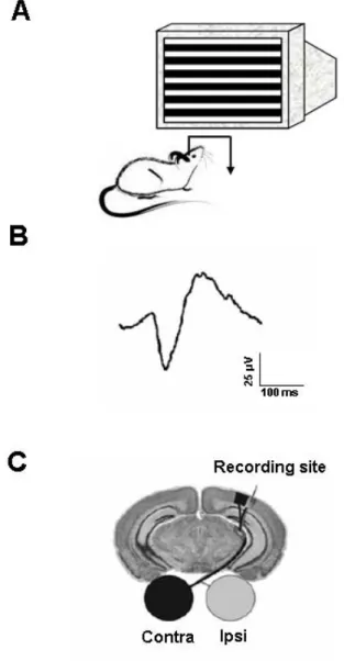
Visual Evoked Potentials (VEPs) and visual acuity
The visual evoked potentials (VEPs) in response to sinusoidal gratings alternated in phase several times per sec have proved to be very useful for the study of visual functions in man and animals. The extensive application of this technique is based on the fact that it does allow to predict either the psychophysical contrast sensitivity or limit of spatial resolution. Indeed, extrapolation to zero amplitude of the regression line obtained by plotting VEP amplitude against log contrast or log spatial frequency of the stimulus grating, gives contrast sensitivity or visual acuity values which are very similar to the psychophysical estimations (Campbell & Maffei, 1970). This has been evidenced not only in man but also in other animal species in which the subjective contrast thresholds or acuity estimations were behaviorally evaluated (Campbell et al., 1973; Nakayama & Mackeben, 1982). Because of the coincidence between contrast threshold estimated from psychophysical and from electrophysiological data, VEPs are assumed to be a reliable technique used to predict the subjective contrast sensitivity in all those cases in which other techniques are difficult to apply (Maffei & Fiorentini, 1990).
The pattern-reversal VEPs have also been useful to study the postnatal development of visual contrast sensitivity in infants (Pirchio et al., 1978) and to investigate the physiological mechanisms underlying the processing of spatial information in man as well. In particular, evidence has been found supporting the existence in the human visual cortex of orientation channels (Maffei & Campbell, 1970) and of spatial frequency channels (Fiorentini et al., 1983). In addition, methods based on the recording of pattern VEPs have been successfully applied in infants to study the postnatal development of orientation and spatial frequency selectivity (Fiorentini et al., 1983). Moreover, VEP techniques have become an important tool in clinical practice both in adults and young children with neurological diseases.
Initial studies aimed at identifying the site of origin of the pattern reversal VEP suggested that pre- and post-synaptic mechanisms very likely contributed to VEPs (Maffei & Fiorentini, 1990). The contribution of post-synaptic components in the generation of VEPs is highlighted by the fact that VEPs share some of the properties of visual cortical cells such as, for instance, orientation selectivity (Hubel & Wiesel, 1962)