Case Report
Asymptomatic carotid acute dissection following focal status epilepticus
Fedele Dono
b,⁎
, Francesca Anzellotti
a, Mirella Russo
b, Claudia Carrarini
b, Stefania Nanni
a, Camilla Ferrante
b,
Maria Vittoria De Angelis
a, Marco Onofrj
ba
Department of Neurology, SS Annunziata Hospital, Chieti, Italy b
Department of Neuroscience, Imaging and Clinical Sciences, University G. d'Annunzio of Chieti-Pescara, Italy
a r t i c l e i n f o
Article history: Received 4 February 2019
Received in revised form 4 March 2019 Accepted 11 March 2019
Available online 17 March 2019
1. Introduction
Carotid artery dissection (CAD) is one of the most important predis-posing factors of cerebral ischemia in young adults. Incidence rate for CAD has been reported to be 1.72 per 100,000[1]. Various clinical symp-toms associated with CAD may be benign (e.g., headache, neck pain, Horner's syndrome, cranial-nerve palsy) or more severe such as stroke and transient ischemic attack (TIA). Seizures rarely represent a predis-posing cause of CAD, although carotid artery dissection has been re-ported in association with minor trauma.
2. Case report
A 45-year-old right-handed man, with personal history of epilepsy beginning at the age of 10, was admitted to our emergency department because of sudden onset of jerking movements of the head to the right associated with blinking of the right eye with narrowing of the homolateral palpebralfissure and right hemi-facial myoclonus starting approximately 30 min prior to arrival. During the neurological examina-tion, the patient was restless and incapable to follow commands and an-swer questions. A 21 channel EEG recording demonstrated continuous left fronto-temporal spike–wave complexes. The patient was then treated with a bolus of lorazepam 8 mg and underwent continuous EEG monitoring. Over the next 10 min there was a complete normaliza-tion of the EEG with a resolunormaliza-tion of all neurological symptoms. General examination was unremarkable. Brain Magnetic Resonance Imaging
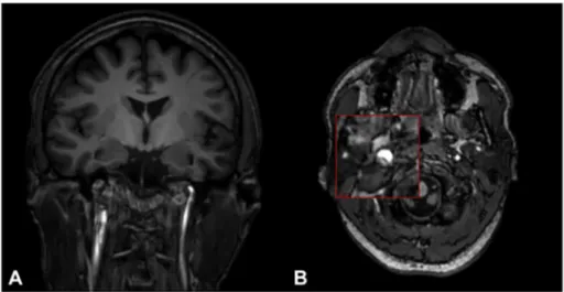
(MRI) and magnetic resonance angiogram (Angio-MRI) performed 24 h after recovery, revealed a right skull base carotid artery dissection without infarction (Fig. 1, A and B). Pre-morbid brain MRI had demon-strated normal carotidflow voids.
3. Discussion
CAD may represent a severe complication associated with epilepsy and seizures. In the previous literature, a case report of acute symptom-atic CAD after a generalized tonic–clonic seizure was described[2].
However, we report how focal motor seizures with the involvement of the neck may also represent a risk condition of developing CAD, in particular if the patient presents with status epilepticus or a series of focal motor seizures. Several anamnestic data could be useful in predicting the risk of CAD: trauma to the neck, infections, migraine, hy-perhomocysteinemia and 5,10-methylenetetrahydrofolate reductase gene (MTHFR) 677TT mutation represent the most well-known risk fac-tors associated with CAD[3]. Nevertheless, our patient's medical history was unremarkable with focal seizures representing the only predispos-ing risk factor for CAD.
4. Conclusion
We describe thefirst case of asymptomatic CAD to our knowledge associated with focal status epilepticus. We therefore speculate that CAD is an underestimated phenomenon in patients with epilepsy. Peri-odic eco-color Doppler of the carotids should be considered in patient with epilepsy following motor seizures, especially when the neck is traumatized in order to identify the potential life-threatening condition of CAD.
Funding
This research did not receive any specific grant from funding agen-cies in the public, commercial, or not-for-profit sector.
Declaration of interest None.
Conflicts of interest
The authors affirm that they do not have conflicts of interest.
Epilepsy & Behavior Case Reports 11 (2019) 120–121
⁎ Corresponding author at: Department of Neuroscience, Imaging and Clinical Sciences, University G. d'Annunzio of Chieti-Pescara, Via dei Vestini, 66100 Chieti, Italy.
E-mail addresses:[email protected](F. Dono),[email protected](M. Onofrj).
Contents lists available atScienceDirect
Epilepsy & Behavior Case Reports
j o u r n a l h o m e p a g e :w w w . e l s e v i e r . c o m / l o c a t e / e b c r
https://doi.org/10.1016/j.ebcr.2019.03.002
References
[1]Lee VH, Brown RD, Mandrekar JN, Mokri B. Incidence and outcome of cervical artery dissection: a population-based study. Neurology 2006;67:1809–12.
[2]Child Nicholas D, Cascino Gregory D. Carotid dissection following a generalized tonic– clonic seizure. Neurology May 2013;80(20):1911.
[3]Debette S, Leys D. Cervical-artery dissections: predisposing factors, diagnosis, and outcome. Lancet Neurol 2009;8:668–78.
Fig. 1. Magnetic Resonance Imaging (MRI) and Magnetic Resonance Angiography (MRA)of the brain performed after SE recovery. In A) Coronal T1 sequence and in B) Axial T1 sequence showing right skull base carotid artery dissection without infarction.
121 F. Dono et al. / Epilepsy & Behavior Case Reports 11 (2019) 120–121