Isolation and Partial Characterization of a Novel Cytorhabdovirus from Citrus Trees
Showing Foliar Symptoms in Iran
Mohammad Sadegh Sadeghi, Dr. Alireza Afsharifar,and Keramatollah Izadpanah, Professor, Plant Virology Research Center, College of Agriculture, Shiraz University, Shiraz, Iran; Giuliana Loconsole and Angelo De Stradis, Istituto per la Protezione Sostenibile delle Piante, UOS Bari, Consiglio Nazionale delle Ricerche, 70126 Bari, Italy; Giovanni P. Martelli, Dipartimento di Scienze del Suolo, della Pianta e degli Alimenti, Universit`a degli Studi di Bari Aldo Moro, 70126 Bari, Italy; and Maria Saponari, Istituto per la Protezione Sostenibile delle Piante, UOS Bari, Consiglio Nazionale delle Ricerche
Abstract
Sadeghi, M. S., Afsharifar, A., Izadpanah, K., Loconsole, G., De Stradis, A., Martelli, G. P., and Saponari, M. 2015. Isolation and partial character-ization of a novel cytorhabdovirus from citrus trees showing foliar symptoms in Iran. Plant Dis. 99:1-6.
Citrus ringspot is a graft-transmissible disease, and at least two taxonom-ically distinct viral species are associated with this syndrome: Citrus pso-rosis virus (CPsV) and Indian citrus ringspot virus (ICRSV). Neither of these two viruses was detected, however, by serological or molecular as-says in symptomatic tissues from citrus trees in southern Iran, where the ringspot syndrome is widespread. By contrast, electron microscopy and molecular assays revealed the presence of a rhabdovirus-like virus, which
was graft transmitted to several citrus species and mechanically to herba-ceous hosts. Virus particles were bacilliform and resembled rhabdovirus nucleocapsids deprived of the lipoprotein envelope. Partial sequences of the viral nucleoprotein and RNA polymerase genes showed a distant ge-netic relatedness with cytorhabdoviruses. This virus appears to be a novel species, for which the name Iranian citrus ringspot-associated virus (IrCRSaV) is suggested.
Chlorotic to yellow ringspots of citrus leaves were reported on cit-rus in different countries (Bov´e 1995; Roistacher 1993; Timmer et al. 2000). At least two taxonomically distinct RNA viruses have been associated with this symptomatology: Citrus psorosis virus (CPsV), the type member of the genus Ophiovirus, whose particles are naked filamentous nucleocapsids (Milne et al. 2000); and Indian citrus ringspot virus (ICRSV), the type member of the genus Mandarivirus, a virus with flexuous filamentous particles (Rustici et al. 2000, 2002).
A ringspot syndrome of sweet orange and several other citrus spe-cies, characterized by small chlorotic and necrotic rings, mostly along the veins of mature leaves, was observed in Iran in the late 1970s (Izadpanah and Rahimian 1978) and was graft transmitted to a number of citrus species (Amiri et al. 1992). This disease is wide-spread in the southern part of the country, where natural infections occur on sweet orange (Citrus sinensis), sweet lime (C. limetta), lemon (C. limon), citron (C. medica), and bakraee (C. reticulata hy-brid). Serological and molecular tests for the presence of CPsV in symptomatic trees were consistently negative, although the possible causal agent is thought to be an RNA virus (Yassaie and Izadpanah 2000).
Here, a study encompassing biological assays, electron micros-copy, and partial sequencing of the viral genome was carried out on sweet orange trees exhibiting ringspot syndrome. This study revealed the presence of a novel rhabdovirus-like virus associated with infected trees. A provisional name of Iranian citrus ringspot-associated virus (IrCRSaV) was proposed for this novel virus species.
Materials and Methods
Source of virus isolates.In 2012, 10 sweet orange trees with typ-ical symptoms were selected in 25- to 30-year-old groves in the Khafr region (100 km south east of Shiraz, Iran). Buds from each
tree were grafted on sour orange seedlings and maintained in a greenhouse (24 to 28°C) until symptoms appeared on the new leaves. Newly developed, symptomatic tissues were used in the ex-periments described hereafter. One of the 10 sources, designated CR-Ir, was selected for electron microscopy and molecular investigations.
Biological assays.Tissues from symptomatic leaves were macer-ated in the presence of 0.01 M potassium phosphate buffer, pH 7, and the extract was used for mechanical inoculation of 21 different her-baceous hosts in the families Amaranthaceae, Solanaceae, Chenopo-diaceae, Cucurbitaceae, Fabaceae, Malvaceae, and Geraniaceae (Table 1). Briefly, the plant sap was rubbed onto the surface of leaves previously dusted with carborundum. Leaves were then rinsed with water.
In addition, buds from symptomatic shoots were used for graft in-oculating a triplet set of 8- to 12-month-old seedlings of sweet orange (C. sinensis), Mexican lime (C. aurantifolia), sour orange (C. auran-tium), grapefruit (C. paradisi), and trifoliate orange (Poncirus trifo-liata). Inoculated plants and mock-inoculated controls of the same species were maintained in a greenhouse at 24 to 28°C, and observed for symptom appearance.
Electron microscopy.A Philips Morgagni 282D electron micro-scope was used for the observation of (i) leaf dips from symptom-atic tissues of the source CR-Ir and infected French bean mounted in 2% uranyl acetate (Milne 1993) and (ii) thin-sectioned leaf tis-sue fragments from the CR-Ir source, infected grapefruit, and healthy controls, processed as described by Martelli and Russo (1984).
Assays for known citrus viruses. Total nucleic acids were extracted from symptomatic leaves of each source, using a cetyltri-methylammonium bromide-based protocol (Saponari et al. 2013) and subjected to conventional or real-time polymerase chain reaction (PCR) or reverse-transcription (RT)-PCR assays to screen trees for the presence of the main citrus-infecting viruses that occurred in this region: CPsV, ICRSV, Citrus tristeza virus (CTV), Citrus leaf blotch virus, Citrus variegation virus, Citrus chlorotic dwarf-associated vi-rus (CCDaV), and Citvi-rus yellow vein clearing vivi-rus (CYVCV). Am-plification conditions were as previously described (Loconsole et al. 2010, 2012a,b; Roy et al. 2005; Ruiz-Ruiz et al. 2009; Sharma et al. 2009). Genus-specific primers were also used in RT-PCR assays for the identification of putative potyviruses (Ha et al. 2008), Corresponding author: M. Saponari; E-mail: [email protected]
Accepted for publication 20 June 2015.
http://dx.doi.org/10.1094/PDIS-02-15-0136-RE © 2015 The American Phytopathological Society
ophioviruses (Vaira et al. 2003), betaflexiviruses (Dovas and Katis 2003), and begomoviruses (Deng et al. 1994).
Random cDNA synthesis, cloning, and sequencing.To gain in-sight into the molecular properties of the putative viral agent, a random-primed cDNA library was constructed. Total RNA (1mg) was recovered from a partially purified preparation (Jackson and Christie 1977) from symptomatic leaves of French bean mechanically inoculated with CR-Ir. Briefly, first-strand cDNA was synthesized with random decamer nucleotides as primers (RETROscript Kit; Life Technologies). cDNA templates were then synthesized, converted into double-stranded DNA, blunt-ended with T4 DNA polymerase, and ligated into the plasmid pUC19. Thirty recombinant clones containing PCR products of 500 bp in size or larger were selected and sequenced in both directions.
Sequence analysis.Nucleotide sequences were compared with the nonredundant GenBank database using the BLASTX and BLASTN algorithms, and searched for conserved protein domains using the National Center for Biotechnology Information (NCBI) Conserved Domain Database (www.ncbi.nlm.nih.gov/cdd). Sequences that showed significant genetic homology to plant viral genomes were selected for further analyses. Phylogenetic analysis was done by the distance-based Clustal W method using the software MEGA 5 (Tamura et al. 2011).
Specific detection of IrCRSaV by conventional and quantitative RT-PCR.Two sets of specific primers (IrF-5¢-TCAT TAGACTGGGATAGAGC-3¢/IrR-5¢-CCAACTTCCCTGGCTTCT AG-3¢ and IrPol-F-5¢-CCCAGAACACATCTAGAGAGCAGGA-3¢/IrPol-r-5¢-CATATCGGGGGTTTGAAGGA) yielding PCR pro-ducts of 260 and 676 bp were designed based on the nucleoprotein (clone Ir90, GenBank accession number KP255975) and RNA po-lymerase (clone Ir119, GenBank accession number KP255976) gene sequences, respectively. Total RNA was isolated from the leaves using a commercial RNA isolation kit (DENAzist Asia). For conventional PCR, RT was carried out in a total reaction vol-ume of 25 ml at 42°C for 60 min using 5 ml of total RNA (at a
concentration of 50 ng/ml), adding 10 mM each dNTP, 200 U of M-MuLV (Fermentas), and 0.5mM reverse primer. PCR was then carried out in a 25-ml reaction volume following standard amplifi-cation conditions.
A quantitative one-step SYBR Green I-based RT-PCR system was also developed using the aforementioned primers IrF and IrR. Amp-lification was carried out in a 12.5-ml reaction mixture containing 1× SYBR Select Master Mix (Applied Biosystems), 320 nM each primer, and 0.15ml of M-MuLV (Fermentas). The thermal profile for one-step SYBR Green-based RT-PCR consisted of a 15-min RT step at 42°C and 10 min at 95°C, followed by 40 cycles at 95°C for 10 s, 55°C for 30 s, and 72°C for 30 s. Following amplifi-cation, a melting curve analysis was done to verify the correct prod-uct by its specific melting temperature (Tm). Melting curves of PCR amplicons were obtained with temperatures ranging from 65 to 90°C. Data were recorded for every 0.2°C of temperature at 10-s intervals. Ten-fold serial dilutions were prepared using the total nucleic acids extracted from the selected source CR-Ir, subjected to quantitative (q)RT-PCR, and the resultant quantitation cycles were used to deter-mine the efficiency of the assay.
All PCR reactions included positive, negative, and nontemplate controls. The specificity of the assay was verified by testing samples collected from the citrus virus collection at the University of Bari, which included CTV, CPsV, CCDaV, and CYVCV. Conventional and real-time PCR assays were validated on a panel of 40 field sam-ples, consisting of mature leaves collected from 30 symptomatic sweet orange trees and 10 symptomless trees. Assays were also ex-tended to assess the presence of the putative novel virus in young, symptomless leaves collected from symptomatic sour orange (C. aurantium) indicator plants.
Results
Field symptoms.Typical ringspot symptoms observed in most lo-cal sweet orange cultivars in Khafr consisted of small rings, etched lines, and chlorotic spots on both sides of the mature leaves (Fig. 1). The rings often followed the lateral veins and turned necrotic as the leaves aged. No specific symptoms were found on fruit. Not all branches of a tree were symptomatic but affected trees were less vig-orous than the healthy ones.
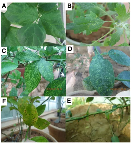
Host range and graft transmission.Among herbaceous hosts, Phaseolus vulgaris reacted with systemic vein clearing and extensive leaf mottling (Fig. 2A) 14 days postinoculation (dpi), whereas Che-nopodium quinoa (Fig. 2B) and C. amaranticolor showed chlorotic local lesions 10 dpi but no systemic symptoms.
Within 3 months from graft inoculation, chlorotic flecking, rings, and line patterns were observed on the leaves of trifoliate orange (Fig. 2C), Mexican lime, grapefruit, and sour orange (Fig. 2D). These le-sions later transformed into necrotic spots (Fig. 2E). Yellow mottling Table 1.Reaction of herbaceous hosts mechanically inoculated with Iranian
citrus ringspot-associated virus
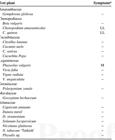
Test plant Symptomsa
Amaranthaceae Gomphrena globosa − Chenopodiacea Beta vulgaris − Chenopodium amaranticolar LL C. quinoa LL Cucurbitaceae Citrullus lanatus − Cucumis melo − C. sativus − Cucurbita Pepo − Leguminosae Phaseolus vulgaris M Vicia faba − Vigna radiata − V. unguiculata − Geraniaceae Pelargonium zonale − Mavalaceae Gossypium herbaceum − Solanaceae Capsicum annuum − Datura metel − D. stramonium − Solanum lycopersicum − Nicotiana glutinosa − N. tabacum‘Turkish’ − Physalis sp. −
aLL = local lesion, M = mosaic, and− = no symptoms.
BW
in
Fig. 1. Ringspot symptoms on mature leaves of sweet orange in the orchards of Fars Province, Iran.
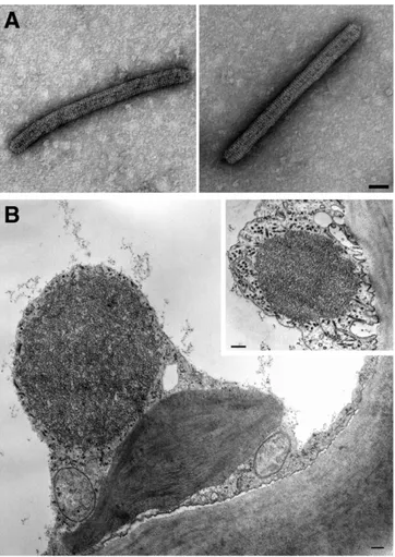
and ringspots were also seen on the stems of young shoots of trifoli-ate orange (Fig. 2F). Mock-inocultrifoli-ated seedlings remained symptom-less. All symptomatic hosts were positive for the presence of IrCRSaV when tested by RT-PCR using the specific primers IrF/IrR. Electron microscopy.Leaf dips from CR-Ir source and French bean showed the presence of bacilliform particles about 450 by 30 nm in size, showing prominent transverse striations and re-sembling very much nonenveloped rhabdoviral nucleocapsids (Fig. 3A). The particle size represents the mean of the measurements performed on 48 and 55 viral particles from citrus and bean, re-spectively. Measurements were performed using the software IMAGE 1.38, and statistic evaluations using KALEIDAGRAPH 3.2. In thin-sectioned samples, the most conspicuous cytopatho-logical feature was the presence in the cytoplasm of mesophyll cells of rounded electron-dense inclusion bodies with a finely fibrillar-granular texture, close to or apposed to the cell wall (Fig. 3B). These bodies were surrounded by dark dots and elon-gated structures (Fig. 3B, inset), which were interpreted as trans-versally and tangentially sectioned virus particles, respectively. On this account, these inclusions were tentatively identified as viroplasm matrices. No such inclusions were seen in the healthy controls.
Sequencing and phylogenetic analysis.Of the 30 nucleotide se-quences assembled from the selected clones, none shared significant similarity with the viral sequences from the NCBI database following BLAST analysis. However, the predicted amino acid sequences of two clones, denoted Ir90 (500 bp, GenBank accession number KP255975) and Ir119 (2,825 bp, GenBank accession number KP255976), appeared of possible viral origin because they shared an amino acid identity of 27 to 39% with nucleoproteins (N protein) and RNA polymerase (L protein) of the following cytorhabdovi-ruses: Northern cereal mosaic virus (NCMV), Lettuce yellow mottle
virus (LYMV), Lettuce necrotic yellows virus, and Barley yellow striate mosaic virus (BYSMV) (Table 2).
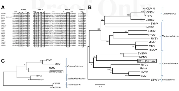
Furthermore, multiple alignment of the L protein genes identi-fied three conserved domains in the clone Ir119: (i) the catalytic do-main III (cl15638) of the L proteins of Mononegavirales and other nonsegmented negative-strand RNA viruses, which starts at nucle-otide position 2 and terminates at nuclenucle-otide position 2,290; this domain comprised the four distinct conserved motifs A, B, C, and D (Poch et al. 1990) (Fig. 4A), thought to play an essential role for RNA polymerase activity (Schnell and Conzelmann 1995); (ii) the mRNA-capping domain V (cl16796) of L RNA polymerase be-longing to nonsegmented negative-sense RNA viruses and Mono-negavirales, which included the motif G(SY)T(k)(n) HR from nucleotide position 2,578 to nucleotide position 2,810; and (iii) the mRNA capping C-terminal region (TIGR04198) shared by paramyxovirus-like RNA-dependent RNA polymerases (L protein).
In a phylogenetic tree constructed with the deduced amino acid se-quences of the partial L protein, IrCRSaV clustered with the cyto-rhabdoviruses BYSMV and NCMV on a branch starting from the same node but on a separate clade (Fig. 4B). Likewise, a phylo-genetic analysis based on the partial N protein genes positioned IrCRSaV among cytorhabdoviruses in the same clade as NCMV (Fig. 4C).
IrCRSaV detection.Total nucleic acids subjected to RT-PCR as-says amplified DNA bands of the expected sizes, 260 bp using the primers IrF/IrR (Fig. 5A) and 646 bp using the primers Ir-Pol-f/Ir-Pol-r (not shown), from all symptomatic indicator plants and the panel of 30 selected field trees showing typical disease symptoms. In addition, positive results were obtained for the five samples con-sisting of symptomless young leaves harvested from symptomatic graft-inoculated sour orange plants.
BW
in
Fig. 2. Symptoms in host plants mechanically inoculated with Iranian citrus ringspot-associated virus. A, Mottling and vein clearing in Phaseolus vulgaris; B, chlorotic local lesions in Chenopodium quinoa; C and D, chlorotic flecks, rings, and line patterns on the leaves of Poncirus trifoliata and Citrus aurantium; E, mottling on stems of P. trifoliata; F, necrotic spots appearing in the late stage of the infections.
The same samples tested negative when assayed for the presence of other known citrus viruses, further confirming that neither CPsV nor ICRSV are involved in the Iranian citrus ringspot disease.
In addition, IrCRSaV was readily detected using the one-step qRT-PCR protocol developed in this study, with resultant quantita-tion cycles of 18 to 23 from tissue prepared from symptomatic indi-cator plants and field samples (Fig. 5B). No signals were detected in the healthy and nontemplate controls, or in the samples harvested from symptomless trees. Following the Tm analysis, a single and spe-cific melting peak, with a Tm of 80 to 50°C, was obtained from all positive samples (Fig. 5C) The efficiency of the qRT-PCR assays was about 90%, close to the optimum value, as determined on the
basis of the regression coefficient (r2= 0.993) of the standard curve generated by 10-fold serial dilutions of the total nucleic acid prepa-ration (data not shown).
Discussion
Sequences of a novel viral pathogen (IrCRSaV) were consistently found in Iranian citrus trees affected by a ringspot syndrome, in ac-cordance with previous indications (Yassaie and Izadpanah 2000) that an unidentified graft-transmissible agent unrelated to CPsV and ICRSV is associated with these symptoms.
IrCRSaV has a genome sharing very little sequence homology with members of the family Rhabdoviridae, and has nonenvel-oped bacilliform particles resembling those of members of this family. Two nucleorhabdoviruses, Citrus necrotic spot virus and nuclear Citrus leprosis virus, with nonenveloped bacilliform par-ticles (40 to 50 by 100 to 110 nm) and a bipartite genome have been found in citrus trees (Cruz-Jaramillo et al. 2014; Roy et al. 2015). Both viruses share a high sequence identity (>90%) with Orchid fleck virus, and their inclusion in Dichorhavirus, a new genus in the family Rhabdoviridae (Dietzgen et al. 2014), has been proposed.
Phylogenetic analysis using the partial amino acid sequences of the N and L proteins showed that IrCRSaV clusters with cytorhab-doviruses and is genetically distant from the two putative dichorha-viruses reported previously in citrus (Cruz-Jaramillo et al. 2014; Roy et al. 2015). However, distinct differences between IrCRSaV and cytorabdoviruses include the size (450 by 30 nm versus 130 to 350 by 45 to 100 nm) and appearance (nonenveloped versus enveloped) of the virions and the presence of viroplasm matrices. Wide differences in particle appearance, genome structure, and cytopathological features exist also with members of other plant vi-rus genera such as Cilevivi-rus (Locali-Fabris et al. 2012) and Dicho-rhavirus (Dietzgen et al. 2014), both of which have nonenveloped smaller bacilliform particles (120 to 130 by 50 to 55 nm for cilevi-ruses and 100 to 150 by 40 to 45 nm for dichorhavicilevi-ruses) and a bi-partite genome.
Taken together, these results provide evidence that IrCRSaV is an undescribed virus with a distant phylogenetic relationship with members of the genus Cytorhabdovirus. Further work involving metagenomic approaches (i.e., next-generation sequencing tech-nology), purification, and insect vector transmission tests are planned for determining the taxonomic position of IrCRSaV, eluci-dating its biological, serological, and molecular features and its role in the etiology of the ringspot syndrome with which it is associated.
Because little information was available regarding the causa-tive agent of this ringspot disease, and its spread and incidence in Iran, no preventive or control measures have been undertaken. Therefore, the diagnostic protocols developed herein represent valuable tools for the implementation of control strategies involv-ing (i) surveys to estimate disease incidence and demarcation of the infected areas, (ii) virus detection in candidate insect vectors, (iii) studies on disease epidemiology, (iv) evaluation of tolerance Fig. 3. A, Negatively stained virus particles from symptomatic leaves of sweet orange
(left) and French bean (right). Bars = 50 nm. B, Electron-dense inclusion body with a granular-fibrillar texture in a mesophyll leaf cell of sweet orange. Bar = 200 nm. Inset: Inclusion body in a mesophyll cell in a grapefruit leaf. Strongly electron-opaque dots and elongated structures surrounding the body are interpreted as cross- and tangentially sectioned virus particles. Bar = 100 nm.
Table 2.BLAST X search for amino acid sequence similarity (%) between the two viral sequences (clones Ir90 and Ir119) from ringspot-affected citrus trees and the most closely related plant rhabdoviruses
Sequence identity (%)a
Genus Virus name (GenBank accession number) Ir90 (KP255975) N protein Ir119 (KP255976) L protein
Cytorhabdovirus Northern cereal mosaic virus (ADE61677) 29 (2e-09) 39 (0.0)
Lettuce yellow mottle virus (YP_002308376) 29 (2e-05) 37 (3e-172)
Lettuce necrotic yellows virus (YP_425092) 27 (8e-04) 37 (1e-177)
Barley yellow striate mosaic virus (ACT21686) None 39 (0.0)
Nucleorhabdovirus Taro vein chlorosis virus (YP_224083) 24 (0.051) 32 (2e-126)
Potato yellow dwarf virus (YP_004927971) None 31 (2e-125)
Dichoravirus Citrus necrotic spot virus (AGQ21972 - AGQ21977) None 32 (3e-128)
Citrus leprosis virus nuclear type (AIH07332 - AGN91973) None 32 (2e-129)
BW
in
Fig. 4. A, Multiple alignment of the L proteins showing four very distinct conserved motifs among members of the family Mononegavirales. Conserved residues are boxed. B, Phylogenetic tree generated by the neighbor-joining method from the alignment of the amino acid sequence of the viral polymerase L protein of representative members of the families Rhabdoviridae and Varicosavirus. C, Phylogenetic tree constructed with the partial amino acid sequence of nucleoprotein N. Bootstrap values for 1,000 replicates at the main branches of the phylogenetic trees. Branch length is proportional to number of amino acid changes. Accession numbers of reference protein sequences are A and B, Barley yellow striate mosaic virus (BYSMV, ACT21686); Citrus leprosis virus nuclear type (CiLV-N, AGN91973); Citrus necrotic spot virus (CiNSV, AGQ21977); Coffee ringspot virus (CoRSV, AHH44830); Eggplant mottled dwarf virus (EMDV, AHL89007); Lettuce big-vein associated virus (LBVaV, YP_002308576); Lettuce necrotic yellows virus (LNYV, YP_425092); Lettuce yellow mottle virus (LMV, YP_002308376); Maize fine streak virus (MFSV, YP_052849); Maize Iranian mosaic virus (MIMV, YP_002308459); Maize mosaic virus (MMV, YP_052855); Northern cereal mosaic virus (NCMV, ADE61677); Orchid fleck virus (OFV, BAH97108); Persimmon virus A (PeVA, YP_006576506.2); Potato yellow dwarf virus (PYDV, YP_004927971); Raspberry vein chlorosis virus (RBCV, CBL76312); Rice yellow stunt virus (RYSV, BAI79332); Sonchus yellow net virus (SYNV, NP_042286); Taro vein chlorosis virus (TaVCV, YP_224083); and C, Citrus leprosis virus nuclear type (CiLV-N, AIH07332); Citrus necrotic spot virus (CiNSV, AGQ21972); Lettuce necrotic yellows virus (LNYV, CAG34090); Lettuce yellow mottle virus (LMV, YP_002308371); Maize Iranian mosaic virus (MIMV, YP_002308454); Northern cereal mosaic virus (NCMV, NP_057954); Taro vein chlorosis virus (TaVCV, AHM10390).
Fig. 5. Results of Iranian citrus ringspot-associated virus (IrCRSaV) detection using reverse-transcription polymerase chain reaction (RT-PCR). A, Agarose gel electrophoresis of RT-PCR products of IrCRSaV amplified using the primers IrF/IrR. Lane 1: Gene ruler 100-bp DNA ladder (Thermo Scientific); lanes 2 to 10: field-grown symptomatic trees; lanes 11 to 13: graft-inoculated Duncan grapefruit, Mexican lime, and sour orange; lanes 14 to 17: field-grown symptomless trees; lane 18: symptomatic Phaseolus vulgaris; lane 19: healthy sour orange; lane 20: nontemplate control. B, Amplification plot and C, derivative melting curves (dRFU/dT) generated by one-step SYBR Green I-based RT-PCR assays using the abovementioned panel of samples. Positive control reported in red, with curve segments associated with triangles; negative control reported in blue, with curve segments associated with squares
Q:7 .
or sensitivity of the most commonly used citrus varieties, and (v) the production and use of virus-free propagating materials for nursery stock.
Acknowledgments
This work was supported by grants from CNR CISIA:“Valorizzazione delle risorse genetiche di colture mediterranee attraverso approcci di metagenomica, tra-crittomica e analisi funzionale per la caratterizzazione di germoplasma autoctono, endofiti, agenti di biocontrollo e fitopatogeni (METARGERM)”, and experiments were conducted using the equipment of“Rete di Laboratori Pubblici SELGE: Regione Puglia (cod. 14)”.
Literature Cited
Amiri, F., Rahimian, H., and Izadpanah, K. 1992. Graft transmission of Iranian citrus ring spot. Iran. J. Plant Pathol. 29:226.
Q:1
Bov´e, J. M. 1995. Virus and Virus Like Disease of Citrus in the Near East Region. FAO, Rome.
Cruz-Jaramillo, J. L., Ruiz-Medrano, R. R., Rojas-Morales, L., L´opez-Buenfil, J. A., Morales-Galv´an, O., Chavar´ın-Palacio, C., Ram´ırez-Pool, J. A., and Xoconostle-C´azares, B. 2014. Characterization of a proposed Dichorhavirus associated with the citrus leprosis disease and analysis of the host response. Viruses 6:2602-2622.
Deng, D., Mcgrath, P. F., Robinson, D. J., and Harrison, B. D. 1994. Detection and differentiation of whitefly-transmitted geminiviruses in plants and vector insects by the polymerase chain reaction with degenerate primers. Ann. Appl. Biol. 125:327-336.
Dietzgen, R. G., Kuhn, J. H., Clawson, A. N., Freitas-Ast ´ua, J., Goodin, M. M., Kitajima, E. W., Kondo, H., Wetzel, T., and Whitfield, A. E. 2014. Dichorhavirus: A proposed new genus for Brevipalpus mite-transmitted, nuclear, bacilliform, bipartite, negative-strand RNA plant viruses. Arch. Virol. 159:607-619.
Dovas, C. I., and Katis, N. I. 2003. A spot nested RT-PCR method for simultaneous detection of members of the vitivirus and foveavirus in grapevine. J. Virol. Methods 107:99-106.
Ha, C., Coombs, S., Revill, P. A., Harding, R. M., Vu, M., and Dale, J. L. 2008. Design and application of two novel degenerate primer pairs for the detection and complete genomic characterization of potyviruses. Arch. Virol. 153:25-36. Izadpanah, K., and Rahimian, H. 1978. Virus-like diseases of citrus in Fars. Pages
173-191 in: Rep. Act. Coll. Agric. Res. Cent. No. 4. Shiraz University. Q:2
Jackson, A. O., and Christie, S. R. 1977. Purification and some physicochemical properties of sonchus yellow net virus. Virology 77:344-355.
Locali-Fabris, E. C., Freitas-Ast ´ua, J., and Machado, M. A. 2012. Genus Cilevirus. Pages 1139-1142 in: Virus Taxonomy: Classification and Nomenclature of Viruses. Ninth Report of the International Committee on Taxonomy of Viruses. A. M. Q. King, M. J. Adams, E. B. Carstens, and E. J. Lefkowitz, eds. Elsevier, San Diego, CA.
Loconsole, G., Onelge, N., Potere, O., Giampetruzzi, A., Bozan, O., Satar, S., De Stradis, A., Savino, V., Yokomi, R. K., and Saponari, M. 2012a. Identification and characterization of citrus yellow vein clearing virus, a putative new member of the genus Mandarivirus. Phytopathology 102:1168-1175.
Loconsole, G., Saldarelli, P., Doddapaneni, H., Savino, V., Martelli, G. P., and Saponari, M. 2012b. Identification of a single-stranded DNA virus associated with citrus chlorotic dwarf disease, a new member in the family Geminiviridae. Virology 432:162-172.
Loconsole, G., Saponari, M., and Savino, V. 2010. Development of real-time PCR based assays for simultaneous and improved detection of citrus viruses. Eur. J. Plant Pathol. 128:251-259.
Martelli, G. P., and Russo, M. 1984. Use of thin sectioning for the visualization and identification of plant viruses. Methods Virol. 8:143-224.
Milne, R. G. 1993. Electron microscopy of in vitro preparations. Pages 215-131 in: Diagnosis of Plant Virus Diseases. R. E. F. Matthews, ed. CRC Press, Boca
Raton, FL. Q:3
Milne, R. G., Garcia, M. L., and Grau, O. 2000. Genus Ophiovirus. Type species citrus psorosis virus (CPsV). Pages 627-631 in: Virus Taxonomy, 7th Rep. Int. Comm. Taxonomy of Viruses. M. H. V. Van Regenmortel, C. M. Fauquet, H. L. Bishop, D. et al., eds. Academic Press, San Diego, CA. Q:4
Poch, O., Blumberg, B. M., Bougueleret, L., and Tordo, N. 1990. Sequence comparison of five polymerases (L proteins) of unsegmented negative-stranded RNA viruses: Theoretical assignment of functional domain. J. Gen. Virol. 71:1153-1162.
Roistacher, C. N. 1993. Psorosis—A review. Pages 139-154 in: 12th IOCV Conf. Q:5
Roy, A., Fayad, A., Barthe, G., and Brlansky, R. H. 2005. A multiplex polymerase chain reaction method for reliable, sensitive and simultaneous detection of multiple viruses in citrus trees. J. Virol. Methods 129:47-55.
Roy, A., Stone, A. L., Shao, J., Otero-Colina, G., Wei, G., Choudhary, N., Achor, D., Levy, L., Nakhla, M. K., Hartung, J. S., Schneider, W., and Brlansky, R. H. 2015. Identification and molecular characterization of nuclear Citrus leprosis virus, a member of the proposed Dichorhavirus genus infecting multiple citrus species in Mexico. Phytopathology 105:564-575.
Ruiz-Ruiz, S., Ambr ´os, S., Vives, M. del C., Navarro, L., Moreno, P., and Guerri, J. 2009. Detection and quantitation of Citrus leaf blotch virus by TaqMan real-time RT-PCR. J. Virol. Methods 160:57-62.
Rustici, G., Accotto, G. P., Noris, E., Masenga, V., Luisoni, E., and Milne, R. G. 2000. Indian citrus ringspot virus: A proposed new species with some affinities to potex-, carla-, fovea- and allexiviruses. Arch. Virol. 145:1895-1908. Rustici, G., Milne, R. G., and Accotto, G. P. 2002. Nucleotide sequence, genome
organisation and phylogenetic analysis of Indian citrus ringspot virus. Arch. Virol. 147:2215-2224.
Saponari, M., Loconsole, G., Liao, H.-H., Jiang, B., Savino, V., and Yokomi, R. K. 2013. Validation of high-throughput real time polymerase chain reaction assays for simultaneous detection of invasive citrus pathogens. J. Virol. Methods 193: 478-486.
Schnell, M. J., and Conzelmann, K. K. 1995. Polymerase activity of in vitro mutated rabies virus L protein. Virology 214:522-530.
Sharma, S., Singh, B., Nagpal, A., Virk, G. S., and Zaidi, A. 2009. Indexing tools for Indian citrus ring spot virus. Open Biol. J. 2:27-31.
Tamura, K., Peterson, D., Peterson, N., Stecher, G., Nei, M., and Kumar, S. 2011. MEGA5: Molecular evolutionary genetics analysis using maximum likelihood, evolutionary distance, and maximum parsimony methods. Mol. Biol. Evol. 28: 2731-2739.
Timmer, L. W., Garnsey, S. M., and Graham, J. H. eds. 2000. Compendium of Citrus Diseases, 2nd ed. American Phytopathological Society, St. Paul, MN. Vaira, A. M., Accotto, G. P., Costantini, A., and Milne, R. G. 2003. The partial
sequence of RNA1 of the ophiovirus Ranunculus white mottle virus indicates its relationship to rhabdoviruses and provides candidate primers for an ophiovirus-specific RT-PCR test. Arch. Virol. 148:1037-1050.
Yassaie, M., and Izadpanah, K. 2000. Electrophoretic profiles of protein and nucleic acid; attempts for mechanical transmission of Fars isolate of Citrus ring spot virus and its comparison with certain foreign isolates. In: 14th Iran.