Synthesis of High Relaxivity Gadolinium AAZTA Tetramers as
Building Blocks for Bioconjugation
Martina Tripepi,
†Federico Capuana,
†Eliana Gianolio,
†Flávio Vinicius Crizóstomo Kock,
‡Amerigo Pagoto,
†Rachele Stefania,
*
,†Giuseppe Digilio,
§and Silvio Aime
††Department of Molecular Biotechnology and Health Sciences, University of Torino, Via Nizza 52, 10126-Torino, Italy
‡São Carlos Institute of Chemistry, São Paulo University, Av. Trabalhador São Carlense, 400, 13566-590, São Carlos, São Paulo, Brazil §Department of Science and Technological Innovation, Università del Piemonte Orientale “A. Avogadro”, Viale T. Michel 11, 15121
Alessandria, Italy
*
S Supporting InformationABSTRACT: Molecular imaging requires the specific accumulation of contrast agents at the target. To exploit the superb resolution of MRI for applications in molecular imaging, gadolinium chelates, as the MRI contrast agents (CA), have to be conjugated to a specific vector able to recognize the epitope of interest. Several Gd(III)-chelates can be chemically linked to the same binding vector in order to deliver multiple copies of the CA (multimers) in a single targeting event thus increasing the sensitivity of the molecular probe. Herein three novel bifunctional agents, carrying one functional group for the bioconjugation to targeting vectors and four Gd(III)-AAZTA chelate functions for MRI contrast enhancement (AAZTA = 6-amino-6-methylperhydro-1,4-diazepinetetraacetic acid), are reported. The relaxivity in the tetrameric derivatives is 16.4± 0.2 mMGd−1s−1
at 21.5 MHz and 25°C, being 2.4-fold higher than that of parent, monomeric Gd(III)-AAZTA. These compounds can be used as versatile building blocks to insert preformed, high relaxivity, and high density Gd-centers to biological targeting vectors. As an example, we describe the use of these bifunctional Gd(III)-chelates to label afibrin-targeting peptide.
■
INTRODUCTIONLow-molecular-weight (LMW) Gd(III)-chelates are used as contrast agents (CAs) in about 40% of the magnetic resonance imaging (MRI) clinical scans worldwide. The efficacy of Gd(III)-complexes as MRI CAs is attributed to their ability to accelerate the water proton relaxation rate in tissues, adding valuable pathophysiologic contrast information to the inherent anatomic resolution of MR images. Besides the well-established applications of Gd-based chelates in clinical MRI, interesting perspectives are expected also in the field of diagnostic X-ray based computed tomography (CT) techniques. Spectral photon counting computed tomography (SPCCT) is a recent X-ray based medical imaging technique that allows the quantitation of electron-dense elements in a specific ROI by exploiting their specific K-edge absorption.1 Gadolinium has been shown to have almost ideal physicochemical properties in terms of K-edge absorption energy and specific absorption coefficient to behave as a CA for SPCCT.2,3
However, it is already clear that the new applications will rely on the attainment of high local concentrations of the Gd-containing systems. Currently, CAs used in the clinics are LMW Gd-chelates that extravasate aspecifically into tissues. Some of the clinically relevant LMW Gd-chelates contain hydrophobic groups that endow them with a high binding affinity to serum albumin, hence a prolonged lifetime in the bloodstream and suitable properties for angiographic applica-tions (blood pool agents).4−6 It was soon realized that Gd-chelates could be functionalized with epitopes having high binding affinity and specificity for molecular targets other than albumin, paving the way to molecular imaging by MRI.7,8 Molecular imaging of biological targets requires the specific accumulation of suitable amounts of contrast agents at the target. However, the tissue density of the molecular target might be inherently low, and MRI has an intrinsically low Received: February 15, 2018
Published: February 22, 2018
pubs.acs.org/bc
sensitivity as compared to other molecular imaging modalities, such as nuclear medicine (NM) or optical imaging (OI). As a consequence, MR molecular imaging applications may be hampered by the amount of CA that can be accumulated with a 1:1 gadolinium-to-target stoichiometric ratio.9,10
An approach to increase the density of the contrast agent at the targeting sites is to link several Gd-chelates to the same binding vector in order to accumulate multiple copies of the CA in a single target binding event. This can be done by attaching a number of Gd-chelates to a scaffold with multimeric/dendrimeric structure, which must then be conjugated to the target binding vector.11,12The target binding moieties used in MR molecular imaging belong to widely heterogeneous classes of chemical structures. Proteins, polypeptides, antibodies, DNA, polysaccharides, and lipids have been used as vectors.13,14 Often, the targeting vector is displayed on the surface of nanoparticles or liposomes.15As the synthesis and/or purification of such systems can demand time and effort, it is of paramount importance to develop highly sensitive, multimeric Gd-chelates carrying functional groups for a prompt conjugation to the targeting vectors. The availability of such methods will provide molecular imaging scientists with a modular approach to easily assemble the needed imaging labeled targeted probe.
In this paper, we describe the synthesis of novel Gd-tetramers ending up with different functional groups ready for the bioconjugation to the targeting vectors. The selected metal chelator is the heptadentate AAZTA ligand (AAZTA = 6-amino-6-methylperhydro-1,4-diazepinetetraacetic acid). The
[Gd(III)-AAZTA]− complex (shortly, Gd-AAZTA) shows
very good relaxivity properties (7.1 mM−1 s−1 at 21.5 MHz and 25°C), as the heptadentate AAZTA chelator leaves room for two rapidly exchanging water molecules (q = 2,τM= 90 ns)
in the inner coordination sphere of Gd(III).16 Despite the relatively low denticity of AAZTA, the Gd-complex shows very good thermodynamic stability and kinetic inertness as compared to complexes with other octadentate polyaminocar-boxylic ligands.17 Moreover, its mesocyclic structure should ensure a higher metabolic stability as compared to linear chelators.18,19 These properties make Gd-AAZTA very attractive for clinical translation20and for the development of
dendrimers with enhanced relaxivity.21,22 Four AAZTA units were linked through amide bonds to a lysine-based dendrimer (dK3), to obtain the AAZTA-tetramer (L1). The latter
compound was functionalized with three different functional groups for conjugation, to obtain, namely, (i) compound L2, bearing the maleimide conjugation group able to react with free thiols to yield a thioester bond; (ii) compound L3, bearing an aromatic aldehyde able to react with an aromatic hydrazine-labeled molecule to yield a bis-aryl hydrazone-conjugate; (iii) compound L4 bearing a dibenzocyclooctyne (DBCO) moiety able to react with an azide-labeled molecule to yield a triazole conjugate via Cu(I)-free click chemistry.23The L2−4 ligands were complexed with Gd(III) ions to yield the corresponding Gd-tetramers (namely, Gd-L2, Gd-L3, and Gd-L4), ready for bioconjugation. As an example, the protocol for conjugation of the Gd-L2 tetramer to a fibrin-targeting peptide is also reported.
■
RESULTS AND DISCUSSIONDesign, Synthesis, and Characterization. Ligand L1 was designed to contain a single free amino group at the end of a spacer for further conjugation with a range of functional groups. Four AAZTA-units were connected to form a tetrameric compound through a branched trilysine scaffold. The spacer and the trilysine scaffold are expected to ensure a high flexibility within the tetramer structure. Such a flexibility is sought for minimizing steric hindrance issues with regard to the conjugation reaction to targeting vectors. Moreover, flexibility of the MRI label and suitable spacing from the targeting vector are also thought to minimize possible interference in the molecular recognition of the biological target. As a drawback of suchflexibility, some limitation to the attainable relaxivity may also be expected.
To synthesize precursor L1 (Scheme 1), the bis(2-amino-ethyl)-ether trityl resin and 9-luorenylmethyloxycarbonyl (Fmoc) protection chemistry were chosen for the setup of solid phase peptide synthesis (SPPS) approach. After coupling thefirst lysine residue (under the form of Nα,Nε-di-Fmoc-L
-lysine) to the resin, the resin was treated with acetic anhydride in DMF to acetylate the residual free amino groups exposed on its surface. Then, after removal of the Fmoc group by adding Scheme 1. Synthesis Scheme of Ligand L1 and Complex Gd-L1ar
a(i) Fmoc-Lys(Fmoc)-OH 2.5 equiv, PyBOP and DIPEA in DMF; (ii) acetic anhydride-DMF 20%; (iii) 40% piperidine-DMF (5 min) and 20%
piperidine-DMF (15 min); (iv) Fmoc-Lys(Fmoc)-OH 5 equiv, PyBOP and DIPEA in DMF; (v) AAZTA(tBu)4C4COOH 4.2 equiv, PyBOP and DIPEA in DMF; (vi) TFA, DCM, and TIS 49:49:2 (5 min, three times), and TFA 100% (12 h); (vii) GdCl3in wate.
20% piperidine in DMF, two more lysine residues were attached. The coupling reactions were performed with 2.5-fold excess Fmoc-Lys(Fmoc)-OH, dissolved in DMF, with PyBOP and DIPEA as the activators, following Fmoc deprotection. The branched trilysine core anchored on the resin provided four amino groups (twoα-amino and two side-chain amino groups) that were used to synthesize the tetrameric AAZTA-based ligand L1. The latter coupling was performed with a small excess of the AAZTA derivative AAZTA(tBu)4C4COOH (Figure 1), whose synthesis has been described previously.24
The coupling reaction between AAZTA and the trilysine scaffold turned out to be a tricky step, as the final yield was strongly dependent upon the activating agent. PyBOP was found to be the most efficient activator for this synthetic step. After cleavage from the resin and purification by semi-preparative HPLC, an overall yield of about 60% and purity of 98% of L1 were obtained.
When HBTU was used instead of PyBOP, a decrease of the yield by 30% and a higher amount of a side-product containing only three AAZTA units coupled to the lysine scaffold was found. This impurity arises from the transfer of a tetramethyluronium moiety from HBTU to the amino group, with the formation of a stable tetramethylguanidine deriva-tive.25Such a side reaction is likely favored by the high density
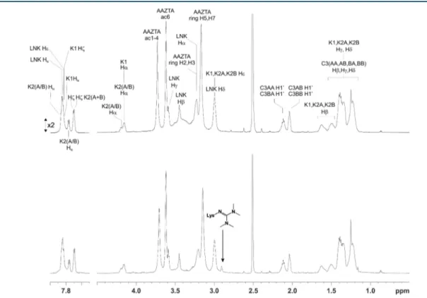
of the amino groups in the trilysine scaffold and by the slow rate of coupling of the fourth AAZTA unit due to an increasing steric hindrance. Besides the decrease of the final yield, the formation of such an impurity induced problems in the purification step by semipreparative reverse phase HPLC. The impurity gave a characteristic signal at 2.90 ppm in 1H NMR spectra (Figure 2) and characteristic ESI+ MS peaks at m/z 938.6 [M+2H]2+ and 625.9 [M+3H]3+, corresponding to the
molecular formula C81H139N19O31.
The amine end-group in the molecular structure of L1 was easily converted to maleimide (L2), benzaldehyde (L3), or cyclooctyne (L4) by reacting the L1 ligand with a 3-fold molar excess of succinimidyl-3-maleimido propionate, or N-succinimidyl-4-formylbenzoate, or dibenzocyclooctyne-N-hy-droxysuccinimidyl ester, respectively, in phosphate buffer 50 mM and CH3CN 3:1. HPLC-MS (ESI+) analysis indicated a total conversion of L1 into the L2, L3, or L4 compounds (Scheme 2). The excess reagents and salts were removed by size exclusion chromatography, without a significant loss of the product. The fully assigned NMR spectra and HPLC-UV/MS (ESI+) chromatograms of ligands L2, L3, and L4 are reported in theSupporting Information.
The GdIIIcomplexes were prepared by mixing stoichiometric amounts (1:4) of the AAZTA-tetramer and GdCl3 solution.
While this step occurred without any problem for L1, L3, and L4, a substantial loss of L2 may take place because of the hydrolysis of the maleimide group. However, we have found that complete complex formation can be attained, within 1 h reaction, when the pH is carefully maintained at 6.5 (with the additions of NaOH).
As an example of practical application, we applied our bifunctional agent to label afibrin-binding peptide. We chose the EP-2104R fibrin-binding peptide (FibPep), that was
Figure 1.Structure of the AAZTA derivative AAZTA(tBu)4C4COOH.
Figure 2.1H NMR spectrum of L1 synthesized by using PyBOP (top) or HBTU (bottom) as the activator in SPPS, after purification by RP-HPLC. The arrow marks the signal at 2.90 ppm of the side product arising from the transfer of a tetramethyluronium group from HBTU to one lysine, yielding a tetramethylguanidine. SeeSupporting Informationfor full assignment of the1H NMR spectrum of L1 and atom nomenclature.
originally introduced and comprehensively characterized by Caravan and co-workers.26,27 Fibrin targeting with para-magnetic agents has an outstanding diagnostic potential, as fibrin is a major component of blood clots and plays an important role in thrombi-related pathologies such as deep venous thrombosis, pulmonary embolism, and atherosclerosis. In fact, several examples of peptide-based fibrin targeted molecular probes have been reported.28−30FibPep is a cyclic 11-amino-acid peptide that showed significantly higher binding to fibrin than other reported peptides. The peptide was synthesized on Rink amide 4-methylbenzhydrylamine using standard Fmoc solid-phase peptide synthesis. FibPep was modified with a glycine at the C-terminal and with two glycines at the N-terminal as spacers and then conjugated to Gd-L2 through the thiol/maleimide chemistry. Thus, N-succinimidyl-S-acetylthioacetate (SATA) was reacted to the N-terminal and then the peptide cleaved from the resin. Disulfide bridge was then formed between Cys5 and Cys10 and the acetylthio group was removed using hydroxylamine. Finally, the thiol-bearing peptide was reacted with the maleimide of Gd-L2 at a molar ratio of 1:1 at pH 6.5 to yield the Gd-labeled peptide (FibPep-Gd-L2) in 80% yield. It is worth emphasizing that, by applying this procedure, Gd-chelates are directly conjugated to the targeting vector, contrary to the typical two-step approach that involves the conjugation of the chelator and then complexation with gadolinium. Thus, the approach presented herein eliminates the “free” gadolinium issue (i.e., Gd(III) ions being weakly coordinated to the secondary coordinating sites potentially offered by the peptidic structure).31−33
This approach may be particularly useful when the targeting vector has the chemical complexity of proteins, antibodies, and polysaccharide-based systems.
Relaxivity. The Gd-L1 complex showed a relaxivity, in water, of 16.4 ± 0.2 mMGd−1 s−1 (0.5 T, 25 °C). It is
significantly higher than that of monomeric Gd-AAZTA (7.1 mM−1s−1),16resulting in an overall relaxivity per molecule of 65.6 ± 0.8 mM−1 s−1. The nuclear magnetic resonance dispersion (NMRD) profile of Gd-L1 (Figure 3, Table 1) as compared to that of Gd-AAZTA shows higher relaxivities throughout the magnetic field strength studied and a smooth relaxivity peak centered at around 40−50 MHz.
This profile is consistent with an increase of the reorienta-tional correlation time, τR, expected on the basis of the
increased molecular size due to the tetrameric structure (macromolecule effect). However, the relaxivity is not as high as one could expect on the basis of the size of the tetrameric system. In fact, the observed relaxivity is well below the straight line obtained by plotting the relaxivity of known Gd(III) complexes, with two inner sphere water molecules, versus their molecular weight (Figure 4).
This can be ascribed to relatively fast internal motions that partially decouple the reorientation of each of the four Gd-AAZTA chelates from the overall global tumbling motion. To get further insights, a quantitative view of the parameters governing the relaxivity of Gd-L1 has been obtained by multiparametricfitting of the NMRD profile according to the
Solomon−Bloembergen−Morgan (SBM) theory for inner
sphere paramagnetic relaxation, combined with the Freed theory for outer sphere paramagnetic relaxation,41and modified according to the Lipari-Szabo model-free approach for the description of the rotational dynamics.42This model allows one to take into account the presence of a certain degree of internal motions superimposed on the overall tumbling motion. These Scheme 2. Synthesis of Ligands Bearing Functional Groups for Bioconjugation (L2, L3, and L4) and of Their Gd(III)-Complexes (Gd-L2, Gd-L3, and Gd-L4)a
a(i) N-Succinimidyl-3-maleimido propionate (3 equiv) in buffer phosphate 50 mM at pH 7 and CH
3CN; (ii) N-succinimidyl-4-formylbenzoate (3 equiv) in buffer phosphate 50 mM at pH 7 and CH3CN; (iii) dibenzocyclooctyne-N-hydroxysuccinimidyl ester (3 equiv) in buffer phosphate 50 mM at pH 7 and CH3CN; (iv) GdCl3in water.
Figure 3. NMRD profiles of Gd-L1 (□) and FibPep-Gd-L2 (■) compared to those of Gd-AAZTA (★). Experimental data points were measured on 1 mM aqueous solutions of the Gd-complexes at 298 K, pH = 7. Lines are bestfitting to the theory.
two types of motions, a relatively fast local rotation of the coordination cage, superimposed on the global reorientation of the system, are characterized by different correlation times, namely, τRl and τRg, respectively. The degree of correlation
between global and local rotations is given by the parameter S2,
which takes values between zero (completely independent motions) and one (entirely correlated motions). Such analysis yielded correlation times for both the local and the global motion that are significantly increased as compared to the τR
value of parent monomeric Gd-AAZTA (74 ps; seeTable 1). However, the S2 value is very low (S2 = 0.068) as a consequence of the great flexibility of the Gd-coordination cages around the linkers that interconnect the four AAZTA chelating units. The relaxivity values of Gd-L2, Gd-L3, Gd-L4 were very similar to those of Gd-L1 (Table 2).
The relaxivity of FibPep-Gd-L2, at 0.5 T in water and 25°C, was 18.5± 0.3 mMGd−1s−1(74± 1 mM−1s−1per molecule). Upon increasing the temperature (Table 2), the relaxivity of all the tetramers decreases as expected on the basis of a shortening of the overall molecular tumbling time. When measured in serum, the observed relaxivities were slightly higher than in water, likely as a consequence of the increased viscosity. More
important, on passing from water to serum the absence of any “quenching” of the observed relaxivity is indicative of the inability of the endogenous anions (e.g., phosphates, carbo-nates, etc.) to replace the two water molecules in the inner coordination sphere. Thus, the tetrameric derivatives maintain this relevant property of the parent Gd-AAZTA complex.16In the case of Gd-L1 and FibPep-Gd-L2, the relaxivity values were measured also at field strength values more relevant for clinical applications (1 and 1.5 T). At both 25 and 37°C the relaxivity slightly increases by increasing the magneticfield in the range 0.5−1.5 T. This favorable behavior is frequently associated with systems endowed with intermediate molecular weight characteristics.43,44
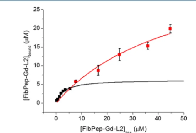
Fibrin Binding. The affinity of the peptide−tetramer conjugate to fibrin was tested by an adaptation of a well-established methodology,26that relies on the thrombin-induced formation of fibrin clots from plasma fibrinogen, in the presence of the binding peptide. The concentration of FibPep-Gd-L2 bound to the clot was obtained by isolating the clot from the plasma supernatant and determining the concentration of gadolinium by ICP-MS analysis (seeMaterials
Table 1. Main Parameters Derived from Fitting of NMRD Profiles Reported inFigure 3a
system r1p(mMGd−1s−1) Δ2b(s−2) τ
Vc(ps) τRld(ps) τRge(ps) S2
Gd-L1 16.4± 0.2 (1.89± 0.97) × 1019 36.3± 3.50 309± 63 741± 116 0.068
FibPep-Gd-L2 18.5± 0.3 (2.08± 0.15) × 1019 30.9± 1.49 305± 58 1080± 310 0.087 aOn carrying out thefitting procedure, the following parameters were kept fixed: r
Gd−H(distance between Gd and protons of the inner sphere water molecule) = 3.1 Å; a (distance of minimum approach of solvent water molecules to Gd3+ion) = 3.8 Å; D (solvent diffusion coefficient) = 2.2 × 10⁻⁵ cm2s−1.bSquared mean transient zero-field splitting (ZFS) energy.cCorrelation time for the collision-related modulation of the ZFS Hamiltonian. dReorientational correlation time, local motions.eReorientational correlation time, global motions.
Figure 4.Molecular weight (MW) dependence of the relaxivity (0.47 T, 298 K) for typical Gd(III) complexes with two inner-sphere water molecules. Data points have been interpolated by linear regression: r1p= 0.011MW + 0.928 (r2= 0.815).34,16,35−40,22
Table 2. Relaxivity per Gadolinium (mMGd−1s−1) of the Different Gd-Tetrameric Systems Measured at 0.5, 1, and 1.5 T and 25
and 37°C in Water and in Seruma
water serum 0.5 T 0.5 T 1 T 1.5 T system 25°C 37°C 25°C 37°C 25°C 37°C 25°C 37°C Gd-L1 16.4± 0.2 12.5± 0.1 18.8± 0.2 14.6± 0.2 19.5± 0.2 15.1± 0.1 19.5± 0.1 15.3± 0.1 Gd-L2 16.0± 0.2 12.5± 0.1 17.6± 0.2 13.7± 0.1 - - - -Gd-L3 16.8± 0.2 12.8± 0.2 18.3± 0.2 14.1± 0.2 - - - -Gd-L4 16.6± 0.2 12.6± 0.1 21.0± 0.2 16.7± 0.2 - - - -FibPep-Gd-L2 18.5± 0.3 14.3± 0.1 22.5± 0.2 18.5± 0.2 23.2± 0.2 18.7± 0.2 21.6± 0.2 16.9± 0.2
and Methods). The data were fit to the classical model for drug−receptor binding (eq 1)
‐ ‐ = × ‐ ‐ ‐ ‐ + B K [FibPep Gd L2] [FibPep Bd L2] [FibPep Gd L2]
bound max free free d (1)
where Bmax is the maximum binding capacity, [FibPep-Gd-L2]freeis the free Gd-tetramer concentration (μM), and Kdis
the dissociation constant (μM). The binding curve reported in
Figure 5displays an unusual course. While the initial part of the
curve is typical of specific binding, the final part does not reach a plateau as expected. Instead, the amount of bound FibPep-Gd-L2 keeps on increasing with increasing FibPep-Gd-L2 concentration in the incubation medium. The overall behavior is indicative of unspecific binding going on together with specific binding. Fitting of the first part of the curve yields a strong affinity constant (Kd = 3.0 ± 0.6 μM, Bmax = 6 ± 1). However, when the full curve is considered for fitting, lower affinity binding sites are found (Kd= 48± 14 μM) with a larger binding capacity (Bmax= 39± 3). The affinity of FibPep-Gd-L2
forfibrin is slightly lower than, but comparable to, that found for systems in which the same binding peptide was conjugated to four Gd-DOTAGA26or GdDTPA45complexes.
Finally, the relaxivity of FibPep-Gd-L2 in thefibrin plasma clots was determined at 25 °C and 0.5 T. The relaxivity per gadolinium was 58± 13 mMGd−1s−1(mean± SD value of 3
experiments), which is ∼150% higher than that measured in serum (Table 2). However, it must be considered that the high relaxivity found in clots arises from both specifically and aspecifically bound species, whose relative contribution to the observed relaxivity cannot be unambiguously disentangled. We speculate that the relaxivity of the aspecifically bound species might be significantly higher than that of the free species, because of the high microviscosity envisaged within the clot microenvironment. This matter certainly needs further investigation. With this limitation in mind, we observe that the∼150% relaxivity enhancement found for our q = 2 FibPep-Gd-L2is comparable to that of EP-2104-R.26The latter is a q = 1 system based on the same target-binding peptide, but bearing four Gd-DOTA-like units. Thus, we envisage that the expected relaxivity loss (in the 20−60 MHz range) due to the high flexibility of Gd(III)-AAZTA chains is compensated by the combined effect of q = 2 hydration number and slow dynamics within the clot.
■
CONCLUSIONIn conclusion, we have presented a protocol for the synthesis of three bifunctional agents, carrying one functional group for bioconjugation to targeting vectors and four Gd-AAZTA like functions as the MRI contrast agent. Despite the high internal flexibility of the Gd(III)-tetramer, the relaxivity per gadolinium shows a 2.4-fold increase as compared to that of monomeric Gd-AAZTA, due to the macromolecular effect. These compounds can be used as building blocks to insert preformed, high-relaxivity, and high-density Gd-centers to biological targeting vectors.16
■
MATERIALS AND METHODSBis(2-aminoethyl)-ether trityl resin, Fmoc-Lys(Fmoc)-OH, and activators HBTU and PyBOP were purchased from Novabio-chem (Darmstadt, Germany). All other Novabio-chemicals were purchased from Sigma-Aldrich. NMR spectra were recorded at 310 K on a Bruker AVANCE 600 spectrometer. Mass spectra with electrospray ionization (ESI) were recorded on a SQD 3100 Mass Detector (Waters). The HPLC-MS analytical and preparative analysis were carried out on a Waters AutoPur-ification system (3100 Mass Detector, 600 Quaternary Pump Gradient Module, 2767 Sample Manager, and 2487 UV/Visible Detector). UPLC analysis was performed using a UPLC Acquity H-Class coupled with the QDa and TUV Detectors. All ligands were characterized by NMR on a Bruker AVANCE 600 NMR spectrometer operating at 14T (corresponding to 600 and 150 MHz1H and 13C Larmor frequencies, respectively). The purified ligands L1−4 were freeze- dried from water solution at pH 2−3 and the powder redissolved in DMSO-d6
for NMR analysis. Either homonuclear (TOCSY, 2D-COSY, 2D-NOESY) or heteronuclear (2D-1H,13C HSQC, 2D-1H,13C HMBC) two-dimensional NMR experiments were
acquired to assign the spectra and confirm the structure of the ligands.
Synthesis of L1. Bis(2-aminoethyl)-ether trityl resin (0.96 mmol; 1.95 g) was loaded into a reaction vessel and dimethylformamide (DMF) (25 mL) was added to swell it for about 10 min. After the elimination of DMF, a mixture of Fmoc-Lys(Fmoc)-OH (2.39 mmol; 1.41 g), PyBOP (2.39 mmol; 1.24 g), and DIPEA (4.78 mmol; 0.83 mL) in DMF (20 mL) was added in the reactor and shaken for 1 h. The amino acid solution was then removed and 20% acetic anhydride− DMF (20 mL) was added and shaken for 15 min. The resin was washedfive times with DMF and then the deprotection of the amino groups from Fmoc was done using 40% piperidine− DMF (20 mL) for 5 min and 20% piperidine−DMF (20 mL) for 15 min. After washingfive times with DMF, the coupling cycle was repeated using a double quantity of Fmoc-Lys(Fmoc)-OH (4.78 mmol; 2.82 g), PyBOP (4.78 mmol; 2.49 g), and DIPEA (9.55 mmol; 1.67 mL) in DMF (20 mL), and the reaction was carried out again for 1 h, followed by DMF washing and piperidine treatment. After washing with DMF, a mixture of AAZTA(tBu)4C4COOH
24
(4.02 mmol; 2.7 g), PyBOP (4.01 mmol; 2.09 g) and DIPEA (8.02 mmol; 1.40 mL) in DMF (20 mL) was added to the reactor and it was shaken for 1 h. The resin was washedfive times with DMF and then thrice with dichloromethane (DCM). Afterward, diethyl ether (Et2O) was used to completely dry the resin before the cleavage. A cocktail solution of DCM, trifluoroacetic acid (TFA), and triisopropylsilane (TIS) in a ratio 49:49:2 (10 mL) was added in the reactor and it was shaken for 5 min. The
Figure 5. FibPep-Gd-L2binding tofibrin clots from human plasma. Solid lines are the fitting curves as described in the text. Data are reported as mean± SD from three independent experiments. Bioconjugate Chemistry
solution was collected in a round-bottomflask and the process was repeated three times. Then the combined filtrate was reduced almost to dryness and fresh TFA was added and stirred overnight until the complete removal of tBu of AAZTA. The mixture was then concentrated in vacuo and cold Et2O (30
mL) was added to let the product precipitate. The precipitate was centrifuged and washed thrice with Et2O. The crude
product was dissolved in 5 mL of water and purified by preparative RP-HPLC using a Waters XTerra prep RPdC8, 5 μm, 19 × 100 mm column by Method 1 and H2O/0.1% TFA
(A) and CH3CN/0.1% TFA (B) as eluents (see theSupporting Information). The volume of injection was 600 μL for every run and the desired product was fractioned, collected, and freeze-dried overnight. The pure product was obtained as a white powder (1.2 g, 57% of yield). The purity of the L1 was evaluated by analytical HPLC using XTerra RPdC8, 5μm, 4.6 × 150 mm column, by Method 2 and H2O/0.1% TFA (A) and
CH3CN/0.1% TFA (B) as eluents: a retention time of 5.84 min and a degree of purity of 98.7% of L1 were estimated (λ = 220 nm) (see theSupporting Information).
ESI-MS (m/z): [M+2H]2+1104.0 (obsd.), 1104.0 (calcd. for C94H156N20O40); [M+3H]3+736.3 (obsd.), 736.4 (calcd.); [M +4H]4+552.6 (obsd.), 552.6 (calcd.). 1H NMR (600 MHz, DMSO-d 6, T = 310 K): 12.20 (br), 7.88 (5H, o, m), 7.79 (2H, d), 7.73−7.72 (2H, o, m), 4.22−4.19 (3H, o, m), 3.78 (16H, s), 3.65 (16H, o, s), 3.62 (2H, o, t), 3.48 (2H, t), 3.27 (18H, o, m), 3.20 (16H, o, br), 3.02 (8H, o, m), 2.15 (4H, o, m), 2.07 (4H, o, m), 1.66−1.53 (6H, o, m), 1.44−1.25 (36H, o, m).
Synthesis of L2, L3, and L4. To a solution of L1 (0.02 mmol; 50 mg) in buffer phosphate (50 mM; 3 mL; pH 7) was added N-succinimidyl-3-maleimido propionate (0.06 mmol; 16 mg) or N-succinimidyl-4-formylbenzoate (0.06 mmol; 15 mg) or dibenzocyclooctyne-N-hydroxysuccinimidyl ester (0.06 mmol; 24 mg) dissolved in 1 mL of CH3CN for compound
L2, L3, and L4, respectively. After stirring 4 h at RT, the organic phase was evaporated and the excess nonbound reagent and buffer products were removed by size exclusion chromatography on a HiTrap Desalting Column 5 mL prepacked with Sephadex G25, using Milli-Q H2O as the
mobile phase, after which the fractions corresponding to the functionalized product were collected. The L2, L3, and L4 compounds were checked with ESI-MS(+) and freeze-dried, obtaining about 70% of yield. The purity of the modified ligands was evaluated by analytical HPLC using XTerra RPdC8, 5 μm, 4.6 × 150 mm column, by Method 2, and H2O/0.1%
TFA (A) and CH3CN/0.1% TFA (B) as eluents (see the
Supporting Information).
L2. ESI-MS (m/z): [M+2H]2+1179.6 (obsd.), 1179.7 (calcd.
for C101H161N21O43); [M+3H]3+786.8 (obsd.), 786.8 (calcd.);
[M+4H]4+ 590.6 (obsd.), 590.2 (calcd.). Retention time 9.90 min, purity 90.0%. 1H NMR (600 MHz, DMSO-d 6, T = 310 K): 12.20 (br), 8.03 (1H, t), 7.94−7.85 (4H, o, m), 7.83 (1H, d), 7.80−7.73 (2H, o, m), 7.00 (2H, s), 4.22−4.13 (3H, o, m), 3.85−3.75 (16H, o, s), 3.61−3.43 (18H, o, br), 3.41−3.35 (4H, o, m), 3.34−3.27 (18H, o, m), 3.23 (16H, s), 3.19−3.14 (2H, o, m), 2.99 (6H, o, m), 2.37 (2H, t), 2.15 (4H, o, m), 2.07 (4H, o, m), 1.66−1.53 (6H, o, m), 1.44−1.25 (36H, o, m).
L3. ESI-MS (m/z): [M+2H]2+1170.0 (obsd.), 1170.0 (calcd.
for C102H160N20O42); [M+3H]3+780.5 (obsd.), 780.5 (calcd.);
[M+4H]4+585.8 (obsd.), 585.5 (calcd.). Retention time 10.05
min, purity 93.3%. 1H NMR (600 MHz, DMSO-d 6, T = 310 K): 12.20 (br), 10.10 (1H, s), 8.81 (1H, t), 8.08 (2H, d), 7.99 (2H, d), 7.95− 7.86 (4H, o, m), 7.85 (1H, d), 7.80−7.75 (2H, o, m), 4.22−4.13 (3H, o, m), 3.85−3.75 (16H, o, s), 3.63 (16H, o, s), 3.57 (2H, t, o), 3.48 (2H, o, m), 3.45 (2H, o, m), 3.33−3.27 (18H, o, m), 3.23 (16H, s) 2.99 (6H, o, m), 2.15 (4H, o, m), 2.07 (4H, o, m), 1.66−1.53 (6H, o, m), 1.44−1.25 (36H, o, m).
L4. ESI-MS (m/z): [M+2H]2+1261.6 (obsd.), 1261.9 (calcd.
for C115H173N21O42); [M+3H]3+841.5 (obsd.), 841.6 (calcd.);
[M+4H]4+631.6 (obsd.), 631.4 (calcd.). Retention time 18.47 min, purity 90.1%. 1H NMR (600 MHz, DMSO-d 6, T = 310 K): 12.20 (br), 8.01 (1H, t), 7.98−7.83 (6H, o, m), 7.80−7.74 (1H, o, m), 7.66 (1H, d), 7.59 (1H, d), 7.52 (1H, t), 7.51−7.46 (2H, o, m), 7.42 (1H, t), 7.38 (1H, t), 7.32 (1H, d), 5.08 (2H, d), 4.24−4.18 (3H, br, o) 3.87 (16H, o, br), 3.66 (16H, o, s), 3.43−3.37 (18H, o, m), 3.36−3.29 (18H, o, s), 3.25−3.13 (4H, o, m), 3.02 (6H, o, m), 2.22−2.13 (6H, o, m), 2.07 (4H, o, t), 1.91 (2H, t), 1.79 (2H, m), 1.66−1.53 (6H, o, m), 1.44−1.25 (38H, o, m). Synthesis of Gd(III)-Complexes (Gd-L1, Gd-L2, Gd-L3, and Gd-L4). An equimolar amount of GdCl3(170 mM water
solution) was slowly added to a 60 mM ligand solution; the solutions were maintained at pH 6.5 with NaOH. The complexes were then desalted by size exclusion chromatog-raphy on a HiTrap Desalting Column 5 mL prepacked with Sephadex G25, using Milli-Q H2O as the mobile phase, and
finally freeze-dried. The amount of residual free Gd3+ions was
assessed by the Orange Xylenol UV method.46All complexes were found to contain less than 0.3% (mol/mol) of residual free Gd3+ions. The purity of Gd complexes was evaluated by analytical UPLC using ACQUITY UPLC Peptide BEH C18 Column, 1.7μm, 2.1 mm × 150 mm by Method 3 for Gd-L2 and Gd-L3 and Method 4 for GdL-4, using H2O/0.05% TFA
(A) and CH3CN/0.05% TFA (B) as eluents (see the
Supporting Information). All the derivatives were obtained in high purity, >90% (see theSupporting Information).
Gd-complexes were also analyzed by direct infusion in ESI-MS in negative-ion mode after dilution in CH3OH−H2O (50/
50).
Gd-L1. ESI-MS m/z: [M-2H]2− 1410.9 (obsd.), 1410.3 (calcd. for C94H140Gd4N20O40); [M-3H]3−940.0 (obsd.), 939.5 (calcd.); [M-4H]4−704.8 (obsd.), 704.1 (calcd.).
Gd-L2. ESI-MS m/z: [M-3H]3−990.8 (obsd.), 990.9 (calcd. for C101H149Gd4N21O43); [M-4H]4− 742.7 (obsd.), 742.9
(calcd.).
Gd-L3. ESI-MS m/z: [M-3H]3−984.5 (obsd.), 984.5 (calcd. for C102H148Gd4N20O42); [M-4H]4− 737.7 (obsd.), 738.4
(calcd.).
Gd-L4. ESI-MS m/z: [M-3H]3− 1045.4 (obsd.), 1045.6 (calcd. for C115H161Gd4N21O42); [M-4H]4− 783.7 (obsd.), 783.9 (calcd.).
Synthesis of FibPep. The peptide sequence GGY-dGlu-C-Hyp-3ClY-GLCYIQG, containing thefibrin binding motif, was synthesized by standard Fmoc solid-phase peptide synthesis on Rink amide 4-methylbenzhydrylamine resin and using a Liberty CEM microwave synthesizer. The coupling reactions were performed with 5.0 equiv excess of Fmoc-amino acids, 5 min at 75°C, 35 W, in the presence of PyBOP and DIPEA in DMF, and the Fmoc-deprotection steps (20% piperidine in DMF) were completed within 3 min at 75°C. At the end of the last cycle, the synthesized peptide was reacted with 2.5-fold excess of N-succinimidyl-S-acetylthioacetate (SATA) in DMF and stirring at RT for 3 h. A cocktail solution of TFA, H2O, Phenol,
and TIS in a ratio 88:5:5:2 (v/v/wt/v, 15 mL) was used to cleave the peptide from the resin and to obtain the side-chain deprotection. After 3 h, the cleavage solution was collected and concentrated to dryness. Et2O was added to the residue to
precipitate the crude peptide, which was collected. The crude linear dicysteine containing peptide was purified by preparative RP-HPLC using a Waters XTerra prep RPdC8, 5μm, 19 × 100 mm column, by Method 5 (see theSupporting Information). Next the peptide was cyclized by dissolution in H2O and
DMSO (9:1, v/v) in a concentration of approximately 2.5 mg of peptide mL−1and stirring the obtained solution at RT for 3 days. Subsequently, after lyophilization the cyclic peptide was purified using preparative RP-HPLC (0.10 g, 60% of yield) by Method 5 (see theSupporting Information). The purity of the peptides was evaluated by analytical UPLC using ACQUITY UPLC Peptide BEH C18 Column, 1.7μm, 2.1 mm × 150 mm, H2O/TFA 0.05% (A) and CH3CN/TFA 0.05% (B) as eluents,
by Method 6. Retention time 5.48 min for linear FibPep, Retention time 5.92 min for cyclic FibPep, purity of the peptides >90% (see theSupporting Information). ESI-MS (m/ z): [M+2H]2+ 845.3(obsd.), 845.1 (calcd. for linear FibPep C72H99ClN16O23S3); [M+2H]2+844.0 (obsd.), 844.2 (calcd. for
cyclic FibPep C72H97ClN16O23S3).
Synthesis of FibPep-Gd-L2. SATA-FibPep (20 mg) was
dissolved in 1 mL of water and 100 μL of 0.5 mol/L
hydroxylamine hydrochloride, 25 mM EDTA solution were added to deprotect a thiol group and to yield the thiolated FibPep. The reaction was monitored by ESI (+)-MS and after stirring 2 h at RT the Sulfhydryl-FibPep was immediately recovered by HPLC separation (Method 5, see Supporting Information). [M+2H]2+ 822.9 (obsd.), 823.1 (calcd. for
sulfhydryl-FibPep C70H95ClN16O22S3). Then, sulfhydryl-FibPep
(5,4 mg, 3,3μmol) was dissolved in sodium acetate 50 mM, pH 6.0, 2 mL containing 30% acetonitrile and under argon atmosphere Gd-L2 (9,0 mg, 3μmol) in 0.2 mL of water was added. The mixture was stirred at RT for 1 h and subsequently purified by gel permeation chromatography on a column packed with Sephadex G10 (1.1× 12 cm), using Milli-Q H2O
as the mobile phase. After freeze-drying the product fractions, 11 mg (80% yield) of a white solid was obtained. ESI-MS (m/ z): The purity of FibPep-Gd-L2 was evaluated by analytical UPLC using ACQUITY UPLC Peptide BEH C18 Column, 1.7 μm, 2.1 mm × 150 mm by Method 7, purity >85%. [M-4H]4−
1153.6 (obsd.), 1153.6 (calcd. for C171H244ClGd4N37O65S3);
[ M - 5 H ]6− 9 2 2 . 5 ( o b s d . ) , 9 2 2 . 7 ( c a l c d . f o r C171H244ClGd4N37O65S3).
Relaxometry. The longitudinal water proton relaxation rates were measured by using a Stelar SpinMaster relaxometer (Stelar, Mede (PV), Italy) operating at 0.5 T (21.5 MHz Proton Larmor Frequency), by mean of the standard inversion−recovery technique. The temperature was controlled with a Stelar VTC-91 air-flow heater equipped with a copper constantan thermocouple (uncertainty 0.1°C). The proton 1/ T1 NMRD profiles were measured at 298 K on a fast
field-cycling Stelar relaxometer over a continuum of magneticfield strengths from 0.00024 to 0.47 T (corresponding to 0.01−20 MHz proton Larmor Frequency). The relaxometer operates under computer control with an absolute uncertainty in 1/T1of
±1%. Additional data points in the range 21.5−70 MHz were obtained on the Stelar SpinMaster relaxometer. The concen-tration of the solutions used for the relaxometric character-ization was determined according to a previously reported relaxometric method.47 Briefly, the solution containing the
Gd(III)-complex is mineralized by transferring 0.1 mL of the Gd chelate solution into a sealable glass ampule, then bringing the volume to 0.2 mL with HCl 37% (analytical grade). The ampule is sealed and the solution heated at 120°C for 18 h. The Gd(III) aquo-ion released is quantitated by measuring the water proton relaxation rate at 20 MHz and 25°C (R1obs), by
the formula: = R − [Gd] 0.5 13.5 1obs (2)
where 13.5 is the relaxivity of free Gd(III) in acidic conditions, and 0.5 is the diamagnetic contribution to the relaxation rate, that is determined on a blank sample.
The concentration of Gd(III) ions in thefibrin plasma clots for the determination of relaxivity value at 20 MHz and 25°C, was determined through ICP-MS analysis of the digested clots. Binding to Fibrin Clots. Fibrin clots were prepared from human plasma (Sigma-Aldrich) as follows: Aliquots of 100μL of plasma were freeze-dried and resuspended in solutions of Gd-L1pep with concentrations in the range 0.5−50 μM. To the plasma suspensions, 2μL of calcium chloride (0.5 M) and 2 μL
thrombin (100 U/mL) were added to reach the final
concentrations of 10 mM and 2 U/mL, respectively. The samples were then incubated for 1 h at 37 °C during the formation of the clots. The experiments were carried out in triplicate. At the end of the incubation time, supernatant was carefully and completely removed, clots were weighed in order to calculate the clot volume (a density of 1 g/mL was considered), and 1 mL of HNO369% was added to the clots.
After complete dissolution of the clots, samples were further digested by applying microwave heating (MicroSYNTH, Microwave labstation equipped with an optical fiber temper-ature control and HPR-1000/6 M six position high-pressure reactor, Milestone, Bergamo, Italy). After digestion, the volume of each sample was brought to 2 mL with ultrapure water, filtered with 0.45 μm filter, and analyzed by ICP-MS, using a Thermo Scientific ELEMENT 2 ICP-MS − Finnigan, Rodano (MI). The quantification was obtained through a calibration curve measured by using four gadolinium absorption standard solutions (Sigma-Aldrich) in the range 0.005−0.1 μg/mL. The concentration of fibrin-bound FibPep-Gd-L2 was calculated through the ratio between the total bound micromoles and the clot volume, while the concentration of the free FibPep-Gd-L2 was calculated as the difference with respect to the total micromoles in the incubation media divided by the incubation volume.
■
ASSOCIATED CONTENT*
S Supporting InformationThe Supporting Information is available free of charge on the
ACS Publications website at DOI: 10.1021/acs.bioconj-chem.8b00120.
1H NMR spectrum, 2D-TOCSY and 2D-NOESY,
HPLC-UV/MS chromatograms, mass spectrum (ESI+)
and ESI (−) of new compounds, preparative and
analytical methods (PDF)
■
AUTHOR INFORMATIONCorresponding Author
*E-mail: [email protected]. Telephone: +39-011-6706452. Fax: +39-011-6706487.
Notes
The authors declare no competingfinancial interest.
■
ACKNOWLEDGMENTSThis project has received funding from the EU’s H2020 research and innovation program under the grant agreement No. 668142 (SPCCT) and AIRC Investigator Grant IG2013 No.14565 Dual MRI-Optical imaging agents in prostatectomy.
■
ABBREVIATIONSNMR, nuclear magnetic resonance; MRI, magnetic resonance imaging; HPLC-MS, high performance liquid chromatography mass spectrometry; CA, contrast agents; AAZTA, 6-amino-6-methylperhydro-1,4-diazepinetetraacetic acid; PyBOP, benzo-triazole-1-yl-oxy-tris-pyrrolidino-phosphonium hexa fluorophos-phate; DIPEA, N,N-diisopropylethylamine; HBTU, O-(benzo-triazol-1-yl)-N,N,N′,N′ tetramethyluronium hexafluorophos-phate; DMF, dimethylformamide; DCM, dichloromethane; TFA, trifluoroacetic acid; tBu, tert-butyl
■
REFERENCES(1) Schlomka, J., Roessl, E., Dorscheid, R., Dill, S., Martens, G., Istel, T., Bäumer, C., Herrmann, C., Steadman, R., Zeitler, G., et al. (2008) Experimental feasibility of multi-energy photon-counting K-edge imaging in pre-clinical computed tomography. Phys. Med. Biol. 53 (15), 4031.
(2) Schirra, C. O., Brendel, B., Anastasio, M. A., and Roessl, E. (2014) Spectral CT: a technology primer for contrast agent development. Contrast Media Mol. Imaging 9 (1), 62−70.
(3) Si-Mohamed, S., Bar-Ness, D., Sigovan, M., Cormode, D. P., Coulon, P., Coche, E., Vlassenbroek, A., Normand, G., Boussel, L., and Douek, P. (2017) Review of an Initial Experience with an Experimental Spectral Photon-Counting Computed Tomography System. Nucl. Instrum. Methods Phys. Res., Sect. A 873, 27.
(4) Caravan, P., Ellison, J. J., McMurry, T. J., and Lauffer, R. B. (1999) Gadolinium (III) chelates as MRI contrast agents: structure, dynamics, and applications. Chem. Rev. 99 (9), 2293−2352.
(5) Aime, S., Chiaussa, M., Digilio, G., Gianolio, E., and Terreno, E. (1999) Contrast agents for magnetic resonance angiographic applications: 1 H and 17 O NMR relaxometric investigations on two gadolinium (III) DTPA-like chelates endowed with high binding affinity to human serum albumin. JBIC, J. Biol. Inorg. Chem. 4 (6), 766−774.
(6) Martin, V. V., Ralston, W. R., Hynes, M. R., and Keana, J. F. W. (1995) Gadolinium(III) Di- and Tetrachelates Designed for in Vivo Noncovalent Complexation with Plasma Proteins: A Novel Molecular Design for Blood Pool MRI Contrast Enhancing Agents. Bioconjugate Chem. 6, 616−623.
(7) Aime, S., Barge, A., Cabella, C., Crich, S. G., and Gianolio, E. (2004) Targeting cells with MR imaging probes based on para-magnetic Gd (III) chelates. Curr. Pharm. Biotechnol. 5 (6), 509−518. (8) Aime, S., Castelli, D. D., Crich, S. G., Gianolio, E., and Terreno, E. (2009) Pushing the sensitivity envelope of lanthanide-based magnetic resonance imaging (MRI) contrast agents for molecular imaging applications. Acc. Chem. Res. 42 (7), 822−831.
(9) Weissleder, R., and Mahmood, U. (2001) Molecular imaging. Radiology 219 (2), 316−333.
(10) Aime, S., Cabella, C., Colombatto, S., Geninatti Crich, S., Gianolio, E., and Maggioni, F. (2002) Insights into the use of paramagnetic Gd (III) complexes in MR-molecular imaging investigations. Journal of magnetic resonance imaging 16 (4), 394−406. (11) Wiener, E. C., Konda, S., Shadron, A., Brechbiel, M., and Gansow, O. (1997) Targeting dendrimer-chelates to tumors and tumor cells expressing the high-affinity folate receptor. Invest. Radiol. 32 (12), 748−54.
(12) Zhang, Z., Greenfield, M. T., Spiller, M., McMurry, T. J., Lauffer, R. B., and Caravan, P. (2005) Multilocus binding increases the
relaxivity of protein-bound MRI contrast agents. Angew. Chem., Int. Ed. 44, 6766−6769.
(13) James, M. L., and Gambhir, S. S. (2012) A molecular imaging primer: modalities, imaging agents, and applications. Physiol. Rev. 92 (2), 897−965.
(14) Hu, H., Arena, F., Gianolio, E., Boffa, C., Di Gregorio, E., Stefania, R., Orio, L., Baroni, S., and Aime, S. (2016) Mesoporous silica nanoparticles functionalized with fluorescent and MRI reporters for the visualization of murine tumors overexpressingαvβ3 receptors. Nanoscale 8 (13), 7094−7104.
(15) Langereis, S., Geelen, T., Grüll, H., Strijkers, G. J., and Nicolay, K. (2013) Paramagnetic liposomes for molecular MRI and MRI-guided drug delivery. NMR Biomed. 26 (7), 728−744.
(16) Aime, S., Calabi, L., Cavallotti, C., Gianolio, E., Giovenzana, G. B., Losi, P., Maiocchi, A., Palmisano, G., and Sisti, M. (2004) [Gd-AAZTA]-: a new structural entry for an improved generation of MRI contrast agents. Inorg. Chem. 43 (24), 7588−7590.
(17) Baranyai, Z., Uggeri, F., Giovenzana, G. B., Bényei, A., Brücher, E., and Aime, S. (2009) Equilibrium and kinetic properties of the lanthanoids (III) and various divalent metal complexes of the heptadentate ligand AAZTA. Chem. - Eur. J. 15 (7), 1696−1705.
(18) Di Gregorio, E., Gianolio, E., Stefania, R., Barutello, G., Digilio, G., and Aime, S. (2013) On the fate of MRI Gd-based contrast agents in cells. Evidence for extensive degradation of linear complexes upon endosomal internalization. Anal. Chem. 85 (12), 5627−5631.
(19) Kanda, T., Osawa, M., Oba, H., Toyoda, K., Kotoku, J. I., Haruyama, T., Takeshita, K., and Furui, S. (2015) High signal intensity in dentate nucleus on unenhanced T1-weighted MR images: association with linear versus macrocyclic gadolinium chelate administration. Radiology 275 (3), 803−809.
(20) Campa, C., Uggeri, F., Paoletti, S., and Flamigni, A. (2008). U.S. Patent Application No. 12/528,685.
(21) Gugliotta, G., Botta, M., and Tei, L. (2010) AAZTA-based bifunctional chelating agents for the synthesis of multimeric/ dendrimeric MRI contrast agents. Org. Biomol. Chem. 8 (20), 4569− 4574.
(22) Gianolio, E., Ramalingam, K., Song, B., Kalman, F., Aime, S., and Swenson, R. (2010) Improving the relaxivity by dimerizing Gd-AAZTA: Insights for enhancing the sensitivity of MRI contrast agents. Inorg. Chem. Commun. 13 (5), 663−665.
(23) Baskin, J. M., Prescher, J. A., Laughlin, S. T., Agard, N. J., Chang, P. V., Miller, I. A., and Bertozzi, C. R. (2007) Copper-free click chemistry for dynamic in vivo imaging. Proc. Natl. Acad. Sci. U. S. A. 104 (43), 16793−16797.
(24) Manzoni, L., Belvisi, L., Arosio, D., Bartolomeo, M. P., Bianchi, A., Brioschi, C., Buonsanti, F., Cabella, C., Casagrande, C., and Civera, M. (2012) Synthesis of Gd and68Ga complexes in conjugation with a conformationally optimized RGD sequence as potential MRI and PET tumor-imaging probes. ChemMedChem 7 (6), 1084−1093.
(25) Dubey, L. V., and Dubey, I. Y. (2005) Side reactions of onium coupling reagents BOP and HBTU in the synthesis of silica polymer supports. Ukrainica bioorganica acta 1, 13−19.
(26) Overoye-Chan, K., Koerner, S., Looby, R. J., Kolodziej, A. F., Zech, S. G., Deng, Q., Chasse, J. M., McMurry, T. J., and Caravan, P. (2008) EP-2104R: A fibrin specific Gadolinium-based MRI Contrast Agent for detection of Thrombus. J. Am. Chem. Soc. 130, 6025−6039. (27) Nair, S. A., Kolodziej, A. F., Bhole, G., Greenfield, M. T., McMurry, T. J., and Caravan, P. (2008) Monovalent and Bivalent Fibrin-specific MRI Contrast Agents for Detection of Thrombus. Angew. Chem., Int. Ed. 47, 4918−4921.
(28) Starmans, L. W. E., van Duijnhoven, S. M. J., Rossin, R., Berben, M., Aime, S., Daemen Mat, J. A. P., Klaas Nicolay, K., and Grüll, H. (2013) Evaluation of111In-Labeled EPep and FibPep as Tracers for Fibrin SPECT Imaging. Mol. Pharmaceutics 10, 4309−4321.
(29) Zhou, Z., Wu, X., Kresak, A., Griswold, M., and Lu, Z. R. (2013) Peptide targeted tripod macrocyclic Gd (III) chelates for cancer molecular MRI. Biomaterials 34 (31), 7683−7693.
(30) Chaabane, L., Tei, L., Miragoli, L., Lattuada, L., von Wronski, M., Uggeri, F., Lorusso, V., and Aime, S. (2015) In vivo MR imaging of
fibrin in a neuroblastoma tumor model by means of a targeting Gd-containing peptide. Molecular Imaging and Biology: MIB 17 (6), 819.
(31) De León-Rodríguez, L. M., and Kovacs, Z. (2008) The synthesis and chelation chemistry of DOTA− peptide conjugates. Bioconjugate Chem. 19 (2), 391−402.
(32) Wu, X., Burden-Gulley, S. M., Yu, G. P., Tan, M., Lindner, D., Brady-Kalnay, S. M., and Lu, Z. R. (2012) Synthesis and evaluation of a peptide targeted small molecular Gd-DOTA monoamide conjugate for MR molecular imaging of prostate cancer. Bioconjugate Chem. 23 (8), 1548−1556.
(33) Gringeri, C. V., Menchise, V., Rizzitelli, S., Cittadino, E., Catanzaro, V., Dati, G., Chaabane, L., Digilio, G., and Aime, S. (2012) Novel Gd (III)-based probes for MR molecular imaging of matrix metalloproteinases. Contrast Media Mol. Imaging 7 (2), 175−184.
(34) Aime, S., Botta, M., Crich, S. G., Giovenzana, G., Pagliarin, R., Sisti, M., and Terreno, E. (1998) NMR relaxometric studies of Gd (III) complexes with heptadentate macrocyclic ligands. Magn. Reson. Chem. 36, S200−S208.
(35) Gugliotta, G., Botta, M., Giovenzana, G. B., and Tei, L. (2009) Fast and easy access to efficient bifunctional chelators for MRI applications. Bioorg. Med. Chem. Lett. 19 (13), 3442−3444.
(36) Aime, S., Botta, M., Geninatti Crich, S., Giovenzana, G. B., Jommi, G., Pagliarin, R., and Sisti, M. (1997) Synthesis and NMR studies of three pyridine-containing triaza macrocyclic triacetate ligands and their complexes with lanthanide ions. Inorg. Chem. 36 (14), 2992−3000.
(37) Aime, S., Gianolio, E., Corpillo, D., Cavallotti, C., Palmisano, G., Sisti, M., Giovenzana, G., and Pagliarin, R. (2003) Designing Novel Contrast Agents for Magnetic Resonance Imaging. Synthesis and Relaxometric Characterization of three Gadolinium (III) Complexes Based on Functionalized Pyridine-Containing Macrocyclic Ligands. Helv. Chim. Acta 86 (3), 615−632.
(38) Aime, S., Gianolio, E., Terreno, E., Giovenzana, G. B., Pagliarin, R., Sisti, M., Palmisano, G., Botta, M., Lowe, M.p., and Parker, D. (2000) Ternary Gd (III) L-HSA adducts: evidence for the replacement of inner-sphere water molecules by coordinating groups of the protein. Implications for the design of contrast agents for MRI. JBIC, J. Biol. Inorg. Chem. 5 (4), 488−497.
(39) Cohen, S. M., Xu, J., Radkov, E., Raymond, K. N., Botta, M., Barge, A., and Aime, S. (2000) Syntheses and relaxation properties of mixed gadolinium hydroxypyridinonate MRI contrast agents. Inorg. Chem. 39 (25), 5747−5756.
(40) Gianolio, E., Cabella, C., Colombo Serra, S., Valbusa, G., Arena, F., Maiocchi, A., Miragoli, L., Tedoldi, F., Uggeri, F., Visigalli, M., et al. (2014) B25716/1: a novel albumin-binding Gd-AAZTA MRI contrast agent with improved properties in tumor imaging. JBIC, J. Biol. Inorg. Chem. 19 (4−5), 715−726.
(41) Lipari, G., and Szabo, A. (1982) Model-free approach to the interpretation of nuclear magnetic resonance relaxation in macro-molecules. 1. Theory and range of validity. J. Am. Chem. Soc. 104 (17), 4546−4559.
(42) Lipari, G., and Szabo, A. (1982) Model-free approach to the interpretation of nuclear magnetic resonance relaxation in macro-molecules. 2. Analysis of experimental results. J. Am. Chem. Soc. 104 (17), 4559−4570.
(43) Tei, L., Gugliotta, G., Gambino, G., Fekete, M., and Botta, M. (2017) Developing High Field MRI Contrast Agents by Tuning the Rotational Dynamics: Bisaqua GdAAZTA-based Dendrimers. Isr. J. Chem. 57, 887−895.
(44) Fulton, D. A., O’Halloran, M., Parker, D., Senanayake, K., Botta, M., and Aime, S. (2005) Efficient relaxivity enhancement in dendritic gadolinium complexes: effective motional coupling in medium molecular weight conjugates. Chem. Commun., 474−476.
(45) Zhang, Z., Kolodziej, A. F., Greenfield, M. T., and Caravan, P. (2011) Heteroditopic binding of Magnetic Resonance Contrast Agents for Increased Relaxivity. Angew. Chem., Int. Ed. 50, 2621−2624. (46) Barge, A., Cravotto, G., Gianolio, E., and Fedeli, F. (2006) How to determine free Gd and free ligand in solution of Gd chelates. A technical note. Contrast Media Mol. Imaging 1 (5), 184−188.
(47) Arena, F., Singh, J. B., Gianolio, E., Stefania, R., and Aime, S. (2011)β-Gal Gene Expression MRI Reporter in Melanoma Tumor Cells. Design, Synthesis, and in Vitro and in Vivo Testing of a Gd(III) Containing Probe Forming a High Relaxivity, Melanin-Like Structure uponβ-Gal Enzymatic Activation. Bioconjugate Chem. 22, 2625−2635. Bioconjugate Chemistry