Abstract. – OBJECTIVE: The vaginal micro-biome is a dynamic environment, depending on the results of a complex interplay between mi-crobiota and the host. In physiological condi-tions, Lactobacillus species are the most rep-resented, regulating glycogen metabolism in order to maintain normal pH. Vaginal flora has been divided into five subtypes. Pattern rec-ognition receptors are present on both squa-mous epithelial cells lining the vagina and co-lumnar cells lining the upper female genital tract. They respond directly to bacterial prod-uct expressed by vaginal microbiome. The va-gina contains different immune related cells and receptors which can recognize and react with the microbial environment. Altered micro-biota and altered interplay between micromicro-biota and immune system underlie several gyneco-logic diseases.
MATERIALS AND METHODS: In this
re-view, literature data related to vaginal microbi-ota, vaginal inflammation, immune system and menopause, preterm labor and miscarriage, were summarized. Relevant publications were retrieved from: PubMed, Medline, Scopus and Web of Science.
RESULTS: The vaginal microbiome and the re-lationship with immune system has been ana-lyzed in different gynecologic conditions. Meno-pause is associated to estrogen loss which causes vaginal atrophy, reduced abundance of Lactobacilli and increased amount of other bac-terial species. Estrogens influence vaginal im-munity through known and unknown mecha-nisms. In bacterial vaginosis (BV), due to many bacterial species, there has been found an in-hibition of the chemotaxis and cytokine
secre-tion. A decreased concentration of Lactobacil-li seems to be playing a role in preterm labor as well as the increased levels of pro-inflammato-ry cytokines. Finally, the disequilibrium in the Th1/Th2 immune adaptive response, with a shift from Th2 to Th1, appears to be playing a role in miscarriage.
CONCLUSIONS: The interplay between micro-biota and the host closely involves the immune system. In particular, the vaginal microbiota is classically characterized by Lactobacilli even if vaginal microbiome of asymptomatic woman of reproductive age includes multiple aerobic and facultative or obligate anaerobic species. The role of microbiota and immune system in deter-mining gynecological and obstetric events has been studied throughout recent years reaching new advancements. Therefore, additional stud-ies are needed to better comprehend the com-plexity of the issue.
Key Words:
Vaginal microbiota, Immune system, Lactobacil-li, Menopause, Inflammation; miscarriage, Preterm la-bor.
Introduction
The vaginal microbiome is a dynamic environ-ment, depending on the results of a complex in-terplay between microbiota (the core of microbial communities) and the host. Several modifying factors are thought to impact the microbiome throughout life. The first contact with microbiota
P. VILLA
1,2, C. CIPOLLA
1, S. D’IPPOLITO
1, I.D. AMAR
2, M. SHACHOR
2,
F. INGRAVALLE
3, F. SCALDAFERRI
4,5, P. PUCA
5, N. DI SIMONE
1,2, G. SCAMBIA
1,21Dipartimento di Scienze della Salute della Donna, del Bambino e di Sanità Pubblica, Policlinico Universitario A. Gemelli IRCCS, Rome, Italy
2Istituto di Ostetricia e Ginecologia, Università Cattolica del Sacro Cuore, Rome, Italy
3Scuola di Specializzazione in Igiene e Medicina Preventiva, Università di Tor Vergata, Rome, Italy 4Centro Malattie Apparato Digerente, Dipartimento Scienze Gastroenterologiche,
Endocrino-Metaboliche e Nefro-Urologiche, Fondazione Policlinico Universitario Agostino Gemelli IRCCS, Rome, Italy
5Istituto di Patologia Speciale Medica, Università Cattolica del Sacro Cuore, Rome, Italy
The interplay between immune system and
microbiota in gynecological diseases:
may begin during late gestation, with the largest exposure at the time of delivery. Over time, the abundance and diversity of the infant microbiome increase with life, stabilize around the time that the infant begins to eat solid foods, and persist throughout adulthood. Several modifying factors are thought to have an influential role in shaping the identity and abundance of the infant microbio-ta throughout life.
Lactobacillus species are the most represented1
in non-pregnant woman reaching the concentra-tion of 107 to 108 CFU/g of vaginal fluid, together
with Lactobacillus Crispatus, Lactobacillus
Iners, Lactobacillus Jensenii, and Lactobacillus Gasseri2.
Lactobacilli regulate the glycogen metaboli-sm, converting glycogen from vaginal epithelial cells into glucose and lactic acid to maintain the typical acidic vaginal pH (pH ≤ 4.5, range 3.8-4.4). Hence, creating an unfavorable envi-ronment for the growth of pathogens or other “un-healthy” bacteria3. Lactobacilli may also
prevent the adherence of pathogenic microorga-nisms to vaginal epithelial cells through ‘com-petitive exclusion’ and ‘bacterial interference’3.
In addition, Lactobacilli produce various me-tabolites, such as bacteriocins and H2O2, which may help to stimulate the immune response du-ring vaginal infections. Lactobacilli reduce lo-cal production of interleukin (IL)-1β, IL-6, and IL-8 and increase anti-inflammatory cytokines, such as IL-2 and IL-173. In addition to
Lacto-bacilli, the vaginal core microbiota account also for other multiple aerobic or facultative aerobic species as well as obligate anaerobic species.
In healthy women, the transition from puber-ty to menopause as well as transient hormonal changes, such as pregnancy and menstruation are characterized by major changes in vaginal mi-crobiome. Furthermore, external factors, such as antibiotic usage, sexually transmitted infections, and vaginal irrigation can affect vaginal flora composition as well4. In particular, estrogens play
a central role. The estrogen environment helps in the maintenance of the right balance among diffe-rent vaginal bacterial communities. Indeed, it has been showed that the hormone replacement the-rapy (HRT) in menopause relieves the symptoms of vulvovaginal atrophy and supports the enrich-ment of vaginal microbiota5.
Vaginal infections are the most common cause of abnormal functional status. Thus, while eva-luating these infections, several characteristics should be considered:
• Bacterial density, which refers to the degree of bacterial distribution. It reflects the total bio-mass of the vaginal flora6.
• Flora diversity, which represents the total num-ber of bacterial species in the vaginal flora. This reflects the vaginal flora variety7.
• Vaginal H2O2 is mainly produced by Lactoba-cilli, such as L. Crispatus, L. Gasseri, L.
Jen-senii and L. Acidophilus. Thus, as these
Lac-tobacilli are often the predominant bacteria in healthy women, H2O2 levels may reflect the function of Lactobacilli8.
• Enzymatic activity, such us leukocyte esterase activity indicates the presence of inflammation in the vagina. Sialidase is a specific marker of BV, whereas β-glucuronidase and coagulase activity may represent bacterial vaginitis9,10.
Patients with the following features are consi-dered to have a normal micro-ecological status: • pH values ranging from 3.8 to 4.5;
• Bacterial density degree II to III; • Flora diversity degree II to III;
• Gram-positive rods as predominant flora; • Nugent and AV score ≤3;
• Absence of pathogens and negative specific en-zymes.
Gajer et al4 and Srinivasan et al11 showed that
the diversity, the composition, and the relative abundance of vaginal microbial species chan-ge dramatically, during different periods of life. Ravel et al14 have demonstrated that
reproducti-ve-aged women can be grouped into five diffe-rent categories referred to as Community State Types (CSTs). Four of these CSTs are dominated by Lactobacilli, namely, L. Crispatus (CST-I), L.
Iners (CST-III), L. Gasseri (CST-II) or L. Jen-senii (CST-V). One category, CST-IV, does not
contain a significant number of Lactobacillus, but is composed of a polymicrobial mixture of stri-ct and facultative anaerobes including species of the genera Gardnerella, Atopobium, Mobiluncus,
Prevotella and other species in the order of Clo-stridiales12-15.
CST IV was recently divided into two subtypes, termed CST IV-A and CST IV-B; by Gajer et al4
CST IV-A is characterized by various species of anaerobic bacteria belonging to the genera
Anae-rococcus, Peptoniphilus, Prevotella, and Strepto-coccus. However, CST IV-B has higher
propor-tions of the genera Atopobium and Megasphaera, among others (Figure 1). Species-specific
diffe-rences in the vaginal microbiota have been shown to be significant, as demonstrated by Srinivasan et al11. In this study, the various bacterial species
were associated differently with each of the four signs constituting the Amsel criteria for the dia-gnosis of BV. This suggests a link between speci-fic vaginal bacteria and clinical signs.
The polymicrobial condition known as BV is compositionally similar to CST-IV since it is de-fined by a loss of Lactobacillus spp., presence of anaerobes, strict anaerobes, and occasionally accompanying clinical manifestations, inclu-ding discharge, odor and irritation. Clinically, the evaluation of vaginal discharge, malodor, clue cells and vaginal pH > 4.5 is necessary for the diagnosis of BV, as defined by the Amsel’s criteria15. The frequency of these CSTs varies
according to different ethnic backgrounds, with CST-IV being more common (40%) in black and Hispanic populations14. Vaginal flora is thought
to be, in some way, correlated with gut micro-biome as well. Some bacterial species have been identified with b-glucuronidase activity that might potentially increase intestinal reabsorp-tion of estrogens into the bloodstream16. This
consideration has led to the idea of the so called “estrogen-gut microbiome axis”.
Data Sources
We searched MEDLINE (PubMed), Web of Science, SCOPUS, and Grey literature (Google Scholar; British Library) from January 1980 to June 2019. We used the terms “vaginal flora”, “vaginal microbiota”, “vaginal microbiome”, “va-ginal bacteria”, as text words and as appropriate medical subject headings or equivalent subject headings/thesaurus terms. These terms were combined with “immune system”, “immune re-sponse”, “immune adaptive rere-sponse”, “immune native response” and terms “gynecological dise-ase”, vaginal disedise-ase”, “vaginal atrophy”. The re-ference lists of all available primary studies were reviewed to identify additional relevant citations.
Screening of Abstract for Eligibility
Abstracts and titles identified from the search were screened by 3 investigators. Disagreements about the inclusion or exclusion of studies were primarily solved by consensus, and when this was not possible, a fourth reviewer resolved them.
Study Selection and Eligibility Criteria
A set of specific criteria were used for se-lection of literature: randomized controlled trials (RCT); prospective or retrospective cohort Figure 1. Microbiome composition is not equally expressed over the female reproductive tract. Components of the female reproductive system and their respective microbiome population; the uterus appears to be mostly occupied by
Lactobacillus spp, the uterine cervix is predominated by non-Lactobacillus spp, and the vagina is normally predominated
by Lactobacillus spp of the Community State Types (CST) I,II, III and V. CST-IV is mainly composed by a polymicrobial mixture of anaerobes. Therefore, a vaginal predominance of CST-IV can manifest clinically as bacterial vaginosis. It was demonstrated that the innate immune response is largely driven by vaginal bacterial community states, with CST-IV potentially having a greater pro-inflammatory response than CST-I or CST-II, and with CST-III triggering an intermediate response. The interplay between the immune system and the microbiome involves diverse immune factors such as leukocyte subsets, plasma cells, IgG , IgM and IgA anti-bodies, interleukins and inflammatory proteins. Note: LB, lactobacillus; CTS, Community State Types.
studies; reviews and meta analyses; internatio-nal societies’ guidelines; and study with cha-racterization of the role of microbiota in gyne-cological and obstetric conditions. Only studies written in English with an available abstract were accepted.
How to Investigate the Vaginal Microflora
The historical and most traditional method of microbiota analysis in gynecology is represented by Pap smear microscopy. This method has been widely used throughout the years both in clinical setting and research, due to its simplicity, quick-ness and effectivequick-ness.
Vaginal swab samples are stained with Gram stain and examined microscopically using the Vaginal Micro-ecology Evaluation System (VMES)6. This tool is mainly composed of
mor-phological and functional micro-ecological indi-cators:
– Morphological indicators include bacterial density, flora diversity, dominant bacterial flo-ra, indicators of inflammation, and pathogenic microorganisms. The system also includes, both Nugent score and Aerobic Vaginitis (AV) score used for bacterial vaginitis and aerobic vaginitis, respectively7,8.
– Functional indicators reflect microbial func-tional status, consisting of three main compo-nents: vaginal factors (pH value), metabolites (for example H2O2) and microbial enzymes, such as sialidase, β-glucuronidase, leuko-cyte esterase, and acetylglucosaminidase. It should be noted that if the functional indica-tors are inconsistent with the morphological indicators, the latter should be taken as refer-ence indicators6.
Pap smear microscopy has been flanked, espe-cially over the last decade, by new methods of mi-crobiota composition analysis. Among these, the most spread one is the 16S rRNA analysis, that al-lows a rapid and effective analysis of diversity, as well phyla, genera and species, giving a complete overall view on the microbiota composition. For this reason, the introduction of this method has led to the so-called Next Generation Sequencing (NGS), with the aim to study and characterize the entire composition of microbiota.
Hong et al17 tested NGS as a diagnostic tool
in vaginitis, finding out a total corresponden-ce between NGS and microbiological culture of
56.7%, whereas a correspondence of 73.1% in detecting Lactobacilli, whose role in maintaining the homeostasis and eubiosis of vaginal flora is long time known.
Virtanen et al18 compared the findings of Pap
Smear analysis and NGS in 50 asymptomatic wo-men, finding high correspondence between the two methods, especially in determining the pre-valence or absence of Lactobacilli.
Smidt et al19 compared culture-based methods,
quantitative PCR and next-generation sequencing (NGS) in detecting Lactobacilli. Good concor-dance for L. Crispatus was also found between the results of the culture-based method and qPCR. Finally, good overlap between the results of the culture-based method and NGS was revealed: in case of a positive NGS result for L. Crispatus, the same species was isolated in 95% of samples. The corresponding percentages were 82% for L.
Jen-senii and 86% for L. Gasseri.
The rRNA analysis, by the NGS methods, has undoubtedly opened new perspectives in the stu-dy of the vaginal microbiota, even if its role in clinical practice is not defined yet.
Although its cost has been decreasing over the last few years, this method still lacks the ri-ght amount of standardization in order to be to-tally inserted in everyday clinical practice. For this reason, classic cultural and microbiological methods keep playing the most important role, especially in clinic, whilst NGS is gradually ope-ning its way thanks to its accuracy in microbiota characterization19.
Immune System and Vaginal Microbiota
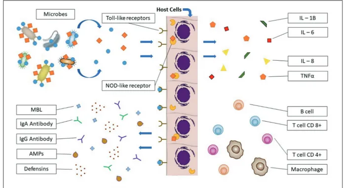
The vagina contains different immune relat-ed cells and receptors which can recognize and react with the microbial environment20.
Surveil-lance for microbes within the female genital tract of both commensal and pathogenic microbes is generally achieved by microbial specimen rec-ognition through pattern recrec-ognition receptors (PRRs), such as toll-like receptors (TLRs), dec-tin-1 receptor, and nucleotide-binding oligomer-ization domain (NOD). These receptors are pres-ent on both squamous epithelial cells lining the vagina and columnar cells lining the upper female genital tract21-25.
Microbial stimulation of PRRs initiates cyto-kine/chemokine signaling cascades, leading to secretion of IL-1β, IL-6, IL-8 and tumor necrosis factor-α (TNF-α), in order to recruit or activate specialized cells, including NK cells, macro-phages, CD4+ helper T-cells, and CD8+ cytotoxic
T-cell lymphocytes, and B lymphocytes. Genetic variants of PRRs, such as the IL-1R antagonist gene, TLR4, TLR9, IL-1R2 and TNF-α may play a role in individual woman’s response to a par-ticular microbial challenge or pregnancy outcome (Figure 2)26,27.
When compared to CST-I, women with CST-IV demonstrate elevated levels of IL-1α, 1β, TNF-α, IFN-γ, 4, 8, 10, IL-12p70, and fms-like tyrosine kinase 3 ligand. Furthermore, significantly higher levels of IFN-γare found in CST-III, relative to CST-I. Particularly, Anahtar et al28 have
demonstrat-ed that Prevotella Amnii, Mobiluncus Mulieris,
Sneathia Amniiand Sneathia Sanguinegens (all
commonly found in CST-IV) were found to induce higher levels of IL-1α, IL-1β, and IL-8 secretion, relative to L. Crispatus dominated communities (in CST-I). In addition, L. Iners dominated communities (CST-III) induced moderate IL-8 levels relative to CST-I. Never-theless, a significant increase in IL-1α, IL-1β and TNF-α was noted during transition from a CST-I, to CST-III and to a CST-IV.
However, mock communities dominated by L.
Crispatus (CST-I) and L. Jensenii (CST-V) on
re-constructed three-dimensional vaginal epithelial models do not cause cytokine IL-1β or IL-8 secre-tion relative to medium control, and also inhibit some pro-inflammatory responses after TLR 2/6 and 3 agonist induction29.
Therefore, these studies demonstrate that the innate immune response is largely driven by vaginal bacterial community states, with CST-IV potentially having a greater pro-in-flammatory response than CST-I or CST-II, and with CST-III triggering an intermediate response.
Other factors contributing to vaginal defense include mannose binding lectin (MBL), immuno-globulin A and G (IgA, IgG) and vaginal antimi-crobial peptides (AMPs):
– MBL binds mannose, N-acetyl-glucosamine and fucose carbohydrate moieties present on microbial cell surfaces. Eventually, this inter-action leads to cell lysis or targeting for the im-mune system30.
Figure 2. The female genital tract is constantly exposed to microbes. Variable defense factors of the innate immune response including mannose binding lectin (MBL), immunoglobulin A (IgA), immunoglobulin G (IgG), vaginal antimicrobial peptides (AMPs) and defensins contribute to clearance of infectious microbes by different mechanisms of action. Surveillance for both commensal and pathogenic microbes is generally achieved by pattern recognition receptors (PRRs) such as toll-like receptors (TLRs) and NOD like receptors (NLR). Microbial stimulation of PRRs initiates cytokine/chemokine signaling cascades, leading to secretion of interleukin and tumor necrosis factor-α (TNF-α), in order to recruit or activate specialized cells including macrophages, CD4+ helper T-cells, CD8+ cytotoxic T-cell and B lymphocytes.
– IgA and IgG may help to prevent adherence to vaginal epithelial cells and subsequent uptake of bacteria, as well as contribute to the neutral-ization and clearance of infectious microbes from the vagina31.
– Vaginal AMPs exist in various classes and may recruit immune cells via chemotaxis and possess anti-endotoxin activity. Defen-sins are a class of cationic and amphipath-ic AMPs with diverse mechanisms of action against common vaginal bacteria as well as pathogenic bacteria and viruses including HIV, herpes simplex virus, and human papil-lomavirus. In organotypic models of the vag-inal epithelium, human β-defensin (HBD)-2 expressions, but not that of HBD-1, was asso-ciated with colonization by L. Iners,
Atopo-bium Vaginae and Prevotella Bivia. Other
studies32,33 with similar experimental in vitro
conditions has shown that L. Jensenii but not
G. Vaginalis induce HBD-2 transcription.
In addition to defensins, other AMPs are found in the human vagina and include the se-cretory leukocyte protease inhibitor (SLPI), hu-man epididymis protein 4 (HE4), LL-37, surfac-tant protein (SP)-A and SP-D. SLPI expression is associated with bacterial vaginitis organisms but not with L. Crispatus, L. Iners, A. Vaginae or P. Bivia. HE4 is associated with G. Vaginalis, and LL-37 inactivates the sexually transmitted pathogen Neisseria Gonorrhoeae while having little or no effect on L. Iners, L. Crispatus, and
L. Jensenii34,35.
Like defensins, SP-A and SP-D contribute to viral inhibition, including HIV. These AMPs bind to the viral protein gp120 and human CD4 receptor, enhancing attachment to dendritic cells and therefore facilitating HIV uptake by immune cells36.
Altered Interplay Between Microbiota and Immune System
Menopause
Menopause is characterized by the loss of estrogen function on vaginal cells metabolism leading to less cell layers, thinner mucus layer, decreased glycogen production. Furthermore, the estrogen deprivation influences the microbiome susceptibility as well as the mucosal immunity. Changes in the immune system accompanying ageing and menopause are known as
immune-se-nescence, the process is characterized by a decre-ase in cell-mediated immune function and humo-ral response37. In fact, in post-menopause, the NK
cell activity decrease significantly and hormone replacement therapy (HRT) was recently shown to restore NK cytotoxicity38.
Several studies have recently assessed the va-ginal microbiome in postmenopausal women. Largely, these studies39-44 sustain the perception
that diversity and abundance of Lactobacilli are declining following menopause. In a portion of these studies, the postmenopausal women were considered healthy (asymptomatic) and not trea-ted with HRT39,40,42.
Gustaesson et al40 found that fertile women had
a great diversity in the subspecies of Lactobacilli present. They concluded that fertile women were more commonly colonized with L. Crispatus, compared to menopausal women (p = 0.0036). Si-milarly, Zhang et al42 observed a lower diversity
of Lactobacillus spp. In postmenopausal relative to premenopausal women (p < 0.05). Mirmonsef et al45 demonstrated similar results, showing that
premenopausal women had significantly higher free glycogen levels and higher Lactobacillus le-vels in comparison with postmenopausal women. In some cases, but not always, the depletion of Lactobacillus spp. and the increase in diver-se microbial species (CST IV-A and CST IV-B), results in symptoms of VVA or the genitouri-nary syndrome of menopause (GSM), which describes a number of menopausal symptoms in relation to changes of the vulva, vagina, and lower urinary tract46.
Many researchers47,48 have begun to investigate
the vaginal microbiome exploring VVA and GSM through the protective features of the vaginal mi-crobiome.
Vaginal microbiota plays an important role in preventing colonization by pathogenic organisms but the predominant connection between the va-ginal microbiome and menopause occurs through the influential action of estrogen. The role of estro-gen has been highlighted in menopause with the estrogen replacement therapy. Gliniewicz et al5
characterized the vaginal bacterial communities of women in three groups: postmenopausal women undergoing HRT who had a vaginal pH ≤ 5 and a vaginal atrophy score ≤ 2; postmenopausal women with a vaginal pH ≥ 5 and vaginal atrophy score ≥ 6, and premenopausal women with a vaginal pH ≤ 5 and vaginal atrophy score ≤ 2. In preme-nopausal women three sorts of communities were commonly found that were dominated by either L.
Crispatus, Gardenerella Vaginalis, or L. Iners. The
vaginal communities of most postmenopausal wo-men receiving HT were dominated by these spe-cies of lactobacilli, whereas this was usually not the case in untreated postmenopausal women. The authors suggest that HRT may lead to preferential enrichment of L. Crispatus. It has been speculated that estrogen stimulates that proliferation of squa-mous epithelial cells, which is accompanied by the increased production of glycogen by these cells. Glucose, maltose, and maltodextrins produced through the hydrolysis of glycogen are thought to serve as carbon sources that support the prolifera-tion of vaginal Lactobacilli5.
It was demonstrated that all routes of estrogen administration (oral, transdermal, and vaginal) are effective for relief of menopausal symptoms.
Ginkelet al48 comparing women receiving
es-trogen replacement therapy to those taking non hormone therapy, showed that women on HRT were less likely to be colonized with anaerobic bacteria. Therefore, a longitudinal study in wom-en treated with oral estrogwom-ens found that after three months of treatment, 20% of women on pla-cebo and 80% of women on oral estrogens treat-ment reported improvetreat-ment in vaginal dryness and irritation concurrent with increased vaginal lactobacilli and lower vaginal pH.
Probiotics (including Lactobacillus spp.) poten-tially work through a variety of mechanisms to re-instate homeostasis by enhancing epithelial barrier function, commensal colonization, blocking adhe-sion of pathogenic bacteria, reduction pH, influenc-ing antimicrobial peptide production/secretion and overall mucosal immunity and vaginal health49-51.
Changes in vaginal microbiota can influence the vaginal microenvironment, and so decrease the efficacy of different potential therapies. Con-sequently, even the local estrogen administration which is effective for relief of menopausal symp-toms may have better result with the probiotics therapy taken at the same time.
Probiotics potentially work to reinstate homeo-stasis by enhancing epithelial barrier function, commensal colonization, blocking adhesion of pathogenic bacteria, reducing pH, influencing antimicrobial peptide production/secretion and overall mucosal immunity and vaginal health52-54.
Both oral and vaginal routes of Lactobacillus (based probiotic formulation) are effective for re-instating vaginal homeostasis55,56
Few studies analyzed the combination treatment of probiotics and estrogen or antibiotics therapy. A recent study included 60 postmenopausal women,
with atrophic vaginitis and chronic recurrent bac-terial cystitis in the acute stage. Patients receiving antibiotic therapy in combination with vaginal lo-cal estriol were compared with patients receiving antibiotics in combination with a lyophilized cul-ture of lactobacilli L. Casei Rhamnosus Doderleini for 3 months. The association therapy of antibiotics and Lactobacilli contributed to the normalization of pH, and reduce the severity of vaginal dryness and burning. The rate of patients with improve-ment of symptoms was significantly higher in the group receiving antibiotics and Lactobacilli than in the group receiving antibiotics and estriol (96.7%
vs. 83.3% respectively)57.
In another study it has been evaluated the ef-ficacy of lyophilized lactobacilli in combination with estriol when compared to metronidazole in the treatment of bacterial vaginal infections. The authors concluded that lyophilized lactobacilli in combination with low dose estriol are equivalent to metronidazole in the short-term treatment of bacterial vaginal infections, but have less effect after 1 month, and so further studies are required to evaluate the long-term efficacy of lactobacilli when applied repeatedly58.
Vaginal Inflammation
The vaginal microbiota can be characterized by different CSTs, with CST-IV lacking a significant number of Lactobacillus spp. Generally, CST-IV can clinically manifest as aerobic vaginitis or BV. Aerobic vaginitis is mainly differentiated from BV by the presence of an inflammatory response predominately associated with aerobes, such as group B Streptococcus, Staphylococcus Aureus,
Escherichia coli, and Enterococcus8.
The aerobic vaginitis inflammatory response is characterized by symptoms, including itch-ing or burnitch-ing, molecular changes, such as in-creased IL-6 and IL-1β, and presence of cells, such as leukocytes or primary blood cells in a microscopic wet mount. In contrast, BV is not characterized by inflammatory responses and therefore, recruitment of neutrophils, redness, itching or burnings assent52.
Several cytokines as well as other immune-re-lated factors [1β, 2, 4, 6, 8, IL-10, IL-12, TNF-α, IFN-γ, chemokine C-C motif ligand 5 (CCL5)] and SLPI have been variably and inconsistently associated with BV. These conflicting findings may be due to different study design different definitions of BV (symptomatic
vs. asymptomatic BV or Nugent BV vs. BV
it is possible that immune-related factors, such as IgA degradation, TLR expression inhibition, or immune-related genetic variants can suppress the inflammatory response in BV53,54.
Bacteria-derived short chain fatty acids (SC-FAs), namely acetate, butyrate, propionate and succinate, some of which exist at relatively higher proportions during BV, can induce a pro-inflam-matory response under the hypothesis that SCFAs may act to ultimately inhibit chemotaxis and in-flammation in BV. Relatively high concentrations (2-20 mM) of acetate and butyrate, but not pro-pionate, induce cytokine IL-6, IL-8 and IL-1β secretions and also induce IL-8 and TNF-α with TLR2 and TLR7 ligand stimulation in a dose- and time-dependent manner in vitro59.
Lactic acid which is produced mainly by vagi-nal microbes inactivates a broad range of BV-as-sociated microbes at pH <4.560. When
Lactoba-cillus spp. dominate the vaginal microbiota, they
acidify the vagina to a highly acidic mean pH of 3.5 ± 0.2 that is likely to help protect against a broad range of infections.
Recent studies aimed to uncover the mecha-nism by which lactic acid can affect host immune functions have found diverse effects, including direct inhibition of pro-inflammatory respons-es IL-6, IL-8 and IL-1RA, induction of the Th17 lymphocyte pathway via IL-23 in a dose-depen-dent manner upon lipopolysaccharide co-stim-ulation, stimulation of mediators from vaginal epithelial cells, and upon transforming growth factor-β, activation of antiviral response53,54,59.
Lactic acid exists in the vagina in both D-(–)- and L-(+)-isomers, with the host contributing only about 4-30% of the total lactate level. Women with BV were found to be deficient in both isomers, while those with vulvovaginal candidiasis have elevated L-(+)-lactic acid as well as CD147 and MMP-8 genes61. L. Iners does not produce D-(–)-lactic acid
and fails to produce as high the L- (+)-lactic acid as seen with L. Crispatus, L. Gasseri. However, L.
Jensenii produces only D-(–)-lactic acid62,
suggest-ing potential Lactobacillus species-specific effects on the host. Consequently, the composition of the vaginal microbiota, and specifically the ability of vaginal microbes to produce D-(–)-lactic acid, may help to inhibit inflammatory responses while also favoring Lactobacillus spp. survival by using host cells resources for carbon source.
Pregnancy
During pregnancy, in parallel to the dramatic hormonal, weight, immunological and metabolic
changes, significant changes in the microbiome occur. These changes affect all the districts where the human microbiota is expressed: gut, vagina, endometrium, and other sites oral cavity. Sever-al authors63 have linked pregnancy complications
with microbial changes. The different body sites harbor different microbial populations according to different pH, oxygen, nutrients and tempera-ture. Pregnancy is associated to an increased gut bacterial load64 and modifications of the
compo-sition of gut microbiota, including reduced α-di-versity (individual richness) and increased ß-di-versity (between-subject diß-di-versity), increased
Actinobacteria and Proteobacteria phyla and
reduced Faecali bacterium and other short-chain fatty-acid producers65. These gut modifications
resemble those observed in metabolic syndrome and have been suggested to play an important contribute to changes in host immunology and in metabolism, via increased absorption of glucose fatty acids, increased fasting-induced adipocyte factor secretion, and stimulation of the immune system. The vaginal microbiome shows a signifi-cant reduction in overall diversity, increased sta-bility and increased abundance of Lactobacillus
species, with the final result of decreased vaginal
pH creating a barrier against pathogenic bacte-ria and viral infections66. It is important to note
that pregnancy is a healthy physiological process in which beneficial microbial alterations are ex-pected. In contrast, pregnancy complications like preterm birth and miscarriage, have been asso-ciated with some bacterial infections, through mechanisms not completely understood. Antibi-otics administered during pregnancy have been shown to affect the microbiome composition and diversity67. However, further research is needed in
order to explain the impact of microbiome chang-es on pregnancy as well as the importance of rec-ommending antibiotic treatments or probiotic for pregnancy complications.
Preterm Labor
The vaginal microbiota in combination with other factors is associated with adverse reproduc-tive and obstetric outcomes.
The association between an abnormal mater-nal vagimater-nal microbiome and an increased risk of preterm birth (PTB) is still controversial. Several studies on the vaginal microbiome and PTB show a small sample size; often there is absence of data collection on vaginal swabs across pregnan-cy, necessary information on spontaneous PTB is lacking, and studies investigating the vaginal
microbiome and PTB show a small sample size. A homogeneous Lactobacillus-dominated micro-biota has been considered a marker of a healthy female reproductive tract. In contrast, a vaginal microbiome with high species diversity, as in BV, has been associated with increased risk of infec-tions, PTB and pelvic inflammatory disease68,69.
DiGiulioet al70 showed that the risk of PTB was
observed to be higher in patients with abundant
Gardnerella or Mycoplasma and poor in
Lacto-bacillus. Otherwise, in a cohort study71 no
corre-lation was observed between absence of Lactoba-cillus and the risk of PTB. Kindingeret al72 have
reported that a dominance of L. Crispatus in the vaginal microbiota seems to be protective against PTB, while L. Iners seems to be a risk factor for PTB in high risk patients. In a paper recently published in Nature73, L. Crispatus was
great-ly reduced in PTB samples, whereas Prevotella,
BVAB1, BVAB-TM7 and Sneathia Amnii, were
more abundant in vaginal PTB samples. Consi-dering that PTB might be related to an ascension of pathogenic microbes from the vagina, these observations suggest that the vaginal microbiome composition, early in pregnancy, might assist in prediction of adverse outcomes and serve as risk marker for PTB.
Nevertheless, it is noteworthy that the associa-tion between the vaginal microbiome and PTB is population-dependent. Specifically, the associa-tion between lower Lactobacillus, higher
Gardne-rella and increased risk of PTB was detected only
in African Americans and in white populations74.
Women of African descent frequently have va-ginal L. Crispatus predominance and they often show an increased vaginal microbial diversity75.
Analysis of vaginal cytokines showed that va-ginal bacterial taxa, generally associated with dysbiosis, are highly correlated with increased le-vels of pro-inflammatory cytokines, which play a role in the induction of labor. Recently, Fettweiss et al76 observed that vaginal inflammatory
cytoki-ne CXCL10 levels were inversely correlated with
L. Crispatus and positively correlated with L. Iners in PTB patients, suggesting a
cytokine/lac-tobacillus ratio as a possible prediction marker of preterm birth.
It is evident that an early prediction of PTB risk is critical for the development of new strategies for prevention and intervention. All the available data support that population-specific studies mi-ght be helpful to assess the impact of the vaginal microbiome on the risk of PBT and to identify high risk vaginal microbiota specific for a subset
of women. Moreover, it is evident that an early prediction of risk for PTB is critical for the de-velopment of new strategies for prevention and intervention75,76.
Miscarriage
The human endometrium displays a crucial im-munological surveillance for the uterus. Indeed, in the human endometrium a complex immune system is able to prevent the risk of infections as well as, when pregnancy occurs, to allow the ac-ceptance of the blastocyst77,78. Combinations of
chemokines are secreted by endometrial stromal and epithelial cells, which act as “sentinels” able to influence leukocyte endometrial expression. Major immune cells in the human endometrium include uterine Natural Killer (uNK) cells, macrophages, dendritic cells (DCs) and T cells. Each of these cell populations demonstrated79-84 a specific role.
uNK are involved in the success of implantation and maintenance of pregnancy 65-70 through their ability both to interact through inhibitor receptors with HLA-G, HLA-E and HLA-C expressed on trophoblast cells and to produce angiogenic fac-tors85,86. Macrophages and DCs are involved in
scavenging and degradative functions associated with menstruation87,88. Moreover, macrophages are
found in the placental bed throughout gestation and likely provide an immediate antigen non-specific host defense to infection, essential for maintaining the integrity of pregnancy89,90.
A further crucial component of the innate im-munity system is the inflammasome, intracel-lular, multiprotein complex involved in the en-dometrial surveillance against possible noxious agents90. Once recruited, inflammasome
increa-ses pro-inflammatory cytokine, such as 1β, IL-18 and IL-33, generating their respective mature secretory forms. These events are necessary for the induction of further systemic responses and to spreading of inflammation91. Of interest, a
si-gnificant increased expression of the inflammaso-me components as well as of IL-1ß and IL-18 in endometrial biopsies obtained from women with recurrent pregnancy loss as compared to controls has been observed.
Uterine T cells represent the most impor-tant component of the adaptive immune system counterpart. They include lymphocytes identi-fied by specific markers, transcription factors, cytokine production, and cytotoxic capacity. Uterine T cells consist of CD8+ cells (66%) and CD4+ cells (33%). The CD4+ T cell po-pulation includes Th1, Th2, regulatory T cells
(Tregs), and Th17 cells, each of which secretes specific cytokines with wide-ranging effects. By simplifying, Th1 cells are implicated in the cell-mediated reactions (cellular immunity), important in resistance to infections caused by intracellular pathogens and viruses and are involved in promoting inflammation92,93.
There-fore, Th1 cells are regarded as potential con-tributors to pregnancy pathologies and major threats to fetal survival. Th2 cells are mostly involved in antibody production (humoral im-munity) and resistance to extra-cellular patho-gen infections93. Th1 and Th2 cells have mutual
inhibitory effect on each other. The existing data linking spontaneous abortion with incre-ased decidual Th1/Th2 ratios94,95 suggest that
pregnancy is a Th2-prevalents phenomenon. It is now well accepted that the human endo-metrium hosts different populations of microor-ganisms, reaching only a 30% of those present in the cervical-vaginal flora96,97. In recent years,
the development of sequencing-based technolo-gies have enabled the evaluation of the endo-metrial microbiota and microbiome, defined as the totality of the microbes and their genomes existing at endometrial level98,99. Moreno et al100
detected up to 191 operational taxonomic uni-ts (OTUs) of bacteria at the endometrial level, with a composition not influenced by steroid hormones fluctuations101. They distinguished a
Lactobacillus-dominated (> 90% Lactobacillus
spp.) and non-Lactobacillus-dominated (< 90%
Lactobacillus with > 10% of other bacteria) mi-crobiota and, they found that a non-Lactobacil-lus-dominated microbiota were associated with a significantly lower rate of implantation (60.7%
vs. 23.1%), pregnancy (70.6 % vs. 33.3%),
on-going pregnancy (58.8% vs. 13.3%), and live birth (58.8% vs. 6.7%) compared to women with a Lactobacillus-dominated microbiota. Verstraelenet al102, performed on a
heterogene-ous group of women with different reproducti-ve history, including subfertility, a unique mi-crobiota dominated by Bacteroides has been documented. More recently, using next-gene-ration sequencing technologies, Kitaya et al103
attempted to characterize the microbiota in the endometrial fluid and vaginal secretions in wo-men with recurrent implantations failure. They found that the endometrial microbiota had hi-gher α-diversity and broader bacterial species than the vaginal microbiota.
These data highlight the efforts made in the re-cent scientific research to better characterize the
endometrial microbiota, recognize the interplay between the vaginal/uterine microbiome and the immune system and to develop predictive mar-kers for possible outcomes.
Additional studies are needed to better under-stand women genital tract milieu and identify new biomarkers and their health consequences. These will improve gynecological evaluation will design new approaches to diagnosis and, ultimately, will lead to better, personalized new therapies.
Conclusions
The study and characterization of the role of microbiota in gynecological and obstetric condi-tions is moving its first steps, following the road of other disciplines (such as gastroenterology), in which it plays a predominant role in explaining the onset and offset of several conditions.
If traditionally vaginal flora has been analyzed through classic microbiologic methods, such as culture or colorations, a new step has to be done: in facts, 16S rRNA sequencing can lead to a more efficient, effective and accurate determination of all the phyla and species determining the diver-sity of microbiome. This method has not got yet the right grade of standardization, and this does not allow its spreading in everyday clinical and research life.
The debate is still controversial on several points, but there are some certainties: healthy vaginal flora is characterized by high concentra-tion of Lactobacilli, responsible for lactic acid production and maintenance of vaginal pH < 4.5, whilst several gynecological pathologies are as-sociated to a reduction in Lactobacilli, with an increase of other bacterial species.
Another important aspect that must be taken into consideration is immune response. Immune response strictly interacts and strictly regulates microbiota itself.
The study and characterization of vaginal flora opens up the road to new perspective of therapy, such as probiotics, prebiotics, antibiotics, hormo-nal therapies that together can shape and modula-te microbiome in order to restore clinical and mi-crobiological eubiosis, fundamental for a correct working of all the reproductive system.
Conflict of Interest
References
1) Vande Wijgert jH, Borgdorff H, VerHelst r, CruCit -ti t, franCis s, Verstraelen H, jespers V. The vaginal microbiota: what have we learned after a decade of molecular characterization? PLoS One 2014; 9: e105998.
2) lamont rf, soBel jd, akins ra, Hassan ss, CHaiW -orapongsa t, kusanoViC jp, romero r. The vaginal microbiome: new information about genital tract flora using molecular based techniques. BJOG 2011; 118: 533-549.
3) petroVa mi, lieVens e, malik s, imHolz n, leB -eer s. Lactobacillus species as biomarkers and agents that can promote various aspects of vagi-nal health. Front Physiol 2015; 6: 81.
4) gajer p, Brotman rm, Bai g, sakamoto j, sCHütte um, zHong X, koenig ss, fu l, ma zs, zHou X, aB -do z, forney lj, raVel j. Temporal dynamics of the human vaginal microbiota. Sci Transl Med 2012; 4: 132ra52.
5) glinieWiCz k, sCHneider gm, ridenHour1 Bj, Williams Cj, song y, farage ma, miller k, forney lj. Com-parison of the vaginal microbiomes of premeno-pausal and postmenopremeno-pausal women. Front Micro-biol 2019; 10: 193.
6) yue Xa, CHen p, tang y, Wu X, Hu z. The dynam-ic changes of vaginal mdynam-icroecosystem in patients with recurrent vulvovaginal candidiasis: a retro-spective study of 800 patients. Arch Gynecol Ob-stet 2015; 292: 1285-1294.
7) CHen Hm, CHang tH, lin fm, liang C, CHiu Cm, yang tl, yang t, Huang Cy, CHeng yn, CHang ya, CHang py, Weng sl. Vaginal microbiome varianc-es in sample groups categorized by clinical crite-ria of bactecrite-rial vaginosis. BMC Genomics 2018; 19: 876.
8) donders gg, VereeCken a, Bosmans e, dekeersmaeCk -er a, salemBier g, spitz B. Definition of a type of ab-normal vaginal flora that is distinct from bacterial vaginosis: aerobic vaginitis. BJOG 2002; 109: 34-43.
9) Wiggins r, CroWley t, Horner pj, sootHill pW, mil -lar mr, Corfield ap. Use of 5-bromo-4- chloro-3-indolyl-α-d-N-acetylneuraminic acid in a nov-el spot test to identify sialidase activity in vagi-nal swabs from women with bacterial vaginosis. J Clin Microbiol 2000; 38: 3096-3097.
10) Wang zl, fu ly, Xiong za, Qin Q, yu tH, Wu yt1, Hua yy, zHang yH. Diagnosis and microecological characteristics of aerobic vaginitis in outpatients based on preformed enzymes. Taiwan J Obstet Gynecol 2016; 55: 40-44.
11) sriniVasan s, liu C, mitCHell Cm, fiedler tl, tHomas kk, agneW kj, marrazzo jm, fredriCks dn. Tempo-ral variability of human vaginal bacteria and rela-tionship with bacterial vaginosis. PLoS One 2010; 5: e10197.
12) fredriCks dn, fiedler tl, marrazzo jm. Molecular identification of bacteria associated with bacterial vaginosis. N Engl J Med 2005; 353: 1899-1911.
13) Campos aC, freitas-junior r, riBeiro lf, paulinelli rr, reis C. Prevalence of vulvovaginitis and bacterial vaginosis in patients with koilocytosis. Sao Paulo Med J 2008; 126: 333-336.
14) raVel j, gajer p, aBdo z, sCHneider gm, koenig ssk, mCCulle sl, karleBaCH s, gorle r, russell j, taCket Co, Brotman rm, daVis CC, ault k, peralta l, for -ney lj. Vaginal microbiome of reproductive-age women. Proc Natl Acad Sci U S A 2011; 108: 4680-4687.
15) moHammadzadeH f, dolatian m, jorjani m, alaVima -jd H. Diagnostic value of Amsel’s clinical criteria for diagnosis of bacterial vaginosis. Glob J Health Sci. 2014; 7: 8-14.
16) Baker jm, al-nakkasH l, HerBst-kraloVetz mm. Es-trogen-gut microbiome axis: physiological and clinical implications. Maturitas 2017; 103: 45-53. 17) Hong kH, Hong sk, CHo si, ra e, Han kH, kang sB,
kim eC, park ss, seong mW. Analysis of the vag-inal microbiome by next-generation sequencing and evaluation of its performance as a clinical di-agnostic tool in vaginitis. Ann Lab Med 2016; 36: 441-449.
18) Virtanen s, rantsi t, Virtanen a, kerVinen k, nieminen p, kalliala i, salonen a. Vaginal microbiota com-position correlates between Pap smear micros-copy and next generation sequencing and asso-ciates to socioeconomic status. Sci Rep 2019; 9: 7750.
19) smidt i, kiiker r, oopkaup H, lapp e, rööp t, truusa -lu k, ŠtŠepetoVa j, truu j, mändar r. Comparison of detection methods for vaginal lactobacilli. Benef Microbes 2015; 6: 747-751.
20) Wira Cr, faHey jV, sentman Cl, pioli pa, sHen l. Innate and adaptive immunity in female genital tract: cellular responses and interactions. Immu-nol Rev 2005; 206: 306-335.
21) CarValHo a, gioVannini g, de luCa a, d’angelo C, Casagrande a, iannitti rg, riCCi g, CunHa C, roma -ni l. Dectin-1 isoforms contribute to distinct Th1/ Th17 cell activation in mucosal candidiasis. Cell Mol Immunol 2012; 9: 276-286.
22) usluogullari B, gumus i, gunduz e, kaygusuz i, si -maVli s, aCar m, oznur m, gunduz m, kafali H. The role of human dectin-1 Y238X gene poly-morphism in recurrent vulvovaginal candidia-sis infections. Mol Biol Rep 2014; 41: 6763-6768.
23) Witkin ss, linHares im, giraldo p. Bacterial flora ofthe female genital tract: function and immune regulation. Best Pract Res Clin Obstet Gynaecol 2007; 21: 347-354.
24) Horne aW, stoCk sj, king ae. Innate immunity and disorders of the female reproductive tract. Repro-duction 2008; 135: 739-749.
25) mitCHell C, marrazzo j. Bacterial vaginosis and the cervicovaginal immune response. Am J Reprod Immunol 2014; 71: 555-563.
26) genC mr, onderdonk aB, VardHana s, delaney ml, norWitz er, tuomala re, paraskeVas l-r; Witkin ss-map study group. Polymorphism in intron 2 of
the interleukin-1 receptor antagonist gene, local midtrimester cytokine response to vaginal flora, and subsequent preterm birth. Am J Obstet Gy-necol 2004; 191: 1324-1330.
27) genC mr, VardHana s, delaney ml, onderdonk a, tuomala r, norWitz e; Witkin ssmap study group. Relationship between a toll-like receptor-4 gene polymorphism, bacterial vaginosis-related flo-ra and vaginal cytokine responses in pregnant women. Eur J Obstet Gynecol 2004; 116: 152-156.
28) anaHtar mn, Byrne eH, doHerty ke, BoWman Ba, yamamoto Hs, soumillon m, padaVattan n, ismail n, moodley a, saBatini me, gHeBremiCHael ms, nusBaum C, HuttenHoWer C, Virgin HW, ndung’u t, dong kl, Walker Bd, fiCHoroVa rn, kWon ds. Cervicovagi-nal bacteria are a major modulator of host inflam-matory responses in the female genital tract. Im-munity 2015; 42: 965-976.
29) roseWa, mCgoWin Cl, spagnuolo ra, eaVes-pyl -es td, popoV Vl, PYLES RB. Commensal bacte-ria modulate innate immune responses of vaginal epithelial cell multilayer cultures. PLoS One 2012; 7: e32728.
30) turner mW. The role of mannose-binding lectin in health and disease. Mol Immunol 2003; 40: 423-429.
31) Wang yy, kannan a, nunn kl, murpHy ma, suBra -mani dB, moenCH t, Cone r, lai sk. IgG in cervi-covaginal mucus traps HSV and prevents vagi-nal Herpes infections. Mucosal Immunol 2014; 7: 1036-1044.
32) doerflinger sy, tHroop al, HerBst-kraloVetz mm. Bacteria in the vaginal microbiome alter the in-nate immune response and barrier properties of the human vaginal epithelia in a species-specific manner. J Infect Dis 2014; 209: 1989-1999. 33) Valore eV, Wiley dj, ganz t. Reversible deficiency
of antimicrobial polypeptides in bacterial vagino-sis. Infect Immun 2006; 74: 5693-5702.
34) orfanelli t, jayaram a, doulaVeris g, forney lj, ledger Wj, Witkin ss. Human epididymis protein 4 and secretory leukocyte protease inhibitor in vaginal fluid: relation to vaginal components and bacterial composition. Reprod Sci 2014; 21: 538-542.
35) monCla Bj, mietzner ta, Hillier sl. In vitro activity of cationic peptides against Neisseria gonorrhoe-ae and vaginal Lactobacillus species: The effect of divalent cations. Adv Biosci Biotechnol 2012; 3: 249-255.
36) gaiHa gd, dong t, palaniyar n, mitCHell da, re -id kBm, Clark HW. Surfactant protein A binds to HIV and inhibits direct infection of CD4+ cells, but enhances dendritic cell-mediated viral transfer. J Immunol 2008; 181: 601-609.
37) gameiro Cm, romão f, Castelo‐BranCo C. Meno-pause and aging: changes in the immune sys-tem‐a review. Maturitas 2010; 67: 316‐320. 38) yang jH, CHen Cd, Wu my, CHao kH, yang ys, Ho
Hn. Hormone replacement therapy reverses the
decrease in natural killer cytotoxicity but does not reverse the decreases in the T-cell subpop-ulation or interferon-gamma production in post-menopausal women. Fertil Steril 2000; 74: 261-267.
39) Burton jp, reid g. Evaluation of the bacterial vag-inal flora of 20 postmenopausal women by direct (Nugent score) and molecular (polymerase chain reaction and denaturing gradient gel electropho-resis) techniques. J Infect Dis 2002; 186: 1770-1780.
40) gustafsson rj, aHrne s, jeppsson B, Benoni C, ols -son C, stjernQuist m, oHlsson B. The Lactoba-cillus flora in vagina and rectum of fertile and postmenopausal healthy Swedish women, BMC Womens Health 2011; 11: 17.
41) petriCeViC l. unger fm, Viernstein H, kiss H. Ran-domized, double-blind, placebo-controlled study of oral lactobacilli to improve the vaginal flora of postmenopausal women. Eur J Obstet Gynecol Reprod Biol 2008; 141: 54-57.
42) zHang r, daroCzy k, Xiao B, yu l, CHen r, liao Q. Qualitative and semiquantitative analysis of Lac-tobacillus species in the vaginas of healthy fertile and postmenopausal Chinese women J Med Mi-crobiol 2012; 61: 729-739.
43) petriCeViC l, domig kj, niersCHer fj, sandHofer mj, krondorfer i, kneifel W, kiss H. Differences in the vaginal lactobacilli of postmenopausal women and influence of rectal lactobacilli. Climacteric 2013; 16: 356-361.
44) Brotman rm, sHardell md, gajer p, fadrosH d, CHang k, silVer mi, VisCidi rp, Burke ae, raVelj, graVitt pe. Association between the vaginal mi-crobiota, menopause status, and signs of vulvo-vaginal atrophy. Menopause 2018; 25: 1321-1330. 45) mirmonsef p, Hotton al, gilBert d, Burgad d, lan -day a, WeBer km, CoHen m, raVel j, spear gt. Free glycogen in vaginal fluids is associated with Lac-tobacillus colonization and low vaginal pH. PLoS One 2014; 9: e102467.
46) muHleisen al, HerBst-kraloVetz mm. Menopause and the vaginal microbiome. Maturitas 2016; 91: 42-50.
47) deVillard e, Burton jp, Hammond ja, lam d, reid g. Novel insight into thevaginal microflora in post-menopausal women under hormone replacement therapy as analyzed by PCR-denaturing gradient gel electrophoresis. Eur J Obstet Gynecol Reprod Biol 2004; 117: 76-81.
48) ginkel pd, soper de, Bump rC, dalton Hp. Vaginal flora in postmenopausal women: the effect of es-trogen replacement. Infect Dis Obstet Gynecol 1993; 1: 94-97.
49) doerflinger sy, tHroop al, HerBst-kraloVetz mm. Bacteria in the vaginal microbiome alter the in-nate immune response and barrier properties of the human vaginal epithelia in a species-specific manner. J Infect Dis 2014; 209: 19891999. 50) CHase d, goulder a, zenHausern f, monk B, HerBst
mi-crobiomes in gynecologic cancers: a review of applications in etiology, symptoms and treatment. Gynecol Oncol 2015; 138: 190-200.
51) yarBrougH Vl, Winkle s, HerBst-kraloVetz mm, An-timicrobial peptides in the female reproductive tract: a critical component of the mucosal mune barrier with physiological and clinical im-plications. Hum Reprod Update 2015; 21: 353-377.
52) CauCi s. Vaginal immunity in bacterial vaginosis. Curr Infect Dis Rep 2004; 6: 450-456.
53) CauCi s, guasCHino s, de aloysio d, driussi s, de santo d, penaCCHioni p, Quadrifoglio f. Interrela-tionships of interleukin-8 with interleukin-1βand neutrophils in vaginal fluid of healthy and bacte-rial vaginosis positive women. Mol Hum Reprod 2003; 9: 53-58.
54) Witkin ss, linHares im, giraldo p, ledger Wj. An al-tered immunity hypothesis for the development of symptomatic bacterial vaginosis. Clin Infect Dis 2007; 44: 554-557.
55) yarBrougH Vl, Winkle s, HerBst-kraloVetz mm. An-timicrobial peptides in the female reproductive tract: a critical component of the mucosal mune barrier with physiological and clinical im-plications. Hum Reprod Update 2015; 21: 353-77.
56) Burton jp, CadieuX pa, reid g. Improved under-standing of the bacterial vaginal microbiota of women before and after probiotic instillation. Ap-pl Environ Microbiol 2003; 69: 97-101.
57) kuzmenko aV, kuzmenko VV, gyaurgieV ta. Expe-rience of application of hormonal and probiot-ic therapy in the complex treatment of women in peri - and postmenopausal with chronic recurrent bacterial cystitis in the background of vulvovagi-nal atrophy. Urologiia 2019; 3: 66-71.
58) donders gg, Van BulCk B, Van de Walle p, kaiser rr, poHlig g, gonser s, graf f. Effect of lyophilized lactobacilli and 0.03 mg estriol (Gynoflor®) on vaginitis and vaginosis with disrupted vaginal mi-croflora: a multicenter, randomized, single-blind, active-controlled pilot study. Gynecol Obstet In-vest. 2010; 70: 264-72.
59) mirmonsef p, zariffard mr, gilBert d, makinde H, landay, spear gt. Short-chain fatty acids induce pro-inflammatory cytokine production alone and in combination with toll-like receptor ligands. Am J Reprod Immunol 2012; 67: 391-400.
60) gong z, luna y, yu p, fan H. Lactobacilli inactivate Chlamydia trachomatis through lactic acid but not H2O2. PLoS One 2014; 9: e107758.
61) BegHini j, linHares im, giraldo pC, ledger Wj, Wit -kin ss. Differential expression of lactic acid iso-mers, extracellular matrix metalloproteinase in-ducer, and matrix metalloproteinase-8 in vaginal fluid from women with vaginal disorders. BJOG 2015; 122: 1580-1585.
62) Witkin ss, mendes-soares H, linHares im, jayaram a, ledger Wj, forney lj. Influence of vaginal bacteria and D- and L-lactic acid isomers on
vaginal extracellular matrix metalloproteinase inducer: implications for protection against up-per genital tract infections. mBio 2013; 6; 4. pii: e00460-13.
63) neuman H, koren o. The pregnancy microbiome. Nestle Nutr Inst Workshop Ser 2017; 88: 1-9. 64) nuriel-oHayon m, neuma H, koren o.
Microbi-al changes during pregnancy, birth, and Infancy. Front Microbiol 2016; 7: 1031.
65) Haro C, garCia-Carpintero s, alCala-diaz jf, go -mez-delgado f, delgado-lista j, perez-martinez p, rangel zuñiga oa, Quintana-naVarro gm, landa BB, Clemente jC, lopez-miranda j, Camargo a, pe -rez-jimenez f. The gut microbial community in met-abolic syndrome patients is modified by diet. J Nutr Biochem 2016; 27: 27-31.
66) raVel j, gajer p, aBdo z, sCHneider gm, koenig ss, mCCulle sl, karleBaCH s, gorle r, russell j, taCket Co, Brotman rm, daVis CC, ault k, peralta l, for -ney lj. Vaginal microbiome of reproductive-age women. Proc Natl Acad Sci U S A 2011; 108 Sup-pl 1: 4680-4687.
67) kHan i, azHar ei, aBBas at, kumosani t, BarBour ek, raoult d, yasir m. Metagenomic analysis of antibi-otic-induced changes in gut microbiota in a preg-nant rat model. Front Pharmacol 2016; 7: 104. 68) fredriCks dn, fiedler tl, tHomas kk, oakley BB,
marrazzo jm. Targeted PCR for detection of vag-inal bacteria associated with bacterial vaginosis. J Clin Microbiol 2007; 45: 3270-3276.
69) CHaVoustie se, eder se, koltun Wd, lemon tr, mitCHell C, nyirjesy p, soBel jd, soBel r, VillanueVa r. Experts explore the state of bacterial vaginosis and the unmet needs facing women and provid-ers. Int J Gynecol Obstet 2017; 137: 107-109. 70) digiulio dB, CallaHan Bj, mCmurdie pj, Costello
ek, lyell dj, roBaCzeWska a, sun Cl, goltsman ds, Wong rj, sHaW g, steVenson dk, Holmes sp, relman da. Temporal and spatial variation of the human microbiota during pregnancy. Proc Natl Acad Sci U S A 2015; 112: 11060-11065.
71) Hyman rW, fukusHima m, jiang H, fung e, rand l, joHnson B, Vo kC, CaugHey aB, Hilton jf, daVis rW, giudiCe lC. Diversity of the vaginal microbiome correlates with preterm birth. Reprod Sci 2014; 21: 32-40.
72) kindinger lm, Bennett pr, lee ys, marCHesi jr, smitH a, CaCCiatore s, Holmes e, niCHolson jk, teoH tg, maCintyre da. The interaction between vaginal microbiota, cervical length, and vaginal proges-terone treatment for preterm birth risk. Microbi-ome 2017; 5:6.
73) fettWeis jm, serrano mg, Brooks jp, edWards dj, girerd pH, parikH Hi, Huang B, arodz tj, edupugan -ti l, glasCoCk al, Xu j, jimenez nr, ViVadelli sC, fong ss, sHetH nu, jean s, lee V, BokHari ya, lara am, mistry sd, duCkWortH ra 3rd, Bradley sp, ko -parde Vn, orenda XV, milton sH, rozyCki sk, mat -VeyeV aV, WrigHt ml, HuzurBazar sV, jaCkson em, smirnoVa e, korlaCH j, tsai yC, diCkinson mr, Brooks jl, drake ji, CHaffin do, seXton al, graVett mg,
ruBens Ce, Wijesooriya nr, HendriCks-muñoz kd, jefferson kk, strauss jf 3rd, BuCk ga. The vaginal microbiota and preterm birth. Nat Med 2019; 25: 1012-1021.
74) CallaHan Bj, digiulio dB, goltsman dsa, sun Cl, Costello ek, jeganatHan p, Biggio jr, Wong rj, druzin ml, sHaW gm, steVenson dk, Holmes sp, rel -man da. Replication and refinement of a vaginal microbial signature of preterm birth in two racially distinct cohorts of US women. Proc Natl Acad Sci U S A 2017; 114: 9966-9971.
75) raVel j, gajer p, aBdo z, sCHneider gm, koenig ss, mCCulle sl, karleBaCH s, gorle r, russell j, taCket Co, Brotman rm, daVis CC, ault k, peralta l, for -ney lj. Vaginal microbiome of reproductive-age women. Proc Natl Acad Sci U S A 2011; 108: 4680-4687.
76) fettWeis, jm, Brooks jp, serrano mg, sHetH nu, girerd pH, edWards dj, strauss jf. tHe Vaginal mi -CroBiome Consortium, jefferson kk, BuCk ga. Differ-ences in vaginal microbiome in African American women versus women of European ancestry. Mi-crobiology 2014; 160: 2272-2282.
77) kalkunte s, CHiCHester Co, gotsCH f, sentman Cl, romero r, sHarma s. Evolution of non-cytotoxic uterine natural killer (uNK) cells. Am J Reprod Im-munol 2008; 59: 425-432.
78) stamenoV g, penkoVa k, CHausHeV t, persenska s, dzHamBazoV B, ilieV i, BaltadjieVa d. Endometri-al CD16+ naturEndometri-al killer cells and sub-endometri-al doppler in unexplained infertility. Int J Gynecol Obstet 2016; 3: 385-390.
79) moffett a, ColuCCi f. Uterine NK cells: active regu-lators at the maternal-fetal interface. J Clin Invest 2014; 124: 1872-1879.
80) gaynor lm, ColuCCi f. Uterine natural killer cells: functional distinctions and influence on pregnan-cy in humans and mice. Front Immunol 2017; 8: 467.
81) fu B, li X, sun r, tong X, ling B, tian z, Wei H. Natural killer cells promote immune tolerance by regulating inflammatory TH17 cells at the human maternal-fetal interface. Proc Natl Acad Sci U S A 2013; 110: E231-E240.
82) li yH, zHou WH, tao y, Wang sC, jiang yl, zHang d, piao Hl, fu Q, li dj, du mr. The Galectin-9/ Tim-3 pathway is involved in the regulation of NK cell function at the maternal-fetal interface in early pregnancy. Cell Mol Immunol 2016; 13: 73-81.
83) loCkWood Cj, Huang sj, CHen Cp, Huang y, Xu j, faramarzi s, kayisli o, kayisli u, koopman l, smedts d, BuCHWalder lf, sCHatz f. Decidual cell regula-tion of natural killer cell-recruiting chemokines: implications for the pathogenesis and prediction of preeclampsia. Am J Pathol 2013; 183: 841-856.
84) lima pd, zHang j, dunk C, lyesj, Croy Ba. Leuko-cyte driven-decidual angiogenesis in early preg-nancy. Cell Mol Immunol 2014; 11: 522-537.
85) CHen sj, lu yl, sytWu Hk. Immunologic regula-tion in pregnancy: from mechanism to therapeu-tic strategy for immunomodulation. Clin Dev Im-munol 2012; 2012: 258391.
86) faas mm, de Vos p. Uterine NK cells and macro-phages in pregnancy. Placenta 2017; 56: 44-52. 87) liu s, diao l, Huang C, li y, zeng y, kWak-kim jyH.
The role of decidual immune cells on human pregnancy. J Reprod Immunol 2017; 124: 44-53. 88) morelli ss, yi p, goldsmitH lt. Endometrial stem
cells and reproduction. Obstet Gynecol Int 2012; 2012: 851367.
89) HiCkey dk, patel mV, faHey jV, Wira Cr. Innate and adaptive immunity at mucosal surfaces of the fe-male reproductive tract: stratification and integra-tion of immune protecintegra-tion against the transmis-sion of sexually transmitted infections. J Reprod Immunol 2011; 88: 185-194.
90) dunne a. Inflammasome activation: from inflam-matory disease to infection. Biochem Soc Trans 2011; 39: 669-673.
91) nold-petry Ca, nold mf, nielsen jW, Bustamante a, zepp ja, storm ka, Hong jW, kim sH, dinarello Ca. Increased cytokine production in interleu-kin-18 receptor alpha-deficient cells is associ-ated with dysregulation of suppressors of cyto-kine signaling. J Biol Chem 2009; 284: 25900-25911.
92) smitH-garVin je, koretzky ga, jordan ms. T cell ac-tivation. Annu Rev Immunol 2009; 27: 591-619. 93) flynn l, Byrne B, Carton j, keleHan p, o’HerliHy
C, o’farrelly C. Menstrual cycle dependent fluc-tuations in NK and T-lymphocyte subsets from non-pregnant human endometrium. Am J Reprod Immunol 2000; 43: 209-217.
94) rapHael i, nalaWade s, eagar tn, forstHuBer tg. T cell subsets and their signature cytokines in au-toimmune and inflammatory diseases. Cytokine 2015; 74: 5-17.
95) sakaguCHi s, miyara m, Costantino Cm, Hafler da. FOXP3+regulatory T cells in the human immune system. Nat Rev Immunol 2010; 10: 490-500. 96) Baker jm, CHase dm, HerBst-kraloVetz mm. Uterine
microbiota: residents, tourists, or invaders? Front Immunol 2018; 9: 208.
97) Quayle aj. The innate and early immune re-sponse to pathogen challenge in the female gen-ital tract and the pivotal role of epithelial cells. J Reprod Immunol 2002; 57: 61-79.
98) CHen C, song X, Wei W, zHong H, dai j, lan z, li f, yu X, feng Q, Wang z, Xie H, CHen X, zeng C, Wen B, zeng l, du H, tang H, Xu C, Xia y, Xia H, yang H, Wang j, Wang j, madsen l, BriX s, kristian -sen k, Xu X, li j, Wu r, jia H. The microbiota con-tinuum along the female reproductive tract and its relation to uterine-related diseases. Nat Commun 2017; 8: 875.
99) payne ms, BayatiBojakHi s. Exploring preterm birth as a polymicrobial disease: an overview of the uterine microbiome. Front Immunol 2014; 5: 595.
100) moreno i, franasiak jm. Endometrial microbiota-new player in town. Fertil Steril 2017; 108: 32-39. 101) moreno i, Codoner fm, Vilella f, ValBuena d,
martinez-BlanCH jf, jimenez-almazan j, alonso r, alamá p, remoHí j, pelliCer a, ramon d, simon C. Evidence that the endometrial microbiota has an effect on implantation success or failure. Am J Obstet Gynecol 2016; 215: 684-703.
102) Verstraelen H, VilCHez-Vargas r, desimpel f, jaure -gui r, VankeirsBilCk n, Weyers s, VerHelst r, de sut
-ter p, pieper dH, Van de Wiele t. Characterisation of the human uterine microbiome in non-preg-nant women through deep sequencing of the V1-2 region of the 16S rRNA gene. Peer J 2016; 4: e1602.
103) kitaya k, nagai y, arai W, sakuraBa y, isHikaWa T. Characterization of microbiota in endometrial fluid and vaginal secretions in infertile women with repeated implantation failure. Mediators In-flamm 2019; 2019: 4893437.