University of Pisa
Research Doctorate School in Biological
and Molecular Sciences
Program: Biology
Congenital Heart Diseases:
parental exposures and
gene-environment interactions
Supervisor:
PhD Student:
Dr. Maria Grazia Andreassi
Dr. Monica Cresci
Academic Year 2012/2013 BIO/11
Index
ABSTRACT Pag. 1
INTRODUCTION Pag. 4
Cardiac transcription factor genes: Genetic Hypothesis Pag. 6 Etiology of Congental Heart Disease: gene-environment interaction Pag. 9 Glutathione S-tranferase enzymes (GSTM1, GSTT1, GSTP1) Pag. 10
AIM Pag. 12
MATERIALS AND METHODS Pag. 13
Study population Pag. 13
DNA extraction Pag. 19
DNA quantitative assessment Pag. 19
Genotyping analysis Pag. 20
Glutathione S-Transferase M1 and T1 (GSTM1 and GSTT1) Pag. 20
Agarose gel electrophoresis Pag. 22
Glutathione S-Transferase P1 (GSTP1) Ile105Val polymorphism Pag. 23
Polyacrylamide gel electrophoresis Pag. 24
ISL1 rs1017 polymorphism (ISL1) Pag. 25
Agarose gel electrophoresis Pag. 26
Statistical analysis Pag. 27
RESULTS Pag. 28
Parental exposures analysis Pag. 28
Gene-environment interactions analysis Pag. 32
Glutathione S-Transferase P1 (GSTP1) Pag. 36
ISL1 rs1017 polymorphism (ISL1) Pag. 38
Genetic analysis Pag. 38
DISCUSSION Pag. 41
Parental exposures analysis Pag. 41
Gene-environmental interactions Pag. 44
Glutathione S-Transferase P (GSTP1) Pag. 45
ISL1 rs1017 polymorphism (ISL1) Pag. 47
CONCLUSIONS Pag. 49
1
ABSTRACT
Congenital heart defects (CHDs) are the most prevalent of all birth defects, arising from the complex interplay of environmental exposures and genes. The molecular causes of most CHDs as well as the modifiable environmental risk factors, (especially for paternal exposure) remain largely unknown. Thus, there is an increasing interest in the study of gene-environment interaction in the pathogenesis of CHDs. The major aim of this project was to expand the knowledge of CHD etiology with specific attention at the identification of genetic and environmental risk factors. The effects of environmental factors might be modified by the genes responsible for the activation and detoxification of toxicant agents, contributing to an increased resistance (or sensitivity) to cardiac teratogenesis. Thus, the knowledge of genetic variants that can modify a person's risk of environmental exposure-induced disease may identify new potential therapeutic targets and appropriate preventive strategies.
In the first part of the study, we analyzed the association between different parental environmental exposures and CHD risk. Moreover, it has been investigated if the presence of specific polymorphisms in genes involved in toxicant metabolism, glutathione-S transferase: GSTM1 and GSTT1, in the children might modulate the risk of CHD associated to toxicant exposure. In a case-control study , 360 parents of a child with CHD and 360 parents of a child without any congenital malformations, were compared in terms of lifestyle habits and toxicant exposures.
The results showed that parental smoking (≥15 cigarettes/day) was significantly associated with CHD risk (OR 2.1, 95% CI 1.3-3.5, p=0.002). Moreover, both maternal (OR 2.6, 95% CI 1.6-4.2, p<0.0001) and paternal (OR 2.5, 95% CI 1.6-3.8,
2
p<0.0001) occupational/environmental exposure to toxicants increased the risk of CHD. In addition, a significant additive risk (OR 4.5, 95% CI 2.5-8.3, p<0.0001) was found when both parents were exposed to toxicants. Regarding to genotype, GSTM1 and GSTT1 polymorphisms were investigated in 180 children with CHD. Both maternal (OR 3.6, 95% CI 1.1-11.2, p=0.03) and paternal (OR 3.3, 95% CI 1.0-10.8, p=0.03) exposure to toxicants increased the CHD risk in children who carried the combined null GST genotypes. The effect for the combined null genotypes was also stronger (OR 6.5, 95% CI 1.5-28.0, p=0.01) when both parents were exposed.
In the second part of the project, we analyzed the joint effect of the glutathione-S transferase P1 (GSTP1) genetic polymorphism (Ile105Val) and maternal environmental exposure, on CHD risk. The GSTP1 gene is highly expressed early in fetal life and is the most abundant phase II xenobiotic metabolism enzyme in a human placenta. Fetal inherited GSTP1 Ile105Val polymorphism may modify the metabolism and excretion of xenobiotics from fetal tissue and increase the risk of CHD. In a case-control study, 190 children with CHD and 190 healthy children were genotyped for the GSTP1 Ile105Val polymorphism. All the mothers completed a structured questionnaire on the demographic as well as the preconceptional and lifestyle exposures.
No significant differences in Ile105Val genotype frequencies were observed between CHD and healthy children (p=0.9) as well as no evidence of significant interaction between the maternal exposure and GSTP1 polymorphism was found.
In the last part of the project, we investigated whether the ISL1 (rs1017) single-nucleotide polymorphism, in 3’-UTR region, conferred susceptibility to CHD. Indeed, the LIM homeodomain transcriptor factor ISL1 is a known marker for undifferentiated cardiac progenitor cells that give rise to both the right ventricle and
3
the inflow and outflow tracts. To date, contradictory findings about the role of the
ISL1 rs1017 single-nucleotide polymorphism on increased risk of CHD have been
reported.
In a case-control study, 309 patients with CHD and 500 healthy controls were genotyped for the ISL1 rs1017 polymorphism. No significant difference in the genotype and variant allele distribution was found between patients and controls. In addition, the ISL1 rs1017 polymorphism was not associated to the risk of CHD neither overall (p=0.7) nor stratifying the population by sex and CHD classification. All these findings suggest that common genetic variants, not necessarily disease-causing, may contribute to increase the risk of CHD, especially interacting with environmental factors. Further studies are required to better define the role of genetic factors and their potential interaction with environmental factors on the risk of CHD.
4
INTRODUCTION
Congenital heart defects (CHDs) are due to an abnormal development of the heart during embryogenesis and fetal life, between the second and ninth week of gestation. CHDs are the most common occurring congenital malformations in newborn, and is the most frequent non-infectious cause of infant death (Hoffman and Kaplan, 2002; Garg, 2006). The incidence of moderate and severe forms of CHD is about 8/1,000 live births (19/1,000 when bicuspid aortic valve is included), and of all forms increases to 75/1,000 live births if tiny muscular ventricular septal defects, present at birth, and other trivial lesions are included (Pierpont et al., 2007; van der Linde, 2011).
About 20% of heart defects can be associated with extracardiac abnormalities, as part of a more complex syndrome, involving chromosomal anomalies, such as the trisomies (e.g. chromosomes 21, 18, and 13), or microdeletion 22q11, which is well-established chromosome cause of DiGeorge syndrome (Oyen et al., 2009; Richards and Garg, 2010).
Nevertheless, many types of CHDs are more frequently diagnosed as isolated and non-syndromic, and single gene mutations have been shown to contribute to the occurrence of malformations (Ware and Jefferies, 2012). To date, more of 30 genes have been linked to non-syndromic forms of CHD and the contribution of which is presumed to be relatively small (Blue et al, 2012).
A family history has been described in both syndromic and isolated defects in 1-6% of the cases (Calcagni et al., 2007). Similarly, an increased risk of pediatric heart disease recurrence in family members of affected individuals has been shown (Garg, 2006; Ransom and Srivastava, 2007). Moreover, if more than one sibling is affected,
5
the recurrence risk can increase to 10% (Nora and Nora, 1988). Furthermore, the most non-syndromic CHDs occur sporadically, without a familial history of disease and a clear Mendelian inheritance. It is estimated that about 80% of CHDs with unknown aetiology has a multifactorial origin with an key interplay of genetic and environmental factors.
Controversy, it has suggested that multiple somatic mutations –mutations present in affected tissue but not in the germline one - may cause sporadic CHDs (Reamon-Buettner and Borlak, 2006). Indeed, the presence of several somatic mutations has been shown in cardiac transcription factors genes, such as NKX2.5, GATA4 and
HAND1, from the Leipzig (Germany) collection of malformed hearts 20-year stored
in formalin (Reamon-Buettner et al., 2004; Reamon-Buettner and Borlak, 2004; Reamon-Buettner and Borlak, 2006; Reamon-Buettner et al., 2007). However, subsequent studies have not replicated this finding (Wang et al., 2001; Draus et al., 2009; Esposito et al., 2011; Salazar et al., 2011).
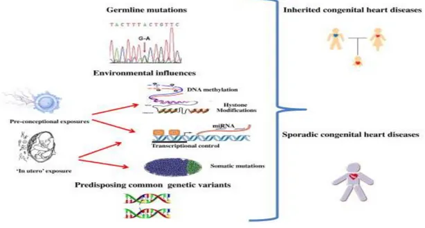
Recently, novel findings supported that epigenetic alterations, such as methylation status, dysregulation of small-non coding RNAs (microRNAs) and histone modification may contribute to understand the molecular basis of CHDs and that congenital heart diseases might be, in part, a consequence of a change in the control of the epigenome induced by the environment (Zhao et al., 2005; Montgomery et al., 2007; Zhao et al., 2007; Movassagh et al., 2010) (Figure 1).
6
Fig. 1: Schematic representation of the pathogenesis of congenital heart defects.
Cardiac transcription factor genes: Genetic Hypothesis
The knowledge about the genetic component of the congenital heart disease is very difficult, due to the complex interplay from different genes in different space and different times (Pierpont et al., 2007).
During the embryonic development, the heart is one of the first organs to form. A high degree of conservation is observed during early-stage heart development in zebrafish, chick, frog, mouse, and human (Brand, 2003).
Because dysregulation of heart development is at root of CHD, a clear picture of how the heart forms is crucial for understanding the genesis of this disease.
In the last decade, it has been suggested that most of the inherited forms of CHD are a result of mutations in genes know to be essential for cardiac development, such as specific transcription factors genes (TFs). These genes, regulate specific events in
7
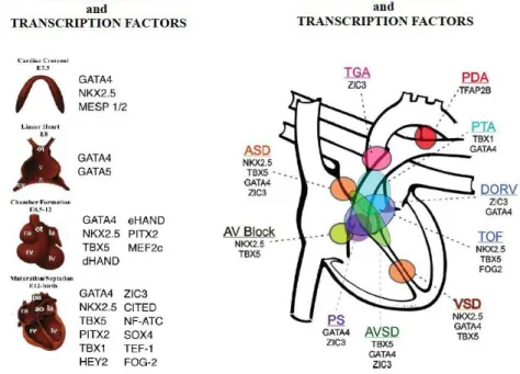
heart morphogenesis, through an intricate process, where TFs regulate each other's expression in order to stabilize and reinforce the cardiac gene program (Olson 2006; Nemer, 2008; Mc Bride et al., 2010; Ware and Jefferies, 2012) (Fig.2).
Fig. 2.: Transcription factors and congenital heart diseases.
Because of their important role in the orchestration of cardiac development, mutations in these genes may induce a significant disruption/dysregulation of downstream gene expression and, thus, lead to cardiovascular malformations, as evidenced by the findings in transgenic mice (Biben et al., 2000; Bruneau et al., 2001; Zhang et al., 2013).
Moreover, the progenitor cells originating from the first heart field, second heart field and cardiac neural crest contribute to the cardiac morphogenesis (Buckingham et al., 2005; Shan et al., 2012).
8
ISL1 is a LIM homeodomain TF considered to be the most important marker of
cardiac progenitor cell lineages in the secondary heart-field differentiation (Moretti et al., 2006; Kang et al., 2009; Stevens et al., 2010; Klaus et al., 2012) (Fig. 3).
Fig.3: ISL1 functions (adapted from Bu et al., 2009)
ISL1 cardiovascular progenitors give rise to right ventricle and inflow and outflow
tracts, which are affected by several cardiovascular malformations, e.g., transposition of the great vessels, tricuspid atresia and tetralogy of Fallot (Laugwitz et al., 2008). Animal experimental models have shown that homozygous mutants for ISL1 developed a severe cardiac phenotype (Laugwitz et al., 2008), whereas mice totally knocked out for this gene were lacking in the outflow tract, in the right ventricle and in several part of the atria (Cai et al., 2003; Lin et al., 2006). Recently, common genetic variants and specific haplotypes in the ISL1 gene have been found to contribute to the risk of CHDs in the white and black/African-American populations (Stevens et al., 2010). On the contrary, a more recent paper (Xue et al., 2012) showed that ISL1 common variant rs1017 did not play a crucial role in conferring
9
susceptibility to sporadic CHD in Chinese population. Moreover, another recent paper, identified the presence of six known and five novel ISL1 variants, investigating the potential contribution of ISL1 in cardiomyopathies. The authors also describes a novel p.Asn252Ser ISL1 gain-of-function variant, which could lead to greater activation of downstream targets, such as Mef2c, which are known to be involved in cardiac development, dilation, and hypertrophy (Friedrich et al., 2013).
Etiology of Congental Heart Disease: gene-environment interaction
In approximately 80% of CHD cases, the cause is multifactorial. Direct evidence regarding environmental exposures and the risk of CHD is very limited (Jenkins et al., 2007; Blue et al., 2012). Unfortunately, less is known about modifiable “non genetic” factors.
Several studies have suggested that some parental occupational and/or environmental exposures may be significantly associated with an increased prevalence of birth defects in offspring, especially for selected congenital heart defects (Dolk and Vrijheid, 2003; Chapin et al., 2009; Rankin et al., 2009; Strickland et al., 2009; Desrosiers et al., 2012). In particular, the maternal environmental risk factor known to influence the incidence of CHD are rubella, pre-gestational diabetes and exposure to teratogens, such as thalidomide, retinoic acid and indomethacin, and exposure to chemicals at work (Øyen et al., 2000).
Every human cell, including the spermatozoa and the oocytes, can suffer DNA mutations due to the exposure to environmental toxicants. Gametic DNA mutations preceding the conception can induce miscarriage, death or congenital defects.
10
Several studies have shown that maternal exposure is associated with a variety of adverse pregnancy outcomes including preterm birth, low birth weight, and birth defects (Brent, 2004; Kuehl and Loffredo, 2005; Jenkins et al., 2007; Patel et al., 2010). Conversely, the information available regarding environmental exposures of father as risk factor for birth defects, in particular for CHD, is very limited (Jenkins et al., 2007).
The toxicity of xenobiotics for embryonic tissues depends on the biotransformation process during which reactive products are formed (phase I) and detoxified (phase II). Several enzymes (and their gene families) are involved in this process. In particular, the impact of parental exposure on birth defects in the offspring might be affected by the presence of polymorphisms in genes responsible for the activation and detoxification of toxicant agents, contributing to an increased resistance or sensibility to cardiac teratogenesis (Loffredo, 2000; Kuehl and Loffredo, 2005; Patel et al., 2010).
Glutathione S-tranferase enzymes (GSTM1, GSTT1, GSTP1)
Genetic polymorphisms in the Glutathione S-Transferase (GST) enzymes, which provide critical defense against numerous toxins, might modulate the effect of toxic agents such as xenobiotics compounds. (Kuehl and Loffredo, 2005; Shi et al., 2008). Eight cytosolic GSTs are known to be expressed in humans: alpha (A), mu (M), pi (P), theta (T), kappa (K), omega (O), sigma (S) and zeta (Z). Each class consists of several distinct subclasses, with some overlap in tissue expression (Hayes and Strange, 2000). The GSTM1 and GSTT1 genes are located in chromosome 1 and 22
11
respectively. Their activity is the detoxification of electrophilic compounds, including carcinogens, therapeutic drugs, environmental toxins and products of oxidative stress, by conjugation with glutathione, which are water-soluble and can be excreted from the body. The gene coding for GSTM1 and GSTT1 exhibits a deletion polymorphism, which in case of homozygozity leads to absence of phenotypic enzyme activity.
The presence of these polymorphisms in association to maternal cigarette smoking has been associated to an increased risk for orofacial defects (Olshan et al., 2005). Moreover, the role of GST polymorphisms and their interaction with environmental pollutants on the risk of birth defects has also been examined (Garlantézec et al., 2012).
The GSTP1 gene, highly expressed in early fetal life, is the most abundant phase II xenobiotic metabolism enzyme in the human placenta (Ahmad et al., 1990; Becket et al., 2000; Raijmakers et al. 2001).
An A to G transition at nucleotide 313 in exon 5 of the GSTP1 gene, which replaces isoleucine (Ile) at codon 105 with valine (Val) within the active site of the enzyme, has been shown to result in altered enzyme activity (Zimniak et al. 1994). Therefore, fetal inherited GSTP1 Ile105Val polymorphism might modify the metabolism and excretion of xenobiotics capable of crossing the placental barrier from fetal tissue.
12
AIM
Most of the known CHDs occur through a heterogeneous and complex process in which predisposing genetic factors interact with environmental factors. The environmental effects may be modified by genes involved in the activation or detoxification of toxicant agents, contributing to an increased resistance (or sensitivity) to cardiac teratogenic substances. Accordingly, recent studies have shown the fundamental role of single-nucleotide polymorphisms (SNPs) and/or mutations in genes critical for detoxification pathway, in the pathogenesis of CHDs. In this framework, the first hypothesis of this project, was that environmental factors interact with genetic predisposing factors in the pathogenesis of CHDs. In order to reach this aim, the specific objectives were:
to examine the association between the environmental exposure of both parents and CHD risk.
to explore the modification effects of metabolizing gene polymorphisms (GSTM1, GSTT1, GSTP1) in association with parental exposure to toxicants.
In the last part of the project, since the importance of TFs genes in the control of heart development, it has been hypothesized that “common” sequence variations in these genes might be one of causative mechanism of CHD. In order to reach this aim, the specific objective was:
to evaluate if the presence of a common variant of TF gene , ISL1 conferred an increased susceptibility to CHD.
13
MATERIALS AND METHODS
Study population
The study population consisted of 309 patients, who were diagnosed with isolated, non-syndromic CHD (197 males [21.3 ±25.2 years], including 200 pediatric [4 ± 5.6 years] and 109 adult patients [52.5 ± 17.2 years] with bicuspid aortic valve, BAV); a control group of 500 healthy subjects [272 males; 15.7 ± 21.3 years] comprising 300 newborn and 200 adult subjects (39.8 ± 13.7 years). Moreover, we enrolled 360 parents of a child with CHD and 360 parents of a child without any congenital malformations. Both case and control parents completed a structured questionnaire on the demographic, preconceptional, and lifestyle exposures. We collected environmental and occupational exposure data from specific questions on potential teratogens/mutagens that have been linked to human reproductive impairment, including ionizing radiation, solvents, pesticides, asbestos and heavy metals. For the parents, the exclusion criteria were mothers who reported inconsistent use of B vitamin and folate supplements in the periconception period; the inability to obtain complete information about the occupational, demographic and lifestyle data from both parents. A sample of blood was obtained from 309 patients with CHD and 500 healthy subjects. The questionnaire model used is shown below.
19
DNA extraction
Genomic DNA was extracted from whole-blood samples through Biorobot EZ1 (Qiagen) that allows to extract DNA, automatically, from both blood and tissue, from 6 together samples in a single step:
sample lysis,
DNA binding to magnetic particles,
washing and elution of the DNA.
DNA quantitative assessment
We have prepared DNA elutions 1:250 on sterilized water for spectrophotometric quantification at 260 nm and 280 nm. The spectrophotometric analysis determines the quantitative concentration of the DNA and qualitative information. In fact, the ratio of the absorbance at 260 nm and 280 nm represent a good index of the sample purity. For the DNA, the well index is 1.7-1.9. The formula for the final concentration of the sample was:
20
Genotyping analysis
Glutathione S-Transferase M1 and T1 (GSTM1 and GSTT1)
The GSTM1 and GSTT1 genotypes were determined using a co-amplification polymerase chain reaction approach, with the GSTM4 gene, which is never deleted, as the internal control to distinguish the null genotypes from aborted polymerase chain reaction. The primers sequences and PCR conditions were:
for the GSTM1 amplification:
forward 5’-CGC CAT CTT GTG CTA CAT TGC CCG-3’ reverse 5’-TTC TGG ATT GTA GCA GAT CA-3’
First cycle denaturing 94° 5 min 35 cycles denaturing 94° 1 min annealing 52° 1 min elongation 72° 1 min Last cycle elongation 72° 10 min
21 for the GSTT1 amplification:
forward 5’- TTC CTT ACT GGT CCT CAC ATC TC -3’ reverse 5’- TCA CCG GAT CAT GGC CAG CA -3’
First cycle denaturing 94° 5 min 35 cycles denaturing 94° 1 min annealing 56° 1 min elongation 72° 1 min Last cycle elongation 72° 10 min
for the GSTM4 amplification:
forward 5’-CGC CAT CTT GTG CTA CAT TGC CCG-3’ reverse 5’-ATC TTC TCC TCT TCT GTC TC-3’
22 Agarose gel electrophoresis
PCR reaction was appears in the 1.5% agarose gel, stained with ethidium bromide 10 mg/ml at final concentration of 0.3%. We have loaded the wells of the gel with 10 µl of PCR product with 3 µl of “loading” buffer (L.B.: 0.25%bromphenol blue, 0.25% cyanol xylene, 15% glycerol) and a DNA marker of 100 bp (PRIME). The
electrophoresis occurred at 100 V in TBE 1X buffer (Tris-base 4 mM, 0.9 M boric acid, 50 mM EDTA, pH 8). The internal standard fragments amplified from the
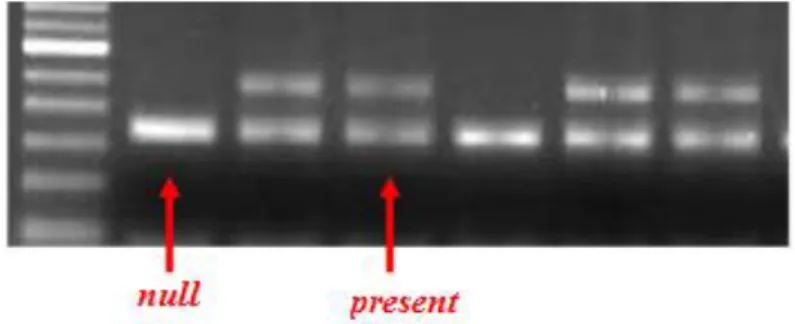
GSTM4 gene was 157 bp. A 230-bp fragment was amplified for the GSTM1 gene,
and a 480-bp fragment was obtained for the GSTT1 gene. The absence of amplified products was consistent with the null genotypes (Fig. 4).
23
Glutathione S-Transferase P1 (GSTP1) Ile105Val polymorphism
A PCR-restriction fragment length polymorphism method was used to determine the allele distribution of the GSTP1 Ile105Val polymorphism. The primers sequences were:
forward 5’-ACC CCA GGG CTC TAT GGG AA -3’ reverse 5’-TGA GGG CAC AAG AAG CCC CT-3’
The PCR condition involved:
First cycle denaturing 94° 5 min 32 cycles denaturing 94° 30 s annealing 64° 50 s elongation 72° 50 s Last cycle elongation 72° 10 min.
The 176-bp amplified GSTP1 gene fragment was subjected to restriction digestion in a 15-µl reaction volume containing 7.5 µl of PCR product, 100 mmol/l of NaCl, 50 mmol/l of Tris-HCl, pH 7.9, 10 mmol/l of MgCl2, 1 mmol/l dithiothreitol, and 5 U of
24 Polyacrylamide gel electrophoresis
The digested PCR products were separated by electrophoresis, occurred at 200 V, using polyacrylamide gel (size: 10.1 cm x 9.7 cm, 1.0 mm thickness) at 8% (acrylamide stock solution 20%; 19:1 acrylamide:bisacrilamide), TBE 1X (Tris-base 4 mM, 0.9 M boric acid, 50 mM EDTA, pH 8), TEMED 0.13% and APS 0.1%. We have loaded the wells of the gel with 15 µl of digestion product with 4 µl of “loading” buffer (L.B.: 0.25% bromphenol blue, 0.25% cyanol xylene, 15% glycerol) and a DNA marker of 20 bp (PRIME). The polyacrylamide gel was stained, though immersion, with a solution composed with 40 µl of ethidium bromide 10 mg/ml and 100 ml TBE 1X, for 30 minutes at final concentration of 2.1%. The genotype was determined by analysis of the bands on the gel as follow: homozygous wild-type for isoleucine (II), one band (176 bp); homozygous mutated for valine (VV), two bands (91 and 85 bp); and heterozygous (IV), three bands (176, 91, and 85 bp) as shown in Fig. 5.
25
ISL1 rs1017 polymorphism (ISL1)
A PCR-restriction fragment length polymorphism method was used to determine the allele distribution of the rs1017 polymorphism. The primer sequences, were designed using the Primer3 program (http://primer3.sourceforge.net/):
forward 5’-CCT TCA GGA AGG TGG AGC TG-3’ reverse 5’-CGC TTG TGG CAA AAT AGA GG-3’. PCR conditions were as follow:
First cycle denaturing 94° 5 min 35 cycles denaturing 94° 30 s annealing 56° 30 s elongation 72° 30 s Last cycle elongation 72° 7 min.
The 248-pb amplified ISL1 gene fragment was subjected to restriction digestion in a 9-µl reaction volume containing 4 µl of PCR product, 100 mmol/l of NaCl, 50 mmol/l of Tris-HCl (pH 7.9), 10 mmol/l of MgCl2, 1 mmol/l dithiothreitol, and 2.5 U of DraI enzyme at 37° overnight.
26 Agarose gel electrophoresis
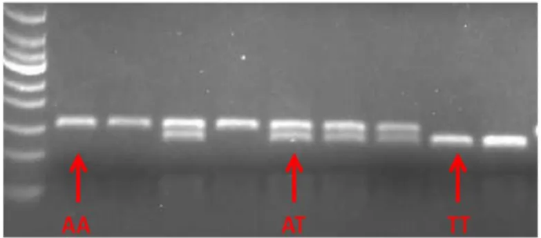
The digested PCR products were separated by electrophoresis using 1.5% agarose gel, stained with ethidium bromide 10 mg/ml at final concentration of 0.3%. We have loaded the wells of the gel with 5 µl of PCR product with 2 µl of “loading” buffer (L.B.: 0.25%bromphenol blue, 0.25% cyanol xylene, 15% glycerol) and a DNA marker of 100 bp (PRIME). The electrophoresis occurred at 100 V in TBE 1X buffer (Tris-base 4 mM, 0.9 M boric acid, 50 mM EDTA, pH 8). A representative gel is shown in Fig. 6.The genotype was determined by analysis of the bands on the gel: homozygous wild-type for adenine (AA), one band (156 bp); homozygous mutated for thymine (TT), one band (134 bp); and heterozygous (AT), two bands (156 and 134 bp).
27
Statistical analysis
Statistical analyses of the data were conducted using the StatView statistical package, version 5.0.1 (Abacus Concepts, Berkeley, California). Data are expressed as the mean ± SD. For statistical analysis, all determinants were dichotomized except for the age variable. Smokers were classified as individuals who smoked at least three cigarettes for day at the time of the conception, ex-smokers as those who stopped smoking at least 6 months before inclusion in the study, and nonsmokers as person who never smoked. Smokers also were divided into medium smokers (3-14 cigarettes a day) and heavy smokers (≥ 15 cigarettes a day). For consumption frequency of alcoholic drinks per day (beer, wine, liquor) and nondrinkers as those who drank less than three drinks for day. Given the relatively small number of exposed mothers in any particular environmental or occupational category, the statistical analysis for toxicant exposure included both environmental and occupational exposure to X-rays, chemicals, anesthetics, industrial cleaning agents and solvents, exhaust and welding fumes, paint/varnish/thinner, asbestos, heavy metals and pesticides. The differences between the mean values of two continuous variables were evaluated using the unpaired Student t test. The differences in non continuous variables and genotype distribution were tested using chi-square analysis. Unconditional logistic regression analysis was used to estimate the odds ratios (ORs) and 95% confidence intervals (CIs) for the association between CHD and parental exposure. The ORs were also adjusted for potential confounding factors. A 2-tailed p value <0.05 was chosen as the level of significance.
28
RESULTS
Parental exposures analysis
We used a prospective and paired case-control study design (1:1). Data obtained from questionnaires showed that the mothers of patients with CHD had a significantly lower age than the controls (p=0.01) but no significant differences between the two groups were found for paternal characteristics.
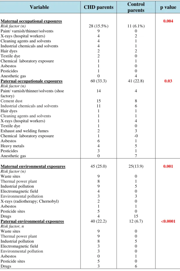
The CHD cases had a significantly lower birth weight (p=0.01) compared to the control children and the children with CHD were also more often conceived with artificial fertilization (p=0.05). In table 1 are summarized the specific occupational and professional risk factors for CHD.
29
Table 1: Occupational/environmental exposure among CHD and control parents
Variable CHD parents Control
parents p value
Maternal occupational exposures 0.004
Risk factor (n) 28 (15.5%) 11 (6.1%)
Paint/ varnish/thinner/solvents 9 0
X-rays (hospital workers) 4 2
Cleaning agents and solvents 4 1
Industrial chemicals and solvents 4 1
Hair dyes 2 2
Textile dye 2 0
Chemical laboratory exposure 1 1
Asbestos 1 0
Pesticides 1 0
Anesthetic gas 0 4
Paternal occupationale exposures 60 (33.3) 41 (22.8) 0.03
Risk factor (n)
Paint/ varnish/thinner/solvents (shoe factory)
14 4
Cement dust 15 8
Industrial chemicals and solvents 11 6
Hair dyes 1 1
Cleaning agents and solvents 1 1
X-rays (hospital workers) 1 4
Textile dye 1 0
Exhaust and welding fumes 2 3
Chemical laboratory exposure 1 -0
Asbestos 6 1
Heavy metals 4 5
Pesticides 3 1
Anesthetic gas 0 7
Maternal environmental exposures 45 (25.0) 25(13.9) 0.001
Risk factor (n)
Waste sites 9 0
Thermal power plant 8 1
Industrial pollution 9 5
Electromagnetic field 4 0
Environmental pollution 3 3
X-rays (radiotherapy; Chernobyl) 2 0
Asbestos 1 1
Pesticide sites 5 0
Drugs 4 15
Paternal environmental exposures 40 (22.2) 12 (6.7) <0.0001
Risk factor, n
Waste sites 9 0
Thermal power plant 9 0
Industrial pollution 8 5 Electromagnetic field 3 0 Environmental pollution 3 0 Asbestos 0 1 Pesticide sites 5 0 Drugs 3 6
30
The parental exposure to environmental factors and CHD risk is listed in Table 2.
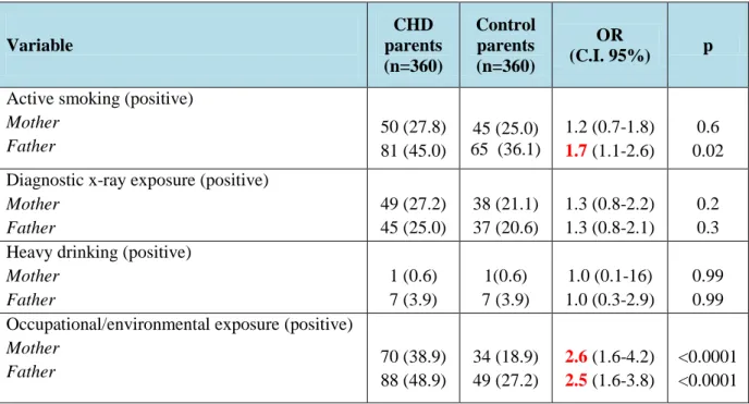
Table 2: Parental exposure to environmental risk factors and CHD risk
Variable CHD parents (n=360) Control parents (n=360) OR (C.I. 95%) p Active smoking (positive)
Mother Father 50 (27.8) 81 (45.0) 45 (25.0) 65 (36.1) 1.2 (0.7-1.8) 1.7 (1.1-2.6) 0.6 0.02 Diagnostic x-ray exposure (positive)
Mother Father 49 (27.2) 45 (25.0) 38 (21.1) 37 (20.6) 1.3 (0.8-2.2) 1.3 (0.8-2.1) 0.2 0.3 Heavy drinking (positive)
Mother Father 1 (0.6) 7 (3.9) 1(0.6) 7 (3.9) 1.0 (0.1-16) 1.0 (0.3-2.9) 0.99 0.99 Occupational/environmental exposure (positive)
Mother
Father 70 (38.9) 88 (48.9) 34 (18.9) 49 (27.2) 2.62.5 (1.6-4.2) (1.6-3.8) <0.0001 <0.0001
Maternal smoking was not significantly associated with an increased risk of CHD (OR 1.2, 95% CI 0.7 to 1.8) but the fathers who were smoking showed an increased risk of CHD in their offspring (OR 1.7, 95% CI 1.1 to 2.6, p=0.02), especially fathers who were heavy smokers (≥ 15 cigarettes/day) had an high risk of having children with CHD (Figure 7). Conversely, parental alcohol use, the existence of a chronic disease or a disease in the first trimester of pregnancy and the exposure to diagnostic radiographs did not show to influence CHD risk.
31 Figure 7: Parental smoking and CHD risk.
0 1 2 3 Mother Father Non-smoking Smoking < 15 cigarettes/day Smoking ≥ 15 cigarettes/day p=0.002 OR
Both maternal (OR 2.6, 95% CI 1.6 to 4.2, p<0.0001) and paternal (OR 2.5, 95% CI 1.6 to 3.8, p<0.0001) occupational/environmental exposure to toxicants increased the risk of CHD. Therefore, the exposure of both parents to toxicants is associated to significant additive risk (OR 4.5, 95% CI 2.5 to 8.3, p<0.0001) of CHD in the offspring (Fig.8).
Fig.8: Interactive effect between maternal and paternal exposures on CHD risk.
0 1 2 3 4 5 Non- exposed parent One exposed parent Both exposed parents p<0.001 OR p=0.04
32
Moreover, on multivariate analysis, a father’s heavy smoking (OR 1.9, 95% CI 1.1 to 3.3) and maternal exposure (OR 2.1, 95% CI 1.2 to 3.6) and paternal exposure (OR 1.8, 95% CI 1.1 to 3.0) to toxicants were the main determinants of CHD risk.
Gene-environment interactions analysis
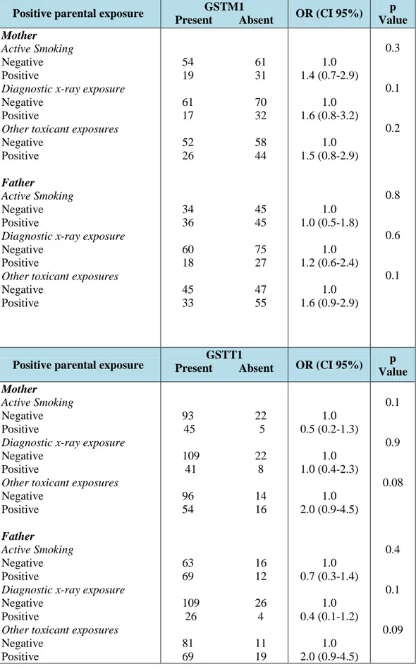
In a case-only approach, we genotyped 180 children with CHD for GST polymorphisms, considering their association to parental exposure. A case-only design was chosen because it is considered the most powerful method to study gene-gene and gene-gene-environment interaction in disease etiology (Gauderman, 2002)
The frequency of GSTM1 null genotype, in our population, was 57.5%, and the frequency of the GSTT1 null genotype was 16.7%. We did not detect an interactive effect of GSTM1 or GSTT1 null genotypes and maternal and paternal exposures (Table 3).
33
Table 3: Gene-environment interactions for combination of glutathione S-transferase (GST) genes and parental exposure.
Positive parental exposure GSTM1
Present Absent OR (CI 95%) p Value Mother Active Smoking Negative Positive
Diagnostic x-ray exposure
Negative Positive
Other toxicant exposures
Negative Positive Father Active Smoking Negative Positive
Diagnostic x-ray exposure
Negative Positive
Other toxicant exposures
Negative Positive 54 61 19 31 61 70 17 32 52 58 26 44 34 45 36 45 60 75 18 27 45 47 33 55 1.0 1.4 (0.7-2.9) 1.0 1.6 (0.8-3.2) 1.0 1.5 (0.8-2.9) 1.0 1.0 (0.5-1.8) 1.0 1.2 (0.6-2.4) 1.0 1.6 (0.9-2.9) 0.3 0.1 0.2 0.8 0.6 0.1
Positive parental exposure Present Absent GSTT1 OR (CI 95%) p Value
Mother
Active Smoking
Negative Positive
Diagnostic x-ray exposure
Negative Positive
Other toxicant exposures
Negative Positive Father Active Smoking Negative Positive
Diagnostic x-ray exposure
Negative Positive
Other toxicant exposures
Negative Positive 93 22 45 5 109 22 41 8 96 14 54 16 63 16 69 12 109 26 26 4 81 11 69 19 1.0 0.5 (0.2-1.3) 1.0 1.0 (0.4-2.3) 1.0 2.0 (0.9-4.5) 1.0 0.7 (0.3-1.4) 1.0 0.4 (0.1-1.2) 1.0 2.0 (0.9-4.5) 0.1 0.9 0.08 0.4 0.1 0.09
34
However, the risk of CHD tended to be greater in children carrying the GSTM1 and
GSTT1 null genotypes who had parents exposed to toxicants.
A significant interaction was found for the combined null GSTs and both maternal (OR 3.6, 95% CI 1.1 to 11.2, p=0.03) and paternal (OR 3.3, 95% CI 1.0 to 10.8, p=0.03) exposure to toxicants.
Finally, children with the combined null GST genotype had a greater risk than children carrying wild-type GST genes when both parents were exposed (Figure 9).
35
Fig.9: ORs stratified by children’s GST genes and (A) maternal, (B) paternal, and (C) parental occupational/environmental exposure to toxicants.
0 1 2 3 4 OR p=0.03 a) GSTM1/GSTT1 +/+ +/- or -/+ 0 1 2 3 4 +/+ +/- or -/+ -/-OR p=0.04 b) GSTM1/GSTT1 0 1 2 3 4 5 6 7 +/+ +/- or -/+
-/-One parent Both parents
OR
p=0.01
p=0.09
c)
36
Glutathione S-Transferase P1 (GSTP1)
Gene-environment interaction analysis
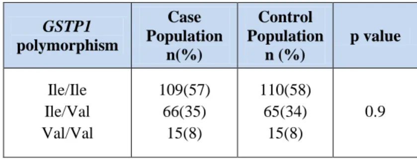
In order to investigate the role of GSTP1 Ile105Val and CHD risk, we used a paired case-control study design (1:1). The frequency of GSTP1 Ile105Val in both CHD cases and control subjects satisfied the Hardy-Weinberg equilibrium and was comparable with that described in the Caucasian population.
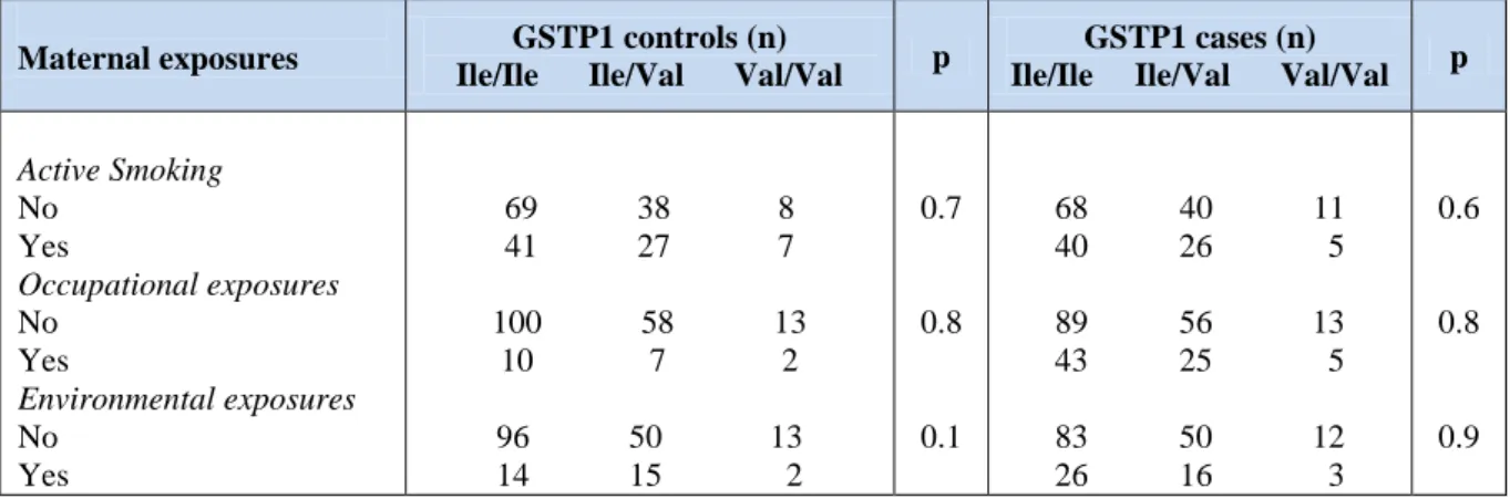
The genotype distribution was not significantly different between the patients and the control subjects (p=0.9), as shown in table 4.
Tab. 4: Genotype distribution of GSTP1 polymorphism.
GSTP1 polymorphism Case Population n(%) Control Population n (%) p value Ile/Ile Ile/Val Val/Val 109(57) 66(35) 15(8) 110(58) 65(34) 15(8) 0.9
37
Moreover, we found no association between GSTP1 Ile105Val polymorphism in children and maternal exposure. Indeed, no significant gene-environment interactions were observed (Table 5). Logistic regression did not show an increased risk for CHD in the presence of positive maternal exposure and GSTP1 polymorphism.
Table 5: Gene-environment interactions for GSTP1 genes and maternal exposure.
Maternal exposures GSTP1 controls (n)
Ile/Ile Ile/Val Val/Val p
GSTP1 cases (n)
Ile/Ile Ile/Val Val/Val p
Active Smoking No Yes Occupational exposures No Yes Environmental exposures No Yes 69 38 8 41 27 7 100 58 13 10 7 2 96 50 13 14 15 2 0.7 0.8 0.1 68 40 11 40 26 5 89 56 13 43 25 5 83 50 12 26 16 3 0.6 0.8 0.9
38
ISL1 rs1017 polymorphism (ISL1)
Genetic analysis
A case-control study design was also used to investigate the association between
ISL1 and CHD. The observed genotype frequency was in agreement with
Hardy-Weinberg equilibrium.
No significant difference between the case and control groups was found in term of genotype and allele distribution, as shown in table 6.
At logistic analysis, the rs1017 AT genotype and the mutated TT genotype were not associated with the risk of CHD (OR 1.0, 95% CI 0.7 to 1.3, p=0.8; OR 1.1, 95% CI 0.7 to 1.7, p=0.6 respectively). Moreover there was no appreciable difference between CHD risk and the presence of T allele (OR 1.0, 95% CI 0.7 to 1.5, p=0.7).
Tab.6: Main effects of ISL1 rs1017 on CHD risk in the case-control study.
Genotypes No. (%) cases (n=309) No. (%) controls (n=500) OR (CI%95) p-Value rs1017 AA AT TT AT + TT Allele A T 142 (46) 130 (42) 37 (12) 167 (54) 414 (67) 204 (33) 229 (46) 204 (41) 67 (13) 271 (54) 662 (66) 338 (34) 1.0 1.0 (0.7-1.3) 1.1 (0.7-1.7) 1.1(0.7-1.5) 1.0 1.2 (0.7-1.7) 0.8 0.6 0.7 0.6
39
Furthermore, when we compared the ISL1 rs1017 genotype distribution among the different groups of patients and controls, no significant difference was also observed (Table 7).
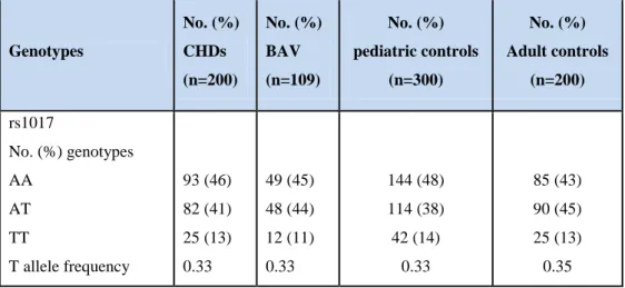
Table 7: ISL1 rs1017 genotype distribution in different groups of patients and controls.
Genotypes No. (%) CHDs (n=200) No. (%) BAV (n=109) No. (%) pediatric controls (n=300) No. (%) Adult controls (n=200) rs1017 No. (%) genotypes AA AT TT T allele frequency 93 (46) 82 (41) 25 (13) 0.33 49 (45) 48 (44) 12 (11) 0.33 144 (48) 114 (38) 42 (14) 0.33 85 (43) 90 (45) 25 (13) 0.35
40
Finally, we evaluated the influence of polymorphism on CHD risk also stratifying the population for sex and CHD classification, but no differences in the various subgroups were found (table 8).
Table 8: Stratified analysis and ISL1 rs1017 polymorphism.
Variables OR (95% CI) p-Value
Overall Sex Male Female
CHD classification Bicuspid Aortic Valve Complex CHD Tetralogy of Fallot Septation defects Patent ductus arteriosus Aortic coarctation Other CHDs 1.1 (0.7-1.5) 0.8 (0.5-1.3) 0.9 (0.7-1.3) 1.0 (0.7-1.6) 0.9 (0.6-1.4) 1.0 (0.6-1.8) 1.3 (0.7-2.4) 0.9 (0.3-2.7) 0.7 (0.3-2.4) 1.5 (0.4-6.0) 0.7 0.4 0.8 0.9 0.6 0.9 0.5 0.9 0.7 0.6
41
DISCUSSION
Our research project shows the importance of cumulative effect of genetic and environmental risk factors in the etiology of CHD. For the first time, we have demonstrated that GSTM1 and GSTT1 polymorphisms mediate the risk of CHD in the presence of a positive history of parental exposure to toxicants. Conversely,
GSTP1 Ile105Val polymorphism showed no association with the maternal toxicant
exposure and the risk to develop a CHD. Moreover, ISL1 rs1017 polymorphism was not associated with an increased risk to develop CHDs. Our data underline the fundamental role of gene-environment interactions in the pathogenesis of congenital heart disease.
Parental exposures analysis
Environmental factors potentially harmful in the aetiology of CHDs are unknown. The best available information comes from the Baltimore-Washington Infant Study (BWIS), conducted in the Baltimore-Washington area between 1981 and 1989 and the Finnish study conducted by the National Public Health Institute (in Helsinki) in cases and controls born during 1982 to 1984 (Källén, 1999; Malik et al., 2008). Several other studies investigated the association between occupational hazards, including the exposure of chemicals, and specific phenotypes of congenital malformation (Thulstrup and Bonde 2006; Snijder et al., 2012). Some of these studies found indication for effects of chemical on fetal development but the evidence remains equivocal.
42
The maternal environmental risk factor known to influence the incidence of CHD are rubella, pre-gestational diabetes and exposure to teratogens, such as thalidomide, retinoic acid and indomethacin, and exposure to chemicals at work (Øyen et al., 2000).
In particular, maternal occupation exposures to solvent-containing products, mineral oil products, dyes, lacquers, paints and pesticides have been associated with an increased risk for CHD (Loffredo 2000; Gilboa et al., 2012).
A number of studies have also investigated maternal cigarettes smoking and congenital heart disease, providing controversial results, probably owing to variations in the method, disease categorization, sample size, or confounding bias (Ferencz et al, 1997; Källén et al., 1999; Woods et al., 2001; Kuciene and Dulskiene, 2008; Patel et al., 2012). An analysis of the BWIS data identified a possible dose-response relationship between the number of cigarettes smoked and non-syndromic atrio-ventricular septal defects (Ferencz et al, 1997; Alverson et al. 2011).
In a population-based case-control study of 3,067 infants with non-syndromic CHD and 3,947 infants without birth defects, the association was stronger for mothers who reported heavier tobacco consumption ( ≥25 cigarettes/day) during pregnancy (Malik et al., 2008). This dose-effect relation was confirmed in another recent studies of children with CHD and maternal smoking exposure (Krapels et al. 2006; Patel et al., 2012).
Few studies have evaluated the effect of paternal exposure to environmental factors in the preconception period. Specifically, paternal exposures to lifestyle substances (Marijuana), paint stripping and ionizing radiation exposure have been associated with certain malformations in the BWIS (Correa-Villasenor et al, 1993; Ewing et al., 1997). An association between parental smoking, particularly when both parents
43
were smokers, and the risk of conotruncal heart defects, including D-transposition of the great arteries, tetralogy of Fallot, double outlet right ventricle and truncus arteriosus has also been reported (Wasserman et al. 1996). A very recent study found that several occupations were associated with an increased prevalence of various birth defects categories (Desrosiers et al., 2012). Moreover, another study found a more than fivefold increase in risk mortality and deaths due to congenital malformation, especially for cardiac defects, for paternal occupational exposures, possibly to organic solvents during preconception period (Sung et al., 2009). In addition, recent studies have revealed that paternal smoking and occupational exposure, are risk factors for congenital defects, such as orofacial, anorectal malformations and CHDs (Krapels et al., 2006; van Rooij et al.,2010; Snijder et al., 2012) supporting the existing data on the great vulnerability of the male reproductive system to environmental exposures. At present, societal concern is growing about the particular sensitivity of the male germ line to genetic transmissible effects, (Cordier, 2008; Gianicolo et al., 2010) that might play a crucial role in the origin of congenital defects in general, and in particular CHD (Kuehl and Loffredo, 2005). It is has been suggested that male exposure may exert a teratogenic effect through toxicant compounds adsorbed to sperm and transmitted to a woman in the ejaculate (Chapin et al., 2004). The contaminant is absorbed by the woman, where it might reach and adversely affect a current pregnancy and, perhaps, remain in the woman’s body to influence future pregnancies (Gianicolo et al., 2010). In contrast, toxicant compounds may act in human seminal fluid as endocrine disrupting agents causing direct germ line DNA damage or epigenetic changes (Cordier, 2008; Gianicolo et al., 2010). Teratogenic, carcinogenic, and endocrine disrupting agents, such as pesticide residues, heavy metals organic solvents (benzene, toluene, and xylene), nicotine,
44
aromatic hydrocarbons and precursors of mutagenic nitrosamines, have all been detected in human seminal fluid (Hales and Robair, 2001; Cordier, 2008; Gianicolo et al., 2010). Recent studies also showed that these environmental toxicants can induce oxidative DNA damage, mutations and chromosomal aberrations, such as DNA strand breaks and aneuploidy, in human seminal fluid (Hales and Robair, 2001; Cordier, 2008; Gianicolo et al., 2010). Therefore, it is plausible that exposure to toxicant agents of both parents during the preconceptional period and 'in utero' during pregnancy, might have a pivotal role in the pathogenesis of CHDs affecting genome and the so-called 'epigenome' (Gianicolo et al., 2010).
Gene-environmental interactions
It is increasing the evidence that people have different susceptibility to develop a disease induced by toxic agents (Kuehl and Loffredo, 2005; Shi et al., 2008). Specifically, the inheritance of particular genotypes for metabolizing systems and DNA repair pathways, might modulate the effect, leading to varying susceptibility to the congenital malformations of toxicants (Kuehl and Loffredo, 2005; Shi et al., 2008). The toxicity of xenobiotics for embryonic tissues depends on the biotransformation process during which reactive products are formed (phase I) and detoxified (phase II). Several enzymes (and their gene families) are involved in this process including glutathione transferases. In particular, the GSTs are the polymorphic super-gene family of detoxification enzymes, that are involved in metabolism of numerous toxins and provide critical defense against xenobiotics (Bolt and Their, 2006).
45
GSTM1 and GSTT1 are the most extensively studied genes in the GST gene
superfamily. There has been some, albeit contradictory, evidence of their enzymatic expression during the early stages of embryonic and fetal development in most tissues (Raijmakers et al., 2001; Shi et al., 2007). For example, GSTM1, appears to be expressed in the fetus early in gestation (Raijmakers et al., 2001), while the
GSTT1 enzyme has been shown to be expressed only in craniofacial structures during
fetal life (Shi et al., 2007). The polymorphism in GSTM1 and GSTT1 gene loci is caused by a deletion which results in the absence of enzyme, especially in individuals with null genotypes. Deletion of the GSTM1 and GSTT1 genes, resulting in loss of functional activity, has been reported in approximately 50 and 20% of the white population, respectively, which might predispose to greater health effects from toxic xenobiotics (Bolt and Their, 2007). Previous studies reported that deletion in the GSTM1 and GSTT1 genes might contribute to the development of congenital malformations, such as oral cleft defects, hypospadias and cardiac congenital defects (Shi et al., 2008; van der Zanden et al., 2012). In particular, an elevated relative risk of cleft palate in infants with the GSTT1 null genotype has been detected and whose mothers were exposed to certain occupational chemicals (Shi et al., 2007).
Glutathione S-Transferase P (GSTP1)
In this research project, the genotype distribution of the GSTP1 Ile105Val polymorphism was not significantly different between the patients and control subjects. Moreover, no adverse effect of GSTP1 Ile105Val polymorphism in the presence of maternal exposure was observed.
46
Studies show that GSTP1 is responsible for the detoxification of benzo(a)pyrene diol epoxide (Robertson et al., 1986). The presence of Ile105Val variant diminishes its enzyme activity (Ali-Osman et al., 1997; Watson et al., 1998) impairing the excretion of toxicants. Hayes and Strange have reported a significant association of valine allele with susceptibility to the development of certain tumors, including bladder, breast, lung, and multiple myeloma (Hayes and Strange, 2000). Cancer research has also found suggestive interaction between the presence of this polymorphism and risk of oral and bladder cancer, especially among smokers (Soya et al., 2007) but the results are contradictory (López-Cima et al., 2012). In addition, the Val105 allele also is reported to increase the risk of asthma and susceptibility to the effects of ozone on breathing difficulties among children with asthma (Romieu et al., 2006). These findings suggest that variable GSTP1 expression may be an important determinant of susceptibility to environmental diseases.
GSTP1 gene is highly expressed early in fetal life and appears to be the predominant
GST present in the human placenta (Ahmad et al., 1990; Beckett et al., 1990; Raijmakers et al., 2001). A recent study showed a significant association between
GSTP1 Ile105Val polymorphism and Hirschsprung disease, a common congenital
intestinal defects (Gao et al., 2011). Conversely, studies on esophageal atresia, orofacial clefts and congenital heart defects have found no association between the Ile105Val variant and the disease (Ramirez et al., 2007; Filonzi et al., 2010). Recently, it has been suggested that the loss of expression of the gene GSTP1 is caused by promoter hypermethylation in several types of cancer, such as prostate and breast cancer (Millar et al., 2000Zhang et al., 2005). The loss of GSTP1 leads to elevated levels of electrophilic intermediates, resulting in increased DNA damage and mutations (Lee et al., 1994).
47
ISL1 rs1017 polymorphism (ISL1)
The formation of the three major cell types of the heart-cardiomyocytes, smooth muscle, and endothelial cell lineages-has been largely ascribed to a set of non-overlapping embryonic precursor derived from distinct origin. Several signaling molecules, included bone morphogenetic proteins and fibroblast growth factors, are required to initiate the cardiomyogenic program. Subsequently, a unique combinatorial subset of transcriptional factors such as GATA, HAND, and TBX interact to generate different cardiac cells types (Kelly and Buckingham, 2002). Several studies revealed that single gene mutations in different cardiac TFs could be responsible for inherited and sporadic forms of CHDs (Hatcher et al., 2003; Pulignani et al., 2011). The LIM homeodomain transcription factor ISL1 is critically involved in embryonic cardiogenesis and is a specific marker for a distinct population of undifferentiated cardiac progenitor cells that give rise to the cardiac segments in secondary heart fielding.
ISL1 function is required for these progenitors contributing to the proliferation,
survival, and migration of cardiac progenitors into the forming heart (Bu et al., 2009; Kang et al., 2009). Its absence is associated with ablation of the entire second heart field, and ISL1+ progenitor cells have been shown to form essential components of the heart, such as the atria, ventricles, coronary arteries, and the conduction system (Cai et al., 2003). Animal experimental studies have shown that both the deficiency and the mis-expression of ISL1 might cause deep developmental defects, growth retardation and death during embryogenesis (at approximately embryonic day [ED] 10.5), thus supporting the importance of a correct regulation of ISL1 gene expression during the fetal life (Ahlgren et al., 1997; Brade et al., 2007; Kappen and Salbaum,
48
2009; Golzio et al., 2012). Histological analysis of mutant hearts from murine fetuses (ED 9.0-9.5) showed that homozygous ISL1 mutants had serious cardiac phenotypes characterized by a severe decrease in tissue mass characterized by loss of some segments (Cai et al., 2003). In addition, ISL1-deficient hearts fail to undergo a correct looping morphogenesis and show a common atrium and an uni-ventricular chamber (Cai et al., 2003). A very recent study describes a new gain-of-function of p.Asn252Ser variant in the human ISL1 gene, which could potentially lead to greater activation of downstream targets involved in cardiac development, dilation, and hypertrophy (Friedrich et al., 2013). Stevens et al recently showed that two different
ISL1 haplotypes contributed to the risk of CHD in the white and
black/African-American populations (Stevens et al., 2010). In particular, two specific polymorphisms, rs1017 and rs3762977, were associated with cardiac congenital defects. The rs1017 SNP (located in 3’UTR) increased the risk of CHD in the United States white population but not in black/African American populations, whereas the rs3762977 SNP (located in 5’UTR) contributed to the risk of CHDs in black/African-American population but not in the white population. Conversely, a more recent article showed that ISL1 common variant rs1017 did not play a crucial role in conferring susceptibility to sporadic CHD in the Chinese population.
Our results are in line with Xue et al., underling the importance of additional studies to better define the association between the genetic variants in ISL1 gene and CHD risk.
49
CONCLUSIONS
Our result showed that paternal smoking and exposure to toxicants for both parents, affects the risk of children with CHD and the polymorphisms in GSTM and GSTT1 genes can modify a person’s risk of toxicant exposure-induced disease. On the contrary, we did not find any association between the presence of polymorphism in
GSTP1 gene and the effect of positive maternal exposure to toxicants, on the risk for
CHD. Similarly, we found no association between ISL1 rs1017 polymorphism and increased risk to develop CHD.
In conclusion, our findings support the notion that cardiac development, regulated by a complex mechanism that involves the role of many different genes, is largely influenced by environmental factors.
Furthermore, understanding the biologic impact of gene-environmental interactions may provide a key insight into the prevention of these congenital malformations in future generations. Understanding the genetic basis and the molecular mechanisms of CHD may allow the identification of family members at risk as well as to identify new possible therapeutic targets and appropriate preventive strategies because environmental factors can be modified in contrast to genetic factors.
Further research based on so-called "omics" technologies, such as transcriptomic, proteomic, metabolomic, exposomic) are fundamental to improve the knowledge of the genetic defects involved in the development of CHD, and to better understand the complex interactions between genes and environment.
50
REFERENCES
Ahlgren U, Pfaff SL, Jessell TM, et al. Independent requirement for ISL1 in formation of pancreatic mesenchyme and islet cells. Nature.1997; 385:257-260.
Ahmad H, Wilson DE, Fritz RR et al. Primary and secondary structural analyses of glutathione S-transferase pi from human placenta. Arch Biochem Biophys. 1990; 278:398-408.
Ali-Osman F, Akande O, Antoun G, et al. Evidence for differential catalytic activity of the encoded proteins. J Biol Chem.1997; 272:10004-10012.
Alverson CJ, Strickland MJ, Gilboa SM, et al. Maternal smoking and congenital heart defects in the Baltimore-Washington Infant Study. Pediatrics. 2011; 127:e647-53.
Beckett GJ, Howie AF, Hume R et al. Human glutathione S-transferases: radioimmunoassay studies on the expression of alpha-, mu- and pi-class isoenzymes in developing lung and kidney. Biochim Biophys Acta. 1990; 1036:176-182.
Biben C, Weber R, Kesteven S et al. Cardiac septal and valvular dysmorphogenesis in mice heterozygous for mutations in the homeobox gene Nkx2-5. Circ Res. 2000; 87:888–895.
51
Blue GM, Kirk EP, Sholler GF et al. Congenital heart disease: current knowledge about causes and inheritance. Med J Aust. 2012; 197:155-159.
Bolt HM, Thier R. Relevance of the deletion polymorphisms of the glutathione S-transferases GSTT1 and GSTM1 in pharmacology and toxicology. Curr Drug Metab. 2006; 7:613-628.
Brade T, Gessert S, Kühl M, et al. The amphibian second heart field: Xenopus islet-1 is required for cardiovascular development. Dev Biol. 2007; 311:297-310.
Brand T. Heart development: molecular insights into cardiac specification and early morphogenesis. Dev Biol. 2003; 258:1-19.
Brent RL. Environmental causes of human congenital malformations: the pediatrician's role in dealing with these complex clinical problems caused by a multiplicity of environmental and genetic factors. Pediatrics. 2004; 113:957-968.
Bruneau BG, Nemer G, Schmitt JP et al. A murine model of Holt–Oram syndrome defines roles of the T-box transcription factor Tbx5 in cardiogenesis and disease. Cell. 2001; 106:709–721.
Bu L, Jiang X, Martin-Puig S, et al. Human ISL1 heart progenitors generate diverse multipotent cardiovascular cell lineages. Nature. 2009; 460:113-118.
52
Buckingham M, Meilhac S, Zaffran S. Building the mammalian heart from two sources of myocardial cells. Nat Rev Genet. 2005; 6:826-835.
Cai CL, Liang X, Shi Y, et al. Isl1 identifies a cardiac progenitor population that proliferates prior to differentiation and contributes a majority of cells to the heart. Dev Cell. 2003; 5:877-889.
Calcagni G, Digilio M, Sarkozy A, et al. Familial recurrence of congenital heart disease: an overview and review of the literature. Eur J Pediatr. 2007; 166:111-116.
Chapin RE, Robbins WA, Schieve LA, et al. Off a good start: the influence of pre- and periconceptional exposures, parental fertility, and nutrition on children’s health. Environmental Health Perspect. 2004; 112:69-78.
Chen Y, Mao J, Sun Y, et al. A novel mutation of GATA4 in a familial atrial septal defect. Clinica Chimica Acta. 2010; 411:1741-1745.
Cordier S. Evidence for a Role of Paternal Exposures in developmental toxicity. Basic Clin Pharmacol Toxicol. 2007; 102:176–181.
Correa-Villasenor A, Ferencz C, Loffredo C, et al. Paternal exposures and cardiovascular malformations. The Baltimore-Washington Infant Study Group .J Expo Anal Environ Epidemiol. 1993; 3:173-185.
53
Desrosiers TA, Herring AH, Shapira SK et al. Paternal occupation and birth defects: findings from the National Birth Defects Prevention Study. Occup Environ Med. 2012; 69:534-542.
Dolk H, Vrijheid M. The Impact of Environmental Pollution on Congenital Anomalies. Br Med Bull. 2003; 68:25-45.
Draus JM Jr, Hauck MA, Goetsch M, et al. Investigation of somatic NKX2-5 mutations in congenital heart disease. J Med Genet. 2009; 46:115-122.
Esposito G, Butler TL, Blue GM, et al. Somatic mutations in NKX2–5, GATA4, and HAND1 are not a common cause of tetralogy of Fallot or hypoplastic left heart. Am J Med Genet A. 2011; 155A:2416-2421.
Ewing CK, Loffredo CA, Beaty TH. Paternal risk factors for isolated membranous ventricular septal defects. Am J Med Genet. 1997; 11:42-46.
Ferencz C, Correa-Villasenor A, Loffredo CA, eds. Genetic and Environmental Risk Factors of Major Cardiovascular Malformations: The Baltimore-Washington Infant Study: 1981–1989. Armonk, NY: Futura Publishing Co; 1997.
Filonzi L, Magnani C, De' Angelis GL, et al. Evidence that polymorphic deletion of the glutathione S-transferase gene, GSTM1, is associated with esophageal atresia. Birth Defects Res A Clin Mol Teratol. 2010; 88:743-747.
54
Friedrich FW, Dilanian G, Khattar P, et al. A novel genetic variant in the transcription factor Islet-1 exerts gain of function on myocyte enhancer factor 2C promoter activity. Eur J Heart Fail. 2013; 15:267-76.
Gao H, He R, He X et al. Correlating of GSTM1, GSTT1, and GSTP1 genetic polymorphisms with the risk and expressions in children with isolated Hirschsprung disease. Int J Colorectal Dis. 2011; 26:117-125.
Garg V. Insight into the genetic basis of congenital heart disease. Cell Mol Life Sci. 2006; 63:1141-8.
Garg V. Molecular genetics of aortic valve disease. Curr Opin Cardiol. 2006; 21:108-111.
Garlantézec R, Chevrier C, Coiffec I, et al. Combined effect of prenatal solvent exposure and GSTT1 or GSTM1 polymorphisms in the risk of birth defects. Birth Defects Res A Clin Mol Teratol. 2012; 94:481-485.
Gauderman WJ. Sample size requirements for association studies of gene-gene interaction. Am J Epidemiol. 2002; 155:478-484.
Gianicolo EA, Cresci M, Ait-Ali L, et al. Smoking and congenital heart disease: the epidemiological and biological link. Curr Pharm Des. 2010; 16:2572-2577.
55
Gilboa SM, Desrosiers TA, Lawson C, et al. Association between maternal occupational exposure to organic solvents and congenital heart defects, National Birth Defects Prevention Study, 1997-2002. Occup Environ Med. 2012; 69:628-635.
Gioli-Pereira L, Pereira AC, Mesquita SM, Xavier-Neto J, Lopes AA, Krieger JE. NKX2.5 mutations in patients with non-syndromic congenital heart disease. Int J Cardiol. 2010; 138:261-265.
Golzio C, Havis E, Daubas P, et al. ISL1 directly regulates FGF10 transcription during human cardiac outflow formation. PLoS One. 2012; 7:e30677.
Hales BF, Robaire B. Paternal exposure to drugs and environmental chemicals: effects on progeny outcome. J Androl. 2001; 22:927–936.
Hatcher CJ, Diman NY, McDermott DA, et al. Transcription factor cascades in congenital heart malformation. Trends Mol Med. 2003; 9:512-515.
Hayes JD, Strange RC. Glutathione S-transferase polymorphisms and their biological consequences. Pharmacology. 2000; 61:154-166.
Hoffman JL, Kaplan S. The incidence of congenital heart disease. J Am Coll Cardiol. 2002; 39:1890-1900.
56
Jenkins KJ, Correa A, Feinstein JA, et al. Noninherited risk factors and congenital cardiovascular defects: current knowledge. AHA Scientific statement. Circulation. 2007; 115:2995-3014.
Kallen K. Maternal smoking and congenital heart defects. EurJ Epidemiol. 1999; 15:731-737.
Kang J, Nathan E, Xu SM et al. Isl1 is a direct transcriptional target of Forkhead transcription factors in second-heart-field-derived mesoderm. Dev Biol. 2009; 334:513-522.
Kappen C, Salbaum JM. Identification of regulatory elements in the Isl1 gene locus. Int J Dev Biol. 2009; 53:935-946.
Kelly RG, Buckingham ME. The anterior heart-forming field: voyage to the arterial pole of the heart. Trends Genet.2002; 18:210-216.
Klaus A, Müller M, Schulz H et al. Wnt/β-catenin and Bmp signals control distinct sets of transcription factors in cardiac progenitor cells. Proc Natl Acad Sci U S A. 2012; 109:10921-10926.
Krapels IP, Zielhuis GA, Vroom F, et al. Eurocran Gene-Environment Interaction Group. Birth Defects Res A Clin Mol Teratol. 2006; 76:613-620.
57
Kuciene R, Dulskiene V. Selected environmental risk factors and congenital heart defects. Medicina. 2008; 44:827–832.
Kuehl KS, Loffredo CA. Genetic and environmental influences on malformations of the cardiac outflow tract. Expert Rev Cardiovasc Ther. 2005; 3:1125-1130.
Laugwitz KL, Moretti A, Caron L, et al. Islet1 cardiovascular progenitors: a single source for heart lineages? Development. 2008; 135:193-205.
Lee WH, Morton RA, Epstein JI et al. Methylation of regulatory sequences near the pi-class glutathione S-transferase gene accompanies human prostatic carcinogenesis. Proc Natl Acad Sci USA.1994; 91:11733-11737.
Lin L, Bu L, Cai CL, et al. Isl1 is upstream of sonic hedgehog in a pathway required for cardiac morphogenesis. Dev Biol. 2006; 295:756-763.
Loffredo CA. Epidemiology of cardiovascular malformations: prevalence and risk factors. Am J Med Genet. 2000; 97:319-325.
López-Cima MF, Alvarez-Avellón SM, Pascual T, et al. Genetic polymorphisms in CYP1A1, GSTM1, GSTP1 and GSTT1 metabolic genes and risk of lung cancer in Asturias. BMC Cancer. 2012; 12:433.
Malik S, Cleves MA, Honein MA, et al. National Birth Defects Prevention Study. Maternal smoking and congenital heart defects. Pediatrics. 2008; 121: e810-816.