Alma Mater Studiorum – Università di Bologna
DOTTORATO DI RICERCA IN
Biologia Cellulare Molecolare e Industriale
Ciclo XXIII
Settore scientifico-disciplinare di afferenza: CHIM/06
DEVELOPMENT OF BIONANOTECHNOLOGICAL
STRATEGIES
FOR SIGNAL ENHANCEMENT IN NUCLEIC
ACIDS BIOSENSORS
Presentata da:
Alessandra Vinelli
Coordinatore Dottorato
Relatore
Prof. Vincenzo Scarlato
Prof. Bruno Samorì
Correlatore
Dott. Giampaolo Zuccheri
Per Luca e
la mia Famiglia
v
A
BSTRACT
Key-words: nucleic acids, amplification, signal enhancement, isothermal reaction, DNA nanotechnology
Nucleic acid biosensors represent a powerful tool for clinical and environmental pathogens detection. For applications such as point-of-care biosensing, it is fundamental to develop sensors that should be automatic, inexpensive, portable and require a professional skill of the user that should be as low as possible. With the goal of determining the presence of pathogens when present in very small amount, such as for the screening of pathogens in drinking water, an amplification step must be implemented. Often this type of determinations should be performed with simple, automatic and inexpensive hardware: the use of a chemical (or nanotechnological) isothermal solution would be desirable.
My Ph.D. project focused on the study and on the testing of four isothermal reactions which can be used to amplify the nucleic acid analyte before the binding event on the surface sensor or to amplify the signal after that the hybridization event with the probe.
Recombinase polymerase amplification (RPA) and ligation-mediated rolling circle amplification (L-RCA) were investigated as methods for DNA and RNA amplification.
RPA mimics the replication machinery of the phage T4 using recombination reaction as alternative mechanism to melt the DNA double helix. It is mediated by the coordinated activity of four proteins (UvsX, UvsY, Gp32 and Bsu Pol I) which work in concert to amplify the target. RPA was found to be a very promising method for point-of-care biosensing application: as low as 100 CFU were amplified and detected in 15 minutes at physiological temperature. The feasibility of RPA as amplification and labeling technique was demonstrated by coupling the reaction with a commercial microarray detection platform.
Ligation-based rolling circle amplification (L-RCA) uses a linear DNA probe in which both ends are juxtaposed by the specific hybridization to a target DNA or RNA sequence. The two ends of the DNA probe are joined by a DNA ligase, and the resulting DNA ring serves as the template for an RCA reaction with a suitable DNA polymerase. In this work, L-RCA was directed for RNA detection. The results obtained showed that
the ligation step was the limiting factor for the strategy: long reaction time and low reaction yield penalize eventual point-of-care applications.
Both reactions were tested using E. coli as a model system, targeting the gene for 16S rRNA (in the case of RPA) or directly the transcript. For the investigation of RPA, our tests were extended also to the detection of the sexually transmitted pathogens
N. gonorrhoeae and N elongata. Optimizations of the reaction conditions, sensitivity and
specificity were evaluated using standard molecular biology technique and a colorimetric/chemiluminescence probe-based assay.
Hybridization chain reaction (HCR) and Terminal deoxynucleotidil transferase-mediated amplification were investigated as strategies to achieve the enhancement of the signal after the surface hybridization event between target and probe.
HCR is an interesting enzyme-free reaction based on DNA nano-assembly. It is founded on the storage of potential energy in the two hairpin species which are stable in this conformation. The binding event between probe and target triggers a cascade of hybridization event and leads to the accumulation of mass which can be easily detected. Sequences belonging to different waterborne pathogens organism as C. parvum,
G. lamblia and HEV were chosen as target and in all the cases the reaction was
successfully tested.
Terminal deoxynucleotidil transferase (TdT) exhibits the unique ability to incorporate nucleotides in a template-independent manner using only single-stranded DNA as nucleic acid substrate. In this work, it has been demonstrated that terminal transferase can catalyze the polymerization of deoxynucleotides starting from the free-3‟-OH end of a DNA target molecule hybridized to a surface probe.
This work provides a proof-of-concept that both HCR and TdT-mediated amplification can occur in different experimental setups. The results on the surface implementation were studied by measuring the fluorescence emission of fluorescently labeled target DNA and hairpin oligonucleotides. For both reactions, we estimated amplification ratios which unfortunately do not appear suitable for biosensing application.
In conclusion, it can be said that only a small subset of the biochemical strategies that are proved to work in solution towards the amplification of nucleic acids does truly work in the context of amplifying the signal of a detection system for pathogens. Amongst those tested during my Ph.D. activity, recombinase polymerase amplification seems the best candidate for a useful implementation in diagnostic or environmental applications.
vii
R
IASSUNTO
Concetti chiave: acidi nucleici, amplificazione, amplificazione del segnale, reazioni isotermiche, nanotecnologie basate sul DNA
I biosensori per acidi nucleici sono degli strumenti molto potenti per la rilevazione di organismi patogeni in ambito clinico ed ambientale. Per poter essere utilizzati in situ, al di fuori del laboratorio, è necessario sviluppare dei sensori che siano economici, portatili e automatici tali che possano essere adoperati facilmente da un operatore non specializzato. In caso di rilevazione di patogeni presenti in piccolissime quantità, come può succedere nell‟analisi delle acque potabili, potrebbe essere necessario aggiungere un passaggio di amplificazione. Considerando che spesso questo tipo di analisi è condotta con degli strumenti molto semplici dal punto di vista hardware, l‟utilizzo di reazioni chimiche (o strategie nanotecnologiche) isotermiche per aumentare la sensibilità dello strumento potrebbero rappresentare una soluzione.
Il mio progetto di ricerca è stato incentrato su lo studio e sul test di quattro reazioni isotermiche che possono essere utilizzate per amplificare l‟acido nucleico analita prima dell‟ibridizzazione con la sonda oppure per amplificare il segnale dopo che il legame con la sonda ha avuto luogo.
La recombinase polymerase amplification (RPA) e la ligation-mediated rolling circle
amplification (L-RCA) sono due reazioni che in questo studio sono state testate come
metodi per amplificare rispettivamente DNA e RNA.
La RPA si basa sull‟apparato replicativo del batteriofago T4 e usa la ricombinazione fra filamenti omologhi come meccanismo alternativo per separare la doppia elica del DNA. La reazione è mediata dall‟attività di quattro proteine (UvsX, UvsY, Gp 32 and Bsu Pol I) che agiscono in modo coordinato per amplificare il DNA bersaglio. In questo lavoro è stato dimostrato che la RPA è un metodo molto promettente per le applicazioni biosensoristiche: una quantità di DNA batterico pari a 100 CFU è stato amplificato in meno di 15 minuti a temperatura fisiologica. La possibilità di utilizzare la RPA come metodo per amplificazione e marcatura è stata inoltre dimostrata accoppiando la reazione con la rilevazione su una piattaforma commerciale di DNA microarray.
La L-RCA usa una sonda lineare di DNA che viene circolarizzata solo a seguito del legame con una sequenza di DNA o RNA specifica. Le due estremità della sonda sono
unite dall‟enzima DNA ligasi e ciò che ne risulta, una sonda circolare, è utilizzata come substrato dalla polimerasi che catalizza la RCA. In questo lavoro la L-RCA è stata utilizzata come reazione per la rilevare RNA. I risultati ottenuti mostrano che il passaggio di ligazione è il fattore limitante per l‟impiego di questa strategia: tempi di reazione molto lunghi e resa di reazione bassa penalizzano la sua applicazione in un biosensore.
Entrambe le reazioni sono state testate utilizzando E. coli come organismo modello. Il gene che codifica per l‟acido ribonucleico ribosomale 16S e il suo trascritto sono stati scelti come sequenze bersaglio per RPA e L-RCA. Nel caso della RPA la reazione è stata testata anche per la rilevazione dei batteri N. gonorrhoeae e N elongata. Le condizioni di reazione, la sensibilità e la specificità delle reazioni sono state valutate tramite tecniche di biologia molecolare standard e con metodo colorimetrico e chemiluminescente.
La Hybridization chain reaction (HCR) e la Terminal deoxynucleotidil transferase (TdT)-mediated amplification sono state testate come strategie per amplificare il segnale dell‟evento di ibridizzazione fra sonda e analita avvenuto sulla superficie del sensore.
HCR è un‟interessante reazione non enzimatica basata sul nano-assemblaggio di DNA. La reazione si basa sul fatto che due filamenti di DNA ripiegati in una conformazione “a forcina” mantengono stabilmente questa struttura fino a quando un altro filamento di DNA, chiamato “iniziatore”, è inserito in soluzione. In questo lavoro il filamento iniziatore corrispondeva all‟analita. Il legame dell‟analita alla sonda innescava una cascata di eventi di ibridizzazione che potevano essere facilmente rilevati. Come bersaglio sono state scelte le sequenze di diversi organismi patogeni quali C. parvum,
G. lamblia il virus dell‟epatite E.
L‟enzima terminal transferasi (TdT), catalizza l‟incorporazione di nucleotidi all‟estremità 3‟- di una molecola di DNA. A differenza della maggior parte delle polimerasi del DNA non richiede un filamento modello. In questo lavoro di tesi è stato dimostrato che la terminal transferasi catalizza la reazione a un DNA bersaglio ibridizzato sulla superficie di un sensore.
È stato dimostrato che sia la HCR e l‟amplificazione mediata dalla TdT funzionano in diverse condizioni sperimentali. L‟implementazione su superficie è stata valutata tramite misure di fluorescenza. Purtroppo, per entrambe le reazioni, è stato calcolato un fattore di amplificazione non sufficiente per poter utilizzare i metodi come strategie per l‟amplificazione del segnale su biosensori.
Per concludere può essere detto che in generale, non tutte le reazioni di amplificazione che si dimostrano efficaci in soluzione, sono adatte all‟applicazione in biosensori per la rilevazione di patogeni. Fra le reazioni testare in questo lavoro di tesi, solo la RPA ha dimostrato di poter essere utilizzata efficacemente per questo scopo.
xi
C
ONTENTS
INTRODUCTION ... 19
NUCLEIC ACID DETECTION IN PATHOGEN DIAGNOSTICS ... 19
PCR VS. ISOTHERMAL AMPLIFICATION METHODS ... 20
ISOTHERMAL STRATEGIES FOR SAMPLE AMPLIFICATION... 21
Transcription Mediated Amplification ... 21
Strand Displacement Amplification ... 22
Loop Mediated Isothermal Amplification (LAMP) ... 23
Isothermal DNA amplification using the T4 replisome ... 24
ISOTHERMAL STRATEGIES FOR SIGNAL AMPLIFICATION ... 26
Enzymatic strategies... 26
Not enzymatic and nanotechnological approaches ... 31
THE CHOICE OF AMPLIFICATION STRATEGY: WORKING WITH DIFFERENT TRANSDUCTION METHOD ... 35
Optical biosensor ... 35
Electrochemical biosensors ... 37
Mass sensitive biosensors ... 38
REFERENCES ... 39
AIM OF THESIS AND OUTLINE ... 45
RECOMBINASE POLYMERASE AMPLIFICATION ... 49
INTRODUCTION ... 49
Taking a look inside the molecular mechanism of RPA. ... 50
Literature overview ... 53
Considerations on RPA reaction: advantages and limitations ... 55
Reasons for the choice of this strategy ... 58
MATERIALS AND METHODS ... 59
Materials ... 59
Bacterial cultures and DNA extraction... 59
Recombinase Polymerase Amplification protocol ... 59
Purification of reaction products ... 59
Agarose gel electrophoresis ... 60
Colorimetric and chemiluminescence assays ... 60
Real Time monitoring ... 60
Characterization of amplicons by means of enzymatic digestion ... 60
Labeling protocol for microarray detection of RPA products. ... 61
RESULTS AND DISCUSSION ... 62
RPA Primer Design... 62
Optimization of the reactions conditions... 67
Reaction time investigation ... 68
Effect of agitation on the reaction yield ... 69
RPA as a suitable amplification method for microarray analysis. ... 70
CONCLUSIONS AND FUTURE PERSPECTIVES ... 72
ROLLING CIRCLE AMPLIFICATION OF RNA ... 77
INTRODUCTION ... 77
Molecular Mechanism principles ...78
RCA–based approaches for DNA detection ...79
RCA-based approaches in RNA diagnostic ...82
Reasons for the choice of this strategy ...85
MATERIALS AND METHODS ... 86
Materials ...86
Bacterial culture and RNA extraction...86
Ligation assay ...86
Rolling circle amplification protocol ...87
Colorimetric assay ...87
RESULTS AND DISCUSSION ... 88
Circularization of the DNA probe on synthetic RNA ...88
Rolling circle amplification of ligation products ...89
Effect of template/probe ratio on RCA ...91
CONCLUSION AND FUTURE PERSPECTIVES ... 93
REFERENCES ... 94
HYBRIDIZATION CHAIN REACTION ... 99
INTRODUCTION ... 99
Molecular Mechanism ...99
Potential applications of HCR in bio-sensing ... 100
Literature overview ... 101
Reasons for the choice of this strategy ... 105
MATERIALS AND METHODS ... 106
Materials ... 106
Hybridization chain reaction in solution ... 106
Preparation of template stripped gold surfaces (TSG) ... 106
Thiol-modified probe reduction ... 106
Preparation of mixed self-assembled monolayers and hybridization chain solution on surface ... 107
RESULTS AND DISCUSSION ... 108
Sequence design ... 108
Investigation of HCR conditions: experiments in solution ... 111
Implementation of HCR on the surface ... 115
CONCLUSION AND FUTURE PERSPECTIVES ... 118
REFERENCES ... 120
TERMINAL DEOXYNUCLEOTIDIL TRANSFERASE MEDIATED SIGNAL AMPLIFICATION... 123
INTRODUCTION ... 123
Terminal deoxynucleotidil transferase reaction as isothermal signal amplification strategy ... 123
TdT as a biochemical tool ... 124
TdT as nanotechnological tool ... 125
Reasons for the choice of this strategy ... 126
MATERIALS AND METHODS ... 127
Materials ... 127
xiii
Preparation of template stripped gold surfaces (TSG) ... 127
Thiol-modified probe reduction and preparation of mixed assembled monolayers... 127
TdT mediated polymerization on gold surface... 128
RESULTS AND DISCUSSIONS ... 129
Terminal transferase reaction optimization: tests in solution... 129
Terminal transferase reaction on the surface ... 132
CONCLUSIONS AND FUTURE PERSPECTIVES ... 135
REFERENCES ... 136
xv
T
ABLE OF ABBREVIATIONS
AGE: agarose gel electrophoresis ATP: adenosine triphosphate AuNPs: gold nanoparticles bp: base pairs
CCD: charge-coupled device
CdSe-Zns: Cadmium Selenide/Zinc Sulfide CFU: colony forming unit
c-NDA: circular nicking endonuclease dependent amplification CoCl2: Cobalt(II) chloride
Cy3-Cy5: cyanine3-cyanine5 DNA: deoxyribonucleic Acid
dNTP: deoxyribonucleotide triphosphate dsDNA: double-stranded deoxyribonucleic acid EDTA: ethylenediaminetetraacetic acid
EtBr: ethidium bromide FAM: carboxyfluorescein
HCR: hybridization chain reaction HRP: horseradish peroxidase Hz: hertz
LAMP: loop mediated isothermal amplification L-RCA: ligation-mediated rolling circle amplification miRNA: micro ribonucleic acid
mRNA: messenger ribonucleic acid Na2HPO4: Disodium hydrogen phosphate
nm: nanometers nt: nucleotides
NTP: nucleotide triphosphate
PAGE: polyacrylamide gel electrophoresis PCR: polymerase chain reaction
pg: picograms POC: point-of-care
QCM: quartz crystal microbalance QD: quantum dots
RCA: rolling circle amplification
RDR: recombination-dependent DNA replication RNA: ribonucleic acid
RPA: recombinase polymerase amplification rRNA: ribosomal ribonucleic acid
SA: streptavidin
SDA: strand displacement amplification
SIEP: surface initiated enzymatic polymerization SIP: surface initiated polymerization
SPR: surface plasmon resonance
ssDNA: single strand deoxyribonucleic acid TAMRA: tetramethylrhodamine
TBE: Tris/Borate/EDTA
TdT: terminal deoxynucleotidil transferase TMA: transcription mediated amplification TMB: 3,3',5,5'-Tetramethylbenzidine
Tris-HCl: Tris (hydroxymethyl) aminomethane hydrochloride tRNA: transfer RNA ribonucleic acid
19
C
HAPTER
1
I
NTRODUCTION
N
UCLEIC ACID DETECTION IN PATHOGEN DIAGNOSTICSThe topic of rapid and reliable detection of pathogenic agents has come to the fore in many different research fields and industries like food and agriculture, healthcare, environmental monitoring, and bio-defense. For example, the development of a fast and sensitive platform for the detection of pathogens in human blood and waste samples is required in order to implement a quick and effective response to an outbreak. The recent pandemic alarms for SARS, avian and swine influenza, motivated academia and industry to move towards this research field1. In the environmental monitoring area, great attention is given to the evaluation of microbial cells in water and environment quality control2,3. In the area of biodefense, in the last 10 years, research projects finalize to the development of alarm systems against bioterrorism has been largely promoted and founded by European Governments4,5. In this setting, diagnostic systems based on nucleic acids represent an attractive tool for pathogenic detection thanks to the intrinsic specificity of nucleic acid interaction. In addition, mechanical and chemical properties of DNA and RNA make these molecules suitable for detection at very low concentration even to the single molecule level.
For applications such as point-of-care biosensing, it is fundamental to develop sensors that should be automatic, inexpensive, reliable and require a professional skill of the user that should be as low as possible. The need of producing a high number of preferably disposable measuring chips, to be read by a relatively inexpensive electronic apparatus implicitly points toward the direction of not employing sophisticated laboratory techniques. Instrumentations such as probe microscopy, fluorescence microscopy or spectroscopy should not be used for the read-out of the biosensors: despite their sensitivity, single-molecule techniques are difficult and expensive even when performed on the scale of the research laboratory, and they require a great deal of professional skill and time for each measurement. This practical need pushes back the limit for the detection of analyte molecules by attainable biosensors by many orders of magnitude with respect to research-level sensitivities. In the case of the detection of nucleic acids in
a solution (being for diagnostic or environmental testing purposes), this problem has been classically tackled by exposing the biosensors or other sensing elements to the result of analyte amplification, usually by means of techniques such as the polymerase chain reaction (PCR). This can be implemented in solution prior to introduction of the specimen in the sensing device or directly in the biosensor through a number of presented on-chip implementations of the PCR protocol6-8.
PCR
VS.
ISOTHERMAL AMPLIFICATION METHODSSince the mid 80's, nucleic acid amplification test based (NAAT) on polymerase chain reaction revolutionized the field of pathogen detection. It was possible to detect virtually at single cell level every bacterial or virus pathogen through the design of specific probe sequences, typically on the order of 10 to 30 base pairs in length. Despite its high sensitivity, some drawbacks limit the use of PCR as amplification strategy in DNA biosensor and move researchers towards looking for some alternatives both for analyte and signal amplification.
Despite it is a well-known mechanism, hugely diffused in genomics, genetics and diagnostics, PCR applications in point-of-care devices are limited in a number of ways. The first limitation of PCR is that it relies on multiple cycles of thermal melting (denaturing) at high temperatures followed by hybridization and elongation at reduced temperatures. To maximize efficiency and minimize noise, complex temperature control of multiple reactions is required. The reaction requires the use of a thermocycler controllable rapid heating/cooling block, or an automatic mechanical device to move samples between temperature-controlled zones. Because of the high temperature required to melt DNA in physiological salt conditions, PCR technology requires the use of thermostable enzymes or to add fresh polymerase for cycle (a practice soon abandoned). In addition, due to its extreme sensitivity, contamination from non-template PCR is a serious problem. As a matter of facts, sample preparation is a key step in PCR amplification and extreme clean environment, dedicated labware and good manual skills are some basic requirements. A surface-integrated PCR could be helpful to reduce sample handling and contamination. Several attempts have been done in this direction9-11.
The use of isothermal methods to achieve strong signal from analyte sample at very low concentration could potentially solve some of the difficulties related to the PCR. Two alternative strategies could be the following:
1. Target amplification with isothermal, non-PCR strategies
2. Signal amplification after the binding of the target with the capture probe. In both cases the results is the same: we can detect theoretically single molecule.
21 In the next section, a number of isothermal amplification strategies and signal amplification methods which has been coupled with nucleic acid biosensors will be described. For each of them, detection limit, amplification factor (if evaluated) and reaction time will be reported in order to give to the reader a more precise idea about the state of the art.
I
SOTHERMAL STRATEGIES FOR SAMPLE AMPLIFICATIONTo amplify a nucleic acid implies the use of a polymerase, RNA or DNA dependent, depending from the type of substrate. Several isothermal methods alternative to PCR have been proposed: they can mimic natural replication machineries, with cloned components which reproduced the system in vitro, or they can be artificial. In the case that they are targeted to DNA amplification they exhibit a mechanism to melt the double strand in alternative to heating.
T
RANSCRIPTIONM
EDIATEDA
MPLIFICATIONTranscription mediated amplification (TMA) is a Gen-Probe proprietary invention12. It is used to amplify RNA (mRNA or rRNA) thanks to the combined activity of two enzymes: RNA polymerase and reverse transcriptase. For the reaction, two primers are required and one of them includes the promoter sequence for the RNA polymerase. The entire reaction takes place at 41°C, after an initial step at 60°C for primer annealing. A scheme of the complete reaction is reported in Fig. 1.1. In the first reaction step, the forward primer, which contains the promoter sequence for the RNA polymerase, hybridizes to the target. The RNA strand in the resulting cDNA-RNA hybrid is degraded by the ribonuclease activity of the reverse transcriptase and the reverse primer can thus hybridizes to the cDNA strand. Reverse transcriptase can then elongate reverse primers leading to the synthesis of a double strand DNA molecule containing the promoter sequence for the RNA polymerase. Starting from this sequence RNA polymerase catalyzes the synthesis of 100-1000 copies of RNA which are used as template for reverse transcriptase. For this reason, it is reported than 10 billions copies of amplicon can be synthesized in one hour13. A series of commercial products is available from Gen-Probe for the detection of several pathogens (see Table 1.1). Several recent articles have reported the efficient use of TMA for pathogens diagnosis in water and biological fluids14-16. TMA is similar to nucleic acid sequence based amplification (NASBA) method which is object of this dissertation (see Chapter 5). TMA appears as a really effective amplification strategy of a RNA target but its application of surface appears difficult since it involves the use of more than one enzyme.
Fig. 1.1: Scheme of transcription mediated amplification (TMA). Pivture modified from the
original published in http://pathology2.jhu.edu/molec/techniques_main.cfm.
S
TRANDD
ISPLACEMENTA
MPLIFICATIONStrand displacement amplification (SDA) is an isothermal DNA amplification method proposed in 1992 by Walker and coworkers17,18. It is a rather complex mechanism based on consecutive events of restriction and amplification. The use of four primers is requested (two amplification primers and two restriction primers) and an initial heating step for template denaturation is necessary. The overall reaction can be divided in two phases: target generation phase and exponential phase (see Fig. 1.2). In the first phase, during the initial melting step, the four primers are annealed to the template. They are elongated simultaneously by the exonuclease-deficient large fragment of E. coli DNA polymerase at 37°C. As result of this phase fragments containing the restriction site for HincII nicking enzyme are generated. During the second phase, the coordinate activity of restriction enzyme and DNA polymerase triggers a cascade of strand displacement events which substitute the denaturation step for template generation.
SDA has been largely used for pathogen detection in solution14, and some of articles reported the use of commercial platform for SDA real time monitoring (Table 1.1). Recently SDA has been coupled with piezoelectric detection for the real time monitoring of human cytomegalovirus20. Quartz crystals have been modified with a specific DNA capture probe: during the strand displacement amplification, RNA products accumulate on the crystal surface changing its resonance frequency. The limit of detection proposed is 1 ng/ml (Fig. 1.3).
23
Fig. 1.2. Scheme of strand displacement amplification. The bold line in S1 and S2 primers
indicates the position of the restriction site for HincII. Figure reproduced from Little et al.19.
Fig. 1.3. Strand displacement amplification can be combined with label free detection of
pathogens. Chen and coworkers combined a quartz crystal microbalance (QCM) biosensor and SDA. Picture reproduced from Chen et al.20.
Despite its seeming complexity has been demonstrated that SDA can efficiently amplify a target molecule present at low concentration both in solution and on surface. The use in combination with a QCM biosensor open the door towards SDA use in lab-on-a chip device lab-on-and render this technique relab-on-ally promising in DNA dilab-on-agnostic.
L
OOPM
EDIATEDI
SOTHERMALA
MPLIFICATION(LAMP)
It is a method proposed ten years ago by Notomi and colleagues21. The LAMP mechanism is largely inspired to SDA and like SDA it is based on cycling strand displacement activity of DNA polymerase. For the reaction a set of four specific primers is necessary. Primer design is a key point of this complex mechanism: as a matter of fact the four primers hybridize to six different specific regions on the template DNA. For
primer annealing an initial heating step is necessary. Primers simultaneously hybridize to DNA target, generating a stem-loop structure for the next amplification cycle (Fig. 1.4).
LAMP is reported to be highly sensitive and fast: it can detect as few as six copies of hepatitis B virus DNA target in solution in 45 minutes21. If combined with a reverse transcription step LAMP can be used for RNA amplification (RT-LAMP)21.
LAMP has been confirmed as a sensitive method for the detection of waterborne pathogens in solution as on a surface. A specific RT-LAMP assay for
Cryptosporidium parvum detection has been developed by Inomata and colleagues22. Using 18S RNA as specific target, they detected a quantity of DNA equivalent to one
Cryptosporidium oocysts genome in 4 ml of solution. Yang et al., have designed specific
primers for the detection of Salmonella enteriditis23. The reaction has been performed at 65°C for 20 min and a limit of detection of 4 CFU/µl has been calculated. A first implementation of LAMP on solid support has been proposed by Maruyama and colleagues for the specific amplification of gene stx2 of E. coli O157:H7 cells24.
Fig. 1.4. Scheme of LAMP mechanism. Figure taken from Maruyama et al.24.
LAMP combines the effectiveness of enzymatic-based amplification with a widely use nanotechnological approach like strand displacement. Base-pair complementarity allows the design of a multitude of engineered and controllable system which can be successfully employed to mechanically unwind the double helix. LAMP and SDA represents two fine example of how DNA nanotechnology can be combined with molecular biology.
I
SOTHERMALDNA
AMPLIFICATION USING THET4
REPLISOMEA new entry in the family of non-PCR based amplification techniques has been proposed by Schaerli and colleagues25. The authors reformed in vitro the bacteriophage T4 replisome amplifying 1100-fold in 1 hour a whole plasmid of 4.7 kb25. Combining the activity of the replisome with a nicking endonuclease (circular nicking endonuclease
25 dependent amplification, c-NDA) they have been able to synthesize linear copies of plasmids avoiding the generation of high molecular weight concatamers (Fig. 1.5). The reaction is totally isothermal: thermal denaturation of the duplex is replaced by helicase activity of some replisome components and the single strand parental strands are stabilized by a single strand binding protein present in solution. In addition, just one primer is required: the nick generated by the endonuclease serves to initiate amplification by the replisome and one synthetic oligonucleotide is required to prime the synthesis of the double strand amplification product (Fig. 1.5). Despite the complexity of the system (the complete T4 replication machinery is formed by 8 proteins) this method is presented by the authors as reliable and quite sensitive. In the same article, they demonstrated that T4 replisome can be used for the whole genome amplification, showing a detection limit of 10 pg of human genomic DNA in solution25. So far no diagnostic application for this method has been proposed. Nevertheless, the authors suggest that the c-DNA could be used in several molecular biology applications like DNA amplification for in vitro transcription and translation protein expression assays or bacterial colonies screening25.
Respect to the aforementioned strategies, the T4 replisome-based amplification is really innovative because it is completely based on a cellular replication machinery. The replisome is perfectly reproduced in vitro and the authors have demonstrated to be able to keep constant the enzymatic efficiency and the reaction yield. Another very interesting isothermal amplification strategy which mimics a natural system is named recombinase polymerase amplification and it is object of this thesis work.
Table 1.1 Isothermal methods for target amplification Method Detection limit or
amplification power Enzymes Suitable for Available commercial kit
TMA ≤ 50 copies/ml26 Reverse
transcriptase and RNA polymerase
RNA TMA Component
System (Siemens) APTIMA® assay kit (Gen-probe)
SDA 100 copies/reaction27 (in
solution) and 1ng/ml20 (if
coupled to mass sensor)
DNA polymerase DNA BDProbeTec™ ET
System (Becton. Dickinson)
LAMP 6 copies/reaction (25 µl)21 Bst DNA
polymerase large fragment
DNA and
RNA Loopamp® (Eiken Chemical Co.)
c-NDA 1100-fold
amplification/hour25
Endonuclease,
Fig. 1.5. Scheme of circular nicking endonuclease dependent amplification (c-NDA) used for
plasmids amplification. The isothermal reaction combines the activity of the bacteriophage T4 replisome and the activity of a nicking endonuclease. The reaction takes place for 1 hour at 37°C and leads to the synthesis of more than 1000 linear copies of a whole plasmids. Figure reproduced from 25.
I
SOTHERMAL STRATEGIES FOR SIGNAL AMPLIFICATIONSignal enhancement strategies take place after analyte recognition by means of the biosensor probes, thus on the biosensor surface and in an independent stage from a target amplification step. A wide literature is present on the topic and some significant example will be described in this paragraph. For simplicity they will be divided in two categories: enzymatic-based strategies or not-enzymatic strategies. For both categories the final goal is to obtain the enhancement factor as high as possible maintaining, if possible, specificity in the detection (even though false positives are generally less harmful than false negatives in pathogen detection). The choice of one method with respect to another one must be done in order to be compatible with the detection technology that is used for the biosensor.
E
NZYMATIC STRATEGIESSome of the most widespread (and commercially available) methods for the enhancement of the hybridization of nucleic acids are based on the presence of enzymes. A single recognition event typically leads to the immobilization of a single enzyme molecule close to the sensing area. There, the enzyme catalyses the conversion of many substrate molecules into an easily detectable product, thus leading to signal enhancement. If the enzyme is a polymerase and the substrates are dNTPs, the event triggers the accumulation of nucleic acids.
27
Signal enhancement strategies based on the use of peroxidase and
alkaline phosphatase
Horseradish peroxidase and alkaline phosphatase are the two of the most used enzymes in nucleic acid biosensor signal enhancement. A number of methods in which the enzyme is finally co-immobilized with the target on the sensing spot have been proposed 28. Usually the enzyme converts an appropriate substrate present into a to-be-sensed product that is thus easily detected in several ways.
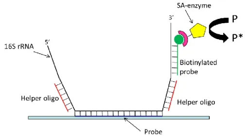
Fig. 1.6. Implementation scheme of the detection of RNA based on alkaline phosphatase.
Helper oligonucleotides can enhance binding by reducing RNA secondary structure and/or increase signal enhancement factor if they can bind the enzyme as well.
Enzymes have been largely used in 16S RNA based-biosensor. In sandwich-type detection, a specific adsorbed oligonucleotide probe hybridizes with the analyte RNA. Other regions of known base sequence of the target can be exploited to bind one to several secondary biotinylated oligonucleotides. These can then bind streptavidin-conjugated alkaline phosphatase molecules. In the presence of alkaline phosphatase, p-aminophenyl phosphate is converted to p-aminophenol, which thus accumulates over time in the case (and in the location) of RNA recognition and binding. p-Aminophenol can be oxidized to the corresponding quinoimide compound. The electric current derived from redox-cycling of this compound can be measured (implementing the chemoelectronic sensor as a series of interdigitated electrodes). The presence of the bound enzyme will make the redox current grow with time due to the progressive accumulation of the electroactive molecule, thus leading to signal enhancement.
Signal enhancement strategies based on the use of DNA polymerase
Phi 29 polymerase has been largely used in signal amplification thanks to its processivity and stability29,30. Like several other types of polymerase which share the same properties, Phi29 catalyzes the so called rolling circle amplification, an isothermal reaction which is extensively described in Chapter 6 of this dissertation. Chengde Mao and co-workers proposed a dual amplification strategy towards the amplification of the hybridization signal between complementary DNA molecules coupling RCA and peroxidase signal enhancement31. The analyte sequence hybridizes on a circular template, and perfect complementarity at the 3‟ end is necessary. The analyte sequence is thus used as a primer for rolling circle amplification by a Phi29 DNA polymerase so that a long piece of single-stranded DNA is produced as a consequence of recognition. This RCA represents the first amplification strategy of this detection method, as the long DNA fragment is easy to detect. On a non-probing section of the circular template strand, additional information content is included, to yield a second amplification step. A sequence is included that will be duplicated many times in case of DNA recognition. This sequence will fold into a G-quadruplex structure with binding capability for hemin, an iron-containing porphyrin that will then work as a peroxidase. The many copies of such DNA peroxidases can catalyse the oxidation of 2,2‟-azino-bis(3-ethylbenzthiazoline)-6-sulfonic acid (ABTS). The reaction product ABTSC+ is blue-green (maximum absorption wavelength, λmax=415 nm) and can serve as a convenient, colorimetric output signal. Thisreaction has multiple turnovers; each enzyme can generate multiple copies of products. This constitutes the second amplification step. The authors report the signal dependence of the colorimetric detection stating that they can clearly distinguish the presence of the analyte strand down to 1 pM. The output of this method depends on the careful tuning of the two amplification steps, as they cannot be optimized (or pushed) independently: too long a RCA amplification will create a highly entangled DNA molecule that will inhibit the diffusion of reactants for the second amplification step.
29
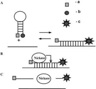
Signal enhancement of fluorescence through the use of a nickase
When a fluorescent probe is used for detecting a target (for example through molecular beacon approach), the cleavage of the probe in case of hybridization can release the target in solution for subsequent binding events. In an implementation of this strategy using a nickase, Zheleznaya and co-workers used molecular beacons and a site specific nickase to obtain an enhancement factor of 10032. Very interestingly, researchers also noted some interference in the assay in case of the presence of extraneous DNA that leads to a decrease in the signal (rather than to an expected increase due to non specific binding) probably due to the binding of the nickase to the extraneous DNA.
Fig. 1.8. Scheme of the nickase amplification. Picture taken from Zheleznaya et al.32.
In a recent application, a nicking enzyme sensing assay was coupled with CdSe/ZnS quantum dots amplification for cymbidium mosaic virus detection33. A thiolated hairpin DNA probe labelled with biotin was immobilized on gold electrode via S-Au bond. The double strand loop of the hairpin contained the restriction site for the endonuclease BfuCI, the nicking enzyme. In absence of the target, the hairpin bounded on the surface was closed and the restriction enzyme could digest the loop. As a consequence, the oligo end labelled with biotin was released in solution and the avidin-QD conjugate could not bind. On the contrary, the presence of a target molecule in solution opened the hairpin, blocking the enzymatic digestion and leading to the binding of avidin-quantum dots conjugate to the biotinylated probe (Fig. 1.10). The excess of QD was removed and the electrochemical detection was performed after a treatment with acid solution to dissolve quantum dots. Stripping voltammetric measurements of the Cd2+ ions were performed using an in situ plated mercury film on a glassy carbon electrode. The sensitivity of the DNA biosensor was calculated to be ≈ 1.0 × 10⁻¹² M.
Fig. 1.10. Quantum dots mediated signal amplification and endonuclease cleavage can be
coupled in a specific assay for DNA and RNA detection. Figure taken from Chen et al.33.
RNase H as a target recycling operator for RNA-based sensors
An alternative strategy that doesn‟t bring to the accumulation of new created material on the biosensor surface is that proposed by Goodrich and coworkers34. They implemented a method to recycle (and not to accumulate) the target DNA molecules by preparing a sensing surface with RNA oligonucleotide probes. After binding with the target (and thus forming a RNA-DNA hybrid) such double strand can be the substrate for RNase H present in solution. Its action leads to the hydrolysis of the bound RNA oligo and to freeing the target in solution again, so that it can bind to another surface-immobilized oligo. Over time, all the specific RNA oligonucleotide probes are digested by RNase, thus leading to signal enhancement (for instance if read through surface plasmon resonance or other techniques). The reported detection limit is 10 fM in a 13 µl sample volume.
Among the different enzymatic methods introduced, those based on the accumulation of easy to detect reaction products look as the most effective. Independently from the type of substrate used this strategy lead to the higher level of amplification. However the use of fluorescent reporter molecules requires the devices as CCD-cameras of fluorescence scanners which cannot be easily integrated in portable devices. In alternative the use of colorimetric substrate appears as an alternative also if less sensitive. RCA-mediated signal amplification lead to the accumulation on the sensor surface of big quantity of DNA that could be coupled to label-free transduction methods like potentiometric or mass sensitive biosensors.
In the final section of the Introduction are described the principles of the main transduction methods.
31
Fig. 1.11. Scheme of the amplification strategy proposed by Goodrich and coworkers. DNA
target is recognized by RNA probes which, after the hybridization event, are digested by RNase H. Target is thus free to bind with another RNA probes, leading to multiple detectable hybridization events. Picture reproduced from Goodrich et al.34.
N
OT ENZYMATIC AND NANOTECHNOLOGICAL APPROACHESSeveral amplification strategies are based on the use of organic and nanocomposite labels. They work in three principal ways: (i) they can be used as carriers, targeting specifically and accumulating at the hybridization site multiple labels which can be easily detected; (ii) they can act as labels themselves, exploiting their chemical and physical protein or (iii) they can work as catalyst, enhancing the chemical reaction that could take place in the hybridization site.
Liposomes as multiple labels carriers
Liposomes can be quite versatile vessels for the transportation of many types of molecules. They are vesicles made by a double layer of phospholipids that can covalently bind molecules on their surface or that can include them in the layer35. They can be used as labels for the detection of nucleic acids in analytical assays36.
Esche et al.37 used liposomes filled with carboxyfluorescein in a sandwich assay. The binding is revealed by fluorescence microscopy with a sensitivity of 0.4 fmoles/μl. Similar results have been obtained in other experimental efforts on different target nucleic acids.
Baeumner and coworkers published several interesting articles focused on detection of E. coli, B. anthracis and C. parvum mRNA transcripts in drinkable water after NASBA amplification (see Chapter 5), using liposome-based sandwich assay38-40.
Fig. 1.12. Scheme for liposome based-assay proposed by Baeumner et al.38. A DNA capture
probe is immobilized on a polyethersulfone membrane. A DNA reporter probe is coupled to the surface of a liposome. When a specific E. coli amplicon from NASBA reaction is present (Figure A), a sandwich is formed between capture probe, RNA and reporter probe. Thus, liposomes are captured in the capture/detection zone. The number of liposomes is directly proportional to the amount of E. coli RNA present. In Figure B it is shown that liposomes are not captured in the detection zone, when a nonspecific RNA molecule. Picture taken from Baeumner et al.38.
Metal nanoparticle and quantum dots as amplification labels
Metal nanoparticles have been used extensively as labelling agents for the detection of DNA hybridization. They are characterized by some interesting and chemical and physical properties which make these compounds suitable for biosensing applications41-44. Particular interests have excited their optical properties: the enhancement effect is due to their stability and to the easy detectability of the plasmonic resonance shift45,46. In addition metal nanoparticle can be easily functionalized47,48 and on them nucleic acid hybridization takes place with very high specificity because of the increased sharpness in the DNA melting transition49.
Storhoff and coworkers50 could avoid the use of PCR target amplification in their detection of genomic DNA. They measured the plasmonic red-shift due to DNA-induced aggregation of nanoparticles41. This method was implemented by using two different nanoparticle-conjugated oligonucleotide probes that are complementary to different portions of the target DNA molecule. The binding event can be evidenced with optical methods. 2 x 10⁵ molecules/μl can be detected (333 zmoles).
33
Fig. 1.13. Scheme of the use of nanoparticle-conjugated probes for the detection of DNA.
Figure taken from Storhoff et al.50.
AuNPs were used by Joung and co-workers to enhance 5500-fold the signal originated from 16s rRNA51. The target was detected by means of a specific PNA capture probe immobilized on a surface plasmon resonance (SPR) sensor. As PNA is characterized by a neutral backbone structure, the hybridization with the 16S RNA led to a change in the ionic charge from neutral to negative. Cationic AuNPs were synthetized and used to amplify locally the signal generated by the target binding. This method was applied on E. coli total RNA extraction showing a sensitivity of 58.2 ± 1.37 pg/ml RNA. For S. aureus detection the method was applied without preliminary nucleic acid extraction. In this case the achieved sensitivity was 7×10⁵ CFU/ml.
As nanoparticle, alternative nanostructures as nanorods are characterized by interesting physical and chemical properties that make them suitable for biosensing and signal amplification52. Recently, Parab and colleagues demonstrated the use gold nanorods (GNR) for the optical detection of a Chlamydia trachomatis DNA in solution. Monitoring the absorption spectra of GNR-capture probe they reliably detected target DNA in the range of 250-50 nM in 100 µl sample53.
Fig. 1.14. Scheme of gold-nanorods based biosensor for C. trachomatis detection. Figure
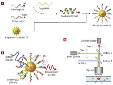
In the last years quantum dots have found large application in DNA, RNA, protein microarray labeling and more frequent is also their use in DNA biosensors54-56. Zhang and colleagues reported a nanosensor based on fluorescence resonance energy transfer (FRET) coupling the use of CdSe–ZnS core–shell nanocrystals as donor and Cy5 dye as acceptor57. In solution, single strand target DNA bound to a biotinylated capture probe and to a reporter probe labeled with Cy5 forming a hybrid sandwich structure. Several hybrids are captured by streptavidin-QD hybrids, accumulating target and Cy5 dyes on it. In this configuration, acceptors dyes were in close proximity to the donor and were able to emit fluorescence by means of FRET after QDs excitation. As a result, fluorescent detection at the emission wavelength of Cy5, indicated the presence of target. The method has reported to be highly sensitive: as the unbound QD produced near-zero background fluorescence while a very clear FRET signal is generated after binding, up to ~ 50 copies of target DNA can be detected.
Fig. 1.15. Scheme of optical biosensor based on FRET. CdSe-ZnS quantum dots and Cy5 dye
are locally concentrated after target detection. In this configuration, acceptors dyes were in close proximity to the donor and were able to emit fluorescence by means of FRET after QDs excitation. Picture taken from Zhang et al.57.
Chad Mirkin and co-workers moreover employed another enhancement step in the nanoparticle-mediated detection of DNA. As their method leads to DNA signals that can be read with a standard flatbed optical scanner, they termed their method “scanometric detection”58
. In their method, ultra-low levels of DNA labelled with oligonucleotide-functionalized nanoparticles are detected by inducing silver reduction on the surface of the nanoparticles. In a sandwich assay, the surface immobilized nanoparticles, bridged by the target DNA, can then be turned into silver microparticles that are detectable even to the naked eye. The authors claim that nanoparticle labelling leads to signals that are 3-4
35 orders of magnitude more intense than fluorophores. 6 × 10⁶ molecules (200 fM in 50 μl) can be detected50.
Besides their optical properties, metal nanoparticles can be used as catalysts, and thus permit the implementation of electrochemical signal enhancements when these are bound to reported oligonucleotide probes. In an example reported by Polsky and co-workers, a sandwich is obtained on the electrode surface by immobilizing the target and a reported oligonucleotide that is labelled with a platinum nanoparticle59. Such nanoparticles can catalyse the conversion of hydrogen peroxide to water at a controlled electrochemical potential, enabling the chronoamperometric measurement of the hybridization of 10 pM DNA.
T
HE CHOICE OF AMPLIFICATION STRATEGY:
WORKING WITH DIFFERENT TRANSDUCTION METHODSThe choice of one amplification method with respect to another must be motivated by the type of biosensor in which it will be integrated. The hybridization event between target and probe is converted in a measurable signal by a transducer which is intimately coupled with the sensing element. The use of fluorescent or redox labels with respect to the use of enzymes or of any other amplification strategy depends on what type of transduction method is used for the signal. In this paragraph, the different types of nucleic acid biosensors will be classified according to their transduction method, illustrating the general principles for each of them.
O
PTICAL BIOSENSOROptical transducers are probably the most common in nucleic acids detection thanks to their sensitivity and specificity. They are usually coupled with signal amplification methods which enhance fluorescence, with methods based on optical properties of nanoparticles or with methods that generate a visible color.
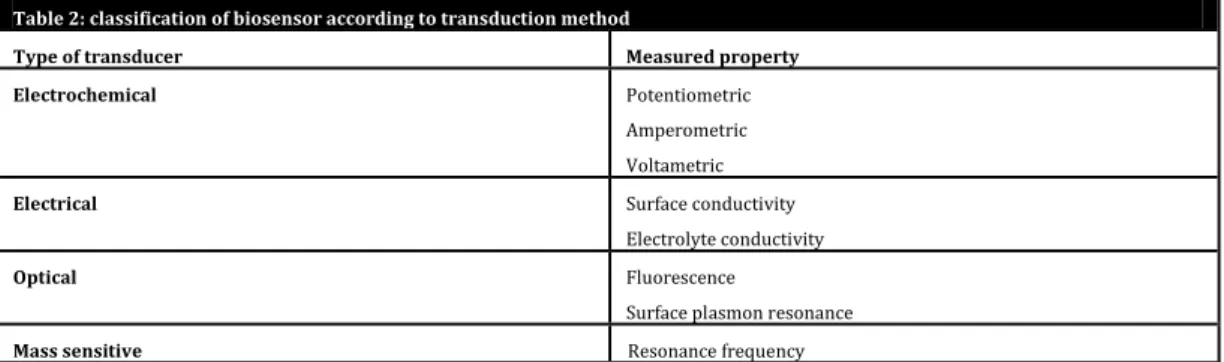
Table 2: classification of biosensor according to transduction method
Type of transducer Measured property Electrochemical Potentiometric
Amperometric Voltametric
Electrical Surface conductivity Electrolyte conductivity
Optical Fluorescence
Surface plasmon resonance
Fluorescence detection
Fluorescence detection works with the monitoring of the emission of light by a compound which goes back from an excited state to the ground energy state. The molecule is excited by the absorption of high energy light (shorter wavelength). When the molecule returns to the original ground state, part of the energy is re-emitted in light at lower energy (longer wavelength) while a part is lost as heat. In order to maximize the signal, it is desirable that the excitation wavelength and the emission wavelength be as far as possible from each other. The use of this type of biosensor implies the use of labels and the use of an excitation source, usually an ultraviolet laser, which must be integrated into the device or that more frequently is part of a scanner. For these reasons, despite their sensitivity, fluorescent detection is not easily integrable in portable devices.
Surface plasmon resonance detection
Surface plasmon resonance (SPR) biosensors are based on the variations in the charge density waves at the surface of metallic structures. Electrons within the conduction band are free to move at the interface between the metal and the external medium. They oscillate collectively, generating surface waves of charge which propagate along the surface. The charge density wave is associated to an electromagnetic wave, which energy is confined mainly to the surface. In SPR biosensors the variations of the reflected light intensity are measured when the surface is illuminated with light beam: the presence of a biological element on the surface like an oligo probe and the consequent accumulation of material after the detection event, modifies the refractive index of the medium, changing the reflection angle. SPR biosensors are quite sensitive: they can detect nanograms of analyte on the surface but their application is limited by the size of the searched molecule. The main drawbacks of this powerful technique lay in its complexity (specialized staff is required), high cost of equipment and large size of most currently available instruments. On the other hand, SPR are prone to be use as label-free detection method for nucleic acid.
Colorimetric biosensors
Colorimetric biosensors are based on the properties of some enzymes to convert a colorless substrate in a reaction product that can be easily detect with a standard photo scanner or even at naked eye. The most typical example is the use of horseradish peroxidase (HRP) which in presence of hydrogen peroxide catalyzes the conversion of suitable substrate, like –tetramethybenzidine (TMB), in a colored product (A blue precipitate in the case of TMB). If the enzyme is localized on the surface in
37 correspondence of the hybridization event between probe and target, the color will be accumulated on the probe feature.
E
LECTROCHEMICAL BIOSENSORSThese devices are mainly based on the observation of current or potential changes due to interactions occurring at the sensor-sample matrix interface. Techniques are generally classified according to the observed parameter: current (amperometric), potential (potentiometric) or impedance (impedimetric). Compared to optical methods, electrochemical biosensor allows the user to work with turbid samples, and the cost of equipment is cheaper. In addition electrochemical biosensors are suitable to perform label-free detection (they can work without the use of a label). On the contrary, they exhibit a slightly lower selectivity and sensitivity than their optical counterparts. Some suggested reviews are those recently proposed by Mir et al.60, Bonanni and del Valle61.
Amperometric biosensors
In amperometric biosensors a potential is applied between two electrodes. They measure the resulting current in oxidation or reduction processes of electro active species present in solution, generally using enzymes as biomarker molecules. The read-out of the enzyme needs the interaction with a specific substrate which is converted in a different molecule, usually a redox molecule that can be detected. In addition, for each catalytic site more than one redox molecule is produced, leading to signal enhancement.
Potentiometric biosensors
DNA potentiometric detectors are based on the use of transistors. They measure the current generated as consequence of the charge accumulation effect at the gate electrode after the specific binding of DNA molecules. Potentiometric DNA biosensors are particularly attractive since they can be fabricated in very small sizes and massively integrated in lab-on-a-chip devices.
Impedimetric biosensors
Impedimetric transduction is based on sensing the electrical resistance and reactance of a medium in an electrochemical cell, applying an alternating current potential. It provides electric information in the frequency domain. Among the different electrochemical transduction methods, electrochemical impedance spectroscopy is particularly suitable for label-free detection of DNA.
M
ASS SENSITIVE BIOSENSORSIn mass-transducers, a mass change on the surface sensor due to the binding of the analyte to the probe determines a variation of the resonance frequency of the device.
Cantilevers
Cantilevers are nanomechanical biosensors. The cantilever surface is usually functionalized with suitable DNA or RNA probes and interactions with the nucleic acid analyte take place only on where probes are present. The bind between probe and analyte bend the cantilever and such bending is usually monitored by electrical readout, since the lever is integrated in a piezoresistor. The binding of the cantilever in presence of the analyte provides information both on analyte concentration than on the kinetics of the interactions because the interactions can be monitored in real time. Cantilevers biosensors work both in air and in liquid environment.
Quartz Crystal Microbalance
Quartz Crystal Microbalance (QCM) is ultra-sensitive weighing device based on the mechanical resonance of piezoelectric single-crystalline quartz. If a voltage is applied to the crystal quartz and the frequency of the applied voltage corresponds to the resonance frequency of the crystal, resonance condition occurs. The presence of mass on the crystal surface changes the frequency of the total oscillating mass. In addition, the presence of water, ions and some type of biomolecules/polymers (like DNA) determine energy dissipation of the oscillating system. Since QCM is sensitive also to the binding of water and ions, it has been found to be more sensitive than optical and electrochemical biosensors.
39
R
EFERENCES1. Diouani, M.F. et al. Miniaturized biosensor for avian influenza virus detection.
Materials Science and Engineering: C 28, 580-583 (2008).
2. Maynard, C. et al. Waterborne Pathogen Detection by Use of Oligonucleotide-Based Microarrays. Journal of Microbiological Methods 71, 8548-8557 (2005).
3. Straub, T.M. & Chandler, D.P. Towards a unified system for detecting waterborne pathogens. Journal of Microbiological Methods 53, 185-197 (2003).
4. Gouvras, G. The far-reaching impact of bioterrorism. What the European Union is doing regarding deliberate releases of biological/chemical agents based on the events in the United States. IEEE Engineering in Medicine and Biology Magazine 21, 112-115 (2002).
5. Tegnell, A. et al. The European Commission’s Task Force on Bioterrorism.
Emerging Infectious Diseases 9, 1330-1332 (2003).
6. Chou, C.F. et al. A miniaturized cyclic PCR device--modeling and experiments.
Microelectronic Engineering 61-62, 921-925 (2002).
7. Kopp, M.U., Mello, A.J.d. & Manz, A. Chemical Amplification: Continuous-Flow PCR on a Chip. Science 280, 1046-1048 (1998).
8. Xiang, Q., Xu, B. & Li, D. Miniature real time PCR on chip with multi-channel fiber optical fluorescence detection module. Biomedical Microdevices 9, 443-449 (2007). 9. Christensen, T.B. & et al. PCR biocompatibility of lab-on-a-chip and MEMS materials. Journal of Micromechanics and Microengineering 17, 1527 (2007).
10. Lagally, E.T., Emrich, C.A. & Mathies, R.A. Fully integrated PCR-capillary electrophoresis microsystem for DNA analysis. Lab on a Chip 1, 102-107 (2001).
11. Nickisch-Rosenegk, M.v. et al. On-chip PCR amplification of very long templates using immobilized primers on glassy surfaces. Biosensors and Bioelectronics 20, 1491-1498 (2005).
12. Ryder, T.B. et al. Methods and kits for determining pre-amplification levels of a nucleic acid target sequence from post-amplification levels of product C12Q1/68; (IPC1-7): C12Q1/68 edn Vol. EP0747488 (A1) (Europe, 1993).
13. Hill, C. Molecular diagnostic testing for infectious diseases using TMA technology. Expert Review of Molecular Diagnostic 1, 445-455 (2001).
14. Bachmann, L.H. et al. Nucleic Acid Amplification Tests for Diagnosis of Neisseria gonorrhoeae and Chlamydia trachomatis Rectal Infections. Journal of Clinical
15. Chelliserrykattil, J. et al. Development of a Quantitative Real-Time Transcription-Mediated Amplification Assay for Simultaneous Detection of Multiple Nucleic Acid Analytes. Journal of Molecular Diagnostics 11, 680-680 (2009).
16. Rao, V. et al. Improved detection of hepatitis C virus infection by transcription-mediated amplification technology in dialysis population. Renal Failure 32, 721-726 (2010).
17. Walker, G.T. et al. Strand displacement amplification: an isothermal, in vitro DNA amplification technique. Nucleic Acid Research 20, 1691-1696 (1992).
18. Walker, G.T., Little, M.C., Nadeau, J.G. & Shank, D.D. Isothermal in vitro amplification of DNA by a restriction enzyme/DNA polymerase system. Proc. Nat. Acad.
Sci. USA 89, 392-396 (1992).
19. Little, M.C. et al. Strand Displacement Amplification and Homogeneous Real-Time Detection Incorporated in a Second-Generation DNA Probe System, BDProbeTecET. Clinical Chemistry 45, 777-784 (1999).
20. Chen, Q.H. et al. Real-time monitoring of the strand displacement amplification (SDA) of human cytomegalovirus by a new SDA-piezoelectric DNA sensor system.
Biosensors & Bioelectronics 24, 3412-3418 (2009).
21. Notomi, T. et al. Loop-mediated isothermal amplification of DNA. Nucleic Acid
Research 28, e63 (2000).
22. Inomata, A. et al. Development and evaluation of a reverse transcription-loop-mediated isothermal amplification assay for rapid and high-sensitive detection of Cryptosporidium in water samples. Water Science and Technology 60, 2167-2172 (2009). 23. Yang, J.L. et al. Simple and rapid detection of Salmonella serovar Enteritidis under field conditions by loop-mediated isothermal amplification. Journal of Applied
Microbiology 109, 1715-1723 (2010).
24. Maruyama, F., Kenzaka, T., Yamaguchi, N., Tani, K. & Nasu, M. Detection of Bacteria Carrying the stx2 Gene by In Situ Loop-Mediated Isothermal Amplification.
Applied and Environmental Microbiology 69, 5023-5028 (2003).
25. Schaerli, Y. et al. Isothermal DNA amplification using the T4 replisome: circular nicking endonuclease-dependent amplification and primase-based whole-genome amplification. Nucleic Acid Research 38, e201 (2010).
26. Sarrazin, C., Teuber, G., Kokka, R., Rabenau, H. & Zeuzem, S. Detection of Residual Hepatitis C Virus RNA by Transcription-Mediated Amplification in Patients With Complete Virologic Response According to Polymerase Chain Reaction-Based Assays. Hepatology 32, 818-823 (2000).
27. Brink, T.L., Thornton, K., Wang, S.S. & Hellyer, T. Detection of Legionella pneumophila by Strand Displacement Amplification on the BDProbeTec™ ET System. in Association for Molecular Pathology (AMP) Meeting (2000).
41 28. Li, J. et al. Amperometric biosensor with HRP immobilized on a sandwiched nano-Au / polymerized m-phenylenediamine film and ferrocene mediator. Analytical and
Bioanalytical Chemistry 376, 902-907 (2003).
29. Blanco, L. et al. Highly efficient DNA synthesis by the phage phi 29 DNA polymerase. Symmetrical mode of DNA replication. The Journal of Biological Chemistry 264, 8935-8940 (1989).
30. Esteban, J.A., Salas, M. & Blanco, L. Fidelity of phi 29 DNA polymerase. Comparison between protein-primed initiation and DNA polymerization. The Journal of
Biological Chemistry 268, 2719-2726 (1993).
31. Tian, Y., He, Y. & Mao, C. Cascade Signal Amplification for DNA Detection.
ChemBioChem 7, 1862-1864 (2006).
32. Zheleznaya, L.A., Kopein, D.S., Rogulin, E.A., Gubanov, S.I. & Matvienko, N.I. Significant enhancement of fluorescence on hybridization of a molecular beacon to a target DNA in the presence of a site-specific DNA nickase. Analytical Biochemistry 348, 123-126 (2006).
33. Chen, J., Zhang, J., Yang, H., Fu, F. & Chen, G. A strategy for development of electrochemical DNA biosensor based on site-specific DNA cleavage of restriction endonuclease. Biosensors and Bioelectronics 26, 144-148 (2010).
34. Goodrich, T.T., Lee, H.J. & Corn, R.M. Enzymatically amplified surface plasmon resonance imaging method using RNase H and RNA microarrays for the ultrasensitive detection of nucleic acids. Analytical Chemistry 76, 6173-6178 (2004). 35. Edwards, K. & Baeumner, A. Optimization of DNA-tagged dye-encapsulating liposomes for lateral-flow assays based on sandwich hybridization. Analytical and
Bioanalytical Chemistry 386, 1335-1343 (2006).
36. Edwards, K.A. & Baeumner, A.J. Liposomes in analyses. Talanta 68, 1421-1431 (2006).
37. Esch, M.B., Baeumner, A.J. & Durst, R.A. Detection of Cryptosporidium parvum Using Oligonucleotide-Tagged Liposomes in a Competitive Assay Format. Analytical
Chemistry 73, 3162-3167 (2001).
38. Baeumner, A.J., Cohen, R.N., Miksic, V. & Min, J. RNA biosensor for the rapid detection of viable Escherichia coli in drinking water. Biosens.Bioelectron. 18, 405-413 (2003).
39. Baeumner, A.J., Leonard, B., McElwee, J. & Montagna, R.A. A rapid biosensor for viable B. anthracis spore. Analytical and Bioanalytical Chemistry 380, 15-23 (2004). 40. Baeumner, A.J., Pretz, J. & Fang, S. A Universal Nucleic Acid Sequence Biosensor with Nanomolar Detection Limits. Analytical Chemistry 76, 888-894 (2004). 41. Storhoff, J.J. et al. What controls the optical properties of DNA-linked gold nanoparticle assemblies? Journal of the American Chemical Society 122, 4640-4650 (2000).