Alma Mater Studiorum – Università di Bologna
DOTTORATO DI RICERCA IN
Scienze Biotecnologiche e Farmaceutiche
Ciclo XXXI
Settore Concorsuale: 05/G1
Settore Scientifico Disciplinare:
BIO/14
TITOLO TESI
"TARGETING EphB1 RECEPTOR IN CELLULAR MODELS:
ANALYSIS OF SIGNALING PATHWAYS AND CROSS-TALK
WITH OPIOID RECEPTORS"
Presentata da
:GABRIELA LISETH VACA ALTAMIRANO
Coordinatore Dottorato
Supervisore
Santi Spampinato
Santi Spampinato
Table of Contents
ABSTRACT
1
1. INTRODUCCION
2
Eph receptors and ephrin ligands 2
Functions and classification 2
Structure 3
EphB and ephrinB system 4
1.1.3.1 Formation and activation 4
1.1.3.2 Bidirectional signaling 6
Forward signaling 7
Reverse signaling 10
Internalization and proteolytic cleavage 12
EphB/EphrinB system in SNC 14
EphB/EphrinB system in early SNC 14
EphB/EphrinB system in adult SNC 18
EphB and EphrinB are involved several pathological processes 20
EphB/EphrinB system in cancer 20
EphB/EphrinB system in inflammation 22
EphB/EphrinB system in neuropathologies 22
1.3.3.1 Pain 22
1.3.3.2 Neurodegenerative diseases 26
Interactions between Eph/ephrin system with other receptors 30
Ion channels receptors 32
1.4.1.1 NMDR 32
1.4.1.2 AMPAR 33
Cell surface receptors 34
1.4.2.1 Crosstalk with others RTKs 34
1.4.2.2 Crosstalk with GPCRs 35
Eph receptor as a therapeutic target 37
Molecules that interfered with EphrinB/EphB signaling 38
1.5.1.1 Soluble form of EphB receptors as part of fusion protein 38
1.5.1.2 Small molecules 39
1.5.1.3 Peptides 40
1.5.1.4 Kinase inhibitors 42
1.5.1.5 Monoclonal antibodies 42
1.5.1.6 siRNAs or antisense oligonucleotides 43
Eph receptor – binding molecules conjugated in the delivery of drugs, toxins or imaging agents 44
Vaccine based immunotherapy 46
3. MATERIALS AND METHODS
51
Plasmid and reagents. 51
Cell culture. 51
Western Blots analysis in SHSY5Y cell model 52
Cells transfection and Western Blot analysis in HEK- 293 cell model. 53
Saturation binding assays 54
Total RNA preparation and real time-polymerase chain reaction (RT-PCR) analysis 54
cAMP measurement 55
Statistical analysis 56
4. RESULTS
57
Downstream molecules involved in occlusive crosstalk observed following the co-administration of morphine
and ephrinB1-Fc ligands. 57
Signaling pathways involved in p42/p44 MAPK activation elicited by ephrinB1 Fc- and Morphine in SHSY5Y
native cells. 58
PI3K signaling pathway is involved in the occlusive croostalk produced by the co-administration of
morphine and ephrinB1-Fc ligands. 59
Blocking PI3K signaling pathway also can recovers completely the activation of PKC protein. 61
cAMP expression is involved in antagonist interaction between MOR and EphB1 receptors. 62
Occlusive crosstalk in PMA-differentiated SHSY5Y cells. 63
Blocking PI3K signaling pathway in PMA-differentiated SHSY5Y cells does not affect p42/44 and PKC
phosphorylation. 64
Occlusive crosstalk in PMA-differentiated SHSY5Y cells exposed to a pro-inflammatory stimulus 65
MOR and EphB1 expression is altered in PMA-Differentiated SHSY5Y cells exposed to TNFα. 66
Occlusive crosstalk occurs in PMA-differentiated SHSY5Y cells exposed to TNF-α. 68
TNF-α added to PMA-differentiated SHSY5Y cells blocks the signaling pathway involved in ephrinB1/EphB1
system activation and recues p42/44 MAPK and PKC activation produced by morphine. 69
Blockade of EphB1 receptor activation by novel peptide antagonist 70
Administration of EphB1 binding peptides in HEK-EphB1 cells do not activate p42/44 MAPK activation. 71 Effects of Tripeptide and pentapeptide on p42/44 MAPK phosphorylation mediated by ephrinB1-Fc in
HEK-EphB1 cells. 73
Effects of hexapeptide on p42/44 MAPK phosphorylation mediated by ephrinB1-Fc in HEK-EphB1 cells 74 Effects of octapeptide on p42/44 MAPK phosphorylation mediated by ephrinB1-Fc in HEK-EphB1 cells. 75
6. CONCLUSIONS
83
1
Abstract
The activation of EphB1 receptors by their membrane bound ligands ephrins in nociceptive
neurons have been implicated in the onset and maintenance of different types of pain and in side
effects produced as consequence of prolonged Mor activation. The use of EphB1 blocking reagents
rescue opioid-induced analgesia suggesting that antagonists targeting EphB1 receptor could
represent a novel class of analgesics.
A potential inverse relation between EphB1 and Mor receptors was displayed in a previous
investigation by Lombardo Sara where showed that the co-administration of Mor and EphB1
agonists resulted in an occlusion of morphine-mediated p42/44 MAPK phosphorylation.
Therefore, the aim of the research was elucidated the signaling pathway and mechanism
responsible for the functional crosstalk triggered by the co-administration of both ligands and at
the same time developed and tested different EphB1 antagonist peptidomimetics.
As results: *ephrinB1/EphB1 activation through PI3K signaling pathways blunted p42/44
activation morphine-mediated and PKC-dependent in SHSY5Y native cells where in addition
EphB1 signaling activation seems to be the responsible for blocking the capacity of the morphine
to modulate adenylyl cyclase activity. *SHSY5Y cells induced to PMA differentiation produced
up regulation of Mor expression and down regulation of EphB1 receptors allowing that morphine
significantly activate p42/p44 MAPK even when ephrinB1 was co-administered. *Conversely, in
PMA-differentiated SHSY5Y cells exposed to TNF-α, EphB1 expression was significantly up
regulated and the morphine failed to increase p42/44 MAPK phosphorylation. * Moreover, were
found two novel antagonist peptides capable to counteract ephrinB1-Fc-mediated activation of
p42/p44 MAPK phosphorylation.
Concluding that when EphB1 is activated morphine no longer triggers PKC-dependent signaling
events downstream of Mor activation, explaining the reasons why opioid analgesics are less
effective in the treatment of different pain states. At this regards, novel EphB1 receptor antagonists
may represent a novel strategy to reduce the negative impact of ephrin system.
2
1. Introduccion
Eph receptors and ephrin ligands
Functions and classification
Eph (erythropoyetin—producing hepatocellular carcinoma) receptors, the largest subfamily of receptor tyrosine kinases (RTKs), and their membrane bound ephrin ligands are cell surface molecules and important mediators of cell–cell communication, that play a fundamental role in cell behavior during embryogenesis and adult life 1–7. Several studies have demonstrated that are involved in important functions as: cell shape, adhesion and repulsion, cell positioning and migration; tissue boundary formation during somatogenesis; development of the vascular system; tumor invasion and metastasis; immune function; hematopoiesis; blood clotting; stem cells development; tissue repair and maintenance; angiogenesis, control of tissue morphogenesis and patterning; cell sorting at compartmental boundaries and bone formation. 1,2,8,9 In nervous system are regulators of axon guidance during development; neural and synaptic plasticity; long term potentiation, synaptogenesis, dendritic filopodia motility, and neural crest cell 1,2,5,10,11
Eph receptors, based on the structure and the ability to bind their membrane attached ephrin ligands, are divided into EphA and EphB receptors. In mammals, there are nine members of EphA class (A1-A8, A10) and five members of EphB class (B1-B4, B6) while Ephrins-As (A1-A5) are bound by a glycophosphatidylinositol (GPI) anchor, and ephrin-Bs (B1-B3) contain a short intracellular signaling domain 5,6,10,11. Furthermore, Ephs and ephrins interact promiscuously within each subclass. Thus cross-class interactions known are EphA4, interacting with B-type ephrins and ephrin-A5 can activate EphB2 as well as EphAs 12.
3
Structure
The extracellular region of all Eph receptors consists in highly conserved N- terminal globular domain called ligand binding-domain which is necessary for ligand recognition and binding site that mediates receptor-ephrin interaction between cells 11,13,14.
The globular domain is followed by a unique cysteine-rich motif (Cys domain, comprising sushi and epidermal growth factor (EGF)-like motifs) and two fibronectin type III motifs, which appear to be involved in receptor–receptor dimerization interactions 13 and facilitate clustering of multiple Eph-ephrin complexes 11,15.
In the juxtamembrane region, there is a highly conserved motif containing two tyrosine residues, which are the major autophosphorylation sites involved in receptor signaling 13,15.
The tyrosine kinase domain has been proposed to serve as a binding site for activators of the small GTPases, which regulate cytoskeletal organization. A conserved region of 60–70 amino acids in the carboxyl-terminal tail of the Eph receptors forms a sterile alpha motif (SAM) domain, which has been implicated in receptor dimerization and mediates cell–cell initiated signal transduction 13,15.
Eph receptors also possess a post- synaptic density protein zona occludens (PDZ)-binding domain. This domain is important in the assembly of Eph or ephrin complexes, that allow interactions with other regulatory molecules (Figure 1), and in their localization to specific sites within the cell, such as membrane raft microdomains and synapses 13,15.
The EphrinB display an extracellular Eph receptor binding domain, a single-pass transmembrane region, a short intracellular domain with several sites for tyrosine and serine phosphorylation, and a C-terminal PDZ-binding motif 16 which is phosphorylated by Src family kinases upon ephrin stimulation by Eph receptors (Figure 1).
Ephrin-Bs domains include three tyrosine residues enabling the recruitment of Src Homology 2/3 (SH2/SH3) adaptor proteins, a PDZ binding domain enabling interaction with adaptor-proteins such as glutamate receptor interacting protein 1 (GRIP1), a D-domain for interaction with Erk/MAPK, and a Grb4 domain enabling interactions with the G protein-coupled receptor kinase-interacting protein 1 (GIT1) 17.
4 Ephrin-As, in contrast, lack a transmembrane domain and are attached to the cell membrane through a GPI-anchor. Therefore, ephrin-As rely on other receptors and mechanisms to transduce intracellular signals, for instance, p75 neurotrophin receptor (p75NTR) and the TrkB neurotrophin receptor tyrosine kinase have been involved in reverse signaling ephrin-A-mediated which is involved in axon guidance 16,18.
Figure 1. Structure of Eph receptors and ephrin ligands. Eph and ephrin signaling relies on specific functional domains; on the extracellular side, Eph receptor is composed by ligand binding domain (LBD) which binds to the receptor-binding domain (RBD) of ephrins, followed by a Cys-rich domain encompassing the sushi and epidermal grown factor (EGF)-like domains, and two fibronectin (FN) domains. The intracellular side of Eph receptor is composed of the transmembrane region (TM), the Tyr kinase domain (TK), the sterile alpha motif (SAM) and the PDZ domain. EphrinsA class is linked to the membrane via a glycosylphosphatidylinositol (GPI) linkage, whereas the B class has a transmembrane domain and an intracellular PDZ domain. Eph–ephrin interaction results in phosphorylation of Tyr residues (P) present in the juxtamembrane domain between the TK and TM domains, as well as in the TK and SAM domains. The juxtamembrane phosphorylation is required for TK function, which is
crucial for many Eph signalling-induced biological responses 15.
EphB and ephrinB system
1.1.3.1 Formation and activation
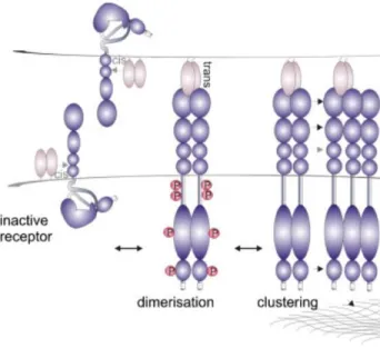
In contrast, to other tyrosine kinases receptors where receptor dimerization is enough to trigger biological activity, Eph receptors need high local density of their ligands and the formation of clusters as pre-requisite to form Eph/ephrin signaling assemblies, which in turn induce a precise downstream signaling pathways and biological response 3,12,19.
5 The formation and activation of Eph/ephrin system is considered a multiple-step process where Eph and ephrin domains interact with each other. Prior to activation, the Eph receptors are loosely distributed on the cell surface and display minimal kinase activity, unless receptor expression levels are considerably elevated 3.
Upon cell–cell contact, ligands and receptors bind each other with 1:1 stoichiometry and nanomolar affinity 20. Eph receptors use two separate interfaces for the assembly of signaling-competent clusters. The first step, in the initiation of Eph-mediated signaling, is the recognition and binding of Eph receptors and ligands located on closely opposed cell surfaces. This process, knowing as DIMERISATION INTERFACE; produce high-affinity dimers that are the predominant form of the complex. 2,20,21 However, is possible that residues of Eph receptor could produce lower-affinity binding, principally inEphrinB2, wich could lead to the association of two homodimers 16.
The second interface, called as CLUSTERING INTERFACE, is produced within the cys-rich domain of the Eph molecules and is responsible for the formation of the higher-order functional tetrameric 2:2 complexes, in which each receptor interacts with two ligands and each ligand with two receptors (Figure 2). For instante, Crystallographic studies of the EphB2– ephrinB2 complex reveal a tetrameric complex forming a ring-like structure. 2,12,20,21. The existence of these two separate and independent interacting interfaces allows the Eph receptors to utilize the so-called ‘seeding’ mechanism for the assembly of clusters 3.
Finally, binding of ephrins to the extracellular part of Eph receptors not only activates their cytoplasmic tyrosine kinase domain, but also leads to the transduction of a reverse signal into the ephrin-bearing cell 3 Importantly, the disruption of ephrin–ephrin homodimers and the Eph–ephrin tetramer formation also results in a repositioning of the B-ephrin transmembrane and cytoplasmic domains, converting them from an inactive to an active configuration 20
Furthermore, is important point out that ligand-independent signaling can also occur if the receptor concentration is not enough, for example, on the surface of tumor cells.
6 Furthermore, Eph assemblies can be independent of ephrin binding for example, EphA4-ECD (ectodomain receptor) can bind FNIII-domain of a neighboring receptor molecule without the presence of ephrins 3
Figure 2. Eph receptor clustering and activation. The inactive Eph receptor present auto-inhibited conformation, released upon phosphorylation and activation. Ephrin binding to one receptor initiates dimerisation into an Eph/ephrin heterotetramer. Further clustering is facilitated by receptor–receptor interactions between multiple domains of adjacent Ephs, including
co-clustering of A and B type Ephs 12.
1.1.3.2 Bidirectional signaling
The binding of ephrin molecules to Eph receptors induces conformational changes in both proteins 13, producing modifications of actin cytoskeleton and microtubular organization22.
A distinguish feature of Eph-ephrin complexes is their ability to generate bidirectional signals that affect both the receptor-expressing and ephrin-expressing cells (Figure 3) 11. Eph receptor “forward” signaling depends principally on the tyrosine kinase domain, which mediates autophosphorylation of the receptor as well as phosphorylation of other proteins and the associations of the receptor with various effector proteins 23.
Ephrin-B “reverse” signaling depends on tyrosine phosphorylation of the ephrin cytoplasmic region (mediated by Src family kinases and some receptor tyrosine kinases) and on associated proteins. For instance at the level of the nervous system, Eph/ephrin bidirectional signaling is important not only for the communication between neurons but also between neurons and glial cells 24
7 Finally, Eph proteins and ephrins can simultaneously act as receptors and ligands, leading to bidirectional or parallel and antiparallel signaling (Figure 3), depending on the distribution of Ephs and ephrins between interacting cells, as well as the direction of signaling in single ephrin–Eph pairs. Ephrins can also induce signaling cascades independently of Eph proteins, a mode of signaling that seems to be evolutionarily conserved 15.
Futhermore, the interaction produced between Eph receptors and ephrin ligands in Eph receptors and ephrins co-expressed cells, is called CIS interaction in which ephrins can attenuate signaling of Eph receptors (Figure 2), for instance in nervous system Cis interaction allow the cell to precisely control the axonal trajectory 25,26; whereas TRANS interaction (Figure 2) is produced between neigboring cells, facilitate forward signaling and, depending on the identity of Eph receptors and ephrins involved, serve as either repulsive or attractant cues for axonal guidance 25.
Figure 3. Eph–ephrin signaling. Forward signaling involves signal transduction from ephrins to Ephs; reverse signalling involves
signalling from Ephs to ephrins; and bidirectional signaling involves the simultaneous activation of pathways downstream of ephrins and Ephs. Parallel signaling occurs when ephrins and Ephs are expressed on the same cell. Anti-parallel signalling is a
special case of simultaneously occurring forward signalling, whereby ephrin–Eph signals are propagated in both directions 15.
Forward signaling
The classic mode of forward signaling is from ephrins to Ephs and frequently results in cell retraction. This repulsive response is particularly important for axon guidance and sorting of Eph-expressing cells during development 15,27.
8 The signaling is initiated by autophosphorylation and Src family kinases-mediated phosphorylation of the intracellular tyrosine residues, resulting in the activation of the tyrosine kinase catalytic domain 16. However the interactions also can be kinase independent, for example, EphB receptor in kinase independent mechanism plays essential roles in retina axon path finding 28. Furthermore, Ephs can be negatively regulated by several protein tyrosine phosphatases and by ubiquitin ligase-mediated internalization and degradation 6.
Once the Eph receptors are phosphorylated, scaffold and adaptor proteins are activated. The adaptor proteins, containing Src-homology 2 (SH2) and Src-homology (SH3) domains, can bind and initiate phosphorylation of downstream substrates and allow protein–protein interactions, via SAM and PDZ-binding motifs, for contribute to signaling 16,29.
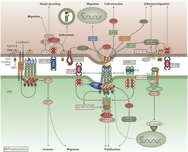
Forward signaling is involved in the activation of several cytoplasmic downstream signaling pathways such as Src family kinases, mitogen- activated protein kinase, p-21 activated kinase, chemokine pathways, heterotrimeric G-protein pathways, and integrin-mediated pathways (Figure 4) 1.
For each pathway to be activated, the recruitment of adaptor and scaffold proteins are necessary. Adaptor proteins involved in forward signaling are: Rho GTPase activating proteins (GAPs) (including RhoA, Cdc42, and Rac) which are involved in the regulation of the actin cytoskeleton and cell shape, movement, and adhesion 16, guanine nucleotide exchange factors (GEFs) (including ephexins, Vav2, Vav3 protein) 15; ADAM10 family proteases 26; non-catalytic regions of Tyr kinase adaptor protein 1 (Nck1) and Nck2 29 and phosphoinositide 3-kinase (PI3K) (FIGURE 4) 15 which can be suppressed by forward signaling, blocking the activation of Akt - mTORC1 pathway 23.
GAPs and GEFs proteins are involved in Eph/ephrin mediated axon guidance events 26, among them α2-chimaerin, which is essential for axon guidance-dependent of EphA4 11, and ephexin induce cytoskeletal collapse in growth cone regions exposed to ephrins. Principally, ephexin is essential for both normal axon outgrowth and ephrin-dependent axon repulsion in LMC neuron, modulating motor axon pathfinding at different stages 26.
9 Furthermore, GTPases protein specifically H-Ras family protein activates a MAP kinase cascade culminating in the phosphorylation and activation of the Erk1/Erk2 MAP kinases (Figure 4). This pathway Ras-MAP kinase is important in cell migration neurite outgrowth and axon guidance 30, for instance, in cultured mouse mesenchymal cells, ephrin-B1-EphB signaling activates Erk to promote proliferation and regulate immediate early gene transcription as well as increase cell migration in P19 embryonal carcinoma cells and microvascular endothelial cells by the recruitment of the adaptors Shc and Grb2 to activate H-Ras in ephrinB- EphB1signaling 23.
Vav family GEFs and ADAM10 family proteases are other kind of adaptor protein which have been proposed to mediate later events in the ephrin-Eph signaling cascade, enhancing endocytosis and proteolysis, which are essential for efficient cell detachment and a complete guidance response and receptor activation 7,26
EphB1, EphB2, EphA2, EphA3 and EphA4 receptors are associated with Nck family protein which has been implicated in the organization of actin cytoskeleton, cell movement, and axon guidance due to SH2 and SH3 domain. For example, SH2 domain of Nck can bind with the first tyrosine residue localized in the juxtamembrane region of EphB1, while its SH3 domains bind proline-rich motifs on downstream target proteins, one of this is paxillin protein 29.
Cask-interactive proteins (Caskins) is a scaffold protein expressed in neurons and involved in the regulation of synaptic function. EphB1 receptor can form a complex with Caskin1 through the adaptor protein Nck participating in the regulation of actin cytoskeleton 29.
10
Figure 4. Downstream molecules activated in forward signaling (ephrin-Eph) and reverse signaling (Eph-ephrin)31.
Reverse signaling
Ephrin reverse signaling, which generally promotes adhesion 27, is also activated following interaction with Eph receptors (Figure 4).
The ephrin-A proteins are localized in lipid rafts, a specific microdomain of the plasma membrane 28, due to the lack an enzymatic domain 23. The lipid raft provides a platform which allows ephrin-A to be constitutively associated and transduce reverse signals by interacting with other similarly localized molecules 28, for example Ephrin A signaling can be initiated when the glycosyl phosphatidylinositol– linked is associated with transmembrane partners, such as the p75NTR neurotrophin receptor, TrkB and
11 Ret receptor tyrosine kinases 32 as well as the Src-family kinase Fyn and Rho family small GTPases that lead to tyrosine phosphorylation of raft membrane proteins, which eventually brings about cytoskeletal rearrangement 28.
Futhermore, Ephrin-As to interact with the EphA or in some cases with EphB2 receptors, can interact in cis mode with other co-expressed membrane-associated molecules. This action interactions may help in the transduction of reverse signals.
Integrins are the principal molecules which interact with ephrin-A in cis mode. Integrins are transmembrane molecules with many subtypes that mediate adhesion through interactions with the extracellular matrix. Some studies have seen that β1-integrin can interact with ephrin-A5 in the lipid raft and help mediate reverse signaling driven cell adhesion upon engagement with EphA receptor. This interaction is sufficient to sustain neurite outgrowth in retinal ganglion neurons 28.
Ephrin-Bs structurally have an extracellular domain, a transmembrane domain and a cytoplasmic domain which enables these molecules to interact not only with membrane exposed proteins but also with intracellular proteins, which allow them to transduce distinct reverse signals 28. In addition ephrinBs can transduce reverse signaling in a tyrosine phosphorylation-dependent or -independent 33manner, as well as PDZ-dependent manner 23.
Reserve signaling have been studied in many biological processes including cell adhesion, migration, and gene expressions 7. For example, the phosphorylation of EphrinB ligands provides a docking site for the SH2 domain-containing adaptor protein Grb4 6,31, this complex is used as signal transducer and activator of transcription STAT3, and subsequent activation of Jak-2-dependent phosphorylation that lead the migration to the nucleus, where regulates a variety of target genes 16. Furthermore, EphrinB–Grb4 complex results in the activation of focal adhesion kinase (FAK) catalytic activity and recruitment of the G-protein-coupled receptor kinase interacting protein (GIT) which is involved in cytoskeleton regulation 16. Another important complex formed in reverse EphrinB signaling is Grb4–Pak1–Dock180 wich is implicated in control axon pathfinding 15.
12 Moreover EphrinB1 activation affects cell adhesion and migration in vitro and in vivo by the interaction with CNK1 scafoold protein. CNK1 - ephrinB1 complex can promote cell migration through RhoA and JNK activity. This finding has demonstrated that the overexpression of EphrinB1 increases cell motility in cancer cell lines, however, CNK1 depletion by siRNA, abrogates ephrinB1-mediated cell migration and JNK activation. 33
Finally, Ephrin-Bs can also interact in cis mode with numerous membranes expressed proteins such as fibroblast growth factor (FGF) receptor, adhesion molecules integrins, claudins, and connexin playing a principal role in cell migration and cell-cell adhesion. For instance, FGF modulates ephrinB1 signaling to regulate the positioning of retinal progenitor cells within the definitive eye field 28.
Internalization and proteolytic cleavage
Eph receptor–ephrin binding can also lead to endocytosis, proteolytic cleavage, or both, generating intracellular Eph/ephrin fragments with distinctive signaling abilities and often leading to proteosomal or lysosomal degradation and signal termination 32. For the attenuation, termination and remotion of Eph/ephrin, the complex formed from the cell surface can be subjected to endocytosis of vesicles containing plasma membrane fragments derived from both cells proteolytic cleavage, by disintegrin, metalloproteinase and γ -secretase 1,7,23, and tyrosine phosphatase activity 1.
In the endocytosis, Eph/ephrin complexes can be internalized into either the Eph receptor- or the ephrin-expressing cells through the formation of vesicles containing plasma membrane fragments derived from both cells 23. The internalization of the receptor-ligand complexes immediately occurred during cell retraction. An implication of this unusual mechanism is that the two cells exchange Eph receptors or ephrins and possibly their associated proteins, which may continue to signal from intracellular compartments 11. For instance, the interactions between EphB1-ephrinB1 induce formation of intracellular vesicles that contain the full-length proteins in a complex; the subsequent endocytosis is mediated by a clathrin-dependent pathway 7,16. The direction of endocytosis depends on the cell type, for example, glial cells are effective at engulfing EphB2, but not ephrin-Bs from neurons 15.
13 Eph–ephrin complexes produced by trans-endocytosis can convert adhesive interactions into cell repulsion by activating metalloproteases, such as ADAM (A Disintegrin And Metalloproteases) family members. EphB receptors also interact with ADAM10 and E-cadherin. these bindings to ephrin-Bs in trans interaction mode provokes shedding of E-cadherin by ADAM10 preferentially in the ephrin-B-expressing cells 23. Other proteins activated in trans-endocytosis are Rac-1 and ubiquitin ligase Cbl proteins; the firt one can removal adhesive complexes from cell–cell contact sites allowing cell separation and repulsive effects whereas the second one can interact with several Eph receptors promoting their ubiquitination 23.
The major proteins involved proteolityc cleavage of ephrin-Eph signaling are ADAMS; these proteins produce cell-cell repulsion that is important for neuronal axon guidance and also for establishment of the arterial and venous vascular networks 34. ADAM10 specifically cleaves the A-class ephrins allowing termination of EphA signaling 3. When ADAM10 cleave ephrin from the opposing cell, only receptor-bound ligand is cleaved, breaking the molecular tethers between the opposing cell surfaces, allowing internalization of the EphA3/ ephrin-A5 complexes into the Eph-expressing cell. ADAM13 is also reported to cleave ephrin-B1 and B2 ligands, and other substrates, including fibronectin (FN) and Cadherin-11 34 EphBs and ephrin-Bs have also been reported to undergo cleavage by MMPs (Matrix-metalloproteases), regulating other MMP functions such as degradation of extracellular matrix facilitating cell migration and invasion. In gastric carcinoma cells, silencing of EphA2 expression inhibits cell proliferation, invasion and expression of MMP 9 in vitro and in vivo 34.
On the other hands, Ephrin-B ligands can also undergo metalloprotease/γ-secretase processing following binding to EphB receptors 23. Moreover, EphB2 receptor can also be processed by γ-secretase, via both ligand dependent and independent pathways. During ligand induced signaling, while the ephrin is sequentially processed by metalloprotease and γ-secretase, the receptor ectodomain cleavage occurs in endosomes, leads to receptor degradation, and is metalloprotease-independent 34
Finally, PTPs (Protein tyrosine phosphatases) can also regulate Eph/ephrin signaling and might be important for controlling the formation or dissolution of the receptor/ligand clusters 3
14
EphB/EphrinB system in SNC
Eph receptor and its membrane anchored ligand ephrin are considered key players in earlier and mature nervous system 1,4,8, both EphA and B receptors as well as ephrins A and B are expressed in the pre-synaptic and post-synaptic neurons as well as in adult olfactory bulb, hippocampus, cerebellum 35 and cortex 24. Further their interactions lead to the bidirectional signaling cascades triggering several signaling pathways 10,36 allowing not only communication between neurons but also between neurons and glial cells 24,35 specifically with astrocytes 18.
In the earlier nervous system Eph and ephrin are higher expressed, while in mature adult system are expressed at lower levels, but can be up-regulated after neural injury on different cell types as astrocytes, neurons and oligodendrocytes 13.
During the last decade, several studies have demonstrated that the activation of Eph receptors in a kinase- independent or dependent manner are involved in different process at nervous system level, for instance, in axon guidance 13, early excitatory synaptogenesis 8 and then later coordinate synaptic function principally synapsis plasticity 36 during learning, memory and in response to injuries 31.
Since several studies have demonstrated that EphB receptors and ephrinB ligands possess a relevant role in earlier and mature nervous system, this chapter will be focused in the involvement of EphB/ephrinB system in SNC.
EphB/EphrinB system in early SNC
The capacity of Ephs and ephrins to act in communications processes of over short distances, such as cell– cell contact, 3,27 and in adhesive and repulsive responses between interacting cells 6 have suggested that this system is involved in nervous system development.
Both forward and reverse signaling of EphB receptors and ephrinB ligands have been involved in the establishment of neuronal connectivity guiding axons to the appropriate targets (axon pathfinding), neuronal cell migration 3,27, regulation of synaptic connections 11,27, neurogenesis 15, proliferation and
15 migration of neural progenitors 15, topographic maps, spine morphogenesis 17, and regulation of neural tube closure 15
Furthermore, in the developing of nervous system, repulsive interactions between Eph receptors and their ligands are required in diverse areas, including anterior commissure formation, spinal cord, motor neuron and neural crest cell migration 13, allowing the interactions between pre-synaptic terminals and postsynaptic sites 6.
In synaptogenesis 18,28 and dendritic spine morphogenesis 17,24,35, the three class of EphB receptors are essential in the formation of up to 40% of excitatory synapses in the developing hippocampus and cortex 35,37. Principally, EphB2 has been considered as indispensable in the maturation of the pre- and postsynaptic sides of excitatory synapses, in fact, EphB2 activation is sufficient to promote the assembly of presynaptic structures even in non-neuronal cells 11. Moreover, Lai and collaborators observed that in triple knockout mice (TKO) (lacking EphB1/B2/B3) fewer synapses were formed producing a failiture in the formation of dendritic spines in the hippocampus 10, suggesting that ephrin-B/EphB signaling promotes spine formation and maturation 13
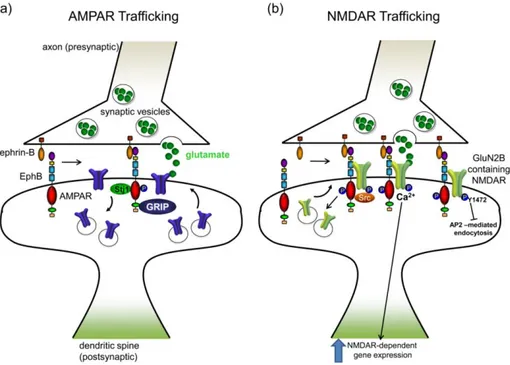
EphB2 receptor in kinase activity manner can interact, clusterize and endocytositade others receptors which in turn are important in excitatory synapsis, such as NMDA and AMPA neurotransmitter receptors11. Likewise, ephrin B by PDZ-binding domain can interact with post-synaptic PDZ domain-containing proteins 10 allowing bind syntenin-1 in synaptogenesis 17 and growth-factor-receptor-bound protein 4 (Grb4) in cytoskeletal remodeling. 7
EphB receptors and ephrinB ligands are expressed at synaptic terminals of hippocampal, cortical neurons, and presynaptic astrocyte 18. During synapse formation in cortical and hippocampal neurons, forward signaling of presynaptic ephrinB2 trigger activation of postsynaptic EphB2 receptors which directly interacts with NMDA receptor, activating Ca2+ influx and gene expression promoting dendritic spine morphogenesis 10,11,15,18
Forward signaling ephrinB1/EphB also triggers the activation of Rho family GTPases among them Cdc 42, Rac1, Kalirin (Kalirin-5 and Kalirin-7), intersectin and Tiam 11. Kalirin and Tiam promote the exchange of
16 GDP for GTP and thereby activate Rac1 activity which promotes actin cytoskeleton rearrangement (FIGURE 5) 10. The activation of Rac1 also acts on PAK (p21 activated kinase) promoting spine formation, maturation and synapse formation (Figure 5) 10,24,38 ,as well as, the ubiquitination and degradation of Rho-GEF Ephexin5 promotes synapse formation, however, the reduction of RhoA activity do not show effects on spine morphology 23 indicating a negative regulation in excitatory synapse 7.
Furthermore, EphB forward signaling can phosphorylate focal adhesion kinase FAK 24 and heparin sulphate proteoglycan syndecan-2 38, which interacts with the synaptic PDZ domain protein caskin and promotes spine maturation 24,25. Specifically, EphB1 receptor can form a complex with Caskin1, through the adaptor protein Nck by its tyrosine 594, to bind SH2 domain, while its SH3 domains may interact with paxillin and Nck-interacting kinase (NIK) participating in the regulation of actin cytoskeleton 29. In addition, EphB1 receptor has been considered as axonal adhesion molecule due to in forward and reverse signaling can regulate cell adhesion by controlling integrin β1 activation in oligodendrocytes (Figure 5) which have a positive effect on the ability of the oligodendrocytes to extend myelin sheets 39.
Reverse signaling has also been involved on axon pruning, synapse formation and dendritic spine morphogenesis in the developing mouse hippocampus through the phosphorylation of adaptor protein Grb4 23,40 as well as in morphological and functional maturation of developing retinotectal synapses in the Xenopus optic tectum due to the activation of ephrin-B by postsynaptic EphB2 receptor 11.
Presynaptic ephrin B1 and ephrinB2 with postsynaptic EphB2 are also necessary in synaptogenesis, in fact reverse signaling allow the formation of normal synapsis due to the interaction with syntenin-1 17 whereas, Ephrin-B3 interact with GRIP1, PICK1 and syntenin-1 proteins regulating the synapse and spine formation in the hippocampus and cortex (Figure 5) 17.
Other important process, in earlier nervous system, in which ephrinB/EphB system has been involved is axon guidance. EphrinB and EphB have the capacity of guide long-distance axonal growth cones. Different studies in mice have demonstrated that the ubiquitous replacement of the intracellular domain of EphB1 and EphB2 by β-galactosidase, mirrors the phenotype of loss of function of the gene encoding ephrin-B1 in the intervening tissues, such as the ventral telencephalon, indicating that the establishment of neural
17 connections between the thalamus and the cortex depends on forward signaling 15. Furthermore, EphB1 expression is sufficient for axon repulsion and for the ipsilateral trajectory of retinal axons. 15
Finally, is important to review the involvement of EphB receptor and ephrinB ligands in other processes in early nervous system. For example, in neuronal migration the lack of ephrinB ligands, in Cajal–Retzius cells, can elicit defects in this process 15 as well as in neurogenesis EphB1 and ephrin-B3 play an important role in migration of neural progenitors in the hippocampus. In fact the lack of EphB1 significantly reduces the number of neural progenitors in the hippocampus, prevents migration and organization of neural progenitors, and affects other aspects of neurogenesis such as polarity, cell positioning and proliferation 35,41. Furthermore, EphB receptor can activate β- catenin, independently of Wnt signal, leading to the upregulation of proneural gene expression, allowing Ephs to influence transcriptional control of cell fate 27. During embryogenesis ephrinBs allowing cell division and cellular differentiation by the alternatively interaction with growth factor receptors, such as the fibroblast growth factor receptor (FGFR), platelet-derived growth factor receptor (PDFGR), epidermal growth factor receptor (EGFR), and the TIE2 receptor 7.
Figure 5. EphB and ephrin-B modulate spine and synapse formation. EphB forward signaling via PAK promotes filopodial
motility. EphB activation by ephrinB-Fc recruits the Rac1 GEF Tiam1 to EphB complexes containing NMDARs. EphBs also modulate the activity of the Rho family GTPases by activating the Rho-GEFs kalirin and intersectin, and signal via FAK to activate RhoA. EphB1 forward signaling involves recruitment of Cdk5 and activation of ephexin1 and RhoA, which regulates actin reorganization. EphrinB reverse signaling may lead to presynaptic differentiation, possibly invo lving the Grb4/GIT1
18
EphB/EphrinB system in adult SNC
EphB and ephrinB persist in presynaptic and postsynaptic sites in the adult brain principally in regions where neuronal circuits continue to be remodeled in response to environmental changes 11 such as hippocampus and cerebral cortex 31, specifically, in pyramidal neurons 8,18, primary sensory neurons, spinal dorsal horn neurons 42 and astrocytes 36.
The principal roles of EphB and ephrinB in the adult brain are the regulation of synaptic transmission and morphology, which in turn also are controlled by synaptic glutamate receptor 8,43, synaptic plasticity 31,36 and in response to nerve injury 31.
Forward signaling has a crucial role in synapse formation and synaptic plasticity in the hippocampus, in fact, changes in synaptic transmission and neuronal morphology are involved in the modulation of learning 11,31 and in memory formation 36.
Long-term potentiation (LTP), long-term depression (LTD) and depotentiation are phenomena underlaying synaptic plasticityinvolved in EphB/ephrinB system 31.
Thus, forward signaling between ephrinB3 31 and EphB1 42, or EphB2 receptors 13 has been involved in long-term potentiation (LTP). The activation of EphB2 can in turn trigger the modulation of NMDA-receptor-mediated calcium influx via a Src- family kinase pathway having direct impact on synaptic transmission 17,30, in fact, in the mature brain EphBs are required for normal levels of synaptic NMDA receptors 37.
In addition, the activation of mediators as RAC1 guanine exchange factor, T lymphoma invasion metastasis-inducing protein 1 (TIAM1) 31, Grb4, Pick1, syntenin 27, MAP kinase and Src family kinases 25 is indispensable for the formation of dendritic spines and establishment of functional synaptic plasticity 27,35.
On the other hand, the structure of ephrinsB play an important role in synaptic plasticity 27 since only PDZ domain-binding site of ephrin-B2 is required in LTP, LTD, depotentiation 11.
Reverse signaling has also been involved in memory formation due to its importance in synaptic transmission, plasticity and neuronal morphogenesis 24,36. The activation of ephrinB signaling, by
EphB2-19 Fc in Xenopus retinotectal system, leads to an enhanced transmission by an early increase in the presynaptic transmitter release and a delayed postsynaptic glutamate responses 44. Other studies in cultured hippocampal neurons have demostrated that the activation of ephrinB by EphB2-Fc promotes spine maturation leading to higher proportion of spines with larger spine heads 10. On the contraty has been also observed that mutation in ephrinB1, either due to the lack the cytoplasmic domain or for mutation of the six intracellular tyrosine residues, leads to formation of immature filopodia indicating that reverse signaling and tyrosine phosphorylation are fundamental in neuronal processes 10,28. Furthermore, upon the activation of ephrinB the activation of downstream pathways such as G-protein-coupled receptor kinase-interacting protein 1 (GIT 1) can regulate synapse formation by the activation of Grb4, in fact, ephrinB/Grb4/ GIT1 complex is necessary in spine morphogenesis and synapse formation 10.
20
EphB and EphrinB are involved several pathological processes
Despite that EphA and B receptors have been studied in cancer, inflammation and in neurological deseases, during these chapter we will focus only in the role of EphB receptors and ephrinB ligands in these pathologies.
EphB/EphrinB system in cancer
Type A and B of receptors and ligands normally act as tumor suppressors, however, in tumor cell arepotentially converted into oncogenic proteins 3 being involved in a wide variety of human cancers 45. They can affect the growth, migration, and invasion of cancer cells in vitro as well as tumor growth, invasiveness, angiogenesis, and metastasis in vivo 36.
Depending on the activated signaling pathway have been involved in the differents tumorigenic process 11 for example, Eph forward signaling promotes cell segregation and is considered as tumor suppressors since can inhibit oncogenic signaling pathways, such as the HRAS-Erk, PI3 kinase-Akt and Abl-Crk pathways 23,46, in colorectal, breast, prostate, and skin cancer cells both in vitro and in vivo 11, whereas reverse signaling is often tumor promoting, driving neoangiogenesis and invasion 31
Abnormal expression of EphB1 receptor has been detected in metastatic gastric carcinoma, colorectal cancer, ovary serous carcinoma and renal cell carcinoma 47–49 ,as well as, in different brain tumors, principally the up-regulation of EphB1expression in medulloblastomas whereas the down-regultion in gliomas 45,47,50.
Interestingly, depending on the cellular context and whether EphB1 receptor forward signaling is dependent or indenpendent of ephrins ligands, EphB1 can act as tumor suppressor or tumor promoter 47.
Alterations in the expression of EphB1 have been involved in 1.49% of the cases of glioma 35. In glioblastoma multiforme (GBM) patients with higher EphB1 expression levels showed longer survival rates 47. Therefore, the overexpression and ligand-dependent EphB1 signaling can inhibit cell migration and invasion upon ephrin-B2 ligand stimulation 35 which is considered as a negative regulator for glioma cell motility and invasion that in turns is a positive predictor for glioma patient survival (Figure 6) 47.
21 Forward signaling ephrinB2/EphB1 activates several adaptor proteins that promotes cell migration, among them EphB1 recruits adaptor protein Grb2, p52Shc and Src whereby can activate MAPK/ERK regulating events involved in cell motility 51, as well as, can induce tyrosine phosphorylation of paxillin and form complex with Grb2, Grb7, integrins, Nck, paxillin, and FAK proteins in a c-Src-dependent manner 35 In medulloblastoma EphB1 expression have an important role in radiation resistance and in cell migration. EphB1 knockdown Daoy cells have demonstrated that the lack of EphB1 receptor reduces cell growth, viability and migration, as well as, the expression of important cell cycle regulators as cyclinE, besides increases the percentage of cells in G1 phase and enhancements radiation sensitivity not only in culture cells but also in a genetically engineered mouse medulloblastoma model. Therefore EphB1 has been considered as therapeutic target 45.
Furthermore, EphB1can interact with other tyrosine kinases receptors as epidermal growth factor receptor (EGFR), contributing to the metastatic behavior of medulloblastoma cells as well as interacts with β1-integrin producing cell migration and chemotaxis via stimulation of Src activity 45,51,52
Figure 6. EphB1/ephrins signaling in brain tumors. EphB1 upon the stimulation of ephrinB2 activate three signaling pathways. In U87 and U251 cell lines, forward signaling ephrinB2/EphB1 supressess cell motility. Whereas, EphB1 expression in
22
EphB/EphrinB system in inflammation
Eph/ephrin interaction has been involved in the movement of inflammatory cells to the site of infection or tissue injury 1 as well as in the disruption of endothelial–epithelial barriers and adhesion of leukocytes to endothelial cells 31.
Principally, EphB/ephrin B system has been involved in the regulation of T cell maturation and proinflammatory gene expression 23,53. For instance, an increase in ephrin-B1/EphB1 expression in peripheral blood lymphocytes has observed in patients with rheumatoid arthritis, as well as, EphB1/2 and ephrin-B1/2 are up-regulated in the intestinal cells of patients with Crohn’s disease 1.
Furthermore, in animal models of rheumatoid arthritis it has observed that the activation of EphB1 receptor by EphrinB1–Fc fusion protein results in an increase of production of TNF-α from lymphocytes and stimulation of the release of interleukin-6 from synoviocytes, as well as, an increase in the number of peripheral blood lymphocytes migration into the joint enhancing lymphocyte migration 1,31.
On the other hands, in intestinal epithelial cells the stimulation of ephrin-B1 or B2, by EphB1-Fc, induced pro-inflammatory genes such as cyclooxygenase-2 and monocyte chemotactic protein-1 1, as well as in inflammatory bowel disease (IBD) this stimulation is considered as a novel protective mechanism that could promote intestinal epithelial wound healing 54
EphB/EphrinB system in neuropathologies
1.3.3.1 Pain
EphBs receptors and ephrinBs ligands are expressed at lower levels in mature nervous system (Figure 7a), but after traumatic or ischemic nervous system injury 1,26,55 and in prolonged MOR activation 56is produced an upregulation in neurons, surrounding astrocytes and oligodendrocytes 13, affecting synaptic remodeling and plasticity13, axon sprouting and repair processes 32. Therefore, have been directly involved in the mediation of spinal nociceptive information and central sensitization 57, in dorsal root ganglia and in spinal dorsal horn neurons 8,58, contributing in sensory abnormalities in persistent pain states 56 and in the formation of an unfavorable environment for regeneration and functional recovery after CNS injury 15.
23 The mechanisms by which EphB/ephrinB system have been involved in the induction and persistence of pain could be principally due to changes in sensory neuron excitability which is produced by an increase in the firing of small-diameter sensory neurons that communicate noxious information to the dorsal horn of spinal cord and an increase in spinal synaptic plasticity 8,59 after inflammation or injury. Once that EphB/ephrinB system is activated can induced peripheral sensitization, which is manifests for hyperalgesia, and allodynia that later could produce central sensitization, which is a major cellular mechanism that converte acute nociceptive injury in chronic pain states 59.
Different molecules contribute to the development of peripheral sensitization inthe EphB/ephrinB system activation and the related downstream59 , among them, MAPKs ,such as, ERK1/2, p38, JNK 59 and p-ERK5, p-CREB a nuclear transcription factor 60, phosphatidy linositol 3-kinase (PI3K) 61, protein kinase A (PKA) 62, protein kinase Cγ (PKCγ) 60 and calpain-1 and caspase-3 63.
On the other hand, the activation of other kind of receptors have been related with EphB-mediated pain states principally N-methyl-D-aspartate receptor (NMDR) and toll-like receptor 4 (TLR4) 9,37.
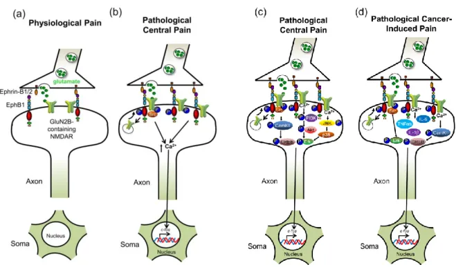
Several studies have demonstrated that the induction of hypersensitivity and pain can occur by enhancing EphB-dependent effects on NMDAR function 37. Forward signaling of EphB1 and EphB2 receptor have been considered as principal regulator of pain processes via NMDA receptors 37 in particular NR2B subunit (Figure 7b)43,64. EphrinB can phosphorylate a single tyrosine (p*Y504) in a highly conserved region of the fibronectin type III (FN3) domain of EphB2 receptor, moduling the EphB-NMDAR interaction in cortical and spinal cord neurons, enhancing, not only, NMDAR localization 37 but also sensitization of nociception in spinal cord synaptic efficiency which is modulated in an NMDR-dependent manner contributing to chronic neuropathic and inflammatory pain states 37 mediated via a MAPK–dependent mechanism 1. While Toll-like receptor 4 (TLR4) contribute with EphrinB/EphB system in the induction of cancer pain in spinal cord 9.
Although, all EphB receptor are involved in pain processes, recently EphB1/ephrinB1 system has been considered as a principal complex involved in acute inflammatory pain 56, neuropathic pain 58,64,65, cancer pain , diabetic pain 66, opiate dependence and tolerance 67, and chronic ocular hypertension 68.
24 In a different studies, have been showed that peripheral nerve injury produced thermal hyperalgesia in wild-type (EphB1+/+) but not in EphB1 receptor homozygous knockout (EphB1-/-) and heterozygous knockdown (EphB1+/-) mice, as well as, the hyperexcitability in dorsal root ganglion neurons was prevented in EphB1-/- and EphB1+/- mice 69. In addition, in chronically morphine-treated mice, the behavioral signs due to morphine administration were diminished in EphB1-/- mice; concluding that EphB1 receptors are required in physical dependence and tolerance of opiates 69, in the thermal and mechanical hyperalgesia and spontaneous pain in a variety of pain models 43,as well, as in microglial activation and in the uregulation of keys proteins involved in pain such as pNR1, pNR2B, pSrc (Tyr418), pERK1/2, p-p38, p-JNK, pCaMKII, pCREB, pNR2B, c-fos 59,70,71.
In CCI and formalin pain models have observed that EphrinB1-Fc activate forward signaling of EphBs receptors 59. The activation of EphBs by EphrinB1-Fc is fundamental during hyperalgesia and allodynia, since induce a dose-dependent increase of spinal Fos protein expression 61 as well as an increase expression of NMDA receptor and up-levels of peripheral and spinal phospho-MAPKs (Figure 7c) 59,71. Furthermore, the precense of sensitizers molecules as TNFα in injury and/or inflammatory tissue could induce peripheral sensitization, which is an important neuronal mechanism underlying primary hyperalgesia at the site of injury or inflammation 59.
Forward signaling between EphB1 postsynaptic receptor expressed in neurons of the spinal cord and ephrinBs presynaptic expressed in pain sensory neurons 32 has demonstrated to be critical in the development of bone cancer pain (Figure 7d) 70. In rats, tumor cell implantation (TCI) or spinal administration of ephrinB2-Fc (an endogenous ligand) can produce bone cancer–related thermal hyperalgesia, mechanical allodynia, and bone destruction, as well as, an increment in the expression of TLR4 and EphB1 receptors and IL-1β and TNF-α cytokines in astrocytes and microglial cells 9. In addition, ephrinB2-Fc increase the phosphorylation of NR1 and NR2B receptors wich in turn also depend on the activation of EphB1 70. However, spinal administration of EphB2-Fc can relieve bone cancer pain and prevents or reverses pain behaviors, disminishing the activation of astrocytes and microglial cells; TLR4, EphB1 9, NR1 and NR2B receptors, and c-Fos protein, Src protein within the N-methyl-D-aspartate receptor
25 complex, and the subsequent Ca2+-dependent signals 70, and also increasing the activity of matrix metalloproteinase (MMP)-2/9 9.
Concluding that spinal blocking or targeted mutation of EphB1 receptor could contribute in the treating bone cancer pain and TLR4 also could be a potential target for preventing or reversing bone cancer pain mediated by ephrinB-EphB receptor signaling 9
In Diabetic neuropathic pain (DNP), streptozotocin (STZ) or alloxan causes significant activation of EphB1 receptor in the spinal cord, as well as, activation of astrocytes, microglial cells and IL-1β and TNF-α which are important in the pathogenesis of DNP 66. However, spinal blocking EphB1 receptor activation can relieve DNP in diabetes induced rats by intraperitoneal injection of streptozotocin (STZ) inhibiting mechanical allodynia and the activation of the astrocytes, IL-1β and TNF-α in the spinal cord 66, concluding that EphB1 receptor activation in the spinal cord is critical to the maintenance, but not in the induction of diabetic pain 66.
Reverse signaling of EphB1 /ephrinB2 in rats can also contribute to retinal ganglion cells (RGC) apoptosis with chronic ocular hypertension (COH) in glial and neuronal elements in the retina which was accompanied by increased protein levels of phosphorylated Src and GluA2. High expression of EphrinB1 and EphB1 in monkeys have also been observed in mild-to-moderate glaucoma, as well as, in cultured astrocytes obtained from human glaucamatous patients, concluding that the upregulation of ephrinB/EphB signaling in the retina may be common in experimental and clinical glaucoma considering that attenuation of EphB/ ephrin B reverse signaling could be an appropriate way for preven the loss of RCGs in glaucoma 68.
On the other hand, the Opiates drugs are used in the treatment of moderate-to-severe post-operative and chronic pain, however, when opiates are administered simultaneously can activate the nocuous mechanism of a sensitization process causing pain hypersensitivity 57. The involvement of EphB1 receptors in these processes have been observed in animal models which the scalating morphine treatment 70 and continuing infusion of remifentanil 57 significantly up-regulates expression of EphB1 receptor. In a rat hindpaw incisional model ephrin B1 ligands in spinal cord produce thermal hyperalgesia and mechanical allodynia
26 57,70, whereas intrathecal administration of EphB2-Fc, used as a blocking reagent, revert the behavioral symptoms and neurochemical signs associated with chronic opiates treatment 70, as well as, reduce the levels of pNR2B, pERK and pCREB in the SC 67,69.
Figure 7. Involvement of ephrinB/EphB in neuropathic pain. a) low expression of ephrin-B and EphB1 in physiological pain conditions. b) In pathological central pain, both ephrin-Bs and EphB1 are upregulated. Activation of EphBs leads to recruitment and activation of Src and consequently NMDARs phosphorylation with an increasing of calcium influx leading to c-fos and CREB gene transcription. c) Pathological peripheral pain, shares with pathological central pain the activation of EphB and NMDR receptors, then the activation of other three signaling pathways caractherize this pain model. Src phosphorylates CamKII, which phosphorylates CREB causing nuclear translocation and Cyclic AMP Response Element (CRE) gene transcription. The activation of PI3K signaling pathway, which phosphorylates Akt and ERK proteins and consequently translocates to the nucleus to activate c-fos gene transcription; and JNK phosphorylated and activated can phosphorylate p-38 and converges to activate ERK. d) in Pathological cancer-induced pain shares the principal caractheristic of pathological central and peripheral pain. However, in cancer pain the phosphorylation of ERK directly by src kinase to activate gene transcription of c-fos and the up- regulation of
inflammatory cytokines as TNFα, IL-6, and IL-1β can lead to hyperalgesia, and hyperexcitability of nerve afferents 8.
1.3.3.2 Neurodegenerative diseases
Aberrant synaptic activity is considered as a major pathological hallmark in neurodegenerative and psychiatric disorders 4. Eph receptor and ephrin ligand are present in the development of the central nervous system and in the adult brain in regions of continued development as hippocampus and amygdala 31
27 regulating synapse formation maintenance and plasticity 4 which are important in learning, memory formation and consequently in normal cognitive function.
Since, Ephs and ephrins play an important role in synaptic efficacy, regulating presynaptic transmitter release, postsynaptic glutamate receptor conductance and trafficking, synaptic glutamate reuptake, and dendritic spine morphogenesis 5,10,17, their deregulation have been directly involved in aberrant synaptic functions associated with cognitive impairment and in long-term memory formation, which can produce alterations of synaptic efficacy modifying neural transmission and morphology 36, producing brain disorders and diseases with memory impairment symptoms including trauma, stroke, epilepsy, psychiatric, anxiety and neurodegenerative diseases as Alzheimer disease, Parkinson disease and Amyotrophic lateral sclerosis (ALS) 18.
Furthermore, various Eph receptors and their ligands have been involved in the excitatory synapses in the hippocampus, including EphA4, EphB1, EphB2, EphB3, ephrinB2 and ephrinB3 4. EphB receptors can coordinate synapses due to a multiple upstream molecules, such as, Rho-GTPases GEFs (guanine-nucleotide exchanging factor) and GAPs (GTPase-activating proteins) 4. In fact, they regulate actin cytoskeleton network through activation of GEF kalirin, intersectin or Tiam1-mediated activation of the Rho GTPases Rac1 and Cdc42 4. In addition, EphB2 can regulate the function of post- synaptic neurotransmitter receptors as NMDAR by the formation of co-clustering of NMDA receptor and specific postsynaptic proteins including calcium/calmodulin-dependent protein kinase II (CaMKII) and Grb1 4. Severe or sustained stress can result in changes of synaptic plasticity which can lead to behavioral changes associated with fear and anxiety disorders. The effects of EphB2 in NMDAR activation allow to think that EphB receptor plays an important role in bipolar disorder and cognitive functions 8.
Deficiency in serine protease neuropsin, an important protein expressed in amygdala and hippocampus, has been also involved in bipolar disorder and cognitive functions 72. EphB2 and neuropsin are upregulated after stress in amygdala which results in cleavage of the EphB2 ectodomain 73. Neuropsin-dependent cleavage of EphB2 increases the dynamics of the EphB-NMDAR interaction, likely explaining the changes
28 in NMDAR currents observed in neuropsin null mice 73 suggesting that targeting neuropsin-dependent cleavage of EphB2 is a potential strategy for treating stress-related and anxiety disorders 8.
Alzheimer's disease (AD) is considering a devastating disease in which is produced a permanent loss of memory and other cognitive functions causing progressive loss of synapses and neurons 32. Senile plaques are a hallmark of AD, which are produced by proteolysis of amyloid precursor protein by the presenilin/γ-secretase intramembrane protease complex, generating the cytotoxic β amyloid (Aβ) peptides 32. Soluble Aβ oligomers can decrease NMDA receptors affecting their equilibrium and activity at synaptic sites producing impairs synaptic plasticity and glutamatergic transmission (Figure 8) 74.
In mice models have demonstrated that beta-amyloid (Aβ) directly binds to the fibronectin repeats domains of EphB2 affecting its function and increasing its proteasomal degradation 74. The reduction of hippocampal EphB2 levels produce memory impairment in early stages of AD due to a decreases of NMDA surface expression with a defective neuronal activation and reduction of LTP (Figure 8) 25. However, since EphB2 expression levels can regulate the amount of NMDARs at synapses, the overexpression of EphB2 acts as a neuroprotector in hippocampal 74.
The involvement of Ephs an ephrins in neurogenesis suggests that could be used in the treatment of Parkinson’s disease 75 which is other type of neurodegenerative disorder characterized by a deep loss of dopaminergic neurons in the Substantia Nigra pars compacta (SNc) accompanied by filamentous protein inclusions termed Lewy bodies (LBs) in the surviving neurons 76. Jing and collaborators have demonstrated that the activation of EphA receptor in lateral ventricle by soluble form of the ephrin-A1 ligand (ephrin-A1 Fc) could promote regeneration of the brain dopaminergic neurons 75
The involvement of EphB1/ ephrinB1 system in Amyotrophic lateral sclerosis (ALS), a fatal disease characterized by a progressive degeneration of motor neurons that produce the breakdown of neuron-glia communication and neuronal dysfunction and death 77, is due to the capacity of EphB1 receptor to produce the activation of protective phenotype of astrocytes in human and mouse ALS models77. EphB1 receptor is up-regulated in injured motor neurons, where, by the activation of ephrinB1 can induce STAT3 (activator
29 of transcription-3) activation which can regulate astrocyte activation 78, producing astrocyte transformation and consequently protective, anti-inflammatory or immunomodulatory pathways 77.
Figure 8. EphBs in AD. The EphB2-Aβ interaction inhibits receptor activation and causes internalization and degradation of both EphBs and NMDARs. Degradation of EphB receptors inhibits their ability to retain NMDARs on the membrane potentially though the EphB-NMDAR interaction. Fewer NMDARs on the cell surface leads to decreased calcium influx and none of the changes in
30
Interactions between Eph/ephrin system with other receptors
The activation of Eph and ephrin proteins can produce a cross-regulation with other communication pathways producing molecular, functional, and genetic interactions with different cell surface signaling pathways controlling broaden range of function in vivo as cell survival, migration and differentiation 3,31,79. Eph/ephrins can produce crosstalk with different cell surface receptor (tyrosine kinases receptors and g-coupled receptor), adhesion molecules, (integrins, claudins and cadherins) and ionotropic receptor (NMDA and AMPA receptors) (Figure 9)79.
Despite, this chapter will be focalized principally in the interaction of Eph/ephrin system with cell surface receptor as ionotropic, tyrosine and g-coupled receptor and their different downstream molecules that are activated, I have believed interesting also review the interactions formed between Eph/ephrin system with other proteins.
Eph receptor can attenuate Ras-MAP- Kinase signaling downstream of other receptors, such as, integrins 80,81 affecting integrin-mediated cell communication with the extracellular environment 11. Endogenous ephrin-B2 expressed in melanoma cells is associated with β1-integrins and their interaction can promote cell adhesion and migration, suggesting a role in tumor progression 11.
PI3K–AKT signaling has been involved in Eph kinase-independent activities and in cell migration in vitro and in vivo. However, in phosphatase and tensin homologue (PTEN)-deficient tumour cells, in which AKT and mTOR are active, the expression of EphA2 can suppres AKT phosphorylation and cell migration 31 Crosstalk between EphB/ephrin-B and Wnt signaling has also been reported. Both EphB receptors and B-type ephrins produce their signal through components of the noncanonical Wnt pathway 11. This interaction produce endocytic removal of EphB receptors from the cell surface, whereas canonical Wnt signaling increase EphB transcripts and decrease ephrin-B transcripts 11, for instance, in intestinal crypts an increasing WNT expression can induce EphB2 and EphB3 expression while represses ephrin B1 and ephrin B2 expression resulting cell–cell repulsion 31.
31 Eph receptor expression and ephrin-dependent activation, in cell-surface localization, can also be regulated by E-cadherin. E-cadherin can be drive to the cell surface by EphB signaling thus promoting the formation of epithelial adherents junctions and enabling EphB/ephrin-B-dependent cell sorting 11 which is important during vascular and neural remodeling 12. In addition adherent junction can be disturb when EphB-ephrin-B binding is inhibited11.
Croostalk between EphA2 or ephrin-B1 with claudins has been implicated in the regulation of cell adhesion and intercellular permeability. Furthermore, claudins can activate ephrin-B1 tyrosine phosphorylation independently of EphB receptors 11.
Besides, EphBs may also interact with acetylcholine receptors (nAChRs) regulating neurotransmitter receptor function 8, in addition, since EphB2 can interact with TNF-α could be considered as neuroprotector 82.