Alma Mater Studiorum – Università di Bologna
DOTTORATO DI RICERCA IN
Scienze Medico-Chirurgiche Gastroenterologiche e dei
Trapianti
Ciclo XXII
Settore Concorsuale di afferenza: 06/E01 Settore Scientifico disciplinare: MED 22
TITOLO TESI
FEASIBILITY AND DIAGNOSTIC EFFECTIVENESS OF NEW CAPSULE ENDOSCOPY TECHNIQUES
Presentata da: Flaminia Cavallaro
Coordinatore Dottorato Relatore
Prof. Andrea Stella prof. Andrea Stella
Correlatore
Prof. Maurizio Vecchi
2 TABLE OF CONTENTS
1. Introduction
2. Patients and methods 3. Results
4. Discussion 5. References 6. Figures 7. Tables
3 INTRODUCTION
The small bowel has been considered for a long time technically difficult to evaluate because of its length, location and tortuosity. Since its approval by FDA in 2001, capsule endoscopy has revolutionized the study of small bowel and its use has been rapidly expanding [1-5]. Several systems have been developed for this purpose. One of the main limitations to its diffusion has been the relatively high cost and thus a questionable cost-effectiveness ratio. More recently, a new videocapsule system (OMOM CE) has been developed in China by Jinshan Science & Technology Company (Chongqing, China) [6,7]and has obtained the CE mark for its marketing in Europe. Its cost is approximately half that of other capsule systems. However, there are few studies addressing the clinical experience with this new videocapsule system and none of them has been performed in the western world.
Aim of the present study was thus to assess the feasibility, safety and diagnostic yield of the OMOM CE in different clinical settings related to possible small bowel disease conditions.
CapsoCam SV1 is a newly introduced device for small-bowel (SB) capsule endoscopy (CE) with wire-free technology, a long-lasting battery life, and 12–20 frames per second captured by four high-resolution cameras located on the capsule sides and facing the four quadrants of the digestive wall. Initial experiences have shown high operative performances, suggesting at least an equal clinical efficacy compared to other frontal view capsules.
4
spontaneous study to assess the performance of CapsoCam SV1 in real life clinical practice.
5 PATIENTS AND METHODS
Patients
A total of 118 patients (61 men, 57 women, mean age 53 years, range 18-86) with suspected small bowel disease underwent OMOM CE in 3 Gastroenterology Units (Gastroenterology & Digestive Endoscopy Unit, IRCCS Policlinico San Donato - University of Milan; Surgery & Digestive Endoscopy Unit, V. Monaldi Hospital, Naples; Santa Barbara Hospital, Iglesias).
Indications to the exam consisted of the following: obscure gastrointestinal bleeding, known or suspected Crohn’s disease, suspected small bowel tumor, familial adenomatous polyposis. The numbers of patients studied for each diagnostic subgroup are reported in table 1. All patients had previously undergone upper and lower gastrointestinal endoscopy. Most of them had also undergone other investigations, such as small bowel follow through, enteroclysis, abdominal computed tomography and magnetic resonance.
In the second part of the study, 50 patients with suspected small bowel disease underwent OMOM CE in 4 Gastroenterology Units.
6 Methods
The OMOM capsule endoscopy (Jinshan Science & Technology Company, Chongqing, China) was used in all patients. This system is made up of three parts: a disposable capsule, an image recorder jacket and an image workstation. The capsule measures 12.5x27.5 mm andweighs < 6 gr. Image features include a 150° field of view and a resolution of 0.1 mm. The capsule has a battery life of approximately 7-9 hours. The pictures are generally taken at a rate of two frames per second, but the rate can be adjusted to needs during the exam, a unique feature of this system. There are 14 receiver elements placed close to the abdomen and to the waist in the recorder jacket. The capsule transmits the acquired images via a digital radio frequency communication channel to the recorder. A portable real-time monitor device allows the endoscopist to follow the progression of the capsule and to send possible commands to the OMOM system: in order to modify rate of frame (2 frames per second, 1 fps or 0.5 fps), flash intensity, conditions of capsule (sleep or awake).
The recorder is later connected to the workstation, in which the images are downloaded and processed.
The main differences between the OMOM capsule endoscopy and the other currently available systems of capsule endoscopy are a slightly bigger size and the use of an antenna-carrying jacket by the OMOM system. Also, further features of the OMOM system are the possibility of modulating frame recording speed and a significantly lower cost. The main features of the system are shown in figures 1-3.
All subjects followed a clear semi-liquid diet on the day before and 2L of PEG (polyethylene glycol solution) in the afternoon before the procedure.
7
out of the stomach into the small bowel. If the capsule had not passed the pylorus after 60 minutes, metoclopramide 10 mg was administered intravenously.
The acquired images were reviewed by two expert gastroenterologists and all videos were classified as: diagnostic, suspicious or negative.
In the second part of the study, patients with suspected SB disorders were consecutively enrolled in 3 Italian centers during 2014 and underwent to CapsoCam SV1 capsule examination. Two expert readers performed a centralized post-hoc revision of those video recordings with undefined findings. The P0/P1/P2 classification proposed by Saurin et al. [50] for obscure gastro-intestinal bleeding (OGIB) was used to assess the clinical relevance of all findings.
8 Study Flow Diagram
Statistical analysis
Quantitative variables were expressed as mean ± SD values. Fisher’s test was used to compare occult OGIB and overt OGIB diagnostic yield.
9 RESULTS
All patients ingested the OMOM capsule very easily and no complications were observed.
All data analyzed were normally distributed. The recording time was 420 to 580 minutes (mean time 514 min, SD 39). Surprisingly, the mean pyloric transit time -defined as the recorded time of the first image of duodenum- was 78 minutes (SD 44) in patients who received metoclopramide and 27 minutes (SD 16) in those who did not receive metoclopramide.
The mean small bowel transit time –defined as the time from the first duodenal image to the time of the first cecal image for patients in whom the capsule reached the cecum - was 241 minutes (SD 123) in patients who received metoclopramide and 235 minutes (SD 73) in those who did not receive metoclopramide. Patients in whom the capsule did not reach the cecum were excluded from analysis of small bowel transit time.
Visualization of the entire small bowel was achieved in 114 patients (97%) and capsule retention without obstruction occurred in 1 patient (0.8%) due to a previously undiagnosed Crohn’s disease stricture at the terminal ileum in a patient with diarrhea but without obstructive symptoms. This patient underwent surgical treatment of the stricture and capsule recovery.
In 4 patients the capsule did not reach the cecum within the time of recording. In 3 of them the capsule failed to reach the cecum because of the impact with a lesion (a jejunal stricture due to a previously unknown Crohn’s disease in one patient, an ileal mass in one patient and a duodenal substenosis in the last one), and in 1 patient the only finding was angiodysplasia. In all cases, capsule was spontaneously expelled in 10 days in all patients except in 1 patient who
10
experienced a retention symptomless and underwent surgical treatment of a previously undiagnosed Crohn’s disease stricture and capsule recovery.
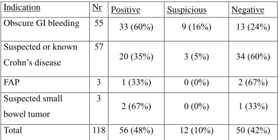
When only positive findings are considered, the overall diagnostic yield was 48%. When also suspicious findings are considered, diagnostic yield increases up to 58%. Diagnostic yield observed in the different subgroups are reported in table 2.
Diagnostic yield in patients with OGIB was 76% (when positive and suspicious findings are considered). It was greater than the yield in the non-OGIB subgroup, confirming that OGIB is the most important indication for capsule endoscopy. When patients were divided according to the type of bleeding (overt vs. occult), the diagnostic yield in OGIB was similar (p=0,7) as shown in table 3.
Angiodysplasia was the most common finding [8] and all these type of lesion were observed in the OGIB subgroup. Other findings included ulcers, erosions, polyps, active bleeding with no recognizable lesion, small-bowel tumors. All the findings are reported in table 4. The main findings are shown in figures 4-7.
Regarding the second part of the study on CapsoCam capsule system, fifty patients underwent SBCE (26 men; median age 67±17 years, range 16-86 years) with the following indications: 35 OGIB (27 occult), 8 iron-deficiency anemia, and 7 suspected Crohn’s disease. No procedure’s failure occurred.
The small bowel completion rate was 96%, the mean mucosal visibility and the video image quality were always scored as optimal. The Vater’s ampulla was identified in 52% with a mean of 2.5 frames for each positive case.
11
ulcerative-enteritis according to he further diagnosis of Crohn’s disease.
To perform the “per lesion” analysis of results, we excluded 125 SB lesions (28 P1, 97 P2) observed in a single woman with a pan-enteric Crohn’s disease; 201 findings were detected in the remaining recordings (26 P0, 81 P1, 94 P2). Most lesions were in the SB (168) and showed relevant clinical potential (14 P0, 63 P1, 91 P2 lesions). Interestingly, thirty lesions were detected in the upper-GI (11 P0, 9 P1, 10 P2). On a per patient analysis, 78% subjects had one or more findings (median=3) with a diagnostic yield of 70% (22% P1 and 48% P2 lesions).
3 patients (2 IDA, 1 OGIB-overt) underwent frontal-viewing SBCE with negative result. These patients underwent to a second look with Capsocam; in 2 patients we found P1 lesions, in 1 patient a P2 lesion (non-bleeding angiodisplasia).
12 DISCUSSION
Since the development of the first model of capsule endoscopy, continuing technological progress has further led to important technical advancement and thus capsule endoscopy has become a very important tool for the evaluation of suspected or known small bowel disease conditions.
However, in times of strict cost containment, the high cost of this procedure has represented the main limitation of its use. A relatively low-cost capsule endoscopy was recently developed and used in large patients populations in China. This is the first study to evaluate the overall performance of OMOM capsule endoscopy in a group of patients of caucasian origin.
In our hands, the system was easy to use and safe. Retention without obstruction occurred in 1 patient due to a previously undiagnosed Crohn’s disease stricture at the terminal ileum. Also in this case, however, retention was symptomless; the patient underwent surgical treatment of the stricture and capsule recovery.
Some features of the system also appear to be very useful in making the procedure more adjustable and tailored to specific clinical needs. In particular, the possibility of modulating flash intensity and the ON/OFF status of the capsule are unique of this system. While the first one might be useful in condition of low visibility (stomach, residues or bleed in the lumen), the second one may be helpful in saving battery life when a distal lesion has to be reached. Another important feature is the possibility of monitoring the pyloric transit in order to decide whether or not to use a prokinetic to fasten it up; in fact, it is known that a delayed gastric time is one of the most frequent causes of failure to reach the cecum [9,10].
In our series, OMOM capsule endoscopy reached the cecum in a very high proportion of patients (97%). This is a much higher figure than that usually
13
reported in the literature for all other capsule systems [11-13]. Although prokinetics may be useful in obtaining this result [10,14], this does not appear to be the case in our study. In fact, when we analyze patients for centers not utilizing either prokinetics injection or real-time viewer, an even higher proportion of reachment of cecum (37/38 patients, 97%) was observed, thus ruling out the possible role of these factors.
The OMOM capsule endoscopy is slightly bigger and heavier than other capsules and this might favor a relatively faster progression along the small bowel. Also, our patient series is characterized by a relatively large proportion of patients with clinical conditions, such as diarrhea and overt OGIB, possibly leading to accelerated peristalsis and a short small bowel transit time. Indeed, the small bowel transit time observed in the present study is quite short but substantially similar to those observed in the literature.
This might be explained considering that transit speed could be affected by multiple variables: completion rate, different definitions of the small bowel transit time used in the literature (many Authors have included patients in whom capsule enteroscopy has never reached the cecum), age of patients, comorbidities and drugs affecting bowel peristalsis (such as diabetes and neuropathies or opiods and prokinetics respectively), in-patients or out-patient, etc. Another possible explanation for this result is the relatively longer lifespan of OMOM capsule endoscopy batteries, allowing a longer duration of recording.
In any case, a more complete visualization of the small bowel could be of importance in obtaining even a higher diagnostic yield than that obtained by current devices.
In our series the diagnostic yield was assessed considering positive findings only (see table 2) and results are similar to those reported in literature with an overall
14
detection rate of 48%. More in details, it was 60% in OGIB and 35% in known or suspected Crohn’s disease. Consistently, as previously described in the literature, the diagnostic yield is rather variable according to the different indications for capsule endoscopy: about 50% for OGIB in a recent series [11,15-16], widely ranging between 33-70% for suspected Crohn’s disease [16-18].
The results of the present study are quite encouraging showing diagnostic figures at least similar to those reported in the literature, although the relatively small number of patients evaluated makes a statistical comparison unfeasible.
In conclusion, OMOM capsule endoscopy appears to be a practical, safe, easy to perform procedure, providing a similar diagnostic yield and an even superior time of observation of the small bowel.
Its significantly lower cost compared to all other systems marketed in Europe should also encourage its diffusion because of a better cost/effectiveness ratio.
The recently introduced CapsoCam SV1 appears to be a very dependable and effective system in the study of patients with SB disorders. In our series, preliminary results showed a high diagnostic yield of this new device and suggested an alternative not only use but also complementary to the capsules in frontal view, in order to further increase the diagnostic value of the survey capsular.
16 REFERENCES
1) Iddan G, Meron G, Glukhovsky A, et al. Wireless capsule endoscopy. Nature 2000; 405- 417.
2) Mishkin DS, Chuttani R, Croffie J, et al. Technology Assessment
Committee, American Society for Gastrointestinal Endoscopy. ASGE Technology
Status Evaluation Report: wireless capsule endoscopy. Gastrointest. Endosc.
2006 Apr; 63 (4):539-45.
3) Pennazio M, Eisen G, Goldfarb N, et al. ICCE consensus for obscure
gastrointestinale bleeding. Endoscopy 2005 ; 37:1046-1050.
4) Mergener K, Ponchon T, Gralnek I, et al. Literature review and
recommendations for clinical application of small-bowel capsule endoscopy,
based on a panel discussion by international experts. Consensus statements for
small-bowel capsule endoscopy, 2006/2007. Endoscopy. 2007 Oct; 39
(10):895-909.
5) Ladas SD, Triantafyllou K, Spada C, et al. ESGE Clinical Guidelines
Committee. European Society of Gastrointestinal Endoscopy (ESGE):
recommendations (2009) on clinical use of video capsule endoscopy to
investigate small-bowel, esophageal and colonic diseases. Endoscopy. 2010
17
6) Liao Z, Gao R, Li F, et al. Fields of applications, diagnostic yields and
findings if OMOM capsule endoscopy in 2400 Chinese patients, World. J.
Gastroenterol. 2010 June 7; 16 (21): 2669-2676.
7) Li C, Zhang B, Chen C, et al. OMOM capsule endoscopy in diagnosis of
small bowel disease, Journal of Zhejiang University Sci. B. 2008 9 (11):
857-862.
8) Pennazio M, Santucci R, Rondonotti E, et al. Outcome of patients with
obscure gastrointestinal bleeding after capsule endoscopy: report of 100
consecutive cases. Gastroenterology 2004; 126: 643-53.
9) Ogata H, Kumai K. Imaeda H, et al. Clinical impact of a newly developed
capsule endoscope: usefulness of a real-time image viewer for gastric transit
abnormality. J. Gastroenterol. 2008; 43: 86-192.
10) Postgate A, Tekkis P, Patterson N, et al. Are bowel purgatives and
prokinetics useful for small-bowel capsule endoscopy? A prospective
randomized controlled study. Gastrointestinal Endoscopy 2009 69, 6:1120-1128.
11) Liao Z, Gao R, Xu C, et al. Indications and detection, completion, and
retention rates of small-bowel capsule endoscopy: a systematic review;
18
12) Selby W. Complete small-bowel transit in patients undergoing capsule
endoscopy: determinating factors and improvement with metoclopramide.
Gastrointestinal Endoscopy 2005; 61:80-5.
13) Rondonotti E, Herrerias J, Pennazio M, et al. Complications, limitations,
and failures of capsule endoscopy: a review of 733 cases, Gastrointestinal
Endoscopy 2005; 62 (5):712-716.
14) Westerhof J, Weersma RK, Koornstra JJ. Risk factors for incomplete
small-bowel capsule endoscopy. Gastrointestinal Endoscopy 2009; 69: 74-80.
15) Estévez E, Gonzalez-Conde B, Vazquez-Iglesias JL, et al. Diagnostic
detection rate and clinical outcomes after capsule endoscopy in 100 consecutive
patients with obscure gastrointestinal bleeding. Eur. J. Gastroenterol. Hepatol.
2006; 18:881-8.
16) Rondonotti E, Villa F, Mulder CJ, et al. Small bowel capsule endoscopy
in 2007: indications, risk and limitations. World J. Gastroenterol. 2007;
14;13(46):6140-9.
17) Marmo R, Rotondano G, Piscopo R, et al. Capsule endoscopy versus enteroclysis in the detection of small bowel involvement in Crohn’s disease: a prospective trial. Clin Gastroenterol. Hepatol. 2005; 3:772-776.
18) Triester SL, Leighton JA, Leontiadis GI, et al. A meta-analysis of the
19
with non-stricturing small bowel Crohn’s disease. Am. J. Gastroenterol. 2006;
101: 954-964.
19) ASGE Technology Committee, Wang A, Banerjee S, Barth BA, Bhat YM, Chauhan S, Gottlieb KT, Konda V, Maple JT, Murad F, Pfau PR, Pleskow DK, Siddiqui UD, Tokar JL, Rodriguez SA. Wireless capsule endoscopy. Gastrointest Endosc. 2013;78(6):805-15.
20) ASGE Standards of Practice Committee, Fisher L, Lee Krinsky M, Anderson MA, Appalaneni V, Banerjee S, Ben-Menachem T, Cash BD, Decker GA, Fanelli RD, Friis C, Fukami N, Harrison ME, Ikenberry SO, Jain R, Jue T, Khan K, Maple JT, Strohmeyer L, Sharaf R, Dominitz JA. The role of endoscopy in the management of obscure GI bleeding. Gastrointest Endosc. 2010; 72(3):471-9.
21) Gerson LB. Capsule endoscopy and deep enteroscopy. Gastrointest Endosc. 2013; 78(3):439-43.
22) Ladas SD, Triantafyllou K, Spada C, Riccioni ME, Rey JF, Niv Y, Delvaux M, de Franchis R, Costamagna G; ESGE Clinical Guidelines Committee. European Society of Gastrointestinal Endoscopy (ESGE): recommendations (2009) on clinical use of video capsule endoscopy to investigate small-bowel, esophageal and colonic diseases. Endoscopy. 2010; 42(3):220-7.
20
23) Bourreille A, Ignjatovic A, Aabakken L, Loftus EV Jr, Eliakim R, Pennazio M, Bouhnik Y, Seidman E, Keuchel M, Albert JG, Ardizzone S, Bar-Meir S, Bisschops R, Despott EJ, Fortun PF, Heuschkel R, Kammermeier J, Leighton JA, Mantzaris GJ, Moussata D, Lo S, Paulsen V, Panés J, Radford-Smith G, Reinisch W, Rondonotti E, Sanders DS, Swoger JM, Yamamoto H, Travis S, Colombel JF, Van Gossum A; World Organisation of Digestive Endoscopy (OMED) and the European Crohn's and Colitis Organisation (ECCO). Role of small-bowel endoscopy in the management of patients with inflammatory bowel disease: an international OMED-ECCO consensus. Endoscopy. 2009;41(7):618-37.
24) Cotter J, Dias de Castro F, Moreira MJ, Rosa B. Tailoring Crohn's disease treatment: The impact of small bowel capsule endoscopy. J Crohns Colitis. 2014. 1;8(12):1610-5
25) Tontini GE, Vecchi M, Neurath MF, Neumann H. Advanced endoscopic imaging techniques in Crohn's disease. J Crohns Colitis. 2014 Apr 1;8(4):261-9 26) Mustafa BF, Samaan M, Langmead L, Khasraw M. Small bowel video capsule endoscopy: an overview. Expert Rev Gastroenterol Hepatol. 2013;7(4):323-9.
27) Koulaouzidis A, Rondonotti E, Karargyris A. Small-bowel capsule endoscopy: a ten-point contemporary review. World J Gastroenterol. 2013 Jun 28;19(24):3726-46.
21
28) Koulaouzidis A, Plevris JN. Detection of the ampulla of Vater in small bowel capsule endoscopy: experience with two different systems. J Dig Dis. 2012;13(12):621-7.
29) Rondonotti E, Marmo R, Petracchini M, de Franchis R, Pennazio M. The American Society for Gastrointestinal Endoscopy (ASGE) diagnostic algorithm for obscure gastrointestinal bleeding: eight burning questions from everyday clinical practice. Dig Liver Dis. 2013 Mar;45(3):179-85.
30) Koh SJ, Im JP, Kim JW, Kim BG, Lee KL, Kim SG, Kim JS, Jung HC. Long-term outcome in patients with obscure gastrointestinal bleeding after negative capsule endoscopy. World J Gastroenterol. 2013 14;19(10):1632-8.
31) Rondonotti E, Herrerias JM, Pennazio M, Caunedo A, Mascarenhas-Saraiva M, de Franchis R. Complications, limitations, and failures of capsule endoscopy: a review of 733 cases. Gastrointest Endosc. 2005; 62(5):712-6.
32) Cañas-Ventura A, Márquez L, Bessa X, Dedeu JM, Puigvehí M, Delgado-Aros S, Ibáñez IA, Seoane A, Barranco L, Bory F, Andreu M, González-Suárez B. Outcome in obscure gastrointestinal bleeding after capsule endoscopy. World J Gastrointest Endosc. 2013 Nov 16;5(11):551-8
33) Svarta S, Segal B, Law J, Sandhar A, Kwok R, Jacques A, Lakzadeh P, Enns R. Diagnostic yield of repeat capsule endoscopy and the effect on subsequent patient management. Can J Gastroenterol. 2010;24(7):441-4.
22
34) Viazis N, Papaxoinis K, Vlachogiannakos J, Efthymiou A, Theodoropoulos I, Karamanolis DG. Is there a role for second-look capsule endoscopy in patients with obscure GI bleeding after a nondiagnostic first test? Gastrointest Endosc. 2009; 69(4):850-6.
35) Kim HM, Kim YJ, Kim HJ, Park S, Park JY, Shin SK, Cheon JH, Lee SK, Lee YC, Park SW, Bang S, Song SY. A Pilot Study of Sequential Capsule Endoscopy Using MiroCam and PillCam SB Devices with Different Transmission Technologies. Gut Liver. 2010; 4(2):192-200.
36) Bar-Meir S, Eliakim R, Nadler M, Barkay O, Fireman Z, Scapa E, Chowers Y, Bardan E. Second capsule endoscopy for patients with severe iron deficiency anemia. Gastrointest Endosc. 2004; 60(5):711-3.
37) Triantafyllou K. Can we improve the diagnostic yield of small bowel video-capsule endoscopy? World J Gastrointest Endosc. 2010 16;2(5):143-6.
38) Rondonotti E, Marmo R, Petracchini M, de Franchis R, Pennazio M. The American Society for Gastrointestinal Endoscopy (ASGE) diagnostic algorithm for obscure gastrointestinal bleeding: eight burning questions from everyday clinical practice. Dig Liver Dis. 2013; 45(3):179-85.
39) Pasha SF. Obscure GI bleeding in the East or West: are capsule and double-balloon enteroscopy the best? Gastrointest Endosc. 2010; 72(2):301-3.
23
40) Pioche M, Vanbervliet G, Jacob P, de Duburque C, Gincul R, Filoche B, Daudet J, Filippi J, Saurin JC; French Society of Digestive Endoscopy (SFED). Prospective randomized comparison between axial- and lateral-viewing capsule endoscopy systems in patients with obscure digestive bleeding. Endoscopy. 2013 Nov 27.
41) Friedrich K, Gehrke S, Stremmel W, Sieg A. First clinical trial of a newly developed capsule endoscope with panoramic side view for small bowel: a pilot study. J Gastroenterol Hepatol. 2013;28(9):1496-501.
42) Koulaouzidis A, Rondonotti E, Giannakou A, Plevris JN. Diagnostic yield of small-bowel capsule endoscopy in patients with iron-deficiency anemia: a systematic review. Gastrointest Endosc. 2012 Nov;76(5):983-92
43) Tontini GE, Cavallaro F, Neumann H, Pastorelli L, Neurath Spina L, Vecchi M. Extensive small-bowel Crohn’s disease identified with the newly introduced 360° panoramic viewing capsule endoscopy system. Endoscopy 2014 46 Suppl 1 UCTN:E353-4
44) Triantafyllou K, Papanikolaou IS, Papaxoinis K, Ladas SD. Two cameras detect more lesions in the small-bowel than one. World J Gastroenterol. 2011 Mar 21;17(11):1462-7.
45) Cotton PB, Eisen GM, Aabakken L, Baron TH, Hutter MM, Jacobson BC, Mergener K, Nemcek A Jr, Petersen BT, Petrini JL, Pike IM, Rabeneck L, Romagnuolo J, Vargo JJ. A lexicon for endoscopic adverse events: report of an
24
ASGE workshop. Gastrointest Endosc. 2010;71(3):446-54. doi: 10.1016/j.gie.2009.10.027.
46) Fleiss JL, Tytun A, Ury HK. A simple approximation for calculating sample sizes for comparing independent proportions. Biometrics 1980; 36:343-346.
47) Moore AD, Joseph L. Sample size considerations for superiority trials in systemic lupus erythematosus (SLE). Lupus. 1999; 8(8):612-9.
48) Sample size calculation in clinical research (2nd ed). Shein-Chung Chow, Jun Shao and Hansheng Wang, Chapman & Hall/CRC, Boca Raton, FL, 2008.
49) Julious SA, Campbell MJ. Tutorial in biostatistics: sample sizes for parallel group clinical trials with binary data. Stat Med. 2012 30;31(24):2904-36.
50) Saurin JC, Delvaux M, Gaudin JL, Fassler I, Villarejo J, Vahedi K, Bitoun A, Canard JM, Souquet JC, Ponchon T, Florent C, Gay G. Diagnostic value of endoscopic capsule in patients with obscure digestive bleeding: blinded comparison with video push-enteroscopy. Endoscopy. 2003 Jul;35(7):576-84.
51) Esaki M, Matsumoto T, Kudo T, Yanaru-Fujisawa R, Nakamura S, Iida M. Bowel preparations for capsule endoscopy: a comparison between simethicone and magnesium citrate. Gastrointest Endosc. 2009;69(1):94-101.
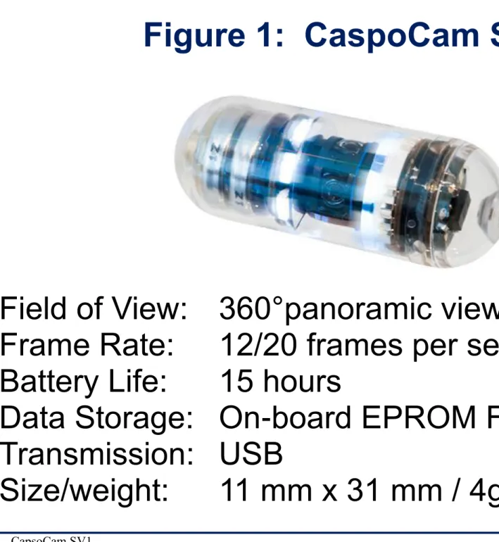
25 Figures 1-3.
26 Videocapsule in front-lateral view
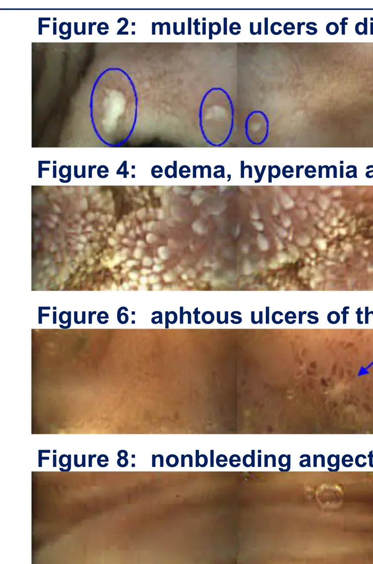
Figures 4-7.
Normal jejunum Red blood in the lumen
27 Figure 8. CapsoCam SV1
Field of View:
Frame Rate:
Battery Life:
Data Storage:
Transmission:
Size/weight:
360°panoramic view
12/20 frames per second
15 hours
On-board EPROM Flash Memory
USB
11 mm x 31 mm / 4gr
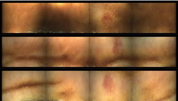
28 Figures 9-10.
29
Figure 2: multiple ulcers of distal jejunum, later diagnosed as CD
Figure 4: edema, hyperemia and lymphangectasia of proximal jejunum
Figure 3: diffuse gastopathy related to portal hypertension
Figure 5: ulcerative enteritis of the ileum, later diagnosed as ishemic
Figure 6: aphtous ulcers of the proximal jejunum
Figure 7: fresh blood and aphtous ulcers in the proximal ileum
30 Pan-enteric Crohn’s disease
5-6mm-large, not-bleeding typical angectasia
Table 1. Indications for capsule endoscopy
Table 2. Diagnostic yield
Indications of patients for capsule
endoscopy Number
Obscure gastrointestinal bleeding 55
Known or suspected Crohn’s disease 57
Familial adenomatous polyposis 3
Suspected small bowel tumor 3
31
Indication Nr Positive Suspicious Negative Obscure GI bleeding 55 33 (60%) 9 (16%) 13 (24%) Suspected or known Crohn’s disease 57 20 (35%) 3 (5%) 34 (60%) FAP 3 1 (33%) 0 (0%) 2 (67%) Suspected small bowel tumor 3 2 (67%) 0 (0%) 1 (33%) Total 118 56 (48%) 12 (10%) 50 (42%)
Table 3. Diagnostic yield in OGIB
Indication Nr Positive Suspicious Negative
OGIB Occult 34 20 (59%) 6 (18%) 8 (23%)
OGIB Overt 21 14 (67%) 1 (5%) 6 (28%)
Table 4. Small-bowel finding in positive patients
Findings Overall OGIB Non OGIB
MAV 15 15 0 Ulcer 20 14 6 Erosions 6 2 5 Polyps 4 3 1 Active bleeding 6 6 0 Stricture 3 1 1 Villous atrophy 2 0 1 Tumor 2 0 1
32