UNIVERSITA’ DEGLI STUDI DI VERONA
DEPARTMENT OFNeurosciences, Biomedicine and Movement Sciences
GRADUATE SCHOOL OF Life and Health Sciences
DOCTORAL PROGRAM IN Biomolecular Medicine
XXXI cycle / 2015-2018
TITLE OF THE DOCTORAL THESIS
Mutant p53-dependent alterations of cancer metabolism and
tumor microenvironment in pancreatic adenocarcinoma cells
S.S.D. BIO/10
Coordinator: Prof.ssa Lucia De Franceschi
Tutor: Prof. Massimo Donadelli
Quest’opera è stata rilasciata con licenza Creative Commons Attribuzione – non commerciale Non opere derivate 3.0 Italia . Per leggere una copia della licenza visita il sito web:
http://creativecommons.org/licenses/by-nc-nd/3.0/it/
Attribuzione Devi riconoscere una menzione di paternità adeguata, fornire un link alla licenza e indicare se
sono state effettuate delle modifiche. Puoi fare ciò in qualsiasi maniera ragionevole possibile, ma non con modalità tali da suggerire che il licenziante avalli te o il tuo utilizzo del materiale.
NonCommerciale Non puoi usare il materiale per scopi commerciali.
Non opere derivate —Se remixi, trasformi il materiale o ti basi su di esso, non puoi distribuire il materiale
3
TABLE OF CONTENTS
1. ABSTRACT.……….………....5
2. SOMMARIO.………..………...7
3. INTRODUCTION………...9
3.1 TUMOR SUPPRESSOR p53: THE GUARDIAN OF THE GENOME…………...9
3.2 MUTANT p53 AND ITS ONCOGENIC ROLE………...12
Chemoresistance………...15
Therapeutic Strategies to Restore Wild-Type Activity to Mutant p53……...16
3.3 CANCER METABOLISM: THE WARBURG EFFECT...……...…………...18
The opposite regulation on Warburg effect by Wild type and Mutant p53...……….19
3.4 NON-METABOLIC FUNCTIONS OF GLYCOLYTIC ENZYMES IN CAN-CER………..…………...20
Multifaceted roles of GAPDH ……….………..20
3.5 THE ENERGY SENSOR AMPK AND ITS REGULATION BY MUTANT p53....22
3.6 THE ONCOGENE AKT AND ITS REGULATION BY MUTANT p53……….….24
3.7 AUTOPHAGY: THE INTRACELLULAR DEGRADATION SYSTEM………....25
3.8 REACTIVE OXYGEN SPECIES IN CANCER………...26
UCP2:a key antioxidant player………....28
Sestrins: crucial role in antioxidant defenes………....30
3.9 THE IMPORTANCE OF SECRETOME AND TUMOR MICROENVIRON-MENT...………..…….31
The role of secreted proteins in cancer………….………..………...32
Mutant p53 and tumour microenviroment……….…...33
4. AIMS OF THE STUDY………...………...……...35
5. MATERIAL AND METHODS………...………..36
6. RESULTS………....49
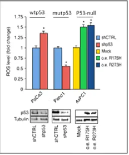
6.1 Mutant p53 and ROS metabolism 6.1.1 Mutant p53 proteins stimulate the production of ROS………....49
4
6.1.2 The oncogenic effects of mutant p53 are mediated by ROS induction…………...51
6.1.3 Mutant p53 downregulates UCP2 expression throughthe inhibition of PGC-1α………...53
6.1.4 Mutant p53-dependent downregulation of the PGC-1α/UCP2 axis is mediated by the blockage of SESN1/AMPK signaling………....55
6.1.5 The pro-oxidant and oncogenic effect of mutant p53 by UCP2 inhibition………...57
6.2 Mutant p53 and energy metabolism: the prevention of GAPDH nuclear translocation in PDAC cells 6.2.1 Mutant p53 prevents the nuclear translocation of GAPDH……….59
6.2.2Mutant p53 enhances the glycolytic activity of GAPDH and stimulates the L-lactate secretion………...61
6.2.3 Prevention of nuclear localization of GAPDH by mutant p53 is mediated by regulation of SIRT1:GAPDH complex and of AMPK and AKT pathways…………...62
6.2.4 GAPDH cytosolic stabilization contributes to the oncogenic effects of mutant p53………64
6.2.5 GAPDH cytosolic stabilization confers chemoresistance to gemcitabine and sensitizes cells to 2-deoxyglucose……….67
6.3 Mutant p53 and tumor microenvironment 6.3.1 The oncogenic effects of mutant p53 are also mediated by alterations of the cancer cell secretome……….69
6.3.2 Mutp53-driven secretome stimulates cancer cell migration and epithelial-to-mesenchymal transition (EMT)………. ……72
6.3.3 Biomarkers secreted from mutp53-driven secretome………...75
7. DISCUSSION AND CONCLUSION……….77
8. REFERENCES………81
9. ANNEXES………....92
5
1. ABSTRACT
Pancreatic adenocarcinoma (PDAC) is one of the most aggressive and devastating human malignancies.Late diagnosis is due to an absence of specific symptoms at ini-tial stages. In about 70% of PDACs, the tumor suppressor gene TP53 is mutated gen-erally resulting in conformational changes of mutant p53 (mutp53) proteins, making an important key in the carcinogenesis process not only through loss of wild type ac-tivity, but also through gain of specific mutant functions. In contrast to the tumor suppressive roles of wild-type p53, mutant p53 proteins support cancer progression by enhancing the ability of cancer cells to invade and metastasize, to confer chemo-resistance, and to stimulate genomic instability. We focused our attention on novel molecular mechanisms by which gain of function (GOF) mutant p53 proteins play their oncogenic roles promoting cancer cell proliferation and chemoresistance. The main project is based on intracellular alterations induced by mutant p53 in cancer metabolism and reactive oxygen species (ROS) production, contributing to cancer development and aggressiveness. ROS are highly reactive byproducts of mitochon-drial oxidative phosphorylation and are implicated in a plethora of biological events addressed to sustain each aspect of human cancer being able to act as second mes-sengers in cellular signaling. In particular, we unveiled that mutp53 is able to inhibit SESN1 expression and consequently the amount of SESN1/AMPK complex, result-ing in the downregulation of the AMPK/PGC-1α/UCP2 axis and ROS production. In this way GOF mutant p53 proteins, contrarily to its wild-type p53 counterpart, lead i) antiapoptotic effects, ii) proliferation and iii) chemoresistance in PDAC cells. These oncogenic roles given by GOF mutp53 are also detected through another mechanism that supports glycolytic metabolism in PDAC cells. Indeed, we demonstrated that mutant p53 prevents the nuclear translocation of the glycolytic enzyme glyceralde-hyde-3-phosphate dehydrogenase (GAPDH) stabilizing its cytoplasmic localization, thus supporting glycolysis of cancer cells and inhibiting cell death mechanisms me-diated by nuclear GAPDH. We further show that the prevention of nuclear localiza-tion of GAPDH is mediated by both stimulalocaliza-tion of AKT and repression of AMPK signaling, and is also associated with the formation of SIRT1:GAPDH complex.The blockage of GAPDH mutp53-dependent cytoplasmic stabilization is able to restore
6
the sensitivity of PDAC cells to the treatment with gemcitabine, permitting cancer cells to acquire sensitivity to anti-glycolytic drugs and suggesting a potential person-alized therapeutic approach in human cancers carrying mutant TP53 gene. In addi-tion, we addressed our research on the extracellular roles of mutant p53 in the tumor microenvironment of PDAC cells. The cancer secretome is a rich repository to find useful information for both cancer biology and clinical oncology. A better under-standing of biological features that are common or peculiar to different tumors could allow the identification of specific prognostic/predictive biomarkers for early diagno-sis and tumor progression monitoring. This is particularly relevant for PDAC, which has extremely high mortality rate and is mainly due to lack of recognizable symp-toms and exact assays for early detection. The objective of this study was to recog-nize a specific signature of biomarkers secreted by PDAC cells carrying GOF mutant p53. Comparing the secretome of p53-null PDAC cells before and after ectopic over-expression of R273H-mutp53 and R175H-mutp53, we found 23 differentially secret-ed proteins by both mutant p53 isoforms that might constitute a secretsecret-ed signature driven by the hot-spot p53 mutants in PDAC. Furthermore, we also studied the func-tional effect of mutp53-driven secretome on cancer cells showing its influence on proliferation, chemoresistance, apoptosis, autophagy, and cell migration. These data constitute a prerequisite for the identification of a secreted biomarker signature for the early identification of mutant p53 PDAC patients. In conclusion, the discovery of novel mechanisms by which hot-spot mutant p53 isoforms induce pancreas cancer growth is crucial to identify specific and personalized therapies for PDAC patients bearing mutant TP53 gene, representing a major therapeutic challenge for modern molecular oncology.
7
2. SOMMARIO
L’adenocarcinoma pancreatico duttale (PDAC) è una malattia letale e rappresenta una delle principali cause di morte per cancro. La diagnosi tardiva è dovuta ad un'as-senza di screening efficaci che permettano di diagnosticarlo nei primi stadi della ma-lattia. Ciò si traduce in un frequente ritardo nella diagnosi, che spesso viene diagno-sticata solo quando il tumore è già in uno stadio avanzato e si è diffuso in altre parti del corpo. In più del 70% dei casi è presente un gene TP53 mutato nelle cellule PDAC. P53 è un fattore di trascrizione che regola il ciclo cellulare e ricopre una im-portante funzione di soppressore tumorale. La maggior parte delle mutazioni presenti nel gene che codifica per p53 sono mutazioni missenso che causano l’espressione di isoforme di p53. Così, la proteina mutata non solo perde la sua funzione wild type ma può acquisire nuove proprietà biologiche chiamate gain-of-function (GOF) le quali contribuiscono allo sviluppo della patologia neoplastica. Lo scopo di questa tesi è stato quello di scoprire nuovi meccanismi molecolari attraverso i quali la proteina p53 mutata contribuisce alla progressione tumorale ed alla chemioresistenza al fine di identificare dei potenziali target terapeutici. Uno di questi meccanismi identificati è incentrato sullo studio delle specie reattive dell’ossigeno (ROS), che sono note in-durre instabilità genomica ed altre alterazioni che favoriscono lo sviluppo di patolo-gie quali il cancro. In questa tesi dimostriamo che le diverse isoforme di p53 mutata inducono alti livelli di ROS, attraverso l’inibizione di proteine antiossidanti, quali UCP2, ed identifichiamo una via di regolazione che coinvolge l’asse SESN1/AMPK/PGC-1α/UCP2. In questo modo, dimostriamo che p53 mutata favori-sce la crescita delle cellule tumorali e chemioresistenza. Queste capacità oncogeniche date dalla proteina p53 mutata vengono riscontrate attraverso un secondo meccani-smo incentrato sul metabolimeccani-smo energetico delle cellule tumorali. Infatti, un altro obiettivo della tesi è incentrato sulla regolazione intracellulare dell’enzima glicolitico GAPDH (gliceraldeide 3-fosfato deidrogenasi) da parte della proteina p53 mutata con conseguente modulazione della proliferazione delle cellule tumorali. GAPDH è una proteina multifunzionale, capace di svolgere altri ruoli oltre al suo ruolo princi-pale di enzima glicolitico. I risultati ottenuti dimostrano che p53 mutata stabilizza la proteina GAPDH nel citosol, stimolando l’effetto Warburg e impedendone
8
l’attivazione di meccanismi di morte cellulare indotti dal GAPDH nucleare. Investi-gando come questo avvenga, scopriamo che la stabilizzazione di GAPDH nel citosol data da p53 mutata, avviene atttraverso la stimolazione della proteina chinasi AKT e dalla repressione della chinasi AMPK ed è inoltre associata alla formazione del com-plesso GAPDH:SIRT1. Inoltre, il blocco della traslocazione nucleare del GAPDH da parte di p53 mutata rende più sensibili le cellule PDAC al trattamento di droghe anti-glicolitiche; in questo modo GAPDH potrebbe essere un target terapeutico per questo tipo di cancro che possiede il gene TP53 mutato. Il secondo progetto si basa sullo studio di biomarcatori secreti in cellule di PDAC con p53 mutato. Una migliore comprensione delle caratteristiche biologiche comuni e/o specifiche nei diversi tumo-ri potrebbe consentire l'identificazione di specifici biomarcatotumo-ri prognostici e/o pre-dittivi per la diagnosi precoce e il monitoraggio della progressione tumorale. Questo è maggiormente richiesto in tumori come PDAC a causa della mancanza di sintomi e saggi riconoscibili per la diagnosi precoce. Il secretoma del cancro rappresenta il mi-croambiente tumorale che svolge un ruolo chiave nei processi che promuovono la formazione dei tumori come l'angiogenesi e l'invasione.Confrontando il secretoma di cellule PDAC non esprimenti la proteina p53, con quelle in cui si ha una sovrae-spressione ectopica di R273H-mutp53 e R175H-mutp53, abbiamo trovato 23 protei-ne differenzialmente secrete da entrambe le isoforme mutanti di p53 che potrebbero costituire dei potenziali marcatori specifici per l’identificazione precoce di PDAC con p53 GOF. Inoltre, abbiamo anche studiato l'effetto funzionale del secretoma gui-dato da p53 GOF nelle cellule tumorali mostrando la sua influenza sulla proliferazio-ne, la chemioresistenza, l'apoptosi e l'autofagia, così come sulla migrazione delle cel-lule. In conclusione, la scoperta di nuovi meccanismi mediante i quali p53 mutato stimola la progressione tumorale è importante per identificare terapie specifiche e mirate in tumori, esprimenti geni mutati per p53, che rappresentano una delle princi-pali sfide terapeutiche per la moderna oncologia molecolare.
9
3. INTRODUCTION
Pancreatic cancer is one of the most frequent causes of tumor-associated deaths, and its incidence has recently increased in the western world [1]. There are different types of pancreatic cancer divided into two main groups; exocrine tumours that start in the exocrine cells where enzymes which help food digestion are made; endocrine tumours, also known as neuroendocrine tumours, that start in the endocrine cells which release insulin and other hormones. Most pancreatic cancers are exocrine and ductal adenocarcinomas [2]. Pancreatic ductal adenocarcinoma (PDAC) is the most common type of pancreatic malignancy and has a poor prognosis, with a dismal overall 5-year survival rate of 5% [3]. Late diagnosis due to an absence of specific symptoms at initial stage, together with high metastatic potential, resistance to thera-pies, and a lack of biomarkers and screening methods, are the main causes of poor prognosis in PDAC. Standard treatments for advanced disease include therapy with gemcitabine (2′,2′-difluoro-2′-deoxycytidine; GEM) with a response rate of less than 20% [4]. Therefore, the identification of effective targets and novel therapeutic strat-egies to improve GEM effects in PDAC have been the topic of extensive investiga-tion in the last few years [5]. PDAC presents genetic heterogeneity with a high num-ber of mutations. Among the various important genes alterated in PDAC, TP53 gene is the best-documented ones [6].
3.1 TUMOR SUPPRESSOR p53: THE GUARDIAN OF THE GENOME
P53 is one of the most important protein involved in signaling pathways that prevent tumour formation and progression. It is a transcription factor, with a modular struc-ture formed by distinct functional domains: i) N-terminal transactivation domain (amino acid aa 1-73), which interacts with the transcriptional machinery [7]; ii) pro-line rich-regions (aa 63-97), which is required for p53 stabilization; iii) DNA binding domain (aa 93-312), that binds the responsive element on DNA and proteins that positively or negatively affect p53 activity, such as MDM2 or 53BP1 respectively
10
[8]; iv) oligomerization domain (aa 325-355),which is essential for tetramer for-mation and represents the active form of p53 [9] and v) C-terminal regulatory do-main, containing residues post-translationally modified which are involved in modu-lation of its stability [10] (figure 1).
Figure 1. Multifunctional domains of p53. The p53 monomer consists of various multifunctional
do-mains [11].
A lot of studies shows p53 at the centre of a molecular network that transduces sig-nals deriving by stress conditions [12]. Under the non-stressed condition, the p53 protein is maintained at a low level in cells by the proteasome degradation pathway. MDM2, an E3 ubiquitin ligase, is the most critical negative regulator for p53 [13].In response to a wide variety of stress signals, including DNA damage, nutritional star-vation, hypoxia, the p53 protein is stabilized through post-translational modifications by a variety of enzymes. These enzymes include kinases, phosphatases, acetyltrans-ferases, deacetylases, ubiquitin ligases, deubiquitinases, methylases, and sumoylases [8]. Once activated, p53acts as a transcription factor and itbecomes able to promote the coordinated expression of many target genes through the binding to specific DNA sequence in the regulatory regions of its target genes [14]. In this way, p53 regulates a wide range of cellular biological processes to maintain genomic integrity and prevent tumor formation, including cell cycle arrest, apoptosis, senescence, en-ergy metabolism, anti-oxidant defense, autophagy, etc. (figure 2) [14], [15]. Because of its role as a key integrator in translating diverse stress signals into different cellu-lar outcomes, p53 has been namely the “guardian of the genome”.
11
Figure 2: The p53 pathway in tumor suppression. Activation of the transcription factor p53 in
re-sponse to different types of cellular stress can lead to cell survival as well as cell elimination. [15].
The importance of the p53 pathway in tumor suppression is strongly highlighted by the observation that mutations of the TP53 gene are very frequent in human cancers. Indeed, whereas somatic TP53 mutations contribute to sporadic cancer, germline TP53 mutations cause a rare type of cancer predisposition known as Li-Fraumeni Syndrome (LFS) which is not associated with site-specific tumours, but rather with a variety of tumour types occurring at a relatively early age [16]. Accordingly, the ab-sence of p53 predisposes to spontaneous development of neoplastic disease, as ob-served in p53 knockout mouse models [17]. Furthermore, somatic mutations in the TP53 gene are one of the most common alterations in human cancers, occurring in more than 50% of cancer patients [18]. In patients with wild-type TP53 gene , the p53 pathway is often compromised through the amplification of negative regulators, such as MDM2 [19] or the inactivation of upstream factors, as Chk2, ATM or p14ARF [20], [21].
12
3.2 MUTANT p53 AND ITS ONCOGENIC ROLE
Mutations in the TP53 gene are among the most common gene-specific alterations in human cancers.The frequency of TP53 gene mutations can vary considerably be-tween cancer types, ranging from 10% in haematopoietic malignancies [22] to 50– 70% in ovarian [23] and pancreas [24] cancers.Most of tumor suppressor genes, such as RB (retinoblastoma-associated protein), APC (adenomatous polyposis coli), and VHL (Von Hippel-Lindau tumor suppressor), are frequently inactivated by deletion or truncation mutations in tumors, resulting in the decreased or loss of expression of their proteins. Interestingly, the majority of p53 mutations in human cancer are mis-sense mutationsfor more than 70% of them, which usually result in the expression of full-length mutant p53 proteins [25]. Although p53 mutations have been found in all coding exons of the TP53 gene, the majority of mutations occur in the p53 DNA-binding domain, resulting in the loss of DNA-DNA-binding activity of mutant p53. In gen-eral, the p53 missense mutations can be classified into two main categories which are commonly referred to as “DNA‑contact” and “conformational” mutations [26]. The first group includes mutations in residues directly involved in DNA binding, such as R248Q and R273H. The second group comprises mutations that cause local (such as R249S and G245S) or global (such as R175H and R282W) conformational distor-tions. The majority of p53 missense mutations occur at six ‘mutational hot-spots’ in the DNA-binding domain of p53, including residues R175, G245, R248, R249, R273, and R282 (figure 3)[26], [27].
13
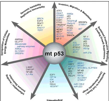
While wild-type p53 protein is kept at a low level in cells by the proteasome degra-dation pathway under non-stressed conditions, mutant p53 protein usually accumu-lates to a high level in tumors, and the underlying mechanisms are not fully under-stood [27]. It has been well-documented that many tumor-associated mutant p53 pro-teins not only lose their tumor suppression functions, but also gain new oncogenic functions, totally independent on wtp53, which is termed the gain-of-function (GOF) of mutant p53, acquiring different GOF activities, including promoting cell prolifera-tion, anti-apoptosis, metabolic changes, migraprolifera-tion, invasion, angiogenesis, and me-tastasis (figure 4) [28], [29].
Figure 4: Selected oncogenic properties of mutant p53 proteins and their underlying mechanisms.
Key cancer hallmarks and selected molecules that are associated with mutant p53 GOF [28].
Mutant p53 GOF contributes to cancer progression through direct interaction with proteins altering their function or through the transcriptional activation or repression of target genes and downstream molecules (figure 5) [29].For example, mutant p53 has been shown to interact with the transcription factor NF-Y, and to up-regulate the expression of NF-Y target genes [30]. Furthermore, p53 mutants can bind and inacti-vate two homologues of the p53 family, p63 and p73.Given that they share amino
ac-14
id sequence identity in the DNA-binding domain, p53, p63 and p73 should have re-dundant functions in the regulation of gene expression [31].
Figure 5: Mutant p53 and its interaction network. As part of its gain of function, mutant p53 interacts
with different proteins to enhance or inhibit their activities. TF, transcription factor; X, any protein other than a transcription factor or transcriptional cofactor; MAR, matrix attachment region DNA el-ement; mp53, mutant p53 [29].
The interaction between mutant p53 and p63/p73 are related with many aspects of the GOF of mutant p53, such as chemoresistance, migration, invasion, and metastasis [31]. In addition, most of mutant p53 isoforms are able to oligomerize with the wild-type protein encoded by the second allele, inhibiting its function, forming a hetero-tetramer unable to bind DNA, revealing a dominant negative function of mutant p53 [28]. An additional mechanism by which mutant p53 induces cancer progression is the up-regulation or down-regulation of a number of genes involved in different as-pects of tumorigenesis, such as c-Myc, Fos, PCNA, IGF1R, EGR1, NF-κB, BCL-xL, IGF2, VEGFA, and others [28]. Furthermore, mutant p53 is also able to regulate non-coding RNA ,such as miRNAs, inducing or repressing the expression of certain
15
miRNAs to mediate new oncogenic activities [32]. Taken together, the modulation of gene transcription and the interference with pivotal signalling pathways are im-portant mechanisms by which p53 mutants exert their oncogenic functions.
Chemoresistance
Chemoresistance causes reversion disease and metastasis, contrasts the development of the clinical outcome for the cancer patients, and remains the main obstacle to can-cer therapy [33]. Several mechanisms are involved in chemoresistance, one of which is the involvement of oncogenes. One typical feature of mutant p53 proteins is their ability to confer an elevated drug resistance to cancer cells. The overexpression of various tumour-associated p53 mutants can render cancer cells more resistant to the effect of chemotherapeutic drugs [34], [35] whereas knockdown of endogenous mu-tant p53 sensitizes cancer cells to killing by such molecules [36].The correlation be-tween p53 mutation status and sensitivity to cytotoxic drugs has been confirmed by a large study conducted by the National Cancer Institute, USA, where 60 cell lines and more than 100 anticancer drugs were examined [37]. However, the way in which p53 influences drug resistance depends on different parameters including the way of ac-tion of the drug, genetic variaac-tions during carcinogenesis, and the kind of cancer [38]. For example, our group and others demonstrated that the treatment with the drug gemcitabine stabilizes mutant p53 in the nuclei of the cells and induces the ex-pression of mutp53-target genes, as CdK1 (cyclin-dependent kinase 1) and CCNB1 (G2/mitotic-specific cyclin-B1), which are both involved in mitosis and cell prolifer-ation, leading to gemcitabine resistance in pancreatic cancer cells [35], [39]. In te-mozolomide-resistant glioma cells, a correlation between mutant TP53 gene and MGMT (O6-methylguanine DNA-methyl-transferase) expression was detected. While temozolomide kills cells by alkylating O6-guanine, MGMT in turn repairs al-kylation. Therefore drug resistance may be caused by MGMT up-regulation [40]. In conclusion, mutant p53 is not only a crucial player in carcinogenesis, but it is also re-lated with resistance to recognized cytotoxic anticancer drugs, such as gemcitabine, cisplatin, epirubicin, 5-fluorouracil, methotrexate and many other chemotherapeutics.
16
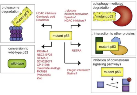
Therapeutic Strategies to Restore Wild-Type Activity to Mutant p53 Many different mutations and phenotypes allow a variety of strategies are being
ex-plored to target tumors expressing mutant p53s (Figure 6) [41]. Re-folding of this mutated and accumulated p53 leads to restoration and activation of defective pro-teins, resulting in high levels of active p53 with wild-type functionality and apoptotic cell death [42]. A variety of compounds that might restore wild-type p53 confor-mation and function have been characterized [42].
Figure 6:Strategies restoring p53 wild-type function. These strategies include promotion of mutant
p53 degradation through the proteasome and autophagy pathways, restoration of wild-type p53 activi-ty, interference with the interaction between mutant p53 and other proteins, and interference in signal-ing pathways downstream of mutant p53 [41].
The compound that represents an innovation in the reactivation of p53 is P53-Reactivation and Induction of Massive Apoptosis-1 (PRIMA-1).Treatment with this compound upregulated wtp53-target genes, such as BAX, PUMA and NOXA [43] and induced activation of caspases -2, -3 and -9 [44]. Under physiological conditions, PRIMA-1 and its methylated analogue PRIMA-1Met (also known as APR246) are converted into a reactive intermediate compound, MQ, which covalently binds to the core domain of p53 [45]. Due to their aberrant folding, mutant p53 proteins may ex-pose cysteine residues, which are hidden in the core domain of wt-p53 [44]. This can lead to the formation of inter- and intramolecular disulfide bonds, locking mutant
17
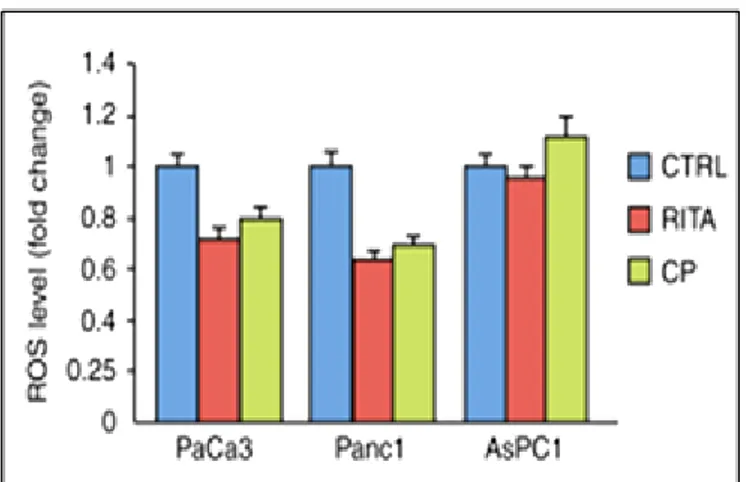
p53 in an inactive conformation and causing protein aggregation. Thiol modification by reactive compounds, such as MQ, prevented the formation of such disulfide bonds and thus promoted correct folding and restoration of the wild-type function [45]. Alt-hough the fact that nuclear levels of both p53 and MDM2 are normally kept at low levels due to a regulatory circuit, a deregulated MDM2/p53 balance (e.g. by overex-pression of MDM2) reduces the tumor suppressive functions of p53 [12]. Due to this antagonizing effect of MDM2 on p53, small molecules mimicing p53-binding resi-dues can block the MDM2-p53 interaction. With this interaction blocked, p53 is no longer controlled by MDM2 and is reactivated in tumor cells harboring wt-p53 [46]. However, MDM2 is not the only a negative regulator of p53 and therefore other ef-fects have to be considered. Again, p53 is involved in a complex network with sever-al other actors. For example, it is sever-also regulated by the proteins SirT1 (Sirtuin 1) [47] or Wip1 (wt-p53 induced phosphatase/PPM1D) [48]. Numerous MDM2 inhibitors have been developed during the past few years [49]. For example, RITA (Reactiva-tion of p53 and Induc(Reactiva-tion of Tumor cell Apoptosis) bound to the N-terminus of p53 (residues 1-63) and induced a conformational change, which transmitted from the N-terminus to the core and to the C-terminal domain, promoting the disruption of p53 and MDM2 complex [50]. This led to p53 accumulation and induction of p53-dependent apoptosis in a variety of tumor cell lines of different origin. Autophagy al-so plays a role in mutant p53 degradation. The degradation of mutant p53 was pro-moted by proteasomal inhibition and depended on functional autophagy machinery [51]. However, mutant p53 can inhibit autophagy [52], [53], indicating that the rela-tionship between autophagy and mutant p53 is complex and the inhibition of autoph-agy by mutant p53 can further improve the overexpression of this oncognenic pro-tein. Another small molecule named RETRA has been suggested to destabilize the interaction between p73 and mutant p53 [54]. Instead of targeting mutant p53 direct-ly, another approach is to identify common pathways regulated by mutant p53 pro-teins in order to target these downstream pathways for therapeutic intervention [41].
18
3.3 CANCER METABOLISM: THE WARBURG EFFECT
Tumorigenesis is dependent on the reprogramming of cellular metabolism as both di-rect and indidi-rect consequence of oncogenic mutations. The alterations in intracellular and extracellular metabolites that can accompany cancer-associated metabolic repro-gramming have effects on gene expression, tumor microenvironment and the genera-tion of signaling molecules such as reactive oxygen species (ROS) [55]. The com-mon feature of this altered metabolism in cancer cells is increased glucose uptake and fermentation of glucose to lactate. Cellular metabolism of glucose to CO2 and H2O in normal adult cells is accomplished through the oxidative phosphorylation pathway that leads to the generation of ATP in the presence O2. The Warburg effect, also called aerobic glycolysis, is the best known metabolic shift that occurs in cancer cells to support the biosynthetic requirements of uncontrolled proliferation [56]. Dur-ing this phenomenon, even in the presence of oxygen and totally functionDur-ing mito-chondria, tumour cells adopt glycolysis for their energy necessities and undergo both high rate glucose uptake and lactate production, as compared with normal cells (fig-ure 7) [57], [58]. Per unit of glucose, aerobic glycolysis is an inefficient way of gen-erating ATP compared to the amount obtained by mitochondrial respiration [59]. However, the rate of glucose metabolism through aerobic glycolysis is higher such that the production of lactate from glucose occurs 10-100 times faster than the com-plete oxidation of glucose in the mitochondria [60]. In this situation, the increased glucose consumption is used as a carbon source for anabolic processes needed to support cell proliferation [61]. In fact, this excessive organic substrate is also used for the de novo generation of nucleotides, lipids, and proteins. Furthermore, having a rate-limiting demand for ATP, proliferating cells are in an increased need of reducing equivalents in the form of NADPH obtained by the penthose phosphate pathway (PPP) [61]. A proposed mechanism for considering the biosynthetic function of the Warburg Effect is the regeneration of NAD+ from NADH in the pyruvate to lactate step that concludes aerobic glycolysis. This process may also influence the homeo-stasis of ROS generation by affecting the concentration of reducing equivalents in the mitochondria [62]. Finally, Warburg effect is also able to induce acidification of the microenvironment and other metabolic crosstalk, favoring cancer cell growth [62].
19
Figure 7: Warburg effect. Schematic representation of the differences between oxidative
phosphory-lation, anaerobic glycolysis, and aerobic glycolysis [61].
The opposite regulation on Warburg effect by Wild type and Mutant p53
Recent studies have shown that regulating energy metabolism is a critical role of wtp53 in tumor suppression [63]. Indeed, wt-p53 is described to regulate glycolysis, mitochondrial oxidative phosphorylation, pentose phosphate pathway (PPP), and li-pid metabolism in cells. Functionally, wt-p53 represses glycolysis and the Warburg effect through multiple mechanisms as transcriptional regulation of genes involved in the glycolytic metabolism, including TIGAR (TP53-induced glycolysis and apoptosis regulator) and Parkin [64], [65]. For example, p53 transcriptionally blocks the ex-pression of glucose transporters, as GLUT 1-4, and induces the exex-pression of TIGAR which decreases the intracellular concentrations of fructose-2,6-bisphosphate, and thus reduces glycolysis and deflects glucose catabolism to the PPP [65]. On the con-trary, tumor-associated mutant p53 was reported to promote tumor metabolic chang-es as a novel gain-of-function in promoting tumor development [66]. Mutant p53 en-courages the Warburg effect both in cultured cells and mutp53 knock-in mice. This effect mainly occurs through promoting the translocation of GLUT1 (glucose trans-porter 1) to the plasma membrane, which is mediated by activated RhoA/ROCK sig-naling [66]. In addition, mutant p53 was reported to induce the expression of glyco-lytic enzyme hexokinase II, which could promote glycolysis [67].
20
3.4 NON-METABOLIC FUNCTIONS OF GLYCOLYTIC ENZYMES IN CAN-CER
In addition to their canonical roles in glycolysis, recent studies progressively un-covered some non-metabolic functions of glycolytic enzymes in tumorigenesis, becoming emerging targets for therapeutic intervention [68]. Emerging evidence showed that most glycolytic enzymes are deregulated in cancer cells and play im-portant roles in tumorigenesis and all essential glycolytic enzymes can be translo-cated into nucleus where they participate in tumor progression independently of their canonical metabolic roles [69]. These non-canonical functions include anti-apoptosis, regulation of epigenetic modifications, modulation of transc ription fac-tors and co-facfac-tors, DNA repair activity, suggesting that these multifaceted glyco-lytic enzymes not only function in canonical glycoglyco-lytic metabolism but also direct-ly link metabolism to epigenetic and transcription programs implicated in tumor-igenesis [70]. The trafficking of metabolic enzymes to the nucleus could be caused by covalent modifications or by forming new protein-protein interactions or pro-tein complexes [71], [72]. However, the precise mechanisms at the basis of the regulation of these non-metabolic functions of the glycolytic enzymes are still largely unknown.
Multifaceted roles of GAPDH
Glyceraldehyde-3-phosphate dehydrogenase (GAPDH) is a glycolytic enzyme that catalyzes the reversible conversion of glyceraldehyde-3-phosphate (G-3-P) to 1,3-diphosphoglycerate in the cytosol of the cells. GAPDH acts as a homo tetramer containing four identical 37 kDa subunits [73]. It initially was identified as a gly-colytic enzyme involved exclusively in cytosolic energy production [73]. Howev-er, emerging evidence indicates that GAPDH is a multifunctional protein display-ing diverse activities distinct from its conventional metabolic role [74]. Its plei-otropic role is largely affected by ability of GAPDH to bind different macromole-cules in the cell and by post-translational modifications in different amino acid residuessuch as phosphorylation, ADP ribosylation, and acetylation [75]. Specifi-cally, several studies have demonstrated that the GAPDH has a variety of other
21
functions, including DNA repair, transcriptional regulation, membrane fusion and transport, autophagy, cell death and nuclear tRNA export [75], [76]. The multi-functional roles of GAPDH are strictly associated to its intracellular localization, which is not restricted to the cytosol for glycolytic energy production [76]. Indeed, after specific stimuli GAPDH can translocate in other subcellular compartments, such as nuclei in which it exerts a critical role in the regulation of cell death-related gene transcription, stimulation of apoptosis and modulation of cell fate. GAPDH has been also observed in the mitochondria, in which its binding to the voltage-dependent anion channel (VDAC) has been suggested to promote the release of proapoptotic proteins, such as cytochrome c (CytC) and apoptosis -inducing factor (AIF) [77]. On the other hand, GAPDH is involved in a series of age-related neurodegenerative disorders [78] and in tumor development and pro-gression [79], promoting tumor-specific GAPDH transcriptional/post
-transcriptional regulation and aging as prosurvival factor [80], [81]. For example, a study showed that GAPDH may translocate to the nucleus when cells encoun-tered certain stress conditions such as oxidative stress. Specifically, oxidative stress-induced S-nitrosylation of GAPDH could promote translocation of GAPDH to the nucleus, where it could interact with Sp1 under oxidative stress conditions, and activate SNAIL transcription, promoting EMT and metastasis through its non-enzymatic function in the nucleus [82]. Our group demostrated that the inhibition of antioxidant uncoupling proteins UCP2 triggers PDAC cell death by ROS-dependent nuclear translocation of GAPDH [83] and later, we discovered that the synergistic PDAC cell growth inhibition given by everolimus and genipin treat-ment was due to a massive GAPDH nuclear translocation observed both in vitro and in mice xenograft [84]. Thus, understanding the biological functions of GAPDH beyond glycolysis will improve the ability to effectively target this en-zyme in cancer therapy.
22
3.5 THE ENERGY SENSOR AMPK AND ITS REGULATION BY MUTANT p53.
AMP-activated kinase (AMPK) is a highly conserved serine/threonine protein ki-nase complex and a central metabolic sensor located at the crossroad between metabolic and signaling networks [85]. AMPK is a heterotrimeric complex com-posed of a catalytic α-subunit and two regulatory subunits, β and γ [86]. The γ-subunit enables AMPK to respond to changes in the ATP-to-AMP ratio as it con-tains domains that bind adenine nucleotides. Upon changes in the ATP-to-AMP or ATP-to-ADP ratio, AMPK is activated by an allosteric mechanism that stimulates its kinase activity phosphorylating downstream targets to redirect metabolism to-wards increased catabolism and decreased anabolism [86]. The two major up-stream kinases responsible for AMPK activation are the tumor suppressor LKB1 and Ca2+/calmodulin-dependent protein kinase kinase 2 (CaMKK2). LKB1 acti-vates AMPK during energy stress, whereas CaMKK2 activity is induced by in-creased intracellular Ca2+ levels, regardless of the energy status of the cells [87]. AMPK restores ATP levels during metabolic stress by inhibiting ATP-consuming biosynthetic pathways while simultaneously activating pathways that regenerate ATP through the breakdown of macromolecules. In particular, AMPK phosphory-lates several transcription factors (or co-factors) that are themselves master regula-tors of biosynthetic pathways (figure 8). In this way, AMPK can acutely restore energy balance but also reprogram cell metabolism transcriptionally in response to prolonged energetic decreases [87]. For example, AMPK promotes glucose uptake by phosphorylating TBC1D1 (TBC domain family, member 1) and TXNIP (thi-oredoxin-interacting protein), which control the translocation and cell-surface lev-els of glucose transporters GLUT4 and GLUT1, respectively [88]. It also promotes autophagy through several mechanisms like the activation of ULK1 (unc-51-like autophagy-activating kinase 1), which triggers the initiation of the autophagic cas-cade [88]. Regarding mutp53, it is able to inhibit AMPK signaling in head and neck cancer cells directly binding to the AMPKα subunit, thus gaining its onco-genic function and stimulating anabolic growth of cancer cells, in contrast to its wild type counterpart [89].
23
Figure 8: Substrates of AMPK regulate multiple metabolic processes in cells. AMPK is
phos-phorylated and activated by LKB1 and CAMKK2 in response to several stimuli. Its phosphoryla-tion induces metabolic changes through the phosphorylaphosphoryla-tion of substrates [87].
Mutp53s can directly interact with both AMPKα1 and AMPKα2 mainly through their DNA-binding domain (DBD), while the N-terminus of mutp53s is responsi-ble for blocking the interaction between AMPKα and its upstream kinase LKB1 inhibiting its Thr172 phosphorylation and consequently preventing its activation [89]. Finally, our group revealed another mechanism by which mutp53 is able to inhibit AMPK-signaling [52]. In particular, p53 blocks AMPK by down-regulating Sestrin1/2 expression which are a class of proteins that can directly interact with AMPK subunits favoring their phosphorylation by upstream kinases and thereby resulting in AMPK signaling stimulation [52], [90]. Importantly, AMPK was re-cently identified as a negative regulator of the Warburg effect through inhibition of the hypoxia-induced factor 1 (HIF-1) pathway [91]. Therefore, the inhibition of AMPK by GOF mutp53s, which relieves the suppression of HIF-1 by AMPK, is expected to increase HIF-1 protein expression and thus lead to increased glucose influx and glycolysis. These phenomena could represent a further mechanism of GOF mutp53 to promote the Warburg effect and drastic metabolic changes in can-cer cells.
24
3.6 THE ONCOGENE AKT AND ITS REGULATION BY MUTANT p53
Protein kinase B or Akt (PKB/Akt) is a serine/threonine kinase, which in mam-mals comprises three highly homologous members known as PKBalpha (Akt1), PKBbeta (Akt2), and PKBgamma (Akt3) [92]. PKB/Akt is activated in cells ex-posed to diverse stimuli such as hormones, growth factors, and extracellular ma-trix components. A well-known upstream target of Akt is phosphatidylinositol-3 kinases, PI3Ks, which is a lipid kinase family and a key enzyme in the generation of the second messenger phosphatidylinositol-3,4,5-trisphosphate (PI-3,4,5-P3) [93]. This allows the translocation of Akt from the cytoplasm to the plasma mem-brane, altering its conformation to allow subsequent phosphorylation by the phos-phoinositide-dependent kinase-1 (PDK-1) [94]. PKB/Akt is then released from the membrane and translocates to other subcellular compartments. Phosphorylation of Akt results in full activation of Akt kinase activity and the subsequent regulation of multiple cellular processes, including the transmission of growth factor-dependent survival signal [95]. In particular, Akt inhibits apoptosis by phosphory-lating and inactivating pro-apoptotic Bcl-2 family members, as Bad, and by inhib-iting the release of cytochrome c. In addition, Akt changes mitochondrial mem-brane potential induced by multiple apoptotic stimuli, in a caspase-independent manner, and it acts maintaining mitochondrial integrity [96], [97]. In addition, Akt-regulated signalling plays a critical role in numerous processes which are known to be hallmark of cancer cells [98]. Akt regulates the transcription of death genes by phosphorylating forkhead family transcription factors or increasing the transcription of survival genes by activating NF-κB and CREB transcription fac-tors [98]. Furthermore, it regulates energy metabolism by multiple mechanisms, including the expression and membrane translocation of glucose transporters. Akt may also indirectly activate the important rate-controlling enzyme phosphofructo-kinase-1 (PFK1) by directly phosphorylating and activating phosphofructokinase-2 (PFK2) [99] whose principal reaction product, fructose-2,6-bisphosphate (Fru-1,6-P2), is a potent allosteric activator of PFK1. Furthermore, Akt affects not only glycolysis, but is also able to improve mitochondrial respiration and oxidative phosphorylation [100], [101]. In fact, inhibiting Akt activation decreases ATP
25
production; activities of complexes I, II, and III; the mitochondrial membrane po-tential (ΔΨm); and F0F1-ATPase activity [100].
Intriguingly, there is a connection between p53 and Akt. Phosphorylation of MDM2 by Akt has been reported to result in the translocation of MDM2 to the nu-cleus, where it promotes the ubiquitination of p53, reducing wtp53 levels and promoting tumour growth [102]. Mutant p53-R273H is able to regulate PI3K/AKT signaling pathway, inducing Akt expression [103].DAB2IP is a negative modulator of PI3K/AKT signaling, because it binds AKT limiting its activation in response to various stimuli [104]. It has been discovered that mutp53 binds and inhibits DAB2IP, favoring insulin-induced AKT activation in cancer cells [105]. In vitro studies have also shown increased transformation in cells having a combination of PI3K/Akt pathway activation and mutp53, as well as increased invasiveness of tumour cells [106]. Thus, Akt is an excellent candidate master regulator responsible for the classical biochemical features of cancer cells. Furthermore, it may constitute a “Warburg kinase” that can be specifically targeted to alter cancer cell energy me-tabolism for therapeutic benefit.
3.7 AUTOPHAGY:THE INTRACELLULAR DEGRADATION SYSTEM
Autophagy is a cellular catabolic degradation response to starvation or stress whereby cellular proteins, organelles and cytoplasm are digested and recycled [107]. Basal autophagy also has an important homeostatic function, maintaining protein and organelle quality control. Autophagy is controlled mainly by the ki-nase mammalian target of rapamycin (mTOR; also known as FRAP1), which is a downstream component of the PI3K pathway [107]. Autophagosomes are double-membrane vesicles that sequester cytoplasm and organelles. The autophagy-regulated or Atg proteins are required for the activation of autophagy, the for-mation of autophagosomes, the sequestration of intracellular constituents, and the targeting and fusion of autophagosomes to lysosomes [108]. One of the most common events in human cancer is the downstream kinase mTOR activation of the PI3K pathway. Autophagy was initially thought to be a tumor-suppression mecha-nism. Indeed, autophagy deficiency causes oxidative stress, activation of the DNA
26
damage response promoting cancer cell growth [109]. Nevertheless, cancer cells also rely on autophagy in many cases due to the increased metabolic and biosyn-thetic demands imposed by deregulated proliferation [110]. Wild type p53, in or-der to react to genotoxic or environmental stimuli, triggers autophagy in cancer cells through various mechanisms, as the stimulation of the nutrient energy sensor AMP-activated protein kinase (AMPK), the inhibition of the mammalian target of rapamycin (mTOR)[111]. On the contrary, mutant p53 proteins counteract autoph-agy through several mechanisms as the stimulation of Akt/mTOR pathway or through ATG12 repression [112]. Thus, the impact of autophagy regulation in the development of cancers and in response to therapies assumes increasing im-portance.
3.8 REACTIVE OXYGEN SPECIES IN CANCER
Reactive oxygen species (ROS) are highly reactive radicals, ions or molecules that can readily oxidize other molecules including lipids, amino acids, proteins, and nucleic acids [113]. ROS can appear from numerous intracellular sources; among them, the most important are mitochondria [114]. In mitochondria, ROS are pro-duced as an inevitable byproduct of oxidative phosphorylation. The electron transport chain encompasses complexes I-IV and ATP synthase on the mitochon-drial inner membrane. Superoxide is generated at complexes I and III and released in the intermembrane space or in the mitochondrial matrix [115]. Under normal physiological conditions, the intracellular levels of ROS are steadily maintained to prevent cells from damage. Detoxification from ROS is facilitated by non -enzymatic molecules (i.e. glutathione, flavenoids and vitamins A, C and E) or through antioxidant enzymes like superoxide dismutases (SODs), catalase or glu-tathione reductase, which specifically scavenge different kinds of ROS. Reactive oxygene species have been detected in almost all cancers where they promote many aspects of tumor development and progression [116]. In cancer cells high levels of reactive oxygen species can result from increased metabolic activity, on-cogene activity, increased cellular receptor signaling and other events [117], [118]. Indeed, ROS in cancer are involved in in a plethora of biological events addressed
27
to sustain each aspect of cancer progression summarized in figure 9, like cell cycle progression and proliferation, cell survival and apoptosis, energy metabolism, cell motility, angiogenesis and maintenance of tumor stemness [116], [119]. Reactive oxygen species, particularly hydrogen peroxide, can act as second messengers in cellular signaling [120]. Indeed, ROS generation can activate several signaling pathway like the PI3K/Akt signaling, MAPK/Erk1/2pathway [116].The non-radical ROS hydrogen peroxide H2O2 regulates protein activity through reversible oxidation of its targets including protein tyrosine phosphatases or kinases, receptor tyrosine kinases and transcription factors [121]. Lipids are others cellular targets of ROS attacks. ROS react with polyunsaturated or polydesaturated fatty acids to initiate lipid peroxidation [122]. However, the role of ROS in cancer biology is ambiguous, indeed despite many studies attributed to ROS a pivotal role in pro-moting many events, many others have highlighted that a severe increase in ROS can induce cell death following a “non-specific” damage of macromolecules such as the irreversible oxidation of lipids, proteins or DNA [119]. Therefore, ROS rep-resent an “Achilles heel” of cancer cells and new therapeutic improvement could be reached playing on this sophisticated redox cellular balance.
28
Figure 9: ROS play multiple roles in the hallmarks of cancers. Contribution of oxidants is indicated
for each point [123]
UCP2: a key antioxidant player
The UCPs belong to the superfamily of anion transport carriers of the mitochondrial inner membrane [124] and some of them are involved in thermogenesis and regula-tion of mitochondrial ROS. UCP2 has been found in several tissues, including liver, brain, pancreas, adipose tissue, immune cells, spleen, kidney, and the central nervous system [125]. UCP2 acts as an important sensor of mitochondrial oxidative stress controlling the production of mitochondrial ROS. As revealed by studies with UCP2-null mice, its antioxidant function is generally implicated in cyto-protective activities [126]. The uncoupling of oxidative phosphorylation is a short circuit in which the transport of protons from the intermembrane space to the matrix bypasses ATP syn-thase resulting in a decrease of mitochondrial inner membrane potential; leakage of electrons from electron transport chain ETC and ROS generation (figure 10). Minor increases in the mitochondrial membrane potential induce ROS formation, whereas slight decreases can substantially diminish their production, without greatly lowering
29
the efficiency of oxidative phosphorylation [127]. Hence, the mild uncoupling of mi-tochondrial oxidative phosphorylation may represent the first line of defense against oxidative stress [127]. According to this pattern, UCP2 can dissipate the proton gra-dient to prevent the proton-motive force from becoming excessive, thus decreasing ROS produced by electron transport [128]. Mitochondrial superoxide ion is consid-ered the initial and leading molecule of ROS signaling and is generally converted in-to hydrogen peroxide (H2O2) by superoxide dismutases. In addition, upon reaction with H2O superoxide ion can generate hydroxyl radicals (•HO) implicated in lipid damage and protein oxidation [128]. Thus, UCP2 acts as a sensor of mitochondrial oxidative stress controlling the production of mitochondrial ROS and regulating re-dox-sensitive cytosolic signaling pathways. In addition to its antioxidant role, UCP2 may acts as a direct metabolic regulator contributing to the Warburg phenotype, promoting pyruvate efflux from mitochondria, restricts mitochondrial respiration, and increases the rate of glycolysis in cancer cells [129].
Figure 10. Uncoupling protein 2 uncoupling activity in oxidative phosphorylation. ROS: Reactive
ox-ygen species; UCP2: Uncoupling protein 2; SOD: Superoxide dismutase; Mn-SOD: Manganese-superoxide dismutase [130].
The regulation of UCP2 can occur at various levels as transcriptional, translational and protein turn over regulation or post-transcriptional modifications [131]. One of
30
the most important mechanisms of the transcriptional regulation of UCP2 is mediated by the peroxisome proliferator-activated receptor gamma coactivator1-alpha (PGC-1α) [132], that has been shown to stimulate Ucp2 gene expression via two thyroid hormone response elements TREs located in the proximal Ucp2 promoter region [133]. PGC-1α can also indirectly induce Ucp2 gene expression by the link with sterol regulatory element-binding protein (SREBP) [134]. SREBP isoforms are known to regulate Ucp2 gene expression via either one of the two E-box motifs pre-sent on Ucp2 promoter [134].
Sestrins: crucial role in antioxidant defenses
The Sestrins constitute a family of stress-inducible proteins upregulated in cells ex-posed to a variety of environmental stresses including DNA damage, oxidative stress and energy deficiency. They contribute to redox homeostasis through the regulation of adenosine monophosphate-dependent protein kinase (AMPK)-mammalian target of rapamycin (mTOR) signaling, leading to inhibition of cellular anabolism and augmentation of catabolic processes such as beta-oxidation [135]. Sestrin-dependent activation of AMPK and suppression of mTORC1 activity are also critical for main-taining basal autophagy [136]. Thus, Sestrins can be important for autophagic elimi-nation of dysfunctional mitochondria that leak electrons and produce pathogenic amounts of ROS. All members of Sestrin family are induced by oxidative stress, alt-hough they are subject to different induction mechanisms (figure 11) [137], [138]. Sesn1 is induced by hydrogen peroxide in a p53-dependent manner [139], whereas induction of Sesn2 by oxidative stress is only partially p53-dependent [140]. Silenc-ing of either Sesn1 or Sesn2 in human fibroblasts significantly inhibit cell prolifera-tion and accelerate cell senescence triggered by ROS accumulaprolifera-tion [137]. The Sestrins were also shown to mediate the antioxidant activities associated with the p53 and FoxO transcription factors. While high levels of oxidative stress can lead to cell death through p53- and FoxO-dependent apoptotic gene transcription, low levels of oxidative stress cause moderate activation of p53 and FoxO that can induce Sestrins to reduce oxidative stress and prevent cell death [139], [141]. Despite their involve-ment in tumor suppression and genome protection, Sestrins are still expressed in
31
many cancers and might actually be required for maintaining the viability of cancer cells as part of the antioxidant defense system under certain conditions [142].
Figure 11: Regulation of Sestrin expression by oxidative stress. p53,Nrf2 and AP-1 are required for
Sestrins induction upon oxidative stress [143]
3.9 THE IMPORTANCE OF SECRETOME AND TUMOR MICROENVIRON-MENT
Secretome is referred to as the rich, complex set of molecules secreted from living cells, and is a vital aspect of cell–cell communication in eukaryotes. Proteins, lipids, micro-RNAs (miRNAs) and mRNAs are secreted into the extracellular space by a cell, tissue, organ, or organism at any given time and conditions [144]. Chronic per-turbations in the secretome are often associated with altered cellular phenotypes dicative of pathological conditions [145], including obesity, diabetes, chronic in-flammation and cancer. The widest application of secretome has been in the devel-opment of new diagnostic biomarkers for human disease classification like cancer [146]. Proteins of secretome play a key role in cell signaling, homeostasis, immune response, communication and migration [147]. Examples of secretory proteins in-clude hormones, digestive enzymes, cytokines, chemokines, interferons (IFNs), col-ony-stimulating factors (CSFs), growth factors, and tumor necrosis factors (TNFs). The secretome of many cell types, including cancer cells, is released by distinct se-cretory processes. The best characterised of these pathways, the ‘classical’ or
endo-32
plasmic reticulum (ER)-Golgi pathway has traditionally been considered responsible for the majority of protein secretion. Proteins secreted through this pathway contain an N-terminal signal peptide that is crucial for recognition by the secretory apparatus. Classically secreted proteins are synthesised in the rough ER before transport to the Golgi apparatus in COPII-containing transport vesicles [148]. During passage through the Golgi, cargo proteins may be modified by processes such as proteolytic cleavage or glycosylation. Cargos are then sorted into secretory vesicles at the trans-golgi network (TGN), and secretion is achieved when Golgi-derived vesicles con-taining the secreted proteins are trafficked to and fuse with the plasma membrane, re-leasing their contents to the extracellular environment [149]. Whilst the majority of cytokines, growth factors and extracellular matrix components are believed to be classically secreted, several factors are known to be secreted via Golgi-independent, ‘non-classical’ pathways. These non-classical secretion pathways are, in general, less characterised than classical secretory events [150]. The need for developing more ef-fective cancer biomarkers and therapeutic modalities has led to the study of cancer cell secretome as a means to identify and characterize diagnostic and prognostic markers and potential drug and therapeutic targets [151].
The role of secreted proteins in cancer
In addition to various pathological events, such as genomic instability and induction of oxidative stress, tumorigenesis and cancer progression may also strongly depend on extrinsic factors secreted by cancer cells. Secreted factors play a critical role in the tumour–microenvironment communications, representing a signal to cells at dis-tant sites and affecting their phenotype (figure 12) [144]. The development of an ad-verse tumour microenvironment as the consequence of the crosstalk between cancer and stromal cells is one of the causes of low efficiency to cancer treatments. Indeed, it is reported that solid tumours take advantage of a co-evolution of neoplastic and stromal cells and that the extracellular matrix (ECM) plays a dynamic role in cancer invasion and migration. This complex cancer-microenvironment system is also strongly influenced by impaired vascularization and by interaction with the immune system cells [152]. Many biological pathways, such as NF-κB, MAPK, IL-1 are in-volved to orchestrate this complex crosstalk system [144], [153]. Those secreted
fac-33
tors, i.e. cytokines/chemokines, proteases, growth and angiogenic factors may regu-late the crosstalk between stroma–cancer cells and tumour microenvironment. The amount of any constituent of the secretome can be regulated by alterations to de novo synthesis, to its half-life and to trafficking processes [154]. Furthermore, many of cancer cell-secreted proteins/enzymes/molecules can stimulate drug resistance through autocrine/paracrine mechanisms [155]. Thus, it’s becoming fundamental to study specific secreted biomarkers which may clinically predict resistance mecha-nisms to specific drugs.
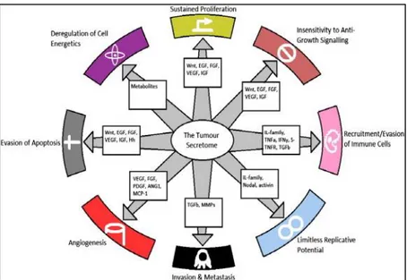
Figure 12: The tumour secretome and the hallmarks of cancer. The well-known secreted factors and
the biological pathways involved. The secreted factors contribute to the hallmarks of cancer shown in the figure [144].
Mut p53 and tumour microenviroment
A novel mechanism to drive invasion by mutant p53 is the manipulation of tumour microenviroment [156]. Mutp53 proteins trigger the production and release of pro-inflammatory and immunomodulatory cytokines to stimulate an pro-inflammatory can-cer-associated microenvironment and to repress the immune system [156]. Indeed, mutp53 proteins, contrary to the wild-type counterpart, upregulate the expression of chemokines like CXCL5, CXCL8 and CXCL12 through the NF-κB-dependent path-way, highlighting a further molecular mechanism by which mutp53 proteins exert
34
their oncogenic activity [157]. Loging and Reisman showed that mutp53 proteins are able to repress the transcription of TIMP-3, which is crucial in the extracellular ma-trix (ECM) turnover and construction, contributing to increased activity of secreted MMPs in the ECM and, subsequently, to tumour invasion and metastasis [158]. Toschi et al. showed that, in human melanoma cells carrying mutp53 proteins, the re-introduction of wtp53 overcomes the mutp53 GOF and reduces cancer cell invasion into the ECM due to inhibition of secreted MMP-2 [159]. Another crucial element supporting the motility of tumour cells and their metastatic potential is covered by extracellular pH decrease [160]. This phenomenon is mainly related to the stimula-tion by mutp53 proteins of secreted lactate and implies an alterastimula-tion of metabolism of cancer cells carrying mutant TP53 gene. Tumour-derived lactate is able to induce in-flammation and immune deficiency [160]. Low extracellular pH can induce expres-sion and activity of many proteases, including MMP-2, MMP-9, cathepsin-B and ca-thepsin-L, enabling cancer cells to invade the surrounding tissue and generate metas-tasis.Lactate secretion can increase IL-17A production by both T-cells and macro-phages, promoting chronic inflammation in tumour microenvironment [161], [162] Several studies attribute to mutp53 proteins also a critical role in tumour–stroma in-teraction [163] and the role of mutp53 proteins in the induction of the pro-angiogenic extracellular mediator VEGF [164]. Thus, mutp53 proteins play a crucial role in the promotion of Warburg effect in cancer cells that, through both stimulation of lactate production and reduction of extracellular pH, makes the tumour microenvironment suitable to cancer cell invasion and tumour dissemination.
35
4. AIMS OF THE STUDY
PDAC is the fourth leading cause of cancer-related deaths due to disease presentation at an advanced stage, early metastasis and generally a very limited response to radio and chemotherapy [1]. Mutations in the TP53 gene occur in over 50% of human can-cers, where most of them are missense mutations resulting in the expression of mu-tant forms of p53 [2]. In addition, p53 mutated proteins acquire new biological prop-erties referred to as gain-of function (GOF) that contribute to the induction and maintenance of cancer [14]. Despite different models have been proposed to explain the GOF activities of mutant p53 in cancer, the detailed mechanisms remain largely unknown. The main aim of my PhD research was to discover novel molecular mech-anisms by which GOF mutant p53 proteins promote PDAC cell proliferation and chemoresistance to identify specific and personalized therapies in tumors bearing mutant TP53 gene. Thus, the aims of this thesis can be summarized as follows:
- to investigate novel mechanism by which mutant p53 oncogenic proteins regulate cancer metabolism. In particular, we focused on the ROS production in cancer cell mitochondria,identifying a signaling pathway involved, and on the regulation of the subcellular localization of the glycolytic enzyme glyceraldehyde-3-phosphate dehy-drogenase (GAPDH) in pancreatic cancer cells bearing mutant p53 gene. We studied if mutp53s can support cell proliferation and chemoresistance, by stabilizing the cy-toplasmic localization of GAPDH or by ROS production though SESN1/AMPK/PGC-1α/UCP2 axis.
-to recognize a specific signature of biomarkers secreted by PDAC cells carrying GOF mutant p53. We analyzed PDAC secretome by untargeted MS-analysis and compared secretome of p53-null PDAC cells before and after ectopic overexpression of R273H-mutp53 and R175H-mutp53, as compared to the mock vector used as con-trol. We also investigated the functional role of mutp53-driven secretome, studying its influence on proliferation, chemoresistance, apoptosis and autophagy, as well as cell migration.These data constitute a prerequisite for the identification of a secreted biomarker signature for the early identification of mutant p53 PDAC patients.
36
5. MATERIAL AND METHODS
5.1 Cell culture
Pancreatic adenocarcinoma PaCa3 (WTp53), Panc1 (mutant p53-R273H), and AsPC1 (p53-null) cell lines were grown in in RPMI 1640 medium (Thermo Fisher, Milan, Italy), supplemented with 10% FBS and 50 µg/ml gentamicin sulfate (BioWhittaker, Lonza, Bergamo, Italy), and incubated at 37 °C with 5% CO2. These cell lines were kindly provided by Dr. Aldo Scarpa (University of Verona, Italy). The clones C9 (mock) and H1 (stably expressing mutant p53-R273H) of the p53-null H1299 cells were kindly provided by Dr. Riccardo Spizzo (Centro di Riferimento Oncologico, National Cancer Institute, Aviano, Italy). All the cell lines were routinely tested to confirm lack of mycoplasma infection.
5.2 Drugs and chemicals
Gemcitabine (2’,2’-difluoro-2’-deoxycytidine; GEM) was provided by Accord Healthcare (Milan, Italy) and solubilized in sterile bi-distilled water. 2-deoxyglucose (2-DG) was obtained from Sigma (Milan, Italy), solubilized bi-distillated sterile wa-ter and stored at −80 °C until use. The GAPDH inhibitor (S)-benzyl-2-amino-2-(S)-3-bromo-4,5-dihydroisoxazol-5-yl-acetate (AXP3009) has been designed and synthe-tized in the laboratory of Dr. Paola Conti at the Department of Pharmaceutical Sci-ences (University of Milan, Italy). AXP3009 was solubilized in methanol and stored at -80 ºC. Bruno et al. previously reported the chemical structure and the synthesis of AXP3009 compound [165]. The AKT inhibitor (SH-5) and the AMPK activator (AICAr) were obtained from Sigma, solubilized in DMSO and bi-distillated sterile water, respectively, and stored at -20 °C until use. N-acetyl-L-cysteine (NAC) and CP-31398 dihydrochloride hydrate were obtained from Sigma-Aldrich (Milan, Italy) and solubilized in bi-distilled sterile water. RITA [5,5’-(2,5-furandiyl)bis-2-thiophenemethanol; reactivation of p53 and induction of tumor cell apoptosis] was obtained from Sigma-Aldrich and solubilized in DMSO.
37 5.3 Liposome-mediated transient cell transfection
Exponentially growing cells were seeded in 96-well plates or in 60 mm cell culture plates. The ectopic expression of mutant p53 isoforms in AsPC1 p53-null cells was carried out transfecting pcDNA3-mutp53R273H or pcDNA3-mutp53R175H expres-sion vectors, or their relative mock vector (pcDNA3). Wild-type and mutant p53 pro-tein expression was transiently knocked-down by transfection with pRSUPER-p53 vector or its negative control (pRSUPER), kindly provided by Dr. Agami (The Neth-erlands Cancer Institute, Amsterdam). The silencing transfections were carried out for 48 h using Lipofectamine 3000 (Thermo Fisher), according to the manufacturer's instructions. Knock-down of GAPDH expression was obtained by transfecting cells with specific GAPDH small interfering siRNA or with a siRNA-CTRL (negative control) purchased from Life Technologies. Cells were transfected by siRNA at a fi-nal concentration of 50 nM using Lipofectamine 3000. The ectopic expression of wild-type or dominant negative (DN)-AMPK subunit γ2 was previously described [166].
5.4 Lentiviral cell transduction
To silence R273H mutp53 expression in Panc1 cells, we used plasmid pLKO.1 puro-vector encoding TP53-shRNA (TRCN0000003756 Sigma-Aldrich) indicated as p53-SH1. As negative control we used a non-target shRNA control (SHC016; Sigma-Aldrich) indicated as p53-NT. To generate viral particles, 293FT cells (Thermo Fish-er) were transfected using pLKO.1 shRNA DNA vector together with ViraPower Lentiviral Packaging Mix (pLP1, pLP2 and pLP/VSV-G) (Thermo Fisher). Seventy-two hours later, viral supernatant was collected and transducing units per ml of su-pernatant were determined by limiting dilution titration in cells. A MOI (multiplicity of infection) of 5 to 1 (5 transducing viral particles to 1 cell) was used for transduc-ing cells ustransduc-ing Polybrene (Sigma-Aldrich) at a final concentration of 8 μg/ml to in-crease transduction efficiency. Twenty-four hours after transduction, puromycin se-lection (2 µg/ml) was performed for 48 h and mutant TP53-silenced cells were used for experiments.
![Figure 1. Multifunctional domains of p53. The p53 monomer consists of various multifunctional do- do-mains [11]](https://thumb-eu.123doks.com/thumbv2/123dokorg/8246995.129417/10.892.239.700.287.463/figure-multifunctional-domains-monomer-consists-various-multifunctional-mains.webp)


![Figure 10. Uncoupling protein 2 uncoupling activity in oxidative phosphorylation. ROS: Reactive ox- ox-ygen species; UCP2: Uncoupling protein 2; SOD: Superoxide dismutase; Mn-SOD: Manganese-superoxide dismutase [130]](https://thumb-eu.123doks.com/thumbv2/123dokorg/8246995.129417/29.892.216.733.595.931/uncoupling-uncoupling-phosphorylation-uncoupling-superoxide-manganese-superoxide-dismutase.webp)
![Figure 11: Regulation of Sestrin expression by oxidative stress. p53,Nrf2 and AP-1 are required for Sestrins induction upon oxidative stress [143]](https://thumb-eu.123doks.com/thumbv2/123dokorg/8246995.129417/31.892.235.696.224.434/regulation-sestrin-expression-oxidative-required-sestrins-induction-oxidative.webp)