Abellaite, NaPb2(CO3)2(OH), a new supergene mineral from the Eureka mine, Lleida province, Catalonia, Spain
Running title: Abellaite, a new basic Pb-Na carbonate mineral
Plan of the article: 1. Introduction, 2. Occurrence and general appearance, 3. Physical properties, 4. Powder X-ray diffraction, 5. Raman and FTIR spectroscopies, 6.
Chemical data, 7. Discussion
Corresponding author: Jordi Ibáñez-Insa
Address: Institute of Earth Sciences Jaume Almera (CSIC) Lluís Solé i Sabarís s/n, 08028 Barcelona, Catalonia, Spain E-mail address: [email protected]
Phone number: +34 93 4095410
European Journal of Mineralogy, 29(5): 915-922 (2017) DOI https://doi.org/10.1127/ ejm/2017/0029-2630
Abellaite, NaPb2(CO3)2(OH), a new supergene mineral from the Eureka mine, 1
Lleida province, Catalonia, Spain 2
Jordi Ibáñez-Insa1,*, José J. Elvira1, Xavier Llovet2, Jordi Pérez-Cano1, Núria Oriols3, 3
Martí Busquets-Masó1, Sergi Hernández4 4
1 Institute of Earth Sciences Jaume Almera, ICTJA-CSIC, 08028 Barcelona, Catalonia, 5
Spain 6
*Corresponding author, email: [email protected] 7
2 Scientific and Technological Centres (CCiTUB), Universitat de Barcelona, 08028 8
Barcelona, Catalonia, Spain 9
3 Museu Nacional d’Art de Catalunya (MNAC), Palau Nacional, Parc de Montjuïc, 10
08038 Barcelona, Catalonia, Spain 11
4 Department of Electronics, Universitat de Barcelona, 08028 Barcelona, Catalonia, 12
Spain 13
14
Abstract: The new mineral abellaite (IMA 2014-111), with ideal formula 15
NaPb2(CO3)2(OH), is a supergene mineral that was found in one of the galleries of the 16
long-disused Eureka mine, in the Southern Pyrenees (Lleida province), Catalonia, 17
Spain. Abellaite is found as sparse coatings on the surface of the primary 18
mineralization, and forms subhedral microcrystals not larger than 10 µm as well as 19
larger pseudohexagonal crystals up to ~30 µm. Individual microcrystals usually have a 20
tabular habit and form fairly disordered aggregates. The mineral is associated with a 21
large number of primary minerals (roscoelite, pyrite, uraninite, coffinite, carbon, galena, 22
sphalerite, nickeloan cobaltite, covellite, tennantite, chalcopyrite) and supergene 23
minerals (hidrozincite, aragonite, gordaite, As-rich vanadinite, andersonite, čejkaite, 24
malachite, devilline). Abellaite is colourless to white, with a vitreous to nacreous lustre. 25
The mineral is translucent, has a white streak, and is non-fluorescent. The aggregates of 26
microcrystals are highly friable. The calculated density using the ideal formula is 5.93 27
g/cm3. The chemical composition of the mineral (the mean of 10 electron microprobe 28
analyses) is: Na 3.88, Ca 0.29, Pb 72.03, C 4.17, O 19.47, H 0.17, total 100.00 wt%, 29
obtained with matrix correction calculations. H, C and O have been determined by 30
stoichiometry assuming the ideal formula. On the basis of 7 O atoms, the empirical 31
formula of abellaite is Na0.96Ca0.04Pb1.98(CO3)2(OH). The simplified formula of the 32
mineral is NaPb2(CO3)2(OH). The mineral is hexagonal, space group P63mc, a = 33
5.254(2), c = 13.450(5) Å, V = 321.5(2) Å3, Z = 2. The strongest powder-diffraction 34
lines [d in Å (I) (hkl)] are: 3.193 (100) (013), 2.627 (84) (110), 2.275 (29) (020), 2.242 35
(65) (021,006), 2.029(95) (023). Abellaite has a known synthetic analogue, and the 36
crystal structure of the mineral was refined by using crystallographic data of the 37
synthetic phase. The mineral is named in honour of the mineralogist and gemmologist 38
Joan Abella i Creus (b. 1968), who has longed studied the minerals of the Eureka mine 39
and who collected the mineral. 40
Key-words: abellaite; basic carbonate; lead carbonate; secondary mineral; supergene 41
mineral; uranium mine; Eureka mine, Catalonia, Spain 42
1. Introduction 43
The Eureka mine, located in the Catalan Pyrenees (Lleida province), is an abandoned U 44
mine that provides a useful reference example of Cu-U-V ore mineralizations and their 45
supergene evolution. Today, the Eureka mine is well-known among researchers and 46
amateur collectors for the numerous minerals it features, both from stratabound 47
mineralizations and from the supergene alteration of the ores. Exotic species such as 48
čejkaite, demesmaekerite, metamunirite, or natrozippeite have been reported from this 49
long-disused mine (Abella & Viñals, 2009; Abella & Viñals, 2012; Castillo et al., 50
2009). So far, however, no new mineral species had been found in this location. 51
Abellaite is a new basic Pb-Na carbonate mineral found in one of the galleries of the 52
Eureka mine, further demonstrating the abundance of unusual rare species in this site. 53
The mineral was named in honour of the amateur mineralogist and mineral collector 54
Joan Abella i Creus (b. 1968) from Sabadell, Catalonia, Spain, who collected the 55
mineral and who has long contributed to the mineralogical study of different deposits in 56
Catalonia, including the Eureka mine. The mineral and its name have been approved by 57
the International Mineralogical Association Commission of New Minerals, 58
Nomenclature and Classification (IMA no. 2014-111). 59
Abellaite has a known synthetic analogue (Brooker et al., 1982; Krivovichev & Burns, 60
2000; Belokoneva et al., 2002), which was investigated due to potential applications of 61
lead carbonates in nuclear-waste management and non-linear optics. Co-type material is 62
deposited in the collections of the Natural History Museum of Barcelona, Catalonia, 63
Spain, catalogue number MGB 26.350. 64
2. Occurrence and general appearance 65
The Eureka mine (42° 23' 10'' N, 0° 57' 27'' E) is an abandoned U mine that was subject 66
to a mining exploration project during the 50’s and 60’s of the last century. It is located 67
in the Vall Fosca (Lleida Province), Southern Pyrenees, adjacent to the Flamisell river 68
and very close to the small village of Castell-estaó, which belongs to the municipality of 69
la Torre de Capdella in the Pallars Jussà, Catalonia, Spain. Here we spell Capdella (the 70
locally preferred spelling) instead of Cabdella, the official denomination. 71
The Eureka mine is located in a complex geological area (see Fig. 1), affected by huge 72
tectonic activity during the end of the Paleozoic era and by the development of the 73
Pyrenees between the Late Cretaceous and Oligocene as a consequence of the inversion 74
of rift basins (Vergés & Muñoz, 1990; Muñoz et al., 1992). Owing to the latter, 75
Palaeozoic and Mesozoic materials are displaced by several thrust sheets. The 76
continental record of Carboniferous and Permian periods is discordant with marine 77
organic-rich Devonian sediments, exhibiting intrabasinal discontinuities and volcanic 78
levels. The contact between Permotriassic materials (Buntsandstein facies) shows an 79
erosive discordance with the Lower and Middle Permian sequences, whereas the rest of 80
Triassic (Muschelkalk and Keuper), Jurassic and Cretaceous sediments are 81
paraconcordant (Mey et al., 1968). At the top of the Triassic sediments, small outcrops 82
of late-Triassic ophites can be found, probably related to the Atlantic opening (Béziat et 83
al., 1991). On top of these units, the continental post-tectonic Tertiary conglomerates 84
mark the end of the Pyrenean uplift. 85
The Eureka mine features four distinct stages of mineralization originated at different 86
geological times: (i) the primary (stratabound) U-V-Cu mineralizations, (ii) ore 87
mineralizations due to tectonic activity during the Eocene and Oligocene, (iii) natural 88
supergene enrichment by chemical weathering and redeposition of dissolved species 89
through the oxidized zone down to the enriched zone, and (iv) neoformation of 90
secondary minerals by chemical weathering and redeposition due to post-mining 91
supergene processes on mine walls. 92
The primary U-V-Cu mineralizations are hosted within the fluvial continental 93
Buntsandstein redbeds, and are composed of millimetre-sized grains of different 94
minerals containing Cu-V-U-Bi-Ag-Se-As-Ni-Co, such as sulphides, sulfosalts, 95
selenides, U-V oxides, silicates or native elements minerals (Castillo et al., 2009). The 96
Kupferschiefer deposit (German-Poland) or the uranium deposits at Karoo Basin (South 97
Africa) are similar cases of valuable U-V-Cu deposits associated with redbed sequences 98
and which have been mined for the extraction of these elements (Turner, 1985). The 99
lower panel of Fig. 1 shows the redbed sediments next to the gallery entrance where 100
abellaite was found. Just to the right side of the gallery, the sediments exhibit the 101
characteristic red-brown colour due to the presence of ferric oxides (note that the strata 102
are almost vertical due to tectonic activity). As can be seen in the picture, on top of the 103
gallery entrance the sediments exhibit the typical greyish colour corresponding to a 104
reduced zone, i.e., where U-V-Cu stratabound mineralizations in redbeds mainly occur. 105
In this case, the mineralizations were probably favoured by the underlying presence of 106
organic matter arising from the Devonian limestones, just below the Buntsandtein 107
redbeds (see Fig. 1). 108
The second stage of mineralization in the Eureka mine is related to the end of the 109
Pyrenean orogeny (Eocene-Oligocene). In this case, tectonic efforts due to Alpine 110
deformation gave rise to remobilization of the chemical species, filling small veins, 111
faults and joints with minerals such as quartz, ankerite and some sulphur minerals 112
(Castillo et al., 2009). The two final stages of mineralization are related to supergene 113
processes. On the one hand, natural pseudomorphic replacements of the primary ores 114
gave rise to a large number of secondary oxidized minerals including sulphates, 115
arseniates, phosphates, vanadates or seleniates, many of which contain U (Castillo et al., 116
2009). On the other hand, numerous neoformation, post-mining minerals are deposited 117
on the walls of the mine, occurring as encrustations, coatings or cryptocrystalline 118
efflorescences. 119
Abellaite, similarly to other carbonates such as andersonite or čejkaite found in the 120
Eureka mine, belongs to this latter group of post-mining secondary minerals originated 121
by supergene enrichment. The mineral forms sparse coatings on the surface of the 122
primary mineralization (see Fig. 2a), most often on a substrate of quartzite, with 123
primary-ore minerals such as roscoelite, pyrite, uraninite, coffinite, carbon, galena, 124
sphalerite, native bismuth, Ni-rich cobaltite, covellite, tennantite, or chalcopyrite. 125
Quartz from the detrital sediments and roscoelite are very common in all the samples 126
investigated. 127
The typical coverage of the specimens is of about 1 to 10 mm2, although somewhat 128
larger clusters of about 1 cm2 have also been observed. In general, the mineral forms 129
subhedral microcrystals not larger than 10 μm, as shown in Fig. 2b. However, some 130
larger (from 10 up to 30 μm) idiomorphic, pseudohexagonal crystals with a prominent 131
pinacoid face (and more poorly developed prism faces) are also observed (Figs. 2c and 132
2d). Pyramid-like forms are barely observed. As can be seen in Fig. 2d, the more 133
euhedral microcrystals have a tabular habit and form fairly disordered aggregates. The 134
mineral may also found to exhibit a crown-like texture when it coats pre-existing 135
minerals with globular habit. 136
In addition to the primary ore minerals mentioned above, the individual specimens 137
investigated contain many of the supergene minerals that have been reported from the 138
Eureka mine (Castillo et al., 2009). These include andersonite, calcium carbonate 139
(probably aragonite), gordaite, andersonite, čejkaite, malachite, natrozippeite or 140
devilline. Other unreported supergene minerals such as hydrozincite or As-rich 141
vanadinite have also been found in close association to abellaite (see Fig 3). 142
3. Physical properties 143
The aggregates of abellaite are colourless to white, with a vitreous to nacreous lustre. 144
The mineral is translucent and has a white streak. No fluorescence was observed under 145
short-wave (254 nm) and long-wave (366 nm) ultraviolet (UV) illumination. Chemical 146
tests indicated that abellaite is not soluble in water, but seems to incongruently dissolve 147
in 20% HCl at room temperature, with separation of PbCl2. Due to the small size of the 148
microcrystals and the difficulty to separate the mineral from the substrate, cleavage and 149
fracture are not observable for individual microcrystals. In the synthetic analogue, the 150
crystals were found to exhibit perfect cleavage along the (001)-plane (Belokoneva et al., 151
2002). 152
Similarly, owing to the small size of the microcrystals, the hardness and density of the 153
mineral cannot be measured. The aggregates of microcrystals in the as-collected 154
specimens are relatively soft, with marked friability. The calculated density of the 155
mineral, using the empirical formula and X-ray powder diffraction data, is 5.90 g/cm3. 156
This value is very similar to that obtained with the ideal formula (5.93 g/cm3). 157
On account of the structural properties and crystal structure of abellaite (see below), the 158
mineral is probably uniaxial. However, it cannot be ruled out that the mineral may 159
exhibit anomalous biaxiality due to lattice strains induced by chemical substitutions or 160
defects (Foord & Mills, 1978). A tentative determination of the refractive index of the 161
mineral, using available immersion liquids and by comparison to grains of other 162
minerals like cerussite or corundum, suggested that the average refractive index of the 163
mineral should be below 2 and somewhat larger than 1.8. This observation is in 164
excellent agreement with the value extracted from the Gladstone-Dale relationship, 165
which predicts an average refractive value for abellaite of 1.90. However, given that the 166
available material is polycrystalline, and given that single-crystal fragments are so small 167
and friable (note also that they are adhered to the substrate and associated minerals, see 168
next section), the result obtained with the immersion liquids is probably somewhat 169
fortuitous. For these same reasons, it was not possible to determine optic sign, 170
birefringence, or dispersion of the mineral. No pleochroism was observed under the 171
microscope. 172
173
4. Powder X-ray diffraction 174
Four different types of powder X-ray diffraction (XRD) measurements were performed 175
on the mineral: i) Micro-diffraction measurements on hand-picked grains with a 176
PANalytical X’Pert PRO MPD powder diffractometer (CuKα1 radiation, λ = 1.5406 Å) 177
at the Scientific and Technical Centres of the University of Barcelona, equipped with a 178
linear position-sensitive (PSD) Si strip X’Celerator detector and a primary Ge(111) 179
Johansson-type monochromator; ii) Conventional Bragg-Brentano measurements on 180
large (and as flat as possible) areas of as-collected abellaite aggregates, using a Bruker 181
D5005 powder diffractometer (CuKα1 radiation, λ = 1.5406 Å) at ICTJA-CSIC, 182
equipped with a scintillator detector and a secondary Ge(111) monochromator; iii) XRD 183
measurements on a few tens of hand-picked grains with a Bruker D8-A25 184
diffractometer (CuKα1 radiation, λ = 1.5406 Å) at ICTJA-CSIC, equipped with TWIN-185
TWIN optics and a linear PSD Lynxeye detector, and configured in Bragg-Brentano 186
mode of operation, using a Si single-crystal low-background sample holder and long 187
integration times; iv) XRD measurements on small (~1 mm2) areas of as-collected dense 188
abellaite aggregates, with the same Bruker D8-A25 powder diffractometer (CuKα1 189
radiation, λ = 1.5406 Å) at ICTJA-CSIC, using a Göbel mirror and a 300-µm collimator 190
in the primary optics to reduce the area of analysis. 191
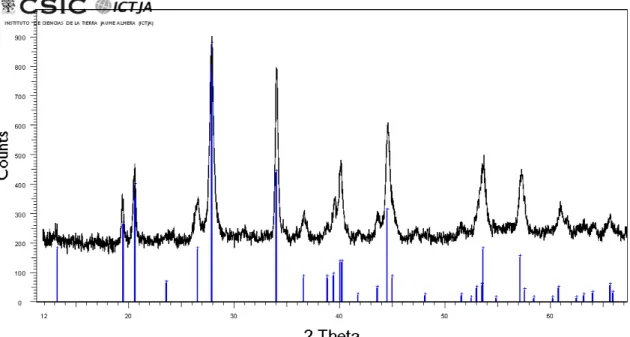
The first micro-diffraction measurements on hand-picked grains (i) allowed us to detect 192
numerous reflections from different phases, including quartz and roscoelite. However, 193
the identification (or indexing) of the unassigned peaks was not possible. We thus 194
concluded that single-crystal XRD studies of abellaite could not be carried out, since the 195
mineral is too-fine grained and cannot be separated from the substrate and associated 196
minerals. 197
Instead, and in spite of the large broadening expected for the conventional Bragg-198
Brentano geometry (ii) due to sample roughness, these measurements turned out to be 199
crucial to identify the mineral. For these measurements, relatively flat areas of the 200
samples were selected to reduce the width of the XRD peaks. Figure S1, which is freely 201
available online as Supplementary Material linked to this article on the GSW website of 202
the journal, http://eurjmin.geoscienceworld.org, shows a diffraction scan of abellaite, 203
together with the diffraction pattern of synthetic NaPb2(CO3)2(OH) from Brooker et al. 204
(1982), where this phase was indexed as hexagonal. As can be seen in the figure, the 205
pattern of the synthetic phase closely matches the XRD scan. Thus, given the chemical 206
data and the results of vibrational spectroscopies (Raman and IR, see below), these 207
XRD measurements indicate that the mineral is the natural analogue of synthetic 208
NaPb2(CO3)2(OH). 209
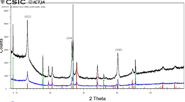
We would like to mention that, after these results, we were able to identify several 210
reflections from the NaPb2(CO3)2(OH) phase in the micro-diffraction scans obtained 211
from hand-picked material (i). In the case of the XRD scans obtained from several 212
hand-picked grains by using a low-background sample holder (iii), the XRD peaks from 213
abellaite are more easily identified, as can be seen in Fig. S2. In such case, and most 214
likely because more amount of the mineral is measured, the peaks from 215
NaPb2(CO3)2(OH) clearly show up in the scans, which further confirms that abellaite is 216
the natural analogue of this synthetic compound. In these scans, however, the (00l) 217
reflections from roscoelite are also visible, which indicates that these measurements are 218
strongly affected by preferential orientation of the grains. 219
The powder XRD measurements obtained with conventional Bragg-Brentano 220
measurements (ii) and those obtained from millimeter-sized areas of dense aggregates 221
of abellaite (iv) allowed us to refine the cell parameters and atomic positions of the 222
mineral. Rietveld refinements were performed with the program TOPAS 4.2, from 223
Bruker. In some cases, weak peaks from associated minerals such as quartz or roscoelite 224
showed up in the scans. These additional features were taken into account in the 225
refinements by including additional peaks. As starting point, here we employ the 226
structural information of NaPb2(CO3)2(OH), space group P63mc, from Krivovichev & 227
Burns (2000a). We would like to note that Belokoneva et al. (2002) synthesized 228
NaPb2(CO3)2(OH) crystals and inferred a P31c space group from the analysis of 229
systematic absences in single-crystal XRD measurements. In the present work we have 230
relied on the data from Krivovichev & Burns (2000a) because application of the 231
Addsym tool from the Platon program (Spek, 2009) indicates that the P31c structure has 232
missing symmetries, which does not occur in the case of the P63mc structure. 233
Table 1 shows the powder diffraction data thus obtained. Note that at low (measured) 234
lattice spacings, dmeas, the observed XRD peaks are fairly broad and weak, and therefore 235
numerous reflections contribute to those features. Thus, below 1.75 Å the dmeas values 236
given in Table 1 correspond to the observed peak maxima. 237
The hexagonal unit-cell parameters refined from the powder data are: a = 5.254(2), c = 238
13.450(5) Å and V = 321.5(2) Å3. These values are close to those reported by Brooker et 239
al. (1982) (a = 5.273(2), c = 13.448(5) Å), Krivovichev & Burns (2000a) (a = 5.276(1), 240
c = 13.474(4) Å) and also to those of Belokoneva et al. (2002) (a = 5.268(4), c = 241
13.48(1) Å). Crystallographic information of abellaite is shown in Table 2. The 242
resulting atomic positions of the mineral, starting from the structural information in 243
Krivovichev & Burns (2000a), are given in Table 3. The values thus obtained are not far 244
from those obtained in that work. The thermal parameters were not refined in the 245
present study, since the errors in these parameters were too large. Thus, the isotropic 246
thermal displacement parameters given in Table 3 are the same as in Krivovichev & 247
Burns (2000a). 248
5. Raman and FTIR spectroscopies 249
Raman spectra were excited with the second harmonic of a continuous-wave Nd:YAG 250
laser (λ = 532 nm). The spectra were acquired with a Horiba Jobin-Yvon LabRam 251
spectrometer coupled to a high-sensitive CCD detector, using a long-working distance 252
50x objective that produced a ~4 µm laser spot on the sample. A notch-filter was 253
employed to filter out the elastically scattered (Rayleigh) radiation, providing access to 254
Raman signals above ~100 cm1. The spectra were directly obtained from denser 255
aggregates of abellaite microcrystals on as-collected specimens. 256
A typical Raman spectrum of abellaite is shown in Fig. 4a. As can be seen in the figure, 257
the spectra are dominated by a sharp band at ~1058 cm1 and a weaker, broader feature 258
at ~1391 cm1 that can be attributed to symmetric (υ1) and asymmetric (υ3) stretching 259
modes of CO groups, respectively (Brooker et al., 1983). As in most carbonate 260
compounds, the υ1 peak is the strongest feature in the Raman spectra. In the high-261
wavenumber region (~3504 cm1), a weak peak arising from O-H stretching vibrations 262
shows up in the spectra, confirming the presence of hydroxyl groups in the crystal 263
structure of abellaite. These two observations support the XRD results, i.e., the 264
assignment of the mineral to a basic carbonate compound. Frequency values for the 265
different spectral features that appear in Fig. 4a are listed in Table 4. For comparison, 266
the Table also shows the Raman features above 200 cm1 reported in Brooker et al. 267
(1983) for the case of synthetic NaPb2(CO3)2(OH). As can be seen by comparing the 268
data from both works, the Raman spectrum of abellaite closely resembles that of the 269
synthetic NaPb2(CO3)2(OH) samples studied in Brooker et al.(1983). It should be noted, 270
however, that a few additional weak features appear in the spectra of abellaite. Most 271
likely, these bands arise from associated minerals, as is the case of the weak, sharp peak 272
at ~465 cm1, which we assign to quartz. However, it cannot be ruled out that some of 273
these peaks actually correspond to abellaite. From one hand, it should be recalled that 274
NaPb2(CO3)2(OH) has 26 atoms in the unit cell and therefore 78 normal modes of 275
vibration, 32 of which correspond to Raman-active optical modes [Γopt=9A1 + 11E1 + 276
12E2, as follows from group-theory analysis of the vibrational modes of 277
NaPb2(CO3)2(OH), space group P63mc, as in Krivovichev & Burns (2000a)]. Thus, 278
given that abellaite is found as aggregates of small microcrystals (while Brooker et al., 279
1983 studied synthetic samples containing much larger crystals), some of the observed 280
peaks might correspond to Raman-active modes of abellaite that would not be observed 281
in single-crystalline material. On the other hand, lattice distortions or incorporation of 282
impurities in the lattice of abellaite could also give rise to these additional modes. 283
The close resemblance between the vibrational properties of abellaite and synthetic 284
NaPb2(CO3)2(OH) is further supported by means of Fourier transformed infrared (FTIR) 285
spectroscopy measurements, which were obtained by using a Spotlight 150 microscope 286
interfaced to a Frontier (Perkin Elmer) spectrometer. For this purpose, hand-picked 287
material in form of microcrystalline aggregates of the mineral was loaded into a 288
diamond anvil cell. FTIR transmission analyses were acquired with 4 cm1 resolution by 289
integrating 32-64 scans from 550 to 4000 cm1. Figure 4b shows a FTIR spectra of 290
abellaite, which is dominated by the characteristic, strong band at ~1425 cm1 arising 291
from the υ3 asymmetric internal stretching modes of the CO group. Weaker bands that 292
can be assigned to the out-of-plane and in-plane bending modes (υ2, υ4) and to the υ1 -293
symmetric stretching modes of CO also appear in the spectra (see Table 5). In 294
addition to these fundamental modes, the spectra also show the characteristic absorption 295
bands of OH-stretching at ~3500 cm1. The band at ~998 cm1 can also be tentatively 296
assigned to PbOH bending vibrations (Brooker et al., 1983). The observation of 297
characteristic absorption bands of CO and OH groups confirms that the mineral is a 298
basic carbonate compound. Table 5 gives measured frequencies in our sample and data 299
from Brooker et al. (1983) and Belokoneva et al. (2002). As in the case of the Raman 300
results, the IR spectra are very similar to those of synthetic NaPb2(CO3)2(OH) (Brooker 301
et al., 1983; Belokoneva et al., 2002). Again, however, it should be noted that some 302
minor differences are observed between the three works (and also between Brooker et 303
al. (1983) and Belokoneva et al. (2002), see Table III). As in the case of the Raman 304
data, the observed differences can be mainly attributed to the origin of the investigated 305
samples (natural vs synthetic) and also to the presence of accessory minerals and 306
impurities in the case of the natural specimens. The present FTIR measurements, 307
however, confirm that abellaite is a basic carbonate compound, very similar to the 308
synthetic phases studied in previous works. 309
6. Chemical data 310
Chemical analyses of abellaite crystals were carried out using a JEOL JXA-8230 311
electron microprobe at the Scientific and Technological Centres of the University of 312
Barcelona. The samples were set in epoxy resin and subsequently polished. The mount 313
thus obtained allowed us to identify around 10 microcrystals of abellaite for chemical 314
analysis. Figure 3 displays backscattered electron (BSE) micrographs showing some of 315
the analysed crystals, with sizes ranging from 2 µm to 10 µm. Wavelength-dispersive 316
spectrometry (WDS) measurements were conducted using a 10 kV accelerating voltage 317
and 2 nA beam current with a defocused 2 to 5 µm spot in order to both minimize 318
surface damage of the crystals (Iizuka et al., 2012) and improve the spatial resolution of 319
the analyses. For this material, the penetration range of the incident electrons drops 320
from 2.75 µm at 20 kV accelerating voltage down to 0.85 µm at 10 kV accelerating 321
voltage (estimation performed using the auxiliary program "Tables" from the 322
PENELOPE distribution (Salvat, 2015)). Counting times were 30 s peak and 15 s 323
background for all analysed elements (Ca, K, Na and Pb). In order to improve counting 324
statistics, Na and Pb were simultaneously measured with two spectrometers, using TAP 325
and TAPH crystals for Na, and PETH and PETL crystals for Pb. No other elements 326
were detected with the WDS scans. Standards used for calibration were: caracolite 327
[Na3Pb2(SO4)3Cl] for Na, cerussite for Pb, orthoclase for K and calcite for Ca. The 328
matrix correction procedure XPP (Pouchou and Pichoir, 1988) was used to convert 329
specimen intensity ratios into concentrations. The matrix correction calculations were 330
carried out by means of O stoichiometry (Lane & Dalton, 1994), considering CO2 by 331
difference to 100%. C-H-N elemental analyses were not performed because it was not 332
possible to unambiguously isolate the hand-picked mineral from the substrate and 333
accessory minerals, as found by XRD measurements. Thus, O, C and H were 334
determined by stoichiometry assuming the ideal formula of the synthetic compound. 335
The average of 10 electron-microprobe determinations and the corresponding ranges are 336
given in Table 6. On the basis of 7 O atoms, the resulting charged-balanced empirical 337
formula for the mineral is Na0.96Ca0.04Pb1.98(CO3)2(OH), while the ideal formula is that 338
of the synthetic phase [NaPb2(CO3)2(OH)]. 339
7. Discussion 340
On the basis of X-ray powder diffraction data, vibrational spectra (Raman and IR) and 341
chemical composition, it can be concluded that abellaite is closely related to the 342
synthetic compound NaPb2(CO3)2(OH) studied by several authors (Brooker et al., 1982; 343
Krivovichev & Burns, 2000; Belokoneva et al., 2002). The fact that the same 344
conclusion is reached with techniques based on different principles (i.e., vibrational vs 345
structural) indicates that the chemical formula of the mineral must be very close to that 346
of the synthetic phases. Although a direct determination of H2O and CO2 contents was 347
not possible due to the small size of the microcrystals, the WDS measurements did 348
confirm that the Pb/Na ratio of the mineral is close to 2 as in the synthetic phase. As 349
suggested by the WDS data, however, abellaite may also contain Ca ions, which are 350
probably incorporated into Pb sites because these two elements may have the same 351
valence (2+). However, taking into account the close ionic radii of Na and Ca, it cannot 352
be ruled out that Ca may also substitute for Na in the mineral. 353
Abellaite adds to the list of uncommon carbonate minerals found in the galleries of the 354
Eureka mine, such as andersonite, čejkaite, bayleyite, liebigite, or schröckingerite 355
(Abella i Creus & Viñals, 2009; Abella i Creus & Viñals, 2012). Following the results 356
of Krivovichev & Burns (2000a), the mineral crystallizes in the hexagonal system, 357
space group P63mc. As discussed by these authors, this phase contains a hexagonal 358
sublattice of Pb atoms and therefore the structure can be related to the structure of other 359
Pb hydroxide carbonates such as plumbonacrite (see also Krivovichev & Burns, 2000b). 360
The crystal structure of abellaite is also closely related to that of the recently approved 361
new mineral grootfonteinite Pb3O(CO3)2 (Siidra et al., 2015). This mineral crystallizes 362
in the space group P63mc with lattice parameters a = 5.303(1) Å and c = 13.770(2) Å, 363
which are only slightly larger than those of abellaite. 364
According to its chemical composition, abellaite is closely related to hydrocerussite, 365
plumbonacrite, and sanrománite. In the New Dana classification, abellaite can be 366
assigned to the carbonates, hydroxyl class (16a) and, similarly to hydrocerussite 367
(trigonal, space group R-3m), to the group of carbonates - hydroxyl or halogen, 368
(AB)3(XO3)2Zq (subgroup 16a.02). In the Nickel-Strunz classification, as in the case of 369
hydrocerussite, the mineral can be classified as a carbonate with additional ions, without 370
H2O, with Pb or Bi (subgroup 5.BE). Both plumbonacrite and sanrománite crystallize in 371
the hexagonal system, P63mc. Sanrománite is an anhydrous carbonate that also 372
incorporates Ca cations, which further suggests that other (basic, hydrated or 373
anhydrous) carbonates with Pb, Ca, Na and also K may also be found in nature. 374
With regard to this, we would like to mention that a recent analysis on carbon mineral 375
ecology applying a Large Number of Rare Events (LNRE) model has predicted that 376
numerous (~145) undiscovered carbon mineral species may exist on Earth (Hazen et al., 377
2016). According to these predictions, a large subset of such undescribed minerals 378
should be compounds of O, H, Ca, and Na. This is the case of NaPb2(CO3)2(OH), which 379
is listed in Hazen et al. (2016) as one of the possible missing carbon minerals. 380
According to the LNRE predictions, other basic or hydrated carbonate minerals, 381
compatible with the oxidizing conditions of secondary mineralizations like those 382
involved in the formation of abellaite, could also be found. More work is thus required 383
to identify and describe possible new carbon minerals and to confirm the predictions of 384
that work. 385
Acknowledgements: This paper is dedicated to the memory of our colleague and friend 386
Prof. Joan Viñals, who passed away on November 2013. Professor Viñals was an expert 387
mineralogist who studied different new species such as barahonite and who initiated the 388
characterization of abellaite. We are grateful to Joan Abella i Creus for supplying us 389
with the mineral samples and for useful information about the mineral and the Eureka 390
mine. We would like to thank Eva Perisé and the Town Hall of Torre de Capdella for 391
valuable support and assistance. Work partially funded by ERDF‐EU Ref. CSIC10‐4E‐ 392
141. 393
References 394
Abella i Creus, J. & Viñals, J. (2009): Čejkaite, arsenuranylite, compreignacite, 395
natrozippeite and other rare uranium minerals in the Eureka mine, Castell-estaó, La 396
Torre de Cabdella, Lleida, Catalonia. Mineral Up, 2, 52–71. 397
Abella i Creus, J. & Viñals, J. (2012): New minerals from the Eureka mine: 398
metamunirite, schröckingerite, boltwoodite and gordaite, Castell-estaó, la Torre de 399
Cabdella, Lleida, Catalonia, Spain. Mineral Up, 3, 14–18. 400
Belokoneva, E.L., Al’-Ama, A.G., Dimitrova, O.V., Kurazhkovskaya, V S., 401
Stefanovich, S.Y. (2002): Synthesis and crystal structure of the new carbonate 402
NaPb2(CO3)2(OH). Crystallography Reports, 47, 217–222. 403
Béziat, D., Joron, J. L., Monchoux, P., Treuil, M., & Walgenwitz, F. (1991): 404
Geodynamic implications of geochemical data for the Pyrenean ophites (Spain-France). 405
Chemical geology, 89(3-4), 243-262. 406
Brooker, M.H., Sunder, S., Taylor, P., Lopata, V.J. (1983): Infrared and Raman spectra 407
and X-ray diffraction studies of solid lead(II) carbonates. Canadian Journal of 408
Chemistry, 61, 494–502. 409
Castillo, M., Torró, L., Campeny, M., Villanova, C., Tauler, E., Melgarejo, J.C. (2009): 410
Mineralogía del Depósito de Uranio Eureka (Castell-estaó, Pirineo, Cataluña). Macla 411
11, 53-54. 412
Foord, E.E. & Mills, B.A. (1978): Biaxiality in ‘isometric’ and ‘dimetric’ crystals. 413
American Mineralogist, 63, 316-325. 414
Hazen, R.M., Hummer, D.R., Hystad G., Downs R.T., J.J. Golden (2016): Carbon 415
mineral ecology: Predicting the undiscovered minerals of carbon. American 416
Mineralogist, 101, 889–906. 417
Iizuka, Y. (2012): Electron microprobe study of otolith: migratory behaviour and habitat 418
of three major temperature species of eels. JEOL News 47, 33-50. 419
Krivovichev, S.V. & Burns, P.C. (2000a): Crystal chemistry of basic lead carbonates. 420
III. Crystal structures of Pb3O2(CO3) and NaPb2(OH)(CO3)2. Mineralogical Magazine, 421
64, 1077-1087. 422
Krivovichev, S.V. & Burns, P.C. (2000b): Crystal chemistry of basic lead carbonates. 423
II. Crystal structure of synthetic ‘plumbonacrite’. Mineralogical Magazine, 64, 1069-424
1075. 425
Pouchou, J.L. & Pichoir, F. (1988): A simplified version of the “PAP” model for matrix 426
corrections in EPMA. In Microbeam Analysis, Newbury D.E. (Ed.), pp. 319-324. San 427
Francisco Press: San Francisco. 428
Lane, S.J & Dalton, J.A. (1994): Electron microprobe analysis of geological carbonates. 429
American Mineralogist 79, 745-749. 430
Mey, P.H.W., Nagtegaal, P.J.C., Roberti, K.J., & Hartevelt, J.J.A. (1968): 431
Lithostratigraphic subdivision of post-Hercynian deposits in the south-central Pyrenees, 432
Spain. Leidse Geologische Mededelingen, 41, 221-228. 433
Muñoz, J. A. (1992): Evolution of a continental collision belt: ECORS-Pyrenees crustal 434
balanced cross-section. In Thrust tectonics (pp. 235-246). Springer Netherlands. 435
Salvat, F. (2015): PENELOPE-2014: A code system for Monte Carlo simulation of 436
electron and photon transport. OECD/NEA Data Bank, Issy-les-Moulineaux, France. 437
Siidra, O.I., Jonsson, E., Chukanov, N.V., Pekov, I.V., Zinyakhina, D.O., Polekhovsky, 438
Y.S. & Yapaskurt, V.O. (2015): Grootfonteinite, IMA 2015-051. CNMNC Newsletter, 439
27, 1226; Mineralogical Magazine, 79, 1229–1236. 440
Spek, A. L. (2009): Structure validation in chemical crystallography. Acta 441
Crystallographica D, 65, 148-155. 442
Turner, B. R. (1985): Uranium mineralization in the Karoo basin, South Africa. 443
Economic Geology, 80(2), 256-269. 444
Vergés, J. & Muñoz, J. A. (1990): Thrust sequences in the southern central Pyrenees. 445
Bulletin de la Société géologique de France, 8, 265-271. 446
TITLES OF TABLES
Table 1. Measured and calculated X-ray powder diffraction data for abellaite. 447
Table 2. Crystallographic data for abellaite. 448
Table 3. Atomic positions for abellaite. The equivalent isotropic thermal displacement 449
parameters correspond to those of the synthetic analogue (Krivovichev & Burns, 2000). 450
Table 4. Raman data for abellaite and for synthetic NaPb2(CO3)2(OH) (Brooker et al., 451
1983). 452
Table 5. FTIR spectroscopy data for abellaite and for synthetic NaPb2(CO3)2(OH) as 453
reported in Brooker et al. (1983) and Belokoneva et al. (2002). 454
Table 6. Electron microprobe analysis of abellaite. 455
TABLES
Table 1
Iobs dobs (Å) Icalc dcalc (Å) hkl
3 6.725 2 6.731 002 7 4.550 9 4.555 010 17 4.310 19 4.315 011 2 3.768 2 3.773 012 9 3.362 15 3.366 004 100 3.193 100 3.197 013 3 2.704 1 2.707 014 84 2.627 91 2.630 110 16 2.447 19 2.450 1-2-2,112 6 2.315 6 2.318 015 29 2.275 27 2.278 020 65 2.243 37 2.246 021 29 2.244 006 <1 2.155 2 2.158 022 19 2.070 22 2.072 1-2-4,114 95 2.029 98 2.031 023 25 2.011 29 2.013 016 1 1.884 4 1.886 024 5 1.770 10 1.772 017 1.706 numerous 1.603 numerous 1.517 numerous 1.420 numerous 1.313 numerous 1.256 numerous 1.215 numerous Table 2
Ideal formula NaPb2(CO3)2(OH) Formula weight 574.41
Crystal system, space group Hexagonal, P63mc Unit-cell dimensions a = 5.254(2) Å
c = 13.450(5) Å
V (Å3) 321.5(2)
Z 2
Table 3
Atom x/a y/b z/c Ueq
Na 1/3 -1/3 0.656(5) 0.021 Pb(1) 2/3 1/3 0.821(5) 0.018 Pb(2) 1/3 2/3 0.993(5) 0.019 C(1) 0 0 0.78(1) 0.015 C(2) 0 0 -0.01(1) 0.016 O(1) 0.70(1) -0.15(1) 0.77(1) 0.020 O(2) 0.25(1) 0.12(1) 0.07(1) 0.025 O(3) 2/3 1/3 0.65(1) 0.046 Table 4 Raman shift (cm-1) This work Raman shift (cm-1)
Brooker et al. (1983) Assignment
202 m 202 m Lattice modes 280 m, br 285 m, br 683 m 681 ms υ4 (CO32-) 695 sh 868 w 868 w υ2 (CO32-) 1038 vw 1036 vw 1058 vs 1052 w, sh 1057.2 vs υ1 (CO32-) 1068.4 w
Most likely overlapped with
band at 1391cm-1 ~ 1350 w, sh 1391 s, br 1392 s υ3 (CO32-) 1695 vw Second orders 1730 vw 1750 vw 3504 vw, br 3500 vw OH stretching
Table 5 Infrared (cm-1) This work Infrared (cm-1) Brooker et al. (1983) Infrared (cm-1) Belokoneva et al. (2002) Assignment 360 vw 455 s 475 m 530 w, br 688 s 693 s 695 s υ4 (CO32-) 825 w 844 m 843 s 847 s υ2 (CO32-) 998 w, br 988 m, br δPbOH 1053 w 1053 w 1055 w υ1 (CO32-) 1425 vs, br 1435 vs, br 1432 vs, br υ3 (CO32-) 1750 w, br 1743 w 1758 w Second order 3500 w, br 3495 w 3480 w, br OH stretching Table 6
Element wt.% Range Nominal
Na 3.88 3.69 ˗ 4.03 4.00 K < DL* - Ca 0.29 0.14 ˗ 0.51 - Pb 72.03 71.14 ˗ 72.7 72.14 C 4.17 4.18 O 19.47 19.50 H 0.17 0.17 Total 100.01 100.00 *Detection limit (DL) for K was in the range 900-1100 ppm
FIGURE CAPTIONS
Figure 1. (a) Location of the Eureka mine in the small town of Castell-estaó, Torre de Capdella (Lleida province), in the Southern Pyrenees. (b) Geological map showing the area around the Eureka mine area (modified from Atlas Geològic de Catalunya 1:5000). (c) Entrance to the gallery where abellaite was found, surrounded by redbed sediments.
Figure 2. (a) Optical photomicrograph of abellaite aggregates. (b) Secondary-electron micrograph showing subhedral abellaite microcrystals. (c) Backscattered electron (BSE) image showing aggregates of idiomorphic, pseudohexagonal abellaite crystals. (d) Detail of the tabular pseudohexagonal crystals, showing prominent pinacoidal faces.
Figure 3. (a) and (b) BSE images showing selected abellaite crystals as well as associated minerals. 1: abellaite...obtained from the probe for chemical analyses. 1: abellaite, 2: vanadinite, 3: Pb-ich intergrowth, 4: Co-rich carbonate, 5: quartz, 6: calcium carbonate, 7: hidrozincite, 8: sphalerite: 9: uraninite, 10: unknown arseniovanadate.
Figure 4. (a) Raman spectrum of abellaite. (b) FTIR transmission spectrum of abellaite. In both figures, the asterisks indicate spectral features that have not been reported in synthetic NaPb2(CO3)2(OH).
FIGURES
SUPPLEMENTARY MATERIAL
Figure S1. Powder X-ray diffraction scan from dense abellaite aggregates. For comparison, the X-ray diffraction pattern of synthetic NaPb2(CO3)2(OH) from Brooker
Figure S2. Powder X-ray diffraction scans of abellaite grains on a low-background Si sample holder. For comparison, X-ray diffraction pattern of synthetic NaPb2(CO3)2(OH)