Trivandrum-695 023 Kerala, India
Recent Advances in Pharmaceutical Sciences III, 2013: 23-43 ISBN: 978-81-7895-605-3 Editors: Diego Muñoz-Torrero, Amparo Cortés and Eduardo L. Mariño
2. Antifungal compounds from plants
Roser Vila, Blanca Freixa and Salvador Cañigueral
Unitat de Farmacologia i Farmacognòsia, Facultat de Farmàcia Universitat de Barcelona, E-08028 Barcelona, Spain
Abstract. Due to the increase of the incidence of fungal infections
in humans and the limitations of the available antimycotic drugs, among which the emergence of resistant strains, there is a need for the discovery of new antifungal agents. Plants, especially those used in Traditional Medicine, linked to an unmatched chemical diversity, either as pure compounds or as plant extracts, provide unlimited opportunities for the development of new antifungals. In recent years, compounds from different phytochemical groups have been described as having antifungal activity, including polyphenols, saponins, or peptides, among others, as well as essential oils and their constituents. After screening of ethnopharmacologically selected plants, mainly from Latin-America, followed by bio-guided isolation, our group has described the antifungal activity of different types of plant constituents, such as sesquiterpenes, triterpenes, flavonoids, lignans, fatty acids and essential oils.
Introduction
Fungal infections arise as an increasing health problem with a high
economic cost. A recent study of the epidemiology of sepsis caused by fungi in USA showed that between 1979 and 2000 they increased by 207%.
Correspondence/Reprint request: Dra. Roser Vila, Unitat de Farmacologia i Farmacognòsia, Facultat de Famàcia, Universitat de Barcelona, E-08028 Barcelona, Spain. E-mail: [email protected]
The morbidity and mortality associated with these infections are substantial:
deaths due to mycoses increased from 1557 in 1980 to 6534 in 1997, in the USA, and they were mainly associated with Candida, Aspergillus and
Cryptococcus sp. infections [1-3].
The most frequent fungal pathogens include the yeasts Candida sp., (mainly C. albicans and other species especially virulent, like C. glabrata) and Cryptococcus sp., filamentous fungi like Aspergillus sp., Fusarium sp. and Rhizopus sp., and the dermatophytes Trycophyton sp., Microsporum sp. and Epidermophyton sp. Particularly, Candida and Aspergillus represent 70-90% and 10-20%, respectively, of all invasive fungal infections [4].
A number of reasons have led to the significant augmentation of the incidence of fungal infections in humans in recent decades [1,4,5]:
- The increase in the number of immunocompromised patients who frequently develop opportunistic superficial and systemic fungal infections.
- The invasive medical procedures in hospitals (use of catheters, peritoneal dialysis, haemodialysis, parenteral nutrition, etc.).
- The emergence of new and uncommon virulent fungal strains, especially favoured by an increase of the population mobility that facilitates a higher exposure to endemic fungal pathogens.
The evolution of fungal infections is further worsened by the fact that they are often difficult to diagnose, being recognized when they are already at an advanced stage.
Currently, there is a variety of antifungal drugs with different mechanisms of action. However, their effectiveness is limited due to a number of factors, such as fungistatic mode of action, lack of oral and intravenous preparations due to low solubility, drug toxicity and the development of drug resistance [6,7].
From another standpoint, fungi cause significant losses in agronomy, taking in account that, depending on the crop type and the region, approximately 10-20% of the cultivated food plants are destroyed by plant pathogens, among them pathogenic fungi. The use of fungicides is still considered essential for increasing crop yields [8].
1. Search for new antifungals
So, therefore, there is a clear need to search for new antifungal agents providing new mechanisms of action, with a broad spectrum of antifungal activity, fewer dose-limiting side effects, and economic [9,10].
Nature offers a wide chemical diversity and natural products are an important source for the development of new therapeutic agents, in particular anti-infective agents. Some of the antifungal drugs most recently introduced in clinical practice (echinocandines and sordarines) are derived from natural products [4,11].
Among the different natural sources of active principles, the plant kingdom has a special significance:
- Plants offer a wide biodiversity that also involves a high chemodiversity. - It is estimated that less than 10% of the plant secondary metabolites have been isolated. In many cases these substances act as plant's defence mechanisms against attack by microorganisms, insects, etc. [12].
- Plants are an important source of medicinal preparations (extracts, teas, essential oils, etc.) used in traditional medicine since antiquity, and particularly many of them in the treatment of infectious diseases. It is estimated that 14-28% of species of higher plants are used in therapy. In addition, to date, more than 600 herbal drugs showed antifungal activity, but only a small part of them have been studied concerning their active constituents.
Therefore, either as a source of pure compounds or extracts, the plant kingdom offers a huge potential for new drug development.
2. Methods for research of antifungal agents from plants
2.1. Selection of plant species
The first key point is the selection of plant species to be studied, from which the success of the research depends greatly.
The ethnopharmacological selection is based on existing knowledge about the traditional use of plants to treat diseases and it has shown to increase the probability of success in drug discovery, reducing empiricism. The isolation and characterization of bioactive molecules from ethnopharmacologically selected plant species allows not only their use as drug or as chemical or pharmacological leads to produce new analogues, but also, and not less important, to set a scientific basis for the development and use of herbal drug preparations and to validate the use of traditional medicinal preparations.
Therefore, there is a strong interest in performing screenings of antifungal activity from medicinal plants used in traditional medicine for the treatment of fungal diseases [13-24].
The first step in the ethnopharmacological-based research is the collection of data (most used plant species, part of the plant, forms of preparation and administration, uses, etc.) from indigenous population and their analysis. It is essential that the collected plant material is correctly authenticated by a botanist and that a voucher specimen is included in an internationally recognized herbarium.
Most of the research conducted by our group has been performed on Latin American plants. Latin America is a very suitable source of plant material for drug discovery because [14]:
- It has a high biodiversity, one of the highest in the world in terms of vascular plants.
- Many of the plant species have never been investigated for the research of bioactive agents.
- There is a rich tradition in the use of medicinal plants and this ethnopharmacological knowledge has been relatively well preserved by the indigenous population.
2.2. Preparation of crude extracts
Both, the appropriate extraction method and the most suitable solvents should be carefully selected. Concerning the method, usually plant extracts are obtained by maceration or percolation. Since the chemical nature of the active principles to be isolated is not known, the plant material is often subjected to a successive extraction with increasing polarity solvents (for example, Cl2CH2 and MeOH 95%) to obtain a wide range of compounds of
different polarities. Additionally, an extract equivalent to the preparation commonly used in traditional medicine (usually aqueous or alcoholic extract) should be prepared. In the particular case of aromatic herbs, it is useful to obtain the essential oil by hydrodistillation.
2.3. Screening of the antifungal activity
For the screening of antifungal activity and its monitoring during the isolation and purification of the active principles, test methods based on diffusion are mostly used, whereas to quantify the activity, both, dilution and diffusion methods may be employed. Reviews on the different techniques used for the evaluation of the antifungal activity of natural products can be found in Cos et al. [25], Das et al. [26], Engelmeier and Hadacek [8], Jacob and Walker [27] and Ríos et al. [28].
One of the most widely employed methods to assess the antifungal activity of plant extracts is the agar disk diffusion assay [29].
In our research, a number of plant extracts and essential oils, mainly coming from plants traditionally used for the treatment of skin diseases, were subjected to a screening of antifungal activity using the agar disk diffusion assay against a panel of fungal strains including both yeasts and filamentous fungi. Among them, there were opportunistic and skin and mucosal membranes pathogenic fungi, as well as phytopathogenic strains that can cause significant losses in agriculture and hinder food conservation [15,16]. The most active extracts were selected for subsequent bio-guided fractionation with the aim of isolating and characterizing the active principles.
2.4. Bio-guided isolation and identification of active constituents
The most time-consuming step is the isolation and purification of the active compounds, which is mainly achieved through bio-guided fractionation. It is based on the combined use of preparative chromatographic techniques (e.g. column chromatography, medium- and high-pressure liquid chromatography, classical and centrifugal thin-layer chromatography, counter current chromatographic techniques) [30] together with bioautography with the aim of selecting in each step of the chromatographic fractionation the fractions containing the active compounds for further purification.
Contact bioautography allows the detection of active constituents in a complex mixture within a reasonable time. It is an agar diffusion method in which the components of a sample (raw extract or fraction) previously separated by thin-layer chromatography (TLC) diffuse from the stationary phase into the culture medium inoculated with the fungus that has been more sensitive to the crude extract in the antifungigram [31].
To do this, the already developed and dried TLC plate is placed overlaid
on the inoculated Sabouraud dextrose agar medium, leaving enough time (1 h at 4ºC) so that diffusion takes place. Then, the TLC plate is removed and
the inoculated agar incubated at 30ºC for 48 h. The growth inhibition zones observed are related to the substances separated on a TLC reference plate developed with the same mobile phase. In this way, active compounds can be detected on the TLC plate and the fractionation can be directed to the isolation of these compounds.
Some considerations should be taken into account when evaluating the results of a bioautography: a) due to the limited exposure time (usually 1 h) of the inoculum to the potential antifungal agents the sensitivity is lower than in the antifungigram, b) according to their polarity the components of the sample will diffuse differently through the agar, and c) the result may be affected by the stability of the product on the TLC plate [27].
Nonetheless, bioautography is a useful technique which complements the antifungigram, since it allows the detection of active substances that in the initial raw extract are present in a very low concentration, or that coexist with others that antagonize their activity.
Furthermore, it is necessary to bear in mind that during the fractionation of an active extract a loss of activity may occur due to a degradation of active principles during isolation, or to the separation of different components of the extract acting synergistically.
Finally, once the active compounds are purified, their chemical structure should be established by using different standard spectroscopic techniques,
such as UV-Vis, IR, different types of mass spectrometry, and a variety of experiments of 1H-NMR and 13C-NMR, involving both mono- and
bidimensional methods.
Particularly, in the case of essential oils, identification of the constituents
of the oil and their fractions is usually achieved by means of qualitative and quantitative analysis by GC-FID, GC-MS [32] and also, if necessary, by
13C-NMR [33-35].
2.5. Evaluation of the antifungal activity
The antifungal activity of the extracts, essential oils and pure isolated compounds should be quantitatively evaluated through determination of their minimal inhibitory concentration (MIC) and minimal fungicidal concentration (MFC).
The MIC is defined as the minimal concentration of a product that inhibits the visible growth of microorganisms. It allows quantification of the sensitivity of a certain fungal strain to an antifungal agent. MIC is usually determined using either broth macrodilution or microdilution methods, the latter being the most used, through a standard two-fold dilution technique [36]. In the case of lipophilic substances that are dissolved in solvents poorly soluble in water, addition of Tween 80 as an emulsifier is recommended.
Because samples may not be 100% antifungal at their MIC, viable fungal cells may remain in the wells in which fungal growth is not appreciated by sight. Thus, the determination of the MFC is also necessary to establish their fungicidal activity. The MFC can be established technically by extending the MIC (microdilution method) [37].
3. Major chemical groups with antifungal activity
A number of compounds isolated from plants are reported to have antifungal activities. However, development of useful antifungal drugs from
the majority of them has not yet been possible, even though mainstream medicine is increasingly receptive to the use of antimicrobial and other drugs derived from plants, because of the resistances developed against traditional antibiotics [12].
One of the reasons is the fact that most of the antifungal plant products, i.e. extracts, essential oils or pure constituents, have only been tested for their in vitro activities, their effectiveness in animals and/or humans remaining unknown. Pharmacokinetics, undesirable effects, interactions and toxicity should also be studied [38].
The interest of finding new antifungal compounds from plants is that they can constitute precursors leading to more active antifungal drugs [12]. In addition, potential benefits of combination therapy which include enhanced potency of antifungal efficacy, reduced selection of resistant organisms (particularly for flucytosine), and reduced toxicities due to lower dosing, are important to be taken into account.
Several chemical groups of plant constituents have been reported for their antifungal activity. Among them: some fatty acids, peptides and alkaloids, but it should be especially highlighted the groups of polyphenols and terpenoids which have furnished a huge variety of active structures. Interesting reviews have been published [9,12,38-40].
Examples of active compounds with different chemical structures, some of them the result of research conducted by our group, are discussed below.
3.1. Antifungal fatty acids
Some fatty acids have shown antifungal activity mainly due to their capacity to disrupt the bacterial membrane. They are able to interfere in the cell membrane structure displacing phospholipids and increasing its permeability. A well-known example is undecylenic acid, a semisynthetic product prepared from ricinoleic acid obtained from castor oil, the oil from seeds of Ricinus communis. Undecylenic acid is mainly used in the treatment of superficial mycoses.
During the search for new antifungals from Paraguaian plants, a series of fatty acids were isolated by our group from the bark of Calycophyllum spruceanum var. multiflorum (Griseb.) Chodat & Hassl. (Rubiaceae). Bio-guided fractionation of the dichloromethane extract afforded a mixture of eight fatty acids which were identified through GC-MS, HPLC-MS, 1H- and
13C-NMR analysis. Five of them were acetylenic: 6-hexadecinoic acid,
6-heptadecinoic acid, 6-octadecinoic acid, 6-nonadecinoic acid and 6-eicosinoic acid, whereas the other three were saturated: palmitic acid,
found in nature, and particularly three of them constituted new natural products: 6-heptadecinoic, 6-nonadecinoic and 6-eicosinoic acids. The mixture was active against the dermatophytes Microsporum gypseum and Tricophyton mentagrophytes with MIC and MFC values of 0.25 μg/mL, lower than those of the reference drugs nystatin (MIC and MFC values from 1.25 to 2.50 μg/mL) and amphotericin B (MIC and MFC values from 0.31 to 0.63 μg/mL) [41].
3.2. Polyphenols
Many polyphenols, including simple phenols, flavonoids, coumarins, tannins and quinone derivatives, have been reported to have antifungal activity, which is partly dependent on the number and position of hydroxyl groups. They may act by inhibiting microorganism enzymes, possibly through reaction with sulfhydryl groups or through less specific interactions with proteins [12].
The antifungal spectra, potency and fungicidal properties of two chalcones from Zuccagnia punctata Cav. (Fabaceae), a plant used in traditional medicine in Argentina as antiseptic and wound-healing poultices, evidenced that they could have potential as antifungal agents for human beings. Particularly, dihydroxy-3’-methoxychalcone and
2’,4’-dihydroxychalcone (Fig. 1) evidenced very strong activities against several clinical strains of the dermatophytes T. mentagrophytes and T. rubrum with MIC values in the range of 1.9-15.6 µg/mL and MFC values between 1.9 and 7.8 µg/mL. In addition, 2’,4’-dihydroxychalcone showed a moderate activity against clinical isolates of the yeasts Candida sp. and Cryptococcus neoformans. Regarding their mode of action, both chalcones appeared to be fungicidal rather than fungistatic; moreover, 2’,4’-dihydroxychalcone did not disrupt the fungal membranes up to 4×MFC and did not act by inhibiting the synthesis of polymers of the fungal cell wall. So, their mechanism would be different than that of most antifungal drugs in clinical use, i.e. amphotericin B, azoles or echinocandins [42].
In the course of our search for new antifungals from Latin-American plants different active polyphenolic structures were identified in our laboratory, among which flavonoids, caffeic acid derivatives and lignans.
Two isoflavones, genistein and biochanin A, were isolated from the bark of Andira surinamensis (Boudt.) Splitz (Leguminosae) collected in Perú. It is a high tree mainly distributed in tropical America, whose bark is used in traditional medicine for different purposes. The dichloromethane extract, which showed a good activity against several fungal strains in the agar disk diffusion assay [15], was subjected to activity-guided fractionation yielding the two isoflavones. Particularly, biochanin A (Fig. 1) evidenced antifungal
activity especially against yeasts and dermatophytes. Both isoflavones have previously been isolated from other Andira sp. [43] and are known to have antifungal activity [44]. In addition, the activity of the methanol extract against the dermatophyte M. gypseum and T. mentagrophytes was mainly related to the presence of condensed tannins [45].
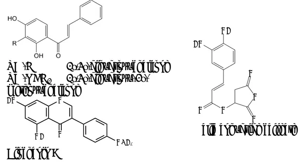
R= -H 2’,4’-dihydroxychalcone R= -OCH3 2’,4’-dihydroxy-3’-methoxychalcone O O O O O OH HO
Malic anhydride caffeate
O O OCH3 OH HO Biochanin A
Figure 1. Some antifungal polyphenols isolated from Latin-American plants
[41,42,45].
The isolation and identification of a new antifungal caffeic acid derivative was performed from the methanol extract of the aerial parts of Geophila repens (L.) I. M. Johnston from Paraguay, a Rubiaceae used by the Quechuas for the treatment of fungal infections [46]. In this case, the active methanol extract was subjected to partition with increasing polarity solvents, namely dichloromethane, ethyl acetate and butanol, the fraction obtained with the last solvent being the most active against C. neoformans, M. gypseum and T. mentagrophytes, its activity even persisting for one week in the agar disk diffusion assay. Fractionation by successive column chromatography on Sephadex® LH-20 led to the isolation of an ester of caffeic acid and malic
anhydride identified as 2,5-dioxotetrahydrofuran-3-yl caffeate (malic anhydride caffeate) (Fig. 1), a compound not previously described, as the main responsible for the activity [47].
Piper fulvescens C. DC. is an herbaceous plant growing in Paraguay whose leaves are used in traditional medicine [48]. Activity-guided fractionation of their dichloromethane extract, which showed growth inhibitory activity against the yeasts Candida albicans and Saccharomyces
cerevisiae, as well as against the dermatophytes M. gypseum and T. mentagrophytes [15], led to the isolation of three neolignans identified as
conocarpan, eupomatenoid 5 and eupomatenoid 6 (Fig. 2), for which antifungal activity had not previously been described. Conocarpan showed the broadest spectrum of activity, being active against yeasts and dermatophytes, whereas eupomatenoid 6 showed the strongest activity
against M. gypseum and T. menthagrophytes with MIC values of 0.5 and 1.0 µg/mL, respectively. Moreover, eupomatenoid 5 turned out to be the least
active. These results suggest that the absence of a methoxy group at 3’ position of the phenyl-propenyl-benzofuran structure plays an important role in the antifungal activity, whereas the saturation of the C2-C3 bond influences the selectivity [49].
R= -CH3 Eupomatenoid 5
R= -H Eupomatenoid 6
Conocarpane
Figure 2. Antifungal neolignans from the leaves of Piper fulvescens [49].
3.3. Terpenoids
Among terpenes a variety of chemical structures showing interesting antifungal activities have been reported in recent years [9]. Their mechanism of action is not fully understood although it has been suggested to involve membrane disruption due to their lipophilic nature [12].
A high-throughput whole animal assay for the identification of antifungal compounds which uses the nematode Caenorhabditis elegans as a heterologous host [50] was performed to screen the activity of 2560 natural products against C. albicans. From all of them, those compounds that produced higher survival rates of the infected nematodes were polyglycosylated saponins with different types of skeletons. Two of them, a spirostanic one (aginoside) and a triterpenic one derived from barrigenol, were selected for further studies concerning their mechanisms of action. They
inhibited C. albicans isolates at relatively low concentrations (16 and 32 µg/mL), including isolates resistant to clinically used antifungal agents,
formation were also disrupted by these two saponins at concentrations below the MIC. Furthermore, no hemolysis of erythrocytes was observed at three-fold the MIC for C. albicans, suggesting the saponins may have a preference for binding to fungal ergosterol when compared to cholesterol of the erythrocyte membranes [51].
One of the few antifungal products from plants that have been subjected to clinical trials is the methanol extract from leaves of Solanum chrysothrichum Schldh. (Solanaceae), a herbal drug used in Mexico for the treatment of fungi-associated dermal and mucosal infections, particularly recommended to cure Tinea pedis. In vitro assays showed antifungal activity against T. mentagrophytes, T. rubrum and M. gypseum, and a preliminar clinical study performed with the methanolic extract at 5% in a cream applied topically on patients affected of Tinea pedis showed clinical success with no side effects [52]. Later on, five new antimycotic spirostanic saponins were isolated and identified [53,54]. Clinical trials with a standardised methanol extract demonstrated not only the therapeutic efficacy and tolerability when applied topically as a cream against T. pedis [55] but also on Pityriasis capitis [56] and as suppository on Candida sp.-associated vaginal infection [57].
The screening on plant products with antifungal activity based on an ethnopharmacological approach, carried out by our research group,
allowed the characterization of several antimycotic terpene compounds, for which antifungal activity had not previously been reported. Among them, the diterpene lactone acanthoaustralide and the sesquiterpene ester 6-cinnamoyloxy-1-hydroxyeudesm-4-en-3-one (Fig. 3) were respectively
isolated through bio-guided fractionation from the dichloromethane extracts
OH O O O 6-Cinnamoyloxy-1-hydroxy- eudesm-4-en-3-one O OH O OH Acanthoaustralide
Figure 3. The sesquiterpene ester 6-cinnamoyloxy-1-hydroxy-eudesm-4-en-3-one and
the diterpene lactone acanthoaustralide are the antifungal principles of the roots of
Vernonanthura tweedieana and the leaves of Acanthospermum australe, respectively
from leaves of Acanthospermum australe (Loefl.) O. Kuntze and from roots of Vernonanthura tweedieana (Baker) H. Rob. (=Vernonia tweedieana Baker), two Asteraceae traditionally used in Paraguay for the treatment of skin diseases such as eczema and itching. Both compounds evidenced a strong activity against T. mentagrophytes with the same MIC and MFC values: 2 μg/mL in the case of acanthoaustralide and 4 μg/mL in the case of the sesquiterpene derivative [58,59].
3.4. Essential oils
Special consideration should be deserved to essential oils, many of which have been used for centuries for their antiseptic activity. Antimicrobial essential oils are nowadays used in the treatment of dermal and mucosal infections, such as acne, onychomycosis, vaginal infections, gingivitis or teeth and gums healthcare, as well as food preservatives and in the control of crop diseases.
A number of papers dealing with the antifungal activity of many essential oils have been published during the last decades, some of them discussing relationships between structures and activities and with interesting contributions on their mechanism of action, which is complex and has not yet been fully explained [39,40,60-66].
Different types of constituents of the essential oils have proven antimycotic properties (Fig. 4). Those that have reported the strongest activity are
phenolic monoterpenes (e.g. thymol and carvacrol), phenylpropanoids (such as eugenol), alcoholic monocyclic monoterpenes (e.g. α-terpineol and terpinen-
4-ol), and bicyclic monoterpene hydrocarbons (e.g. α-pinene) and ketones
Thymol Carvacrol Terpinen-4-ol α-Terpineol
Eugenol α-Pinene Camphor
(e.g. camphor). The acidic nature of the hydroxyl group of phenols facilitates a hydrogen bond with enzyme active centres, being responsible of their high activity [67,68].
In the course of our research, several essential oils have demonstrated strong antifungal activities. The investigation of their composition together with bio-guided fractionation led to the identification of the main active constituents responsible for the activity.
From the antifungal essential oils from Piper amalago L. collected in Panama, a series of four new 2-acyl-3-hydroxycyclohex-2-en-1-ones, which represented more than 50% and 85% of the oils of the leaves and the stems, respectively, were isolated and their structures were established by EI-MS and NMR spectroscopy as 2-hexanoyl- (1), 2-octanoyl- (2), 2-decanoyl- (3) and 2-dodecanoyl-3-hydroxycyclohex-2-en-1-one (4) (Fig. 5). 1 and 2, the two main constituents of the essential oils, showed a good antifungal activity against different yeasts. The hexanoyl- compound (1) showed the highest activity against C. albicans and S. cerevisiae, whereas the octanoyl- derivative (2) was the most active against Candida lactis-condensi and the decanoyl- one (3) only showed activity against C. lactis-condensi. These findings suggested that the length of the lateral chain modulates the activity [69,70].
The essential oil from fresh leaves of Plinia cerrocampanensis Barrie (Myrtaceae) from Panama, mainly constituted by oxygenated sesquiterpenes (65.9%), especially α-bisabolol (42.8%), bisabolol oxide B (10.3%) and trans-nerolidol (9.4%), was assayed against several fungal strains showing the strongest activity against M. gypseum, T. mentagrophytes and T. rubrum with MIC values from 32 to 125 μg/mL [71].
The unusual compound (-)-5,6-dehydrocamphor was found to be the major constituent in the essential oil from aerial parts of Zuccagnia punctata Cav. (Fabaceae) from Argentina, reaching percentages from 12.3% to 56.5%.
(CH2)n O O OH 1: n=1 2-hexanoyl-3-hydroxycyclohex-2-en-1-one 2: n=3 2-octanoyl-3-hydroxycyclohex-2-en-1-one 3: n=5 2-decanoyl-3-hydroxycyclohex-2-en-1-one 4: n=7 2-dodecanoyl-3-hydroxycyclohex-2-en-1-one
Figure 5. Antifungal acyl-hydroxycyclohexenones of the essential oils from
It was isolated from nature for the first time and its structure was completely elucidated from NMR data. The essential oil showed antifungal activity against the dermatophytes M. gypseum, T. rubrum and T. mentagrophytes with MIC values between 15.6-125 μg/mL, T. rubrum being the most susceptible species [72].
The composition and the antifungal activity of the essential oil from the rhizome and root of Ferula hermonis Boiss. (Apiaceae), a perennial shrub that grows on the Hermon mountain between Lebanon and Syria, evidenced this species as a potential source of interesting antifungal agents. The exhaustive analysis of the essential oil combined with successive bio-guided fractionations afforded different active products among which the unusual acetylenic compound 3,5-nonadiyne (Fig. 6), α-bisabolol and a fraction with 73% jaeschkeanadiol benzoate were the most active against the dermathophytes M. gypseum and T. mentagrophytes. Particularly, the fraction with the latter compound showed the strongest activity with MIC and MFC values superior or equivalent to those of the positive controls amphotericin B and nystatin [32].
The daucane aryl esters jaeschkeanadiol benzoate (teferidin) and jaeschkeanadiol p-hydroxybenzoate (ferutinin) (Fig. 6) were also isolated from hexane and dichloromethane extracts of the rhizome and root of Ferula hermonis. Determination of MIC and MFC values of both compounds evidenced a stronger antifungal activity for ferutinin than for teferidin. Particularly, T. mentagrophytes was the most sensitive strain with MIC and MFC values ranging from 8 to 256 μg/mL [73]. These results suggest that the presence of a phenolic hydroxyl in the acidic moiety of the ester, in the case 3,5-Nonadiyne R= -H Teferidin (jaeschkeanadiol benzoate) R= -OH Ferutinin (jaeschkeanadiol p-hydroxybenzoate)
Figure 6. Antifungal constituents from the rhizome and root of Ferula hermonis
of jaeschkeanadiol p-hydroxybenzoate, enhances the antifungal activity. This would be consistent with the fact that compounds with phenolic structures, like carvacrol, eugenol and thymol show stronger antimicrobial activities than those lacking a phenol group, as for example p-cymene [39,40,74].
Essential oils may induce visible signs of their action against fungi that can be observed as morphological changes both under microscope and as colony macro-features. Thus, eugenol and carvacrol have been found to cause significant deformations of the hyphae in food spoiling mould Cladosporium herbarum, which were related to their action on some cell wall enzymes, such as chitinases and glucanases [75]. Borneol induces morphological changes in cell walls and cellular organelles of Aspergillus fumigatus and Epidermophyton floccosum and inhibits mycelial growth of both filamentous fungi [76]. Furthermore, the essential oil from Thymus vulgaris inhibited mycelial development and germination of sporangiospores in Rhizopus oryzae [77], whereas the oil from Zataria multiflora caused vacuolisation of the cytoplasm and cell swelling, detachment of the cell membrane from the cell wall and deformation of mycelia in Aspergillus flavus [78]. Souza et al. [79] reported on the efficacy of Origanum essential oils against potentially pathogenic fungi, finding that the oil from O. vulgare completely inhibited radial mycelia growth of T. rubrum and M. canis and elicited disruption of cell structure in Aspergillus flavus.
Some studies evidenced that morphological modifications in the fungal cell may be induced even at sub-MIC concentrations [38]. In this way, Zuzarte et al. [62,65] found that essential oils from Portuguese Lavandula sp. completely inhibited filamentation in Candida albicans at concentrations as low as MIC/16. Also, farnesol derivatives prevent the conversion of yeast form into the invasive filamentous growth form of C. albicans at sub-inhibitory levels [80]. These findings could be of potential interest for the treatment of candidiasis.
Concerning their mechanism of action, as complex mixtures of a variety of chemical structures, essential oils do not seem to act on specific targets in fungal cells but to trigger different types of radical reactions [38,67,81].
Degradation of the cell wall and damage to both cytoplasmic and mitochondrial membranes have been proposed as the main mechanisms leading to changes in permeability and subsequent leakage and cell death [81]. It has been reported that lipophilic compounds accumulate in the lipid bilayers of the cell membrane altering its permeability and causing disruption of lipid packing and leakage of inorganic ions, while more hydrophilic constituents can enter the cytoplasm [38]. A mechanism of action related to damage of mitochondrial membranes and subsequent metabolic arrest rather than to direct disruption of cytoplasmic membrane or cell wall has been suggested by Zuzarte [65].
In the last years, investigations on the mechanisms of action of essential oils and their constituents showed that the antifungal activity is mediated through several mechanisms other than membrane disruption and inhibition of cell division. Inhibition of both ergosterol biosynthesis and H+-ATPase leading to disruption of ion homeostasis and cell death [82-86] and induction of oxidative stress [87] have been proposed as main targets of antifungal essential oils. Several terpenoids inhibit microbial oxygen uptake and oxidative phosphorylation, the phenolic and non-phenolic alcohols exhibiting the strongest inhibitory effects, followed by aldehydes and ketones [66].
Synergistic effect between the essential oil constituents or between different essential oil mixtures against yeasts and dermatophytes have been reported by several authors [60,88,89]. But, due to increasing fungal resistance to classical antifungal drugs and their toxicity, what is more interesting from a clinical point of view is that combinations of essential oils or their pure constituents with commercial antifungal drugs used in therapeutics, such as amphotericin B or ketoconazole, can potentiate the activity of the latter and, thus, may lead to a dose reduction and minimization of secondary effects [90-94]. Although the combination of essential oils with other drugs can be very useful it has to be carefully considered, since depending on the percentage of each one antagonistic effects may also occur [40].
4. Conclusions
Despite the enormous progress made in recent decades in medicine, fungal infections are still an unsolved health problem, mainly due to the fact that the available antifungal drugs are of limited effectiveness. The plant kingdom is a source of medicinal preparations used in Traditional Medicine that offers a wide chemical diversity, making it of huge potential for new drug development. The research developed over the last years by different groups has led to the characterization of a variety of chemical structures with outstanding antifungal activity. Unsaturated fatty acids, polyphenols, triterpene derivatives and, particularly, essential oils have been shown as promising groups, some of them showing a synergistic effect with commercial antifungal drugs used in therapeutics. In case of complex mixtures such as essential oils synergy within some of their constituents have also been reported.
Acknowledgements
Part of our research was performed inside the framework of the Iberoamerican Program CYTED. Financial support from Generalitat de
Catalunya, EC FEDER funds and Fundación Roviralta is also greatly acknowledged.
References
1. Pfaller, M. A., Diekema, D. J. 2007, Clin. Microbiol. Rev., 20, 133.
2. Martin, G. S., Mannino, D. M., Eaton, S., Moss, M. 2003, N. Engl. J. Med., 348, 1546.
3. McNeil, M. M., Nash, S. L., Hajjeh, R. A., Phelan, M. A., Conn, L. A., Plikaytis, B. D., Warnock. D. W. 2001, Clin. Infect. Dis., 33, 641.
4. Di Santo, R. 2010, Nat. Prod. Rep., 27, 1084.
5. Mathew, B. P., Nath, M. 2009, ChemMedChem, 4, 310.
6. Bastert, J., Schaller, M., Korting, H. C., Evans, E. G. V. 2001, Int. J. Antimicrob.
Agents, 17, 81.
7. Fromtling, R. A., Rahway, N. J. 1987, Recent Trends in the Discovery, Development and Evaluation of Antifungal Agents. In: Proceedings of an International Telesymposium. J. R. Prous, Barcelona.
8. Engelmeier, D., Hadacek, F. 2006, Antifungal Natural Products: Assays and Applications. In: Advances in Phytomedicine Series. Volume III: Naturally Occurring Bioactive Compounds. M. Rai and M. C. Carpinella (Eds.), chapter 17. Elsevier, Amsterdam.
9. Abad, M. J., Ansuategui, M., Bermejo, P. 2007, Arkivoc, 7, 116. 10. Barrett, D. 2002, Biochim. Biophys. Acta, 1587, 224.
11. Tomishima, M., Ohki, H., Yamada, A., Maki, K., Ikeda, F. 2008, Bioorg. Med.
Chem. Lett., 18, 1474.
12. Arif, T., Bhosale, J. D., Kumar, N., Mandal, T. K., Bendre, R. S., Lavekar, G. S., Dabur, R. 2009, J. Asian Nat. Prod. Res., 11, 621.
13. Damián-Barrillo, L. M., Salgado-Garciglia, R., Martínez-Muñoz, R. E., Martínez-Pacheco, M. 2008, Open Nat. Prod. J., 1, 27.
14. Svetaz, L., Zuljan, F., Derita, M., Petenatti, E., Tamayo, G., Cáceres, A.,
Cechinel Filho, V., Giménez, A., Pinzón, R., Zacchino, S., Gupta, M. 2010,
J. Ethnopharmacol., 127, 137.
15. Freixa, B.; Vila, R., Vargas, L., Lozano, N., Adzet, T., Cañigueral, S. 1998,
Phytother. Res., 12, 427.
16. Portillo, A., Vila, R., Freixa, B., Adzet, T., Cañigueral, S. 2001, J. Ethnopharmacol., 76, 93.
17. López, V., Akerreta, S., Casanova, E., García-Mona, J. M., Cavero, R. Y., Calvo, M. I. 2008, Pharm. Biol., 46, 602.
18. Maregesi, S. M., Pieters, L., Ngassapa, O. D., Apers, S., Vingerhoets, R., Cos, P., Vanden Berghe, D. A., Vlietinck, A. 2008, J. Ethnopharmacol., 119, 58.
19. Webster, D., Taschereau, P., Belland, R. J., Sand, C., Rennie, R. P. 2008,
J. Ethnopharmacol., 115, 140.
20. Chea, A., Jonville, M.-C., Bun, S.-S., Laget, M., Elias, R., Duménil, G., Balansard, G. 2007, Am. J. Chin. Med., 35, 867.
21. Kadam, R. M., Allapure, R. B., Biradar, R. G., Jadhav, B. S. 2008, Eco. Env. &
22. Zabka, M., Pavela, R., Gabrielova-Slezakove, L. 2011, J. Agric. Food Chem., 91, 492.
23. Coelho de Souza, G., Haas, A. P. S., von Poser, G. I., Schapoval, E. E. S., Elisabetsky, E., J. Ethnopharmacol., 90, 135.
24. McCutcheon, A. R., Ellis, S. M., Hancock, R. E. W., Towers, G. H. N. 1994,
J. Ethnopharmacol., 44, 157.
25. Cos, P., Vlietinck, A., Vanden Berghe, D., Maes, L. 2006, J. Ethnopharmacol., 106, 290.
26. Das, K., Tiwari, R. K. S., Shrivastava, D. K. 2010, J. Med. Plants Res., 4, 104. 27. Jacob, M. R., Walker, L. A. 2005. Natural Products and Antifungal Drug
Discovery. In: Methods in Molecular Medicine, Vol. 118: Antifungal agents: Methods and Protocols. E. J. Ernst and P. D. Rogers (Eds.), Humana Press Inc., Totowa, NJ.
28. Ríos, J. L., Recio, M. C., Villar, A. 1998, J. Ethnopharmacol., 23, 127.
29. Barry, A. L., Thornsberry, C. 1991. Susceptibility test: Diffusion test procedures. In: Manual of Clinical Microbiology. A. Balows, W. J. Hausler, K. L. Hermann, H. D. Isenberg, H. J. Shadomy (Eds.), American Society for Microbiology, Washington D.C. pp. 1526-1542.
30. Hostettmann, K., Marston, A., Hostettmann, M. 1998. Preparative Chromatography Techniques: Applications in Natural Product Isolation, 2nd Ed. Springer-Verlag, Berlin Heidelberg.
31. Rahalison, L., Hamburger, M., Hostettmann, K., Monod, M., Frenk, E. 1991,
Phytochem. Anal. 2, 199.
32. Al-Ja’fari, A.-H., Vila, R., Freixa, B., Tomi, F., Casanova, J., Costa, J., Cañigueral, S. 2011, Phytochemistry, 72, 1406.
33. Tomi, F., Casanova, J. 2006, Acta Hort., 723, 185.
34. Tomi, F., Bradesi, P., Bighelli, A., Casanova, J. 1995, J. Magn. Reson. Anal. 1, 25. 35. Rezzi, S., Bighelli, A., Castola, V., Casanova, J. 2002, Appl. Spectrosc., 56, 312. 36. Murray, P. R., Baron, E. J., Pfaller, M. A., Tenover, F. C., Yolken, R. H. 1999.
Manual of Clinical Microbiology, 7th Ed. American Society for Microbiology,
ASM Press, Washington D. C.
37. Balows, A., Hausler, W. J., Hermann, L. K., Isenberg, H. D., Shadomy, H. J.
1991. Manual of Clinical Microbiology, 5th Ed. American Society for
Microbiology, ASM Press, Washington D. C. 38. Pauli, A. 2006, Med. Res. Rev., 26, 223.
39. Dorman, H. J. D., Deans, S. G. 2000, J. Appl. Microbiol., 88, 308. 40. Lang, G., Buchbauer, G. 2012, Flavour Fragr. J., 27, 13.
41. Portillo, A. 2004. Principios antifúngicos de especies vegetales de Paraguay. PhD Thesis. Universitat de Barcelona.
42. Svetaz, L., Agüero, M. B., Álvarez, S., Luna, L., Feresin, G., Derita, M., Tapia, A., Zacchino, S. 2007, Planta Med., 73, 1074.
43. Lock de Ugaz, O., Costa, J., Sanchez, L., Ubillas Sanchez, R. P., Tempesta, M. S. 1991, Fitoterapia, 62, 89.
44. Weidenboerner, M., Hindorf, H., Jha, H.C., Tsotsonos, P., Egge, H. 1990,
45. Freixa, B. 2001, Búsqueda de principios antifúngicos de especies vegetales latinoamericanas. PhD Thesis. Universitat de Barcelona.
46. Schultes, R. E., Raffauf, R. F. 1990. The Healing Forest. Medicinal and Toxic Plants of the Northwest Amazonia. Dioscorides Press, Portland (Oregon).
47. Portillo, A., Vila, R., Freixa, B., Parella, T., Ferro, E., Cañigueral, S. 2004, 2nd
Joint Symposium IOCD-CYTED on Chemistry, Biological and Pharmacological Properties of Medicinal Plants from the Americas. São Pedro (Brasil).
48. Gonzalez Torres, D. M. 1970. Catálogo de Plantas Medicinales (y Alimenticias y Útiles) usadas en Paraguay. Asunción, Paraguay. pp. 430.
49. Freixa, B., Vila, R., Ferro, E. A., Adzet, T., Cañigueral, S. 2001, Planta Med., 67, 873.
50. Okoli, L., Coleman, J. J., Tempakakis, E., An, W. F., Holson, E., Wagner, F., Conery, A. L., Larkins-Ford, J., Wu, G., Stern, A., Ausubel, F. M., Mylonakis, E. 2009, PLoS ONE, 4, e7025.
51. Coleman, J. J., Okoli, I., Tegos, G. P., Holson, E. B., Wagner, F. F., Hamblin, M. R., Mylonakis, E. 2010, ACS Chem. Biol., 5, 321.
52. Lozoya, X., Navarro, V., García, M., Zurita, M. 1992, J. Ethnopharmacol., 36, 127. 53. Alvarez, L., Pérez, M. C., González, J. L., Navarro, V., Villareal, M. L., Olson, J.
O. 2001, Planta Med., 67, 372.
54. Zamilpa, A., Tortoriello, J., Navarro, V., Delgado, G., Alvarez, L. 2002, J. Nat.
Prod., 65, 1815.
55. Herrera-Arellano, A., Rodríguez-Soberanes, A., Rivera, M. A., Martínez-Cruz, E., Zamilpa, A., Alvarez, L., Tortoriello, J. 2003, Planta Med., 69, 390.
56. Herrera-Arellano, A., Jiménez-Ferrer, E., Vega-Pimentel, A. M., Martínez-Rivera, M. A., Hernández-Hernández, M., Zamilpa, A., Tortoriello, J. 2004,
Planta Med., 70, 483.
57. Herrera-Arellano, A., Jiménez-Ferrer, E., Zamilpa, A., Martínez-Rivera, M. A., Rodríguez-Tovar, A. V., Herrera-Alvarez, S., Salas-Andonaegui, M. L., Nava-Xalpa, M. Y., Méndez-Salas, A., Tortoriello, J. 2009, Planta Med., 75, 466.
58. Portillo, A., Vila, R., Freixa, B., Adzet, T., Ferro, E., Cañigueral, S. 2003, 3rd
World Congress on Medicinal and Aromatic Plants for Human Welfare. Chiang Mai, Thailand.
59. Portillo, A., Vila, R., Freixa, B., Ferro, E., Parella, T., Casanova, J., Cañigueral, S. 2005, J. Ethnopharmacol., 97, 49.
60. Pina-Vaz, C., Gonçalves Rodrigues, A., Pinto, E., Costa-de-Oliveira, S., Tavares, C., Salgueiro, L., Cavaleiro, C., Gonçalves, M. J., Martínez-de-Oliveira, J. 2004,
JEADV, 18, 73.
61. Pinto, E., Pina-Vaz, C., Salgueiro, L., Gonçalves, M. J., Costa-de-Oliveira, S., Cavaleiro, C., Palmeira, A., Rodrigues, A., Martínez-de-Oliveira, J. 2006, J. Med.
Microbiol., 55, 1367.
62. Zuzarte, M., Gonçalves, M. J., Cavaleiro, C., Canhoto, J., Vale-Silva, L., Silva, M. J., Pinto, E., Salgueiro, L. 2011, J. Med. Microbiol., 60, 612.
63. Zuzarte, M., Gonçalves, M. J., Canhoto, J. M., Salgueiro, L. 2011.
Antidermatophytic Activity of Essential Oils. In: Science Against Microbial Pathogens: Communicating Current Research and Technological Advances. A. Méndez-Vilas (Ed.). Microbiology Book Series, nº3, vol. 2, pp.1167-1178. Formatex Research Center, Badajoz (Spain).
64. Zuzarte, M., Vale-Silva, L., Gonçalves, M. J., Cavaleiro, C., Vaz, S., Canhoto, J., Pinto, E., Salgueiro, L. 2012, Eur. J. Clin. Microbiol. Infect. Dis., 31, 1359.
65. Zuzarte, M. 2012. Portuguese Lavenders: Evaluation of their Potential Use for Health and Agricultural Purposes. PhD Thesis. Universidade de Coimbra.
66. Griffin, S. G., Wyllie, S. G., Markham, J. L., Leach, D. N. 1999, Flavour Fragr.
J., 14, 322.
67. Knobloch, K., Pauli, A., Iberl, B., Weigand, H., Weis, N. 1989, J. Essent. Oil
Res., 1, 119.
68. Tampieri, M. P., Galuppi, R., Macchioni, F., Carelle, M. S., Falcioni, L., Cioni, P. L., Morelli, I. 2005, Mycopathologia, 159, 339.
69. Vila, R., Santana, A. I., Tomi, F., Mundina, M., Solís, P. N., Iglesias, J., Gupta, M. P., Casanova, J., Cañigueral, S. 2002, Rev. Fitoterapia, 2 (Supl 1), 322.
70. Freixa, B., Vila, R., Santana, A. I., Gupta, M. P., Cañigueral, S. 2005, 53th
Annual Congress of the Society for Medicinal Plant Research. Florence, Italy. 71. Vila, R., Santana, A. I., Pérez-Rosés, R., Valderrama, A., Castelli, M. V.,
Mendonca, S., Zacchino, S., Gupta, M. P., Cañigueral. 2011, Biores. Technol., 101, 2510.
72. Álvarez, S. L., Cortadi, A., Juárez, M. A., Petenatti, E., Tomi, F., Casanova, J., van Baren, C. M., Zacchino, S., Vila, R. 2012, Phytochemistry Lett., 5 ,194.
73. Al-Ja’fari, A.-H., Vila, R., Freixa, B., Costa, J., Cañigueral, S. Phytother. Res., in press.
74. Ultee, A., Bennik, M. H., Moezelaar, R. 2002, Appl. Environ. Microbiol., 68, 1561. 75. Adams, S., Kunz, B., Weidenbörner, M. 1996, J. Essent. Oil Res., 8, 535.
76. Lee, S. Y., Kim, S. H., Hong, C. Y., Park, M.-J., Choi, I.-G. 2013, Flavour
Fragr. J., 28, 129.
77. Lira Mota, K. S., Oliveira Pereira, F., Araújo de Oliveira, W., Oliveira Lima, I., Oliveira Lima, E. 2012, Molecules, 17, 14418.
78. Gandomi, H., Misaghi, A., Akhondzadeh Basti, A., Hamedi, H., Ramezani Shirvani, Z. 2011, Mycoses, 54, e429.
79. Souza, N. A. B., Lima, E. O., Guedes, D. N., Pereira, F. O., Leite de Souza, E., Barbosa de Sousa, F. 2010, Braz. J. Pharm. Sci., 46, 499.
80. Kim, S., Kim, E., Shin, D.-S., Kang, H., Oh, K.-B. 2001, Bioorg. Med. Chem.
Lett., 12, 895.
81. Bakkali, F., Averbeck, S., Averbeck, D., Idaomar, M. 2008, Food Chem.
Toxicol., 46, 446.
82. Bhatia, R., Shreaz, S., Khan, N., Muralidhar, S., Basir, S. F., Manzoor, N., Khan, L. A. 2012, J. Basic Microbiol., 52, 504.
83. Shreaz, S., Bhatia, R., Khan, N., Muralidhar, S., Basir, S. F., Manzoor, N., Khan, L. A. 2011, Fitoterapia, 82, 1012.
84. Khan, A., Ahmad, A., Akhtar, F., Yousuf, S., Xess, I., Khan, L. A., Manzoor, N. 2010, Res. Microbiol., 161, 816.
85. Rao, A., Zhang, Y., Muend, S., Rao, R. 2010, Antimicrob. Agents Chemother., 54, 5062.
86. Ahmad, A., Khan, A., Yousuf, S., Khan, L. A., Manzoor, N. 2010, Fitoterapia, 81, 1157.
87. Khan, A., Ahmad, A., Akhtar, F., Yousuf, S., Xess, I., Khan, L. A., Manzoor, N. 2011, FEMS Yeast Res., 11, 114.
88. Kalemba, D., Kunicka, A. 2003, Curr. Med. Chem., 10, 813.
89. Prasad, C. S., Shukla, R., Kumar, A., Dubey, N. K. 2010, Mycoses, 53, 123. 90. Khan, M. S. A., Ahmad, I. 2011, Appl. Microbiol. Biotechnol., 90, 1083.
91. Silva, F., Ferreira, S., Duarte, A., Mendonça, D. I., Domingues, F. C. 2011,
Phytomedicine, 19, 42.
92. Shin, S., Pyun, M.-S. 2004, Phytother. Res., 18, 827.
93. Rosato, A., Vitali, C., Gallo, D., Balenzano, L., Mallamaci, R. 2008,
Phytomedicine, 15, 635.
![Figure 2. Antifungal neolignans from the leaves of Piper fulvescens [49].](https://thumb-eu.123doks.com/thumbv2/123dokorg/4464676.31527/10.892.108.771.138.491/figure-antifungal-neolignans-leaves-piper-fulvescens.webp)