of July 24, 2018.
This information is current as
the Asymptomatic Stage of Infection
Immune Response That Appears Prevalent in
Induce a Predominant Th1-Type Adaptive
and Barbara Ensoli
Aurelio Cafaro, Fausto Titti, Paolo Monini, Fabrizio Ensoli
Giovanni Barillari, Mauro Magnani, Maria Elena Laguardia,
Stefania Bellino, Vittorio Francavilla, Antonella Caputo,
Scoglio, Barbara Collacchi, Filomena Nappi, Iole Macchia,
Antonella Tripiciano, Maria R. Pavone Cossut, Arianna
Emanuele Fanales-Belasio, Sonia Moretti, Valeria Fiorelli,
http://www.jimmunol.org/content/182/5/2888
doi: 10.4049/jimmunol.0711406
2009; 182:2888-2897; ;
J Immunol
References
http://www.jimmunol.org/content/182/5/2888.full#ref-list-1
, 14 of which you can access for free at:
cites 36 articles
This article
average *
4 weeks from acceptance to publication
Fast Publication!
•
Every submission reviewed by practicing scientists
No Triage!
•
from submission to initial decision
Rapid Reviews! 30 days*
•
Submit online.
?
The JI
Why
Subscription
http://jimmunol.org/subscription
is online at:
The Journal of Immunology
Information about subscribing to
Permissions
http://www.aai.org/About/Publications/JI/copyright.html
Submit copyright permission requests at:
Email Alerts
http://jimmunol.org/alerts
Receive free email-alerts when new articles cite this article. Sign up at:
Print ISSN: 0022-1767 Online ISSN: 1550-6606.
Immunologists, Inc. All rights reserved.
Copyright © 2009 by The American Association of
1451 Rockville Pike, Suite 650, Rockville, MD 20852
The American Association of Immunologists, Inc.,
is published twice each month by
The Journal of Immunology
by guest on July 24, 2018 http://www.jimmunol.org/ Downloaded from by guest on July 24, 2018 http://www.jimmunol.org/ Downloaded from
HIV-1 Tat Addresses Dendritic Cells to Induce a Predominant
Th1-Type Adaptive Immune Response That Appears Prevalent
in the Asymptomatic Stage of Infection
1
Emanuele Fanales-Belasio,* Sonia Moretti,* Valeria Fiorelli,* Antonella Tripiciano,
†Maria R. Pavone Cossut,* Arianna Scoglio,
†Barbara Collacchi,* Filomena Nappi,*
Iole Macchia,* Stefania Bellino,* Vittorio Francavilla,
†Antonella Caputo,
‡Giovanni Barillari,*
Mauro Magnani,
§Maria Elena Laguardia,
§Aurelio Cafaro,* Fausto Titti,* Paolo Monini,*
Fabrizio Ensoli,
†and Barbara Ensoli
2*
Tat is an early regulatory protein that plays a major role in human HIV-1 replication and AIDS pathogenesis, and therefore, it represents a key target for the host immune response. In natural infection, however, Abs against Tat are produced only by a small fraction (⬃20%) of asymptomatic individuals and are rarely seen in progressors, suggesting that Tat may possess properties diverting the adaptive immunity from generating humoral responses. Here we show that a Th1-type T cell response against Tat is predominant over a Th2-type B cell response in natural HIV-1 infection. This is likely due to the capability of Tat to selectively target and very efficiently enter CD1a-expressing monocyte-derived dendritic cells (MDDC), which represent a primary target for the recognition and response to virus Ag. Upon cellular uptake, Tat induces MDDC maturation and Th1-associated cytokines and -chemokines production and polarizes the immune response in vitro to the Th1 pattern through the transcriptional activation of TNF-␣ gene expression. This requires the full conservation of Tat transactivation activity since neither MDDC maturation nor TNF-␣ production are found with either an oxidized Tat, which does not enter MDDC, or with a Tat protein mutated in the cysteine-rich region (cys22 Tat), which enters MDDC as the wild-type Tat but is transactivation silent. Consistently with these data, inoculation of monkeys with the native wild-type Tat induced a predominant Th1 response, whereas cys22 Tat generated mostly Th2 responses, therefore providing evidence that Tat induces a predominant Th1 polarized adaptive immune response in the host. The Journal of Immunology, 2009, 182: 2888 –2897.
T
he definition of the type, strength, and breadth of the im-mune response against HIV-1 is important for the identi-fication of immune correlates of protection in natural HIV infection or after vaccination as well as of virus pathogenesis, i.e., mechanism(s) for eluding or diverting the immune response. The HIV-1 Tat protein is a regulatory protein produced early after in-fection, which plays key roles in HIV replication and AIDS patho-genesis, therefore representing a suitable target for the develop-ment of an anti-HIV vaccine (1– 4). Indeed, in HIV-1-infected individuals the presence of a humoral immune response to Tat is associated with a reduced risk of disease progression (5–9). In preclinical studies performed in mice and monkeys, vaccination with either the biologically active Tat protein or tat DNA wasshown to be safe and immunogenic, inducing both humoral and cell-mediated immune responses capable of controlling virus rep-lication and disease onset upon challenge with pathogenic simian HIV (SHIV)3(10 –13). In natural HIV infection
,however, anti-Tat
Abs are produced by only a small fraction of individuals, predom-inantly in the asymptomatic stage (4). In addition, similar frequen-cies of anti-Tat Ab-positive individuals are found in different geo-graphical regions, which harbor multiple HIV-1 subtypes (6, 7, 14). The reason for such a limited Ab response against Tat in infected individuals is unclear. In particular, it is unknown whether this is due to a limited recognition and response to Tat by the immune system in natural infection or to the predominant induc-tion of Th1 T cells in the generainduc-tion of the anti-Tat adaptive im-mune responses during primary infection. Indeed, Tat possesses adjuvant properties that may play a key role in influencing the development of the primary immune response (15).
In particular, during acute infection of T cells by HIV-1, Tat is released in the extracellular milieu (15–18) and taken up by neigh-bor cells and modulates the expression of several cytokines and chemokines (15–17). These molecules not only alter the cell per-missivity to productive HIV infection but also modulate multiple immune functions affecting the development of the adaptive im-mune response. In particular, native Tat targets and is selectively
*National AIDS Center, Istituto Superiore di Sanita`, Rome, Italy;†
Department of Pathology and Microbiology, Istituti Fisioterapici Ospitalieri—S. Gallicano Hospital, IRCCS, Rome, Italy;‡
Department of Histology, Microbiology and Medical Biotech-nology, University of Padua, Padua, Italy; and§
Institute for Biological Chemistry “G. Fornaini”, University of Urbino, Urbino, Italy
Received for publication June 7, 2007. Accepted for publication January 1, 2009. The costs of publication of this article were defrayed in part by the payment of page charges. This article must therefore be hereby marked advertisement in accordance with 18 U.S.C. Section 1734 solely to indicate this fact.
1
This work was supported by grants from the National AIDS Program, and from the Italian Concerted Action on HIV/AIDS Vaccine Development, Istituto Superiore di Sanita`, Rome, Italy and by the EC Commission under the VI Framework Programme of Research and Technological Development (2002–2006), Project no. LSHP-CT-2004-503487, AIDS Vaccine Integrated Project.
2
Address correspondence and reprint requests to Dr. Barbara Ensoli, National AIDS Center, Istituto Superiore di Sanita`, Viale Regina Elena 299, 00161 Rome, Italy. E-mail address: [email protected].
3Abbreviations used in this paper: SHIV, simian HIV; MDDC, monocyte derived
dendritic cells; wt, wild type; ICS, intracellular cytokine staining; CBA, cytometric bead assay; SFC, spot forming cells.
Copyright © 2009 by The American Association of Immunologists, Inc. 0022-1767/09/$2.00
The Journal of Immunology
www.jimmunol.org/cgi/doi/10.4049/jimmunol.0711406
by guest on July 24, 2018
http://www.jimmunol.org/
internalized by monocyte-derived dendritic cells (MDDC), pro-moting their maturation and Ag-presenting functions via pathways, which are capable of driving Th1-type responses (15). This sug-gested that Tat acts as both Ag and adjuvant in the setting of a primary immune response (15).
Here we show that a Th1-type T cell response against Tat is predominant over a Th2-type B cell response in asymptomatic HIV-1 infection. In vitro studies indicate that Tat targets CD1a-positive MDDC, which represent a primary target for the recog-nition and response to virus Ag, and induces their maturation and Th1-associated cytokines and -chemokines production through the transcriptional activation of TNF-␣ gene expression, address-ing to Th1-type immune responses in vitro. This effect requires the full conservation of Tat transactivation activity since neither MDDC maturation nor TNF-␣ production are found with either an oxidized Tat, which does not enter MDDC, or a transactivation silent Tat protein mutated in the cysteine-rich region (cys22 Tat), which, however, enters MDDC as the wild-type (wt) Tat. Finally, inoculation of monkeys with the native Tat induces a predominant Th1 response, whereas cys22 Tat generates mostly Th2 responses, therefore providing a direct demonstration that, in vivo, native Tat induces a Th1-polarized adaptive immune response.
Materials and Methods
Clinical studies: patients
Seventy-two HIV-1-positive clinically asymptomatic patients were exam-ined in the present study. All were naive for antiretroviral therapy and classified as stage A disease category. Of the 72 individuals, 47 were males and 25 females. The median age was 35 y (range 19 – 67). Mean value of CD4⫹T cell counts was 565.2⫾ 163.9/l. Mean value of plasma HIV-1 RNA was 3.75⫾ 0.88 Log(range 1.69–4.96 Log). No concomitant active opportunistic infections were present at the time of the study. All subjects gave written informed consent according to the Ethical Committee procedures.
Measurement of serum Ab against the Tat protein
Serum samples were tested for Ab to Tat protein by ELISA as previously described (14). To obtain high levels of sensitivity and specificity, two different protocols were applied, which are characterized by different sec-ondary Ab and amplification systems (14). All sera were tested by both methods. Each sample was tested in duplicate with the addition of a control well, coated with the Ag buffer alone, to normalize for unspecific binding (14).
IFN-␥ and IL-4 ELISpot assay on human PBMC
ELISpot assays were performed to quantify the frequency of immune cells releasing IFN-␥ and/or IL-4 by using commercial plates and kits (EL285 and EL204; R&D Systems). In brief, after Ficoll separation, 3 ⫻ 105
PBMC/well were plated in RPMI 1640 10% FBS in the presence of three distinct pools of 15-mer Tat peptides overlapping by 10 aa (each 5g/ml) spanning the entire Tat sequence (aa 1–102) (UFP Service, University of Ferrara, Ferrara, Italy). In particular, pool 1 spans region 1–50, pool 2 region 41–70, and pool 3 region 61–102 of Tat protein. PHA (2g/ml) and anti-CD3 (1 g/ml) (R&D Systems) were used as positive controls and medium alone as negative control. After incubation at 37°C in a humidified 5% CO2chamber (24 h for IFN-␥ and 48 h for IL-4), the detection Ab was
added into wells and incubated at 4°C overnight. Plates were then revealed by a combined treatment with streptavidin-AP and BCIP/NBT Chromogen and, after drying, were read by using an ELISpot Reader (AID ELISPOT Reader System). The tests were considered valid only when positive con-trols PHA and anti-CD3 showed a number of spot-forming cells (SFC)/ wellⱖ100 and 50, respectively.
The cellular IFN-␥ secretion response against a specific Ag was con-sidered positive only when the number of SFC/well wasⱖ9 and fold in-crease over control wasⱖ3. The cellular IL-4 secretion response was con-sidered positive only when fold increase wasⱖ3.
In vitro studies: production and purification of the native HIV-1 Tat protein
HIV-1 Tat from the human T lymphotropic virus type IIIB (HTLV-IIIB) BH-10 (subtype B) was expressed in Escherichia coli and purified by
he-parin-affinity chromatography and HPLC as described previously (15, 17). The different lots of Tat protein (lots 61400, 80801, 80802) were highly purified as assessed by analytical HPLC and fully biological active as as-sessed by the rescue assays on the HLM1 cell line carrying a Tat-defective HIV provirus, as previously published (15, 17), and by uptake assays on MDDC (15). Since Tat oxidizes very easily, the protein was always stored lyophilized at ⫺80°C in the dark, and reconstituted in degassed buffer (PBS-BSA 0.1%) just before use, as described (16, 17). Furthermore, in every experiment the handling of the protein was performed in the dark and on ice at all times and, to prevent attachment of the protein to surfaces, plastic tips and vials were rinsed in degassed buffer before use. Parallel experiments were also performed with the same lots of Tat (lots 80801 and 80802) upon oxidation by exposure to light and air for 18 h, a procedure which leads to the loss of Tat biological activity due to conformational changes, multimerization, and aggregation. Tat mutated in the transacti-vating (cysteine rich) domain by the substitution of cysteine 22 with gly-cine (cys22 Tat) isolated by a long-term nonprogressor individual with high anti-Tat Ab titers was used (19, 20). Cys22 Tat was expressed and purified according to the same procedures reported above for the wt Tat. This protein (lots 49 and 4203) was inactive in transactivation and in rescue assays (19, 20), but conserved the monomeric conformation as evidenced by HPLC and uptake assays (see below). Endotoxin content in the different lots of wt (native or oxidized) Tat (lots 61400, 80801, 80802) and cys22 Tat (lot 4203), determined by a commercially available Limulus amoebo-cyte lysate analysis (Pyrochrome), was always below 0.02 endotoxin unit/g of protein.
Reversed phase HPLC
Reversed phase HPLC analyses of wt (lots 61400, 80801, 80802), oxidized (lot 80801), and cys22 Tat (lot 4203) were performed using a Beckman Gold chromatographic system (Beckman Coulter) equipped with a diode array detection system (model 168). Two hundred microliters of a solution of purified Tat protein (0.05g/l) were injected onto a 5 M Supelcosil LC 318 column (5 cm⫻ 4.6 mM, intradermal) protected by a Supelcosil LC 318 guard column (2 cm⫻ 4.6 mM, intradermal; Supelco). Solvent A was 0.1% (v/v) trifluoroacetic acid (Fluka), whereas solvent B was 0.1% (v/v) trifluoroacetic acid in acetonitrile (Carlo Erba). The gradient of sol-vent B used for protein separation was: 10% in 5 min, 10 – 40% in 30 min, 40 – 80% in 2 min, 80% in 5 min, 80 –10% in 1 min and 10% in 2 min. The flow rate was 1 ml min⫺1, and the protein detection was performed at 214 and 280 nM. The data were elaborated with 32 KARAT software (System Gold Software).
Rescue assay
HLM1 cells, a HeLa-CD4⫹cell line containing an integrated copy of a HIV-1 Tat-defective provirus whose replication is rescued by the addition of exogenous Tat (16, 17) were seeded (7⫻ 104/ml) in 24-well plates in
500l of DMEM containing 10% FBS (Invitrogen). After 24 h, culture medium was replaced with 500l of fresh medium in the presence or in the absence of scalar doses (from 0.5 to 10g/ml) of wt (lots 61400, 80801 and 80802), oxidized (lot 80802), or cys22 (lots 49 and 4203) Tat. Forty eight hours later, the rescue of HIV-1 replication was monitored in the culture supernatants by an enzyme immunoassay for the detection and quantification (as pg/ml) of p24 core Ag of HIV (Innogenetics). Experi-ments were repeated (3 to 5 times) for each lot of protein.
Cell preparation and culture
MDDC were obtained from peripheral blood monocytes of different healthy donors and purified as described (15). Monocytes were induced to differentiate into MDDC by 6 days culture in RPMI 1640 supplemented with 20 mM HEPES (Sigma-Aldrich), 100 U/ml penicillin, 100g/ml streptomycin, 2 mML-glutamine (Invitrogen), and 15% FBS (HyClone) (complete medium) in the presence of GM-CSF (500 U/ml) (Leucomax; Novartis or R&D Systems) and IL-4 (1000 U/ml) (PeproTech or R&D Systems). Differentiation to DC (for review, see Ref. 21), was assessed by morphologic observation using light microscopy and by the detection of characteristic surface markers (CD1a, HLA-DR, CD40, CD80, CD83, CD86) by flow cytometry on cells stained with the following mAb: FITC-or PE-conjugated IgG isotypes (negative control), FITC-conjugated anti-HLA-DR (BD Biosciences), FITC-conjugated anti-CD40, -CD80, -CD83, PE-conjugated anti-CD86 (BD Pharmingen), FITC-conjugated anti-CD1a (Serotec). Since only CD1a-positive cells have been shown to efficiently respond to stimuli inducing cell maturation and production of cytokines leading to Th1 responses (22), MDDC with a high proportion of CD1a-positive cells (over 80%) were used to study the Tat-induced maturation.
by guest on July 24, 2018
http://www.jimmunol.org/
Cellular uptake of the Tat protein
The uptake of Tat by MDDC was performed as described (15). In brief, MDDC from healthy donors were cultured (2⫻ 105/ml) in complete
me-dium in the presence of the HIV-1 Tat (0.1–10,000 ng/ml) or degassed buffer for 10 or 60 min at 37°C in the dark. Cells were then washed with cold medium and treated for 10 min at 37°C with trypsin-EDTA (Invitro-gen) to remove any surface bound protein. After fixation and permeabili-zation, MDDC were stained with affinity-purified rabbit polyclonal anti-Tat IgG Ab (16, 17) or rabbit IgG control Ab (Sigma-Aldrich), followed by FITC-conjugated goat anti-rabbit Ig (Pierce). Stained samples were ana-lyzed by flow cytometry and results were expressed as the percentage of
positive cells compared with isotype-stained samples. Non-permeabilized cells were always used as controls.
Induction of MDDC maturation and blocking experiments MDDC from healthy donors were cultured at the density of 2⫻ 105/ml in
complete medium in the presence or in the absence of serial concentrations (0.3–20g/ml) of native or oxidized wt (lot 80801 or 80802) or cys22 Tat (lot 4203). LPS from E. coli, serotype 055:B5 (1g/ml) (Sigma-Aldrich) was used as the positive control. In selected experiments, cells were cul-tured in the presence of protein G-affinity purified goat Ab blocking TNF-␣, or the relative isotype control (all at 5 g/ml) (anti-human TNF-␣ or normal goat IgG; R&D Systems). After 18 h, cells were washed twice and analyzed for the expression of surface molecules (ABC, HLA-DR, CD40, CD83, and CD86) by staining with specific mAbs and mea-surement, in flow cytometry, of mean fluorescence intensity. The cell cul-ture supernatants were stored at⫺70°C in small aliquots and assayed for the presence of TNF-␣, IL-12 (p40 or p70) and the -chemokines MIP-1␣ and MIP-1 with commercially available kits according to the manufac-turer’s instructions (Cytoscreen TNF-␣ and IL-12 ELISA kits; Biosource International; human IL-12 p70 ELISA Ready Set kit; eBioscience; Quan-tikine MIP-1␣ and MIP-1; R&D Systems).
Intracellular staining of MDDC for TNF-␣
Intracellular staining (ICS) of MDDC for TNF-␣ was performed by mod-ifying a technique originally developed for PBMC (23–25). In brief, MDDC from healthy donors were cultured into propylene tubes (5⫻ 105
cells/ml) for 6 or 18 h in complete medium and in the presence of native wt or cys22 Tat (20g/ml). LPS (1 g/ml) was used as positive control. A protein transport inhibitor containing brefeldin A (GolgiPlug; BD Pharmingen) was added (1l for every 1 ml of cell culture) after the first hour of stimulation. Cells were then washed with cold PBS containing 2% FBS and stained for 30 min at 4°C with a FITC-conjugated anti-CD1a mAb (Serotec) or an isotype-matched control mAb, fixed and permeabilized with 1⫻ FACS lysing and permeabilizing solution (BD Biosciences Immuno-cytometry Systems). Permeabilized cells were then incubated for 30 min at 4°C with a PE-conjugated anti-TNF-␣ mAb (BD Pharmingen) or an iso-type-matched control mAb, washed, and kept at 4°C in 1% paraformalde-hyde until acquisition by flow cytometry. Stained MDDC (at least 50,000 events) were analyzed in flow cytometry (FACScan equipped with CELLQuest software; BD Biosciences Immunocytometry Systems) and the percentage of TNF-␣ producing CD1a⫹MDDC determined.
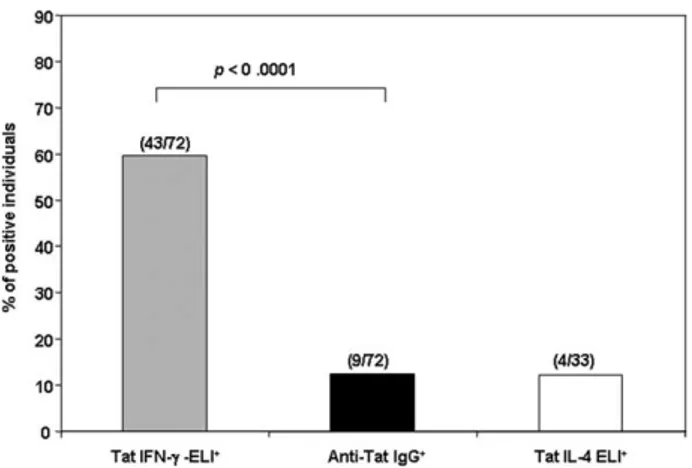
FIGURE 1. Predominant Th1 polarization of the adaptive anti-Tat im-munity in asymptomatic HIV-1 infection. The comparative assessment of Th1/Th2 specific responses to Tat was performed in HIV-1-infected asymptomatic individuals, naive to antiretroviral therapy. A Th1 response against Tat was found in 59.7% of the individuals whereas serum anti-Tat IgG Abs or IL-4 were found in only 12.5% and 12.1% of the individuals, respectively. Statistical analysis, by the McNemar test, indicated a signif-icant disagreement between the frequencies of Th1 and Th2 responses (p⬍ 0.0001) and a complete agreement between the frequencies of Th2 re-sponses (IL-4 rere-sponses and IgG production).
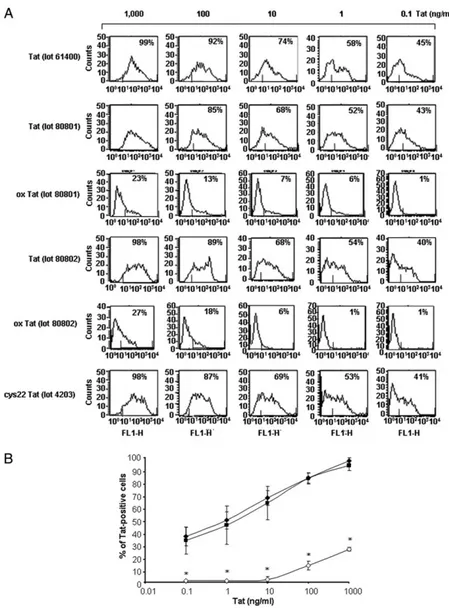
FIGURE 2. Physico-chemical and biological characterization of wt, oxidized and cys22-mutated Tat proteins. A, Elution profile of native wt Tat (lots 61400, 80801, 80802), oxidized wt Tat (lot 80801) (ox Tat), or cys22 Tat (lot 4203) (50g/ml) by reverse HPLC. B, Biological activity of native wt Tat (lots 61400, 80801, 80802) (upper panel), oxidized Tat (lot 80802) (ox Tat) and cys22 Tat (lots 49 and 4203) (lower panel) (0.5 to 10g/ml) expressed as levels of p24 (pg/ml) induced in HLM1 cell culture supernatants after 48 h of exposure to the proteins (mean and SD from three to five different experiments).
2890 GENERATION OF Th1 IMMUNE RESPONSES BY HIV-1 Tat
by guest on July 24, 2018
http://www.jimmunol.org/
In vitro assay for Th lymphocyte polarization pattern
MDDC from six healthy donors were transiently exposed (10 min) to na-tive wt Tat (lots 80802 and 040805) cys22 Tat (lot 4203) (10g/ml), or LPS (1g/ml), washed and cultured for 18 h at 37°C in complete medium. Autologous thawed PBL were then added at a ratio of 10:1 and cultured in complete medium. Culture supernatants at days 2 and 6 were analyzed for IFN-␥ or IL-4 production by commercial ELISA (Cytoscreen IFN-␥ or IL-4 ELISA kits; Biosource International), or by a human Th1/Th2 cyto-metric bead array kit (CBA; BD Biosciences). At day 6 of coculture, IL-2 (50 U/ml) was added and at day 12 cells were analyzed for intracellular IFN-␥ and IL-4 detection by ICS according to in-house established meth-ods. In brief, cells were stimulated with 10 ng/ml PMA and 1g/l iono-mycin for 5 h, adding 1g/ml brefeldin A (GolgiPlug) during the last 4 h of culture. Cells were then harvested, washed, and stained with mAbs FITC-anti-CD3, allophycocyanin-Cy7-anti-CD4, and PerCP-Cy5-anti-CD8 (BD Biosciences), fixed with lysing solution (BD Biosciences), per-meabilized with Perm solution (BD Biosciences), and stained on ice for 30 min with allophycocyanin-labeled anti-IFN-␥ and PE-labeled anti-IL-4 mAb (BD Biosciences). Cell fluorescence was evaluated and analyzed by a FACSCanto flow cytometer equipped with FACSDiva Software (BD Biosciences).
Animal studies: immunization of monkeys
Cynomologus monkeys (Macaca fascicularis) were housed singly accord-ing to European guidelines for non-human primate care (EEC, directive no. 86-609, Nov. 24, 1986). Two groups of five monkeys each (ID: AF874, AC252, AF523, AC017, AC259; and AF737, AC276, AC344, AF597, AC296, respectively) were immunized subcute twice (weeks 0 and 4) with either wt Tat (10g) or cys22 Tat (10 g) mixed in alum (aluminum
phosphate) as adjuvant. A third group of four cynomolgus macaques (ID: 61750, 61777, 61780, and 61785) was inoculated intradermally three times (weeks 0, 4, 8) with the wt Tat protein (6g) without adjuvant. At base-line, corresponding to the day of first Ag administration, and 3 to 4 wk after each immunization, all the animals were clinically examined and blood was drawn from the inguinal vein under ketamine hydrochloride anesthesia (10 mg/kg) for routine blood chemistry tests and immunological determina-tions (anti-Tat Ab titers, IFN-␥, and IL-4 ELISpot).
Detection of anti-Tat Ab titers in vaccinated monkeys
Monkey serum anti-Tat IgG titers were determined by ELISA as already described (10). In brief, 96-well microplates (Nunc) were coated with Tat protein (100 ng in 200l/well of 0.05 M carbonate buffer, pH 9.6) for 12 h at 4°C, and blocked with PBS containing 1% BSA and 0.05% of Tween 20 (block buffer). Duplicates of 2-fold serial dilutions of sera were added to the plates and incubated for 90 min at 37°C. After extensive washing, plates were incubated with an anti-monkey IgG HRP-conjugated Ab (Sigma) for 90 min at 37°C, then washed and incubated with peroxidase substrate (ABTS; Roche) for 50 min at 37°C. The OD of each well was measured with a spectrophotometer and Ab titers were calculated as the highest reciprocal serum dilution giving OD readings⬎3 SD above back-ground levels as calculated using preimmune serum.
IFN-␥ and IL-4 ELISpot assay in monkeys PBMC
The number of Tat-specific IFN-␥ and IL-4-producing PBMC was mea-sured by using commercial kits (monkey IFN-␥ and human IL-4 ELISpot; Mabtech), following the manufacturer’s instructions. In brief, PBMC (2⫻ 105/well, in duplicate) were cultured in complete medium in PVDF 96-well
plates (MAIP S4510; Millipore) previously coated with anti-IFN-␥
FIGURE 3. Native wt and cys22 Tat protein but not oxidized Tat are efficiently taken up by MDDC. MDDC were cultured for 10 min in the presence of different lots of native wt Tat (lots 61400, 80801, 80802), oxidized Tat (lots 80801 and 80802) (ox Tat), or cys22 Tat (lot 4203) (0.1 to 1000 ng/ml) and then processed, washed, fixed, permeabilized and stained with a polyclonal Ab to Tat, as reported in Materials and Methods. Cells ex-pressing intracytoplasmic Tat were evaluated by flow cytometry. A, Data from a representative donor are ex-pressed as the percentage of Tat-positive cells compared with isotype-stained controls; B, cumulative data from 15 (wt Tat lot 80802) (⽧), five (ox Tat lot 80802) (〫), and three (cys22 Tat lot 4203) (f) donors, respectively, are expressed as mean⫾ SD of the percentages of Tat-positive cells.ⴱ, p ⬍ 0.001 vs wt Tat.
by guest on July 24, 2018
http://www.jimmunol.org/
(anti-human-monkey IFN-␥ mAb GZ4) or anti-IL-4 (anti-human-IL-4 mAb 82.4) Abs, in the presence of a pool of 18 overlapping 15-mer Tat peptides (UFP Service, University of Ferrara, Italy) (each 2g/ml) or of PHA (2g/ml, as positive control). After 18 h cells were removed, and the locally produced cytokine was revealed by the addition of a secondary biotinylated anti-IFN-␥ (human-monkey IFN-␥ mAb 7-B6-1), or anti-IL-4 (anti-human-anti-IL-4 mAb 12.1), followed by alkaline streptavidin-con-jugated alkaline phosphatase and enzyme substrate chromogen (BCIP/NBT tablets; Sigma-Aldrich). The spots, corresponding to the cytokine-produc-ing cells, were counted by a specific ELISpot Reader (A.EL.VIS ELISpot Reader and Analysis Software) and expressed as the number of SFC per million of PBMC upon subtraction of the background.
Statistics
Statistical analysis was performed using the McNemar test (26, 27), which is appropriate when both measurements (positive/negative) are made on the same individual subject and summarizes the agreement between the two different sets of results. This test is a non-parametric method used on nom-inal data to determine whether the row and column margnom-inal frequencies are equal (marginal homogeneity), where the information can be displayed in a 2⫻ 2 contingency table. If the test result is significant the marginal frequencies are not homogeneous, otherwise there is no difference in the proportions. This test focuses on the discordant pairs; when the discordant pairs are skewed in one direction (for example, more positive/negative than negative/positive), this is evidence that the overall proportion of positives is higher for one measurement than the other. When the discordant pairs are split evenly, this is evidence that the overall proportion is about the same for both measurements. A two-tailed t test was used for the statistical treatment of data gathered from experiments with MDDC.
Results
Adaptive anti-Tat immunity in natural HIV-1 infection
To investigate the type of adaptive immune response to Tat in natural HIV-1 infection, a comparative assessment of Th1/Th2-specific responses to Tat was performed in 72 HIV-1 infected, asymptomatic individuals, naive to antiretroviral therapy. Th1 re-sponses were determined by assessing the frequencies of Tat-spe-cific IFN-␥-producing cells by ELISpot while Th2 responses were evaluated by the assessment of serum levels of anti-Tat Ab by ELISA as well as by the measurement of Tat-specific IL-4-pro-ducing cells by ELISpot, respectively. As shown in Fig. 1, a Th1 response against Tat was found in 43 of 72 (59.7%) individuals, whereas anti-Tat IgG Abs were found in only 9 of 72 (12.5%)
individuals. This low frequency is consistent with those previously observed in different cohorts of HIV-infected individuals (6, 7, 14). Similarly, cellular Th2 responses against Tat were rare, be-cause Tat elicited IL-4 responses in only 4 of the 33 (12.1%) in-dividuals tested. Statistical analysis confirmed a significant differ-ence between the frequencies of Th1 and Th2 responses ( p ⬍ 0.0001) for the whole group of patients ( p⫽ 0.0023 for the sub-group of 33 individuals by the McNemar test) (Fig. 1 and data not shown). In addition, a complete agreement was found between the frequencies of Th2 responses (IL-4 responses and IgG serum lev-els). Therefore, the frequency of individuals positive for IFN-␥ ELISpot is significantly greater than those positive for anti-Tat IgG Ab and IL-4 ELISpot, suggesting the presence of a strong Th1 polarization of the adaptive immune response against Tat in asymptomatic HIV infection. Since Tat enters efficiently MDDC and possess adjuvant properties (15–17), we further explored the effects of native Tat on MDDC, which represent key cells in the recognition and response to viral Ags in vivo.
MDDC efficiently take up wild-type and cys22 Tat but not oxidized Tat
To investigate the effects of biologically active, native Tat on DC, different lots of the protein were characterized for their purity and biological activity. Purified preparations included both wt Tat (be-fore or after oxidation upon exposure to air and light) and a Tat mutated in the cysteine-rich region (cys22 Tat), which lacks trans-activation activity. As assessed by analytical reversed phase HPLC (Fig. 2A), both recombinant wt (lots 61400, 80801, and 80802) and cys22 (lot 4203) Tat had the same elution profile (i.e., a sharp absorbance peak at the range of 21–23 min of elution), whereas oxidized Tat (lot 80801) displayed a markedly reduced peak, due to change in protein conformation and multimerization. The bio-logical activity of the different Tat proteins was then determined by assessing their capacity of rescuing the replication of a Tat-defec-tive HIV provirus in HLM1 cells by the direct addition of the protein as previously reported (15–17). As shown in Fig. 2B, all lots of wt native Tat induced the production of p24 by HLM1 cells in a reproducible and dose-dependent fashion. In contrast, both
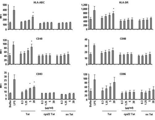
FIGURE 4. MDDC maturation is in-duced by native Tat but not by oxidized or cys22 Tat proteins. MDDC from five healthy donors were cultured in the presence of na-tive wt Tat (lot 80802), oxidized Tat (lot 80802) (ox Tat), or cys22 Tat (lot 4203) (0.3–20 g/ml) and after 18 h the surface expression of HLA-ABC, HLA-DR, CD40, CD83, and CD86 was evaluated by flow cy-tometry. Data are expressed as mean fluo-rescence intensity (MFI) plus SEM of the five-donor group.ⴱ, p ⬍ 0.05 vs buffer.
2892 GENERATION OF Th1 IMMUNE RESPONSES BY HIV-1 Tat
by guest on July 24, 2018
http://www.jimmunol.org/
oxidized and cys22 Tat had no effects, confirming that the trans-activating activity is lost upon protein oxidation or by mutation of the cysteine-rich domain of Tat (15, 19, 20).
Wild-type Tat, oxidized Tat, and cys22 Tat were then used for experiments of uptake by MDDC (15). As shown (Fig. 3), both native wt and cys22 Tat were efficiently taken up by MDDC in a dose-dependent fashion in the pico-nanomolar range. In contrast, the uptake of oxidized Tat was either markedly diminished (at nanomolar doses) or undetectable (at picomolar doses), as com-pared with the native protein, although oxidized Tat was found on the cell surface at levels comparable to those of wt or cys22 Tat in the absence of trypsin treatment indicating a similar binding to the cell membrane (Fig. 3 and data not shown). Thus, cys22 Tat, al-though not capable of transactivation activity, maintains the native protein conformation, which is key for its uptake by MDDC. Con-versely, the oxidation of Tat alters its conformation and markedly affects its uptake by MDDC.
Wild-type Tat, but not oxidized Tat nor cys22 Tat, induces MDDC maturation and cytokine production
Wild-type Tat, oxidized Tat, and cys22 Tat were then assayed for their effects on MDDC maturation. This was performed by eval-uating cell surface molecules expression and cytokine production. For these experiments CD1a-expressing MDDC were selected since this molecule represents a specific hallmark of differentiation (for review, see Ref. 21) and only CD1a⫹-expressing cells have been shown to efficiently respond to stimuli-inducing cell matura-tion and producmatura-tion of cytokines leading to Th1 responses (22). As shown in Fig. 4, native wt Tat induced a significant, dose-depen-dent increase of the surface expression of HLA-ABC, HLA-DR, CD40, CD83, and CD86 on MDDC. In contrast, both oxidized or cys22 Tat had no effects. Similarly, native Tat, but not oxidized Tat nor cys22 Tat, markedly and significantly increased the pro-duction of TNF-␣ and IL-12 and of MIP-1␣, MIP-1, as detected by ELISA in MDDC supernatants (Table I). Thus, only wt native Tat induces both MDDC maturation and production of Th1-type cytokines and chemokines.
Tat mediates MDDC maturation by inducing TNF-␣ production
Among the cytokines and -chemokines induced by Tat in MDDC, TNF-␣ is known to play a key role for MDDC maturation (for review, see Ref. 21). We have previously demonstrated that Tat transactivates TNF-␣ gene expression by a transcriptional ac-tivation of the TNF promoter, which contains a structure similar to the transactivation responsive sequence of the HIV promoter (28 – 30). This requires the full transactivation activity of the protein (28 –30). Consistent with these data, no production of TNF-␣ and no MDDC maturation were observed by the addition to MDDC of the cys22 Tat protein (Table I). Therefore, the role of TNF-␣ in the
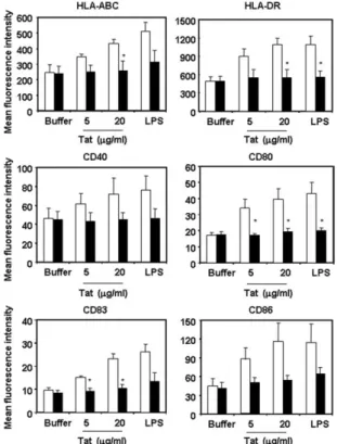
Tat-induced MDDC maturation was further investigated. To this aim, experiments were performed with native Tat in the presence of anti-TNF-␣-neutralizing Ab or an isotype-matched control Ab. As shown in Fig. 5, the increase of HLA-ABC, HLA-DR, CD40, CD80, CD83, and CD86 induced by Tat on MDDC was abolished in the presence of anti-TNF-␣ Ab. In addition, experiments per-formed by ICS (Fig. 6) showed that wt Tat induces a statistically significant increase of the number of TNF-␣-producing CD1a⫹ MDDC ( p⬍ 0.001), whereas cys22 Tat did not. These data sug-gest that TNF-␣ presumably mediates both Tat-induced MDDC maturation and production of cytokines addressing to Th1 lym-phocyte responses. Thus, TNF-␣ production and MDDC matura-tion induced by Tat require the native conformamatura-tion of the protein
FIGURE 5. Tat-induced MDDC maturation is abolished by anti-TNF-␣ neutralizing Ab. MDDC from 4 healthy donors were stimulated with native wt Tat (lot 80802) (5 and 20g/ml) or LPS (1 g/ml) in the presence of anti-TNF-␣ blocking Ab (5 g/ml) (black bars) or isotype control (open bars) and after 18 h the surface expression of HLA-ABC, HLA-DR, CD40, CD80, CD83, and CD86 on MDDC was evaluated by flow cytometry. Mean values plus SEM of receptors surface expression (mean fluorescence intensity) from the experiments with 4 donors are shown.ⴱ, p ⬍ 0.05 vs isotype control.
Table I. The production of cytokines and chemokines by MDDC is enhanced by the native but not by the cysteine mutated or the
oxidized Tat (ox Tat) proteinsa
Buffer LPS Tat cys22 Tat ox Tat
TNF-␣ 18.5⫾ 3.2 392.4⫾ 59.7* 243.3⫾ 78.5* 25.9⫾ 8.5 34.0⫾ 20.0
IL-12 (p40) 195.8⫾ 65.2 2898.3⫾ 596.0* 1791.6⫾ 546.9* 404.1⫾ 148.2 149.5⫾ 114.6
IL-12 (p70) 2.3 390.8 62.3 Not done Not done
MIP-1␣ 121.6⫾ 8.2 13916.1⫾ 1667.3* 7119.0⫾ 2048.3* 319.6⫾ 173.4 355.7⫾ 207.8 MIP-1 302.1⫾ 79.7 6339.3⫾ 638.4* 4512.4⫾ 1520.4 703.3⫾ 344.2 866.0⫾ 723.2
a
MDDC from 12 healthy donors were cultured in complete medium in the presence of native, wt Tat (lot 61400 or 80802), oxidized Tat (lot 80802) and cys22 Tat (lot 4203) (20g/ml). After 18 h, the levels of TNF-␣, IL-12 p40 and p70 (only with 1 donor) and that of MIP-1␣ and MIP-1 were evaluated in culture supernatants by ELISA. The results are expressed as mean concentrations (pg/ml) plus SEM. LPS (1g/ml) was used as the positive control.
ⴱ, p ⬍ 0.05 vs control buffer.
by guest on July 24, 2018
http://www.jimmunol.org/
for cellular entry and the functional interaction of a transcription-ally active Tat.
Tat-conditioned MDDC induce prevalent Th1 response by autologous lymphocytes
The effect of Tat on Th1/Th2 polarization of the immune response was further evaluated in vitro by determining PBL cytokine pro-duction upon coculture with autologous MDDC exposed or not to the Tat protein. Specifically, production of both IFN-␥ and IL-4 was determined in cell culture supernatants and into the cytoplasm of CD3⫹lymphocytes collected from six different donors and after stimulation with PMA and ionomycin. Of note, MDDC treated with LPS (positive control) and wt Tat, but not those treated with cys22 Tat, induced the production of high levels of IFN-␥ by au-tologous PBL (Fig. 7A). Similarly, Tat-conditioned MDDC in-duced a marked production of IFN-␥ but a much less pronounced amount of IL-4 by autologous PBL as evaluated by both ICS and CBA (Fig. 7B). Thus, Tat-treated MDDC address the immune re-sponse toward a prevalent Th1 pattern.
Inoculation of monkeys with native Tat induces a predominant Th1-specific immune response, whereas cys22 Tat induces a predominant Th2 response
The next set of studies was performed to verify whether the effects of native Tat observed in vitro on MDDC would also
reflect the development of the adaptive immune response to the protein in vivo. To this aim, two groups of five cynomolgus macaques were inoculated subcute twice in the presence of alum with either wt Tat or cys22 Tat. The humoral and cellular specific responses were evaluated at baseline and 3 to 4 wk after each immunization. Anti-Tat Ab were induced in both groups of monkeys, peaking at 3 wk after the second Ag inoculation. However, at this time point the median of the titers in the an-imals immunized with cys22 Tat (range 1:1600 –1:25,600) was 4-fold higher than in those inoculated with wt Tat (range 1:200 –1:6400) (Fig. 8A).
Determination of Tat-specific IFN-␥ and IL-4 production by PBMC was performed by ELISpot assay, 3 wk after the second Ag inoculation. In most animals, a specific IFN-␥ and IL-4 production was observed in response to a pool of overlapping Tat peptides. Of note, a predominant production of IFN-␥ in response to Tat peptides was found in the monkeys immunized with the native protein (for IFN-␥ the median SFC/106PBMC
was 68, the range 20 to 163; for IL-4 the median SFC/106
PBMC was 35, range 18 to 45, respectively) (Fig. 8B). The ratio IFN-␥/IL-4 was 1.94, whereas the IL-4/IFN-␥ ratio was 0.52. In contrast, a predominant production of IL-4 was detected in the animals inoculated with the cys22 Tat mutant (for IFN-␥ the median SFC/106PBMC was 58, the range 5 to 253; for IL-4 the
median SFC/106PBMC was 85 with a range 15 to 453) (Fig.
FIGURE 6. CD1a expressing MDDC produce TNF-␣ in response to Tat but not to cys22 Tat protein. MDDC from 12 healthy donors were cultured in the presence of native wt Tat (lot 80802) or cys22 Tat (lot 4203) (20g/ml), or LPS (1 g/ml) for 6 and 18 h, respectively. Cells were stained (FITC) for CD1a expression, fixed, permeabilized, and then stained (PE) for TNF-␣. TNF-␣-producing CD1a-positive cells were determined by flow cytometry by assessing at least 50,000 events. A, Data from a representative healthy donor, of 12 analyzed, are reported as flow cytometry dot plots. The percentages of TNF-␣ producing CD1a⫹cells are indicated in the panels; B, the percentages of TNF-␣ producing CD1a⫹cells from 12 donors are reported as mean and SEM;ⴱ, p ⬍ 0.01 vs medium and cys22 Tat.
2894 GENERATION OF Th1 IMMUNE RESPONSES BY HIV-1 Tat
by guest on July 24, 2018
http://www.jimmunol.org/
8B). The ratio IFN-␥/IL-4 was 0.68, whereas the IL-4/IFN-␥ ratio was 1.46. Thus, wt Tat favored the development of prev-alent Th1-specific responses, as compared with the cys22 Tat mutant, which induced a prevalent Th2 response. Of note, in another experimental vaccination study, 4 animals, inoculated three times intradermally with wt Tat in the absence of alum, displayed an evident Tat-specific IFN-␥ response and no de-tectable Ab production (data not shown), further indicating that, in absence of adjuvant, wt Tat addresses prevalent Th1 immune responses.
Discussion
In this paper we have performed a comparative functional assess-ment of the humoral (Th2 type) and cellular (Th1/Th2 types) arms of the adaptive immune response to Tat in a cohort of HIV-in-fected, asymptomatic, drug naive individuals and investigated at different levels the mechanisms governing the development of the anti-Tat immunity. We found that in asymptomatic HIV-1 infec-tion the adaptive immune response to Tat is strongly skewed to-ward a Th1 type of immunity, which is dominated by cytotoxic T cell responses with a low prevalence of anti-Tat Ab. Of note, these findings are consistent with the low prevalence of anti-Tat Ab reported in HIV-infected individuals, which are found only in a small proportion of asymptomatic individuals (5–9, 14).
These data suggested that the low prevalence of anti-Tat Ab in HIV-1-infected individuals is due to a predominant Th1-type po-larization of the adaptive anti-Tat immune response in vivo. In-deed, previous data showed that native Tat enters MDDC, induces their maturation, and promotes their capacity of presenting Ag through the preferential development of a Th1-type immune re-sponse (15). This body of evidence prompted us to further inves-tigate the apparent intrinsic adjuvanticity of Tat by exploring the effects of the native, wt Tat protein compared with those of cys22 Tat, a naturally occurring Tat mutant originated from a long-term non-progressor individual (31), which induced high titers of anti-Tat Ab (32, 33).
We found that both wt Tat and cys22 Tat efficiently enter MDDC, whereas the uptake of the oxidized protein is indeed poor or absent. However, despite a comparable uptake by MDDC, wt Tat, but not the cys22 Tat mutant, induces MDDC maturation and Th1-associated cytokines and chemokines production, indicating that, although the native conformation of the protein is required for specific targeting and entry of Tat into MDDC, the transactivating function of Tat is key to the induction of cell maturation and Th1 cytokines production. Since Tat induces TNF gene expression via a transcriptional activation of the TNF promoter, which contains a stem-loop transactivation responsive-like structure similar to the HIV promoter (28 –30), and since TNF-␣ has been shown to be key for MDDC maturation (for review, see Ref. 21), we focused on this cytokine. Indeed, wt Tat but not cys22 Tat induced the pro-duction and secretion of TNF-␣, and anti-TNF-␣ neutralizing Ab blocked the effect of native Tat on MDDC maturation, indicating that this cytokine is the main mediator of the effects of Tat. This requires the presence of differentiated CD1a⫹MDDC and may
FIGURE 7. PBL produce mostly IFN-␥ when cocul-tured with Tat-conditioned MDDC. MDDC from six healthy donors were transiently exposed (10 min) to na-tive wt Tat (lots 80802 and 040805) cys22 Tat (lot 4203) (10g/ml), or LPS (1 g/ml), washed and cul-tured for 18 h. Autologous PBL were then added at a ratio of 10:1 and cultured for an additional 12 days. Culture supernatants at days 2 and 6 were analyzed for IFN-␥ and IL-4 production by commercial ELISA or human Th1/Th2 CBA assay and cells at day 12 for in-tracellular production of the same cytokines upon treat-ment with 10 ng/ml PMA and 1g/l Ionomycin for 5 h. A, Levels of IFN-␥ in supernatants of PBL/MDDC cocultures from 6 normal donors at day 2; B, percentage of intracellular cytokine-producing CD3⫹lymphocytes (left panel) and levels of IFN-␥ (gray columns) and IL-4 (white columns) in supernatants of cocultures (right
panels), from a representative donor. ⴱ, p ⬍ 0.01 vs
medium and cys22 Tat.
FIGURE 8. Inoculation of monkeys with wt native Tat (lot 80802) in-duces a predominant Th1-specific response whereas cys22 Tat (lot 4203) induces prevalent Th2 responses. Two groups of five cynomolgus monkeys each were inoculated twice either with wt Tat or cys22 Tat (10g) in alum adjuvant. A, Tat-specific Ab titers were measured by ELISA testing serial dilutions (1/100 –1/102,400) of monkeys sera collected at baseline, 4 wk after the first and 3 wk after the second immunization, respectively. Data are reported as the median plus SEM of the reciprocals of the last positive dilution from animals immunized with wt Tat (⽧) or cys22 Tat (〫). The arrows on top of the panels mark the times of Ag administration. B, Tat-specific cellular responses were determined 3 wk after the second Ag in-oculation by IFN-␥ and IL-4 ELISpot assays. Data are reported as the mean number plus SEM of IFN-␥ or IL-4 SFC/106PBMC from the two groups
of animals immunized with wt Tat (white columns) or cys22 Tat (gray columns).
by guest on July 24, 2018
http://www.jimmunol.org/
explain the absence of Tat-induced maturation described in a pre-vious report (34) in which CD1a⫺cells, which have been shown less responsive to maturation stimuli (22), were used. Of note Tat-conditioned MDDC induced a strong IFN-␥ production by autol-ogous lymphocytes capable of supporting Th1-type immune re-sponses. This effect required Tat transactivation activity since was not induced by the mutant Tat cys22 and it is presumably mediated by TNF-␣.
It is well known that vaccination with soluble proteins and alum induces Th2 responses (for review, see reference (35) and the mechanism has been recently elucidated (36). As expected, mon-keys inoculated twice with cys22 Tat and alum developed a pre-dominant Th2 immune response associated with high titers of anti-Tat Ab. In contrast, a predominant Th1 response was induced upon inoculation with wt Tat and alum. Moreover, in animals immu-nized intradermally with wt Tat in the absence of the adjuvant, a marked IFN-␥ production but not Ab response was observed. Thus, wt Tat can address immune response toward a prevalent Th1 pattern, and this effect is relevant also in the absence of any adjuvant.
Overall these data indicate that, upon interaction with MDDC, wt Tat induces, both in vivo and in vitro, immune responses prev-alently polarized to a Th1 pattern and that this effect requires the conserved conformation of the protein. This has important impli-cations for understanding the mechanisms governing the genera-tion of the adaptive immune response against virus Ags along spe-cific pathways. On this goal, we have recently shown that picomolar concentrations of the native wt Tat protein modify the catalytic subunit composition of the immunoproteasome, changing the hierarchy of heterologous peptide Ags that are generated, pre-sented, and recognized by CTL (37). Consistent with these data, mice vaccinated with the HIV-1 Gag, Env, or V2-deleted Env Ags in combination with Tat showed a broadening of Th1-type and CTL responses against a larger number of T cell epitopes, com-pared with mice vaccinated with these proteins in the absence of Tat. However, Tat did not affect Th2-type responses to these struc-tural HIV proteins (38). Thus, a growing body of evidence indi-cates that Tat has potent immunoregulatory functions, which may play an important role in HIV pathogenesis. Indeed, the capacity of native Tat to target, enter, and induce MDDC maturation toward a prevalent Th1 response is consistent with the in vivo data that show a clear Th1 polarization of the adaptive anti-Tat response both in humans and in animal models. This biological property of Tat may have implications for the setting of the anti-HIV-1 im-mune response in infected individuals. In addition, this property of Tat may be further exploited for vaccination strategies aimed at inducing specific Th1 immune responses against other HIV Ags as well as other intracellular pathogens or even against cancer.
Acknowledgments
We thank M. T. Maggiorella, R. Belli, L. Sernicola, P. Leone, and Erika Olivieri for helpful discussion and technical help; P. Pupino-Carbonelli, E. Iale, F. Incitti, N. Verrone, A. Marini, A. Avitabile, M. Chiodi, and M. Azzetti for hematoclinical analysis of cynomolgus samples and for the handling of the animal facility; and P. Cocco, D. Diamanti, and F. Costa for technical support. We also thank P. Sergiampietri and A. Carinci for the excellent editorial assistance.
Disclosures
The authors have no financial conflict of interest.
References
1. Ensoli, B., A. Cafaro, A. Caputo, V. Fiorelli, F. Ensoli, R. Gavioli, F. Ferrantelli, A. Cara, F. Titti, and M. Magnani. 2005. Vaccines based on the native HIV Tat protein and on the combination of Tat and the structural HIV protein variant DeltaV2 Env. Microbes Infect. 7: 1392–1399.
2. Ensoli, B. 2005. Rational vaccine strategies against AIDS: background and ra-tionale. Microbes Infect. 7: 1445–1452.
3. Ensoli, B. 2005. Criteria for selection of HIV vaccine candidates— general prin-ciples. Microbes Infect. 7: 1433–1435.
4. Ensoli, B., V. Fiorelli, F. Ensoli, A. Cafaro, F. Titti, S. Butto, P. Monini, M. Magnani, A. Caputo, and E. Garaci. 2006. Candidate HIV-1 Tat vaccine development: from basic science to clinical trials. AIDS 20: 2245–2261. 5. Re, M. C., M. Vignoli, G. Furlini, D. Gibellini, V. Colangeli, F. Vitone, and
M. La Placa. 2001. Antibodies against full-length Tat protein and some low-molecular-weight Tat-peptides correlate with low or undetectable viral load in HIV-1 seropositive patients. J. Clin. Virol. 21: 81– 89.
6. Reiss, P., J. M. Lange, A. de Ronde, F. de Wolf, J. Dekker, C. Debouck, and J. Goudsmit. 1990. Speed of progression to AIDS and degree of antibody re-sponse to accessory gene products of HIV-1. J. Med. Virol. 30: 163–168. 7. Rezza, G., V. Fiorelli, M. Dorrucci, M. Ciccozzi, A. Tripiciano, A. Scoglio,
B. Collacchi, M. Ruiz-Alvarez, C. Giannetto, A. Caputo, et al. 2005. The pres-ence of anti-Tat antibodies is predictive of long-term nonprogression to AIDS or severe immunodeficiency: findings in a cohort of HIV-1 seroconverters. J. Infect.
Dis. 191: 1321–1324.
8. Rodman, T. C., S. E. To, H. Hashish, and K. Manchester. 1993. Epitopes for natural antibodies of human immunodeficiency virus (HIV)-negative (normal) and HIV-positive sera are coincident with two key functional sequences of HIV Tat protein. Proc. Natl. Acad. Sci. USA 90: 7719 –7723.
9. Zagury, D., A. Lachgar, V. Chams, L. S. Fall, J. Bernard, J. F. Zagury, B. Bizzini, A. Gringeri, E. Santagostino, J. Rappaport, et al. 1998. Interferon␣ and Tat involvement in the immunosuppression of uninfected T cells and C-C chemokine decline in AIDS. Proc. Natl. Acad. Sci. USA 95: 3851–3856.
10. Cafaro, A., A. Caputo, C. Fracasso, M. T. Maggiorella, D. Goletti, S. Baroncelli, M. Pace, L. Sernicola, M. L. Koanga-Mogtomo, M. Betti, et al. 1999. Control of SHIV-89.6P-infection of cynomolgus monkeys by HIV-1 Tat protein vaccine.
Nat. Med. 5: 643– 650.
11. Cafaro, A., F. Titti, C. Fracasso, M. T. Maggiorella, S. Baroncelli, A. Caputo, D. Goletti, A. Borsetti, M. Pace, E. Fanales-Belasio, et al. 2001. Vaccination with DNA containing tat coding sequences and unmethylated CpG motifs protects cynomolgus monkeys upon infection with simian/human immunodeficiency virus (SHIV89.6P). Vaccine 19: 2862–2877.
12. Caselli, E., M. Betti, M. P. Grossi, P. G. Balboni, C. Rossi, C. Boarini, A. Cafaro, G. Barbanti-Brodano, B. Ensoli, and A. Caputo. 1999. DNA immunization with HIV-1 tat mutated in the trans activation domain induces humoral and cellular immune responses against wild-type Tat. J. Immunol. 162: 5631–5638. 13. Maggiorella, M. T., S. Baroncelli, Z. Michelini, E. Fanales-Belasio, S. Moretti,
L. Sernicola, A. Cara, D. R. Negri, S. Butto, V. Fiorelli, et al. 2004. Long-term protection against SHIV89.6P replication in HIV-1 Tat vaccinated cynomolgus monkeys. Vaccine 22: 3258 –3269.
14. Butto`, S., V. Fiorelli, A. Tripiciano, M. J. Ruiz-Alvarez, A. Scoglio, F. Ensoli, M. Ciccozzi, B. Collacchi, M. Sabbatucci, A. Cafaro, et al. 2003. Sequence con-servation and antibody cross-recognition of clade B human immunodeficiency virus (HIV) type 1 Tat protein in HIV-1-infected Italians, Ugandans, and South Africans. J. Infect. Dis. 188: 1171–1180.
15. Fanales-Belasio, E., S. Moretti, F. Nappi, G. Barillari, F. Micheletti, A. Cafaro, and B. Ensoli. 2002. Native HIV-1 Tat protein targets monocyte-derived den-dritic cells and enhances their maturation, function, and antigen-specific T cell responses. J. Immunol. 168: 197–206.
16. Chang, H. C., F. Samaniego, B. C. Nair, L. Buonaguro, and B. Ensoli. 1997. HIV-1 Tat protein exits from cells via a leaderless secretory pathway and binds to extracellular matrix-associated heparan sulfate proteoglycans through its basic region. AIDS 11: 1421–1431.
17. Ensoli, B., L. Buonaguro, G. Barillari, V. Fiorelli, R. Gendelman, R. A. Morgan, P. Wingfield, and R. C. Gallo. 1993. Release, uptake, and effects of extracellular human immunodeficiency virus type 1 Tat protein on cell growth and viral trans-activation. J. Virol. 67: 277–287.
18. Wu, Y., and J. W. Marsh. 2001. Selective transcription and modulation of resting T cell activity by preintegrated HIV DNA. Science 293: 1503–1506. 19. Balboni, P. G., R. Bozzini, S. Zucchini, P. C. Marconi, M. P. Grossi, A. Caputo,
R. Manservigi, and G. Barbanti-Brodano. 1993. Inhibition of human immunode-ficiency virus reactivation from latency by a tat transdominant negative mutant.
J. Med. Virol. 41: 289 –295.
20. Caputo, A., M. P. Grossi, R. Bozzini, C. Rossi, M. Betti, P. C. Marconi, G. Barbanti-Brodano, and P. G. Balboni. 1996. Inhibition of HIV-1 replication and reactivation from latency by tat transdominant negative mutants in the cys-teine rich region. Gene Ther. 3: 235–245.
21. Bell, D., J. W. Young, and J. Banchereau. 1999. Dendritic cells. Adv. Immunol. 72: 255–324.
22. Chang, C. C., A. Wright, and J. Punnonen. 2000. Monocyte-derived CD1a⫹ and CD1a⫺ dendritic cell subsets differ in their cytokine production profiles, suscep-tibilities to transfection, and capacities to direct Th cell differentiation. J.
Immu-nol. 165: 3584 –3591.
23. Gauduin, M. C., A. Kaur, S. Ahmad, T. Yilma, J. D. Lifson, and R. P. Johnson. 2004. Optimization of intracellular cytokine staining for the quantitation of an-tigen-specific CD4⫹ T cell responses in rhesus macaques. J. Immunol. Methods 288: 61–79.
24. Kaur, A., C. L. Hale, B. Noren, N. Kassis, M. A. Simon, and R. P. Johnson. 2002. Decreased frequency of cytomegalovirus (CMV)-specific CD4⫹ T lymphocytes in simian immunodeficiency virus-infected rhesus macaques: inverse relationship with CMV viremia. J. Virol. 76: 3646 –3658.
25. Waldrop, S. L., C. J. Pitcher, D. M. Peterson, V. C. Maino, and L. J. Picker. 1997. Determination of antigen-specific memory/effector CD4⫹ T cell frequencies by
2896 GENERATION OF Th1 IMMUNE RESPONSES BY HIV-1 Tat
by guest on July 24, 2018
http://www.jimmunol.org/
flow cytometry: evidence for a novel, antigen-specific homeostatic mechanism in HIV-associated immunodeficiency. J. Clin. Invest. 99: 1739 –1750.
26. Armitage, P. 1994. Statistical Methods in Medical Research (3rd Ed.). Blackwell Scientific, Oxford.
27. Fleiss, J. L. 1981. Statistical Methods for Rates and Proportions (2nd Ed.). John Wiley & Sons, New York.
28. Brother, M. B., H. K. Chang, J. Lisziewicz, D. Su, L. C. Murty, and B. Ensoli. 1996. Block of Tat-mediated transactivation of tumor necrosis factor gene expression by polymeric-TAR decoys. Virology 222: 252–256.
29. Buonaguro, L., G. Barillari, H. K. Chang, C. A. Bohan, V. Kao, R. Morgan, R. C. Gallo, and B. Ensoli. 1992. Effects of the human immunodeficiency virus type 1 Tat protein on the expression of inflammatory cytokines. J. Virol. 66: 7159 –7167.
30. Buonaguro, L., F. M. Buonaguro, G. Giraldo, and B. Ensoli. 1994. The human immunodeficiency virus type 1 Tat protein transactivates tumor necrosis factor gene expression through a TAR-like structure. J. Virol. 68: 2677–2682. 31. Huet, T., M. C. Dazza, F. Brun-Vezinet, G. E. Roelants, and S. Wain-Hobson.
1989. A highly defective HIV-1 strain isolated from a healthy Gabonese indi-vidual presenting an atypical western blot. AIDS 3: 707–715.
32. Opi, S., J. M. Peloponese, Jr., D. Esquieu, G. Campbell, J. de Mareuil, A. Walburger, M. Solomiac, C. Gregoire, E. Bouveret, D. L. Yirrell, and E. P. Loret. 2002. Tat HIV-1 primary and tertiary structures critical to immune response against non-homologous variants. J. Biol. Chem. 277: 35915–35919.
33. Peloponese, J. M., Jr., Y. Collette, C. Gregoire, C. Bailly, D. Campese, E. F. Meurs, D. Olive, and E. P. Loret. 1999. Full peptide synthesis, purification, and characterization of six Tat variants. Differences observed between HIV-1 isolates from Africa and other continents. J. Biol. Chem. 274: 11473–11478. 34. Izmailova, E., F. M. Bertley, Q. Huang, N. Makori, C. J. Miller, R. A. Young, and
A. Aldovini. 2003. HIV-1 Tat reprograms immature dendritic cells to express chemoattractants for activated T cells and macrophages. Nat. Med. 9: 191–197. 35. Lindblad, E. B. 2004. Aluminium compounds for use in vaccines. Immunol. Cell
Biol. 82: 497–505.
36. Jordan, M. B., D. M. Mills, J. Kappler, P. Marrack, and J. C. Cambier. 2004. Promotion of B cell immune responses via an alum-induced myeloid cell popu-lation. Science 304: 1808 –1810.
37. Gavioli, R., E. Gallerani, C. Fortini, M. Fabris, A. Bottoni, A. Canella, A. Bonaccorsi, M. Marastoni, F. Micheletti, A. Cafaro, et al. 2004. HIV-1 tat protein modulates the generation of cytotoxic T cell epitopes by modifying pro-teasome composition and enzymatic activity. J. Immunol. 173: 3838 –3843. 38. Gavioli, R., S. Cellini, A. Castaldello, R. Voltan, E. Gallerani, F. Gagliardoni,
C. Fortini, E. B. Cofano, C. Triulzi, A. Cafaro, et al. 2008. The Tat protein broadens T cell responses directed to the HIV-1 antigens Gag and Env: impli-cations for the design of new vaccination strategies against AIDS. Vaccine 26: 727–737.
by guest on July 24, 2018
http://www.jimmunol.org/