UNIVERSITÀ DEGLI STUDI DI ROMA
"TOR VERGATA"
FACOLTA' DI SCIENZE MM.FF.NN.
DOTTORATO DI RICERCA IN
BIOLOGIA CELLULARE E MOLECOLARE
XXI CICLO
TESI
Melanoma cells differentiation through inhibition of p-Mek
activity following treatment with
5,7-dimethoxycoumarin
Dottorando: DANIELA ALESIANI
A.A. 2008/2009
Docente Guida/Tutor: Prof.ssa Antonella Canini
Coordinatore: Prof. Gianni Cesareni
INDEX
1. INTRODUCTION 4
1.1 The coumarins 4
1.1.1 Chemistry of the coumarins 4
1.1.2 The role of the coumarins in the plant 5
1.1.3 Antioxidant activity of the coumarins 7
1.1.4 Anticancer activity of the coumarins 8
1.1.5 Metabolism and toxicity of coumarin in humans 12
1.1.6 The 5,7-dimethoxycoumarin 13
1.2 Melanoma 15
1.2.1 Biology of melanoma 15
1.2.2 Genetics of melanoma: the signaling networks in melanoma
proliferation and progression 17
1.2.3 Therapy of melanoma: Ras/Raf/Mek/Erk MAPKs as
targeted growth signaling pathway 22
2. PURPOSE 25
3. MATERIALS AND METHODS 26
3.1 Test compound 26
3.2 Cell lines and culture conditions 26
3.3 MTT assay 27
3.4 Trypan blue exclusion test 27
3.5 Treatment of B16 with 5,7-dimethoxycoumarin and U0126
and Trypan blue assay 28
3.7 Protoporphyrine IX (PpIX) analysis and phenotypic
characterization with flow cytometry 30
3.8 Melanin synthesis in B16 and A375 cells 30
3.9 Tyrosinase assay 31
3.10 Western blot analyses 32
3.11 Mek 1/2 immunoprecipitation and kinase assay 33
3.12 Statistical analysis 33
4. RESULTS 34
4.1 Reduction of metabolic activity of murine and human cell lines
after treatment with 5,7-dimethoxycoumarin 34
4.2 Effect of 5,7-dimethoxycoumarin on growth and death of
melanoma cell lines 36
4.3 Block of cell cycle in G0/G1 phase after treatment of melanoma
cells with 5,7-dimethoxycoumarin 40
4.4 Effect of Mek 1/2 inhibitor on 5,7-dimethoxycoumarin
antigrowth activity in B16 cell line 42
4.5 Morphological changes in melanoma cells after treatment with
5,7-dimethoxycoumarin 44
4.6 Phosphorylation of ERK1/2 after treatment with
5,7-dimethoxycoumarin 45
4.7 Phosphorylation of MEK 1/2 and inhibition of its kinase
activity in melanoma cells after treatment with
5,7-dimethoxycoumarin 47
4.8 Alteration of phenotypic characteristics in melanoma cells after
treatment with 5,7-dimethoxycoumarin 49
4.9 Melanogenesis process induced in B16 and A375 cells after
treatment with 5,7-dimethoxycoumarin 51
4.10 CREB phosphorylation and expression of Mitf in melanoma
cells after treatment with 5,7-dimethoxycoumarin 53
4.11 PpIX accumulation and expression of porphobilinogen
deaminase (PBG-D) in melanoma cells after treatment with
5,7-dimethoxycoumarin 55
5. DISCUSSION 58
6. REFERENCES 71
1. INTRODUCTION
1.1 The coumarins
1.1.1 Chemistry of the coumarins.
Coumarins are a group of heterocyclic compounds synthesized by numerous plant species as well as by some bacteria and fungi (O’Kennedy & Thornes, 1997), that have been widely used as a fragrance in food and cosmetic products (Lake, 1999).
They are characterized by the presence of fused benzene and α-pyrone rings as a structural nucleus and they can occur as the hydroxylated, alkoxylated or alkylated derivatives of the parent compound, the coumarin (1,2-benzopyrone), along with their glycosides (Hoult & Paya, 1996). These compounds may be located in the peel of seeds, in the roots, in the leaves and stems, but the highest concentration was found in flowers and fruits. The most representative molecule, the coumarin, has been extensively studied both in biochemical and pharmaceutical fields (Hoult & Paya, 1996); also the 7-hydroxycoumarin aroused a great interest, as the first compound derived from the metabolism of coumarin in humans. There are other
derivative molecules with important biological properties, such as furanocoumarins and pyranocoumarins; among the furanocoumarins, we should mention the 8-methoxypsoralen or xanthotoxin, a light-sensitizing agent, which can cause skin diseases in phytophage animals, if they produce abrasions and injuries to plants synthesizing these defense compounds (Timmermann et al., 1999 ).
Coumarin
(1,2-benzopyrone)
Figure 1. Molecolar structure of the coumarin.
1.1.2 The role of the coumarins in the plant.
The role of these secondary metabolites in the plants is mainly related to defense, considering their antimicrobial and deterrent properties, their shielding activity against UV and allelopathic functions (Buchanan et al. , 2003). The scopoletin, or 7-hydroxy-6-methoxycoumarin, is the most interesting coumarin in the study of defense mechanisms in the plants; it is the first phytoalexin to be identified in Hevea brasiliensis L., where it is accumulated in the leaves following infections with several fungi, such as
Mycrocyclus ulei, Colletotrichum gloeosporioides, and Ophiostoma ulmi.
The accumulation of scopoletin in plant tissues is observed also when the plants are exposed to abiotic factors, such as CuCl2 and UV radiation which
trigger a state of physiological stress (Gutierrez et al., 1995). In Corchorus
olitorius L., the synthesis of scopoletin was observed contemporaneously
with that of other coumarins - the fraxinol, the isopimpinellin, the xanthotoxol - after treatment of the leaves with biotic and abiotic stress agents (Abou Zeid, 2002). Even furanocoumarins are known for their antifungal and antibacterial activities, such as 8-methoxypsoralen and 5-methoxypsoralen, identified for the first time in the leaf extract of Ruta
graveolens L; the two phytoalexins have demonstrated a fungistatic activity
against Fusarium solani, Pyrenochaeta lycopersici and Trichoderma viride, inhibiting the mycelial growth (Oliva et al., 1999). 5,8-Di-(2,3-dihydroxy-3-methylbutoxy)-psoralen and other furanocumarine (the psoralidin, the psoralin and the angelicin), extracted from the seeds of Psoralea corylifolia L. (Khatune et al., 2004), showed bacteriostatic effect, particularly against
Shigella sonnei, Shigella flexneri and Staphylococcus aureus. The
antibacterial and antifungineal activities of these plant metabolites have a great importance for human and environmental health; indeed, coumarins may be used instead of antibiotics against pathogens affecting humans, and also as fungicides in agriculture instead of the synthetic preparations (Oliva
et al., 1999, Ojala et al., 2000). The coumarins are known for their
allelopathic activities; the allelopathy was defined by Molisch (1937) as a chemical interaction between two plants, and may be both stimulative and inhibitory. Coumarin, for example, promotes or inhibits the growth of the plant and its allelopathic effect is species-specific and
concentration-are the target of the coumarin: this compound reduces photosynthesis and it is able to inhibit respiration and electronic transport; it interferes with the absorption of nitrates in the seedlings of wheat and reduces nitrogen metabolism in cell cultures of carrot (Abenavoli et al., 2003). Anyway, the major effect of coumarin is the inhibition of radical growth, coupled with the reduction of diameter and branching of the roots (Abenavoli et al., 2004).
1.1.3 Antioxidant activity of the coumarins.
Several coumarins showed a strong antioxidant activity, due to their ability to neutralize reactive oxygen species (ROS) considered the main cause of cardiovascular disease, cancer, atherosclerosis and aging processes. If these highly reactive molecules are not neutralized by antioxidant systems, can cause oxidative damage to cellular components such as membrane lipids, proteins and DNA, resulting in the damage of cellular structures (Kaneko et
al., 2001). The antiradical activity of the coumarins depends on the presence
of hydroxyl groups (OH), linked to benzenic ring (Ng et al., 2000), while the α-pyrone ring contributes in small measure to antioxidant properties of these compounds (Zhang & Wang, 2004). Foti et al. (1996) demonstrated that the coumarins with a catechol group, which is the benzenic ring with two OH groups, had a higher antioxidant power against radical peroxide. Furthermore, it has been observed that the catechol group increases the antiradicalic activity of natural antioxidants, including coumarins; in coumarins class, antiradicalic properties decrease when the OH groups of catechol are replaced with methoxy or sugar groups (Ng et al., 2000; Zhang
& Wang, 2004). Among the coumarins, the 6,7-dihydroxycoumarin, or esculetin, is known to be an inhibitor of lipoxygenase enzyme (Pan et al., 2003); it shows neutralizing activity against the ROS, such as the superoxide and hydroxyl radicals and reduces the oxidative damage induced by butyl hydroperoxide in rat hepatocytes (Kaneko et al., 2003). More recently, by using human endothelial cells, it was shown the protective effect of esculetin and 4-methylesculetin against cytotoxic effects induced with linoleic hydroperoxide (LOOH), the first product resulting from the lipid peroxidation. Some coumarin derivatives, such as 7,8-dihydroxy-4-methylcoumarin and 7-hydroxy-4-7,8-dihydroxy-4-methylcoumarin, inhibited significantly the peroxidation of low density lipoproteins (LDL) (Liu et al., 1999), which is known to contribute in atherosclerosis development (Esterbauer & Ramos, 1995; Klatt & Esterbauer, 1996).
1.1.4 Anticancer activity of the coumarins.
Several coumarins are known for their therapeutic activities and many of these have been purified from medicinal plants. In addition to their pharmacological properties, these plant metabolites showed a significant atoxicity in mammalian cells. The combination of these two characteristics makes coumarin a class of compounds of interest in pharmaceutical field (Hoult & Paya, 1996). Numerous biological activities have been attributed to simple coumarins and analogues as anti-coagulant (Suttie, 1987), hypotensive (Huang et al., 1992), antibacterial (Michaeli et al., 1970), anti-inflammatory (Paya et al., 1992), anticancer (Thornes et al., 1983; Gawron
& Glowniak, 1987; Marshall et al., 1987), antimutagenic (Pillai et al., 1999), anti-viral effects (Kostova et al., 2006) and enzyme inhibition (Ghani et al., 2001; Gnerre et al., 2000), scavenging of reactive oxygen species (Vairagupta et al., 2000), anticarcinogenesis/chemoprevention of cancer properties (Kelly et al., 2000; Prince et al., 2006; Kleiner et al., 2002b; Cai
et al., 1997; Wattenberg et al., 1979; Tanaka et al., 1998; Baba et al., 2002).
Many of the cancer chemopreventive effects of coumarins have been attributed to their abilities to modulate xenobiotic metabolizing enzymes. For example, bergamottin was one of the most potent at suppressing mouse hepatic ethoxyresorufin O-dealkylase activity (Cai et al., 1993); furthermore, it inhibited the formation of the major benzo[a]pyrene (B[a]P)-DNA adduct,
anti-benzo[a]pyrene-7,8-diol,9,10-epoxide-dGuo, in mouse epidermis in vivo, resulting in the block of B[a]P-induced skin tumor initiation in mice
(Cai et al., 1997). The ability of bergamottin to suppress mouse skin tumor initiation by B[a]P is likely mediated by its ability to suppress cytochrome P450 1A1 dependent metabolic activation of B[a]P into the anti-benzo[a]pyrene-7,8-diol,9,10-epoxide. Bergamottin is a weak inhibitor of murine P450 1B1, and was not effective at suppressing P450 1B1-dependent metabolism of 7,12-dimethylbenz[a]anthracene (DMBA) into the syn-DMBA diol-epoxide derived DNA adducts (Kleiner et al., 2002a). In contrast, imperatorin and isopimpinellin were similarly effective at suppressing both P450 1A1 and P450 1B1 activities (Kleiner et al., 2003) and were effective at suppressing DNA adduct formation and skin tumor initiation from both B[a]P and DMBA (Cai et al., 1997; Kleiner et al., 2002b).
Wattenberg and colleagues (Wattenberg et al., 1979; Sparnins & Wattenberg, 1981) showed that coumarin enhanced forestomach glutathione
S-transferase (GST) activity and sulfhydryl levels following 2 weeks of dietary administration to mice. The compound also induced NAD(P)H quinone oxidoreductase (NQO) activity in small intestine of mice, and both coumarin and 3-hydroxycoumarin induced GST activity in the same (McMahon et al., 2001). Moreover, it was shown that coumarin increased the aflatoxin B1 (AFB1) aldehyde reductase, GSTA5, GSTP1, and NQO1 expression in rat liver; in the diet, it protected rats also from AFB1 hepatocarcinogenesis (Kelly et al., 2000). Thus, induction of GSTs and other carcinogen-detoxifying enzymes is one of the mechanisms by which the coumarins can inhibit chemical-induced carcinogenesis. Respect to anti-tumoral activity, different natural and synthetic coumarins are of great interest due to their described properties in sensitive neoplasm. Several authors reported that numerous nitrated, hydroxylated or isopentenylated coumarins and derived pyrano- and furanocoumarins show strong antiproliferative activity and induce apoptosis in various cancer cell lines (Appendino et al., 2004; Finn et al., 2002; Kawaii et al., 2001a; Kawaii et
al., 2001b). Since the 1980s, some in vivo studies were carried out to
investigate the possible use of coumarin in the treatment of kidney and renal carcinoma cells (Marshall et al., 1994; Kokron et al., 1991). Contemporaneously, in vitro experiments demonstrated that coumarin and 7-hydroxycoumarin were powerful cytotoxic and cytostatic agents in carcinogenic kidney cells (Marshall et al., 1994), following the inhibition of DNA, RNA and survival proteins synthesis. Instead, in the case of the bone cancer, coumarin and 7-hydroxycoumarin have demonstrated the properties to inhibit the growth of carcinogenic cells, inducing the cell cycle arrest in
compounds (Lopez-Gonzalez et al., 2004). Coumarin has also shown efficacy in treating skin cancer and was clinically defined as an agent able of preventing the recurrence of malignant melanomas; in vitro studies, about this, have shown that coumarin derivatives, especially the 7-hydroxycoumarin, have an antiproliferative effect both in human and murine melanoma cell lines (Marshall et al., 1994, Weber et al. 1998). Furthermore, Lopez-Gonzalez et al. (2004) showed that the two compounds have cytostatic and proapoptotic actions in non-small cell lung carcinoma. On the other hand, several coumarins show synergistic effects with retinoic acid on the differentiation of human leukemic HL-60 cells (Hofmanova et al., 1998); also the daphnetin behaves as a potent antiproliferative and differentiation agent in human renal cell carcinoma (Finn et al., 2004a). Moreover, it has been reported that a group of natural coumarins exhibit anti-proliferative and differentiation activity in the promonocytic U-937 cell line (Riveiro et al., 2004) and more recently it has been observed that hydroxylated coumarins induce apoptosis in human leukemic cells. Moreover, nitrated and hydroxyl coumarins, the 6-nitro-7-hydroxycoumarin and 7,8-dihydroxycoumarin respectively, demonstrated a selective cytotoxic activity using two human cell lines, kidney adenocarcinoma cells and proximal tubular healthy cells (Finn et al., 2002). It was also studied the selective activity of synthetic nitrate coumarins, the 6-nitro-7-hydroxycoumarin and the 3,6,8-trinitro-7-hydroxycoumarin on malignant melanocytes (SK-MEL-31) and healthy human fibroblasts from skin (Hs 613.sk). The two coumarins have showed an antiproliferative activity against SK-MEL-31 cell line, through an irreversible inhibition of cyclin A expression and a following reduction in DNA synthesis; these results allowed to propose this two nitro-coumarin derivatives as therapeutic drugs to treat melanoma, taking into account their
selective and irreversible cytotoxic properties (Finn et al., 2004b). These studies strongly support the potential therapeutic applications of coumarins, making these molecules attractive for their further evaluation as novel therapeutic agents for cancer treatment. However, the anti-tumor mechanisms of coumarins are not well understood. Several researchers have investigated different intracellular events in an attempt to elucidate the mechanism of action of active coumarins in cancer cells. Cellular events such as the modulation of mitogen-activated protein kinases (MAPKs) (Han
et al., 2005; Finn et al., 2004; Finn et al., 2003a), the inhibition of protein
kinases including EGF receptor tyrosine kinase, PKA and PKC (Yang et al., 1999) or topoisomerase II activity (Finn et al., 2004) have been evaluated. (proapoptotic activity of 7,8-dihydroxycoumarin). The identification of molecular targets of coumarin and its derivatives could facilitate the development of innovative and effective drug for the treatment of the cancer (Riveira et al., 2004).
1.1.5 Metabolism and toxicity of coumarin in humans.
Coumarin is a natural product, widely used in the production of food, alcoholic drinks, soaps (Cohen, 1979); it was also applied in medicine field, considering its therapeutic properties mentioned above, at concentrations higher than 5 g/day. Although the percentage of side effects, following administration of coumarin, is very low, the compound has demonstrated toxic and carcinogenic effect in animal models (Lake, 1999). In humans, the risk of exposure to small amounts of coumarin contained in food or soap, is
nil. This amount has been calculated at around 0.06 mg/kg /day and corresponds to a lower level of 2000/3000 times respect to the dose that triggers liver cancer in rats and lung cancer in mice. Unlike the rats and the mice, where high doses of coumarin may cause toxicity and carcinogenesis, in humans there is not a significant evidence about the toxicity induced by the compound administered at therapeutic doses (Born et al., 2003): infact, cases of hepatotoxicity occurred in a not significant number of patients. It has been proposed that the different effects are due to species-specific metabolism. In humans, coumarin is largely converted in its hydroxylated and not-toxic form, the 7-hydroxycoumarin, which is further metabolized into its derivatives glucuronide and sulfate before its excretion with the urine. Conversely, in mice and other rodents, the 7-hydroxylation is a process that occurs rarely in the coumarin metabolism; in these species, the coumarin is predominantly metabolized in the corresponding 3,4-epoxide (EC) and then in the hydroxyphenylacetaldehyde (o-HPA). The further oxidation of aldehyde leads to the formation of o-hydroxyphenylacetic acid
(o-HPAA), the metabolite present in greater quantities in the urine of these
species (Born et al. 2000; Lake, 1999). It was shown that the production of EC and o-HPA is the cause of coumarin-induced toxicity in rats and mice (Lake, 1999).
1.1.6 The 5,7-dimethoxycoumarin.
A coumarin derivative, the 5,7-dimethoxycoumarin, has been identified in a small group of plant species, among which Euodia borbonica L. var.
borbonica (Valenciennes et al., 1999), Citrus limon L. (Salvatore et al.,
2004), Citrus bergamia L. (Kawaii et al., 1999), Heracleum
mantegazzianum L. (Glowniak et al., 2000), Citrus medica L. var. sarcodactylis (Gao et al., 2002). Recently, our group has identified the
compound also in Carica papaya L. leaves extract, at the concentration of
150 μg/g. The chemical investigation, carried out by using gas
chromatography-mass spectrometry analysis, allowed to detect 5,7-dimethoxycoumarin and other phenolic compounds – such as protocatechuic acid, p-coumaric acid, caffeic acid, chlorogenic acid, kaempferol and quercetin (Canini et al., 2007). This molecule showed a differentiating effect on HL-60 cell line (Kawaii et al., 1999), a significant induction of mouse
hepatic GSTs (Kleiner et al., 2008) and an inhibition activity in DNA adduct
formation (DMBA-DNA), induced by carcinogens, in mouse mammary gland (Prince et al., 2006).
O O
OCH3
H3CO O O
OCH3
H3CO
1.2 Melanoma
1.2.1 Biology of melanoma.
The incidence of melanoma has been increasing at an alarming rate; currently, the lifetime risk of developing melanoma is 1/58 for males in the United States, but has been reported to be as high as 1/25 for Australian males (Jemal et al., 2008). Although melanoma is highly curable, there is currently no effective treatment for metastatic melanoma. Despite over 20 years of trials with various therapeutic combinations, dacarbazine (DTIC) and interleukin-2 (IL-2) remain the standard treatment for advanced melanoma with response rates of only 15–25% (Tsao et al., 2004a). Despite this increase and an overall rise in mortality due to melanoma, the case survival rate for newly diagnosed melanoma has improved substantially; this improvement is largely attributable to earlier detection and surgery. The prognosis of patients with distant metastases not amenable to surgery remains poor and is uninfluenced by any treatment intervention (Eigentler et al., 2003). Thus, advances in understanding melanoma cell biology have fueled hopes for a breakthrough in the therapy of disseminated melanoma (Chudnovsky et al., 2005). Genetic and epigenetic alterations play an important role in the etiology and pathogenesis of this disease. Mammalian cells have numerous safeguards that protect against neoplastic transformation, and only when lesions occur in multiple genes the cancer
should become invasive (Vogelstein & Kinzeer, 2004); consequently, individual mutations are contributors to, rather than the sole causes of, cancer. Transforming mutations in oncogenes may render them constitutively active, or active under conditions in which the wildtype gene is inactive; these activations may result from chromosomal translocations, amplifications, or subtle intragenic mutations affecting crucial residues that regulate the activity of the gene product. Over the past two-and-a-half decades, application of innovative methodologies and technologies has helped to define melanoma as a highly complex, yet potentially comprehensible, disease. Early identification of melanoma oncogenes, such as NRAS, and melanoma tumor suppressor genes, such as CDKN2A, have led to a better understanding of the molecular engine (with the major genes, pathways, and networks) involved in melanoma tumorigenesis, and a more promising future in terms of therapy (sosman & Puzanov, 2006); infact, genetically informed targeted therapies have demonstrated promising results in many early clinical trials of patients with advanced melanoma.
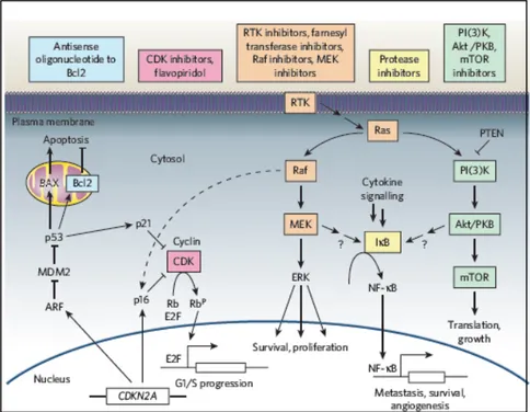
Melanocyte and melanoblast proliferation, survival, differentiation, and migration are all critical elements of normal developmental physiology; the signaling circuitry that regulates these events is often altered at the genetic level by the aspiring melanoma cell. Two large cancer circuits—the growth-promoting Ras signaling network, the tumor-constraining CDKN2A network—and their interaction with a key downstream regulator of apoptosis, the Bcl-2/p53 network are crucial points also in therapeutical studies for melanoma treatment (Figure 3).
Figure 3. Melanoma drugs and their target pathways (Gray-Schopfer et al., 2007. Nature).
1.2.2 Genetics of melanoma: the signaling networks in melanoma proliferation and progression.
The Ras signaling pathway. The complex Ras signaling network regulates cell growth, survival, and invasion through two distinct cascades— the Ras/mitogen-activated protein kinase (MAPK) and the Ras/ phosphatidylinositol-3-kinase (PI3K) signaling streams. Up to 90% of melanomas and benign melanocytic neoplasms carry activating mutations in
one of two key MAPK pathway genes, BRAF or NRAS; similarly, alterations in the PI3K pathway have been reported in up to 50–60% of melanomas (Zhou et al., 2000; Stahl et al., 2004). Functional cloning studies in the early 1980s revealed Ras GTPases as oncoproteins that were frequently mutated in a large number of human cancers. In 1984, Albino et
al. established RAS as the first oncogene in melanoma; notably, the
alterations were mainly in NRAS, a then lesser-known isoform in the RAS family, and activating NRAS mutations are relatively common (26%) (Hocker & Tsao, 2007). As Ras activates both the MAPK and PI3K pathways, mutations in NRAS have been demonstrated to occur in a reciprocal pattern with genes downstream of it in either pathway, such as PTEN and BRAF (Tsao et al., 2004b; Curtin et al., 2005). These data suggest that oncogenic NRAS promotes melanocytic proliferation, but it is not sufficient to yield true malignant transformation. Infact, when the Wellcome-Trust Sanger Institute initiated a project to resequence all relevant human protein kinases in the cancer, one of the first and perhaps most substantial findings was the discovery of a highly recurrent mutation in BRAF among different cancer types including melanoma (Davies et al., 2002). Although BRAF was mutated in less than 20% of most cancers, this gene was altered in 80% of short-term melanoma cell cultures and 66% of uncultured melanoma, thereby making BRAF the single most commonly mutated gene in melanoma. The discovery of a highly recurrent mutation in BRAF launched a new era in melanoma therapeutics and provided validation of the highthroughput resequencing strategy now adopted by the National Cancer Institute’s Cancer Genome Project. Similar to oncogenic NRAS, the BRAF mutation is prevalent in benign nevi supporting a role for BRAF
et al., 2003; Yazdi et al., 2003). This hypothesis has been experimentally
validated using a zebrafish model, where it has been shown that BRAF activation leads only to development of benign nevi, whereas progression to frank melanoma requires concurrent p53 inactivation (Patton et al., 2005); thus, the full oncogenic potential of BRAF appears to be dictated by the presence or absence of other genetic constraints. Although much remains to be understood regarding its exact role in melanoma biology, BRAF is a key element in melanoma tumorigenesis and crucial therapeutic target.
The kinase Ras induces also the membrane translocation and activation of PI3K, which in turn phosphorylates and activates the major signaling element of PI3K-AKT cascade, Akt. Once activated, Akt has several different enzymatic substrates including Mdm2, procaspase 9, NF-kB, mammalian target of rapamycin (mTOR), and p27, many of which contribute to tumor proliferation and survival (Sharma et al., 2005). Although activating mutations in PI3K, itself, are believed to be rare, other downstream components of the PI3K pathway have been shown to be altered in up to 50–60% of melanomas (Zhou et al., 2000; Stahl et al., 2004); more specifically, the expression of PI3K and AKT have been shown to steadily increase during the progression from benign nevi to early melanoma, and lastly, to metastatic disease (Dhawan et al., 2002; Stahl et al., 2004). With regard to resistance to apoptosis, Akt is known to phosphorylate CREB, increase antiapoptotic Bcl-2, and inhibit apoptotic Bad (del Peso et al., 1997). Indeed, it was recently demonstrated that a number of metastatic melanoma cell lines were resistant to independent inhibition of the MAPK or PI3K pathways, but they were susceptible to simultaneous inhibition of these pathways, with growth and invasion blocked in vitro (Smalley et al., 2006).
The CDKN2A/CDK4 network. Patients that possess mutations in genes encoding for tumor suppressor, such as p16INK4A, p14ARF, RB, and TP53, have increased susceptibility to melanoma (Mooi & Peeper, 2006). The CDKN2A gene encodes two proteins involved in melanoma growth and survival (Sharpless et al., 2003): p16INK4A (cyclindependent kinase (CDK) inhibitor 2a) and p14ARF (alternative reading frame) p16INK4A inhibits Cdk4/6-mediated phosphorylation of the retinoblastoma protein; in the hypophosphorylated state, Rb binds and represses the E2F transcription factor and prevents G1-to- S transition. On the other hand, p14ARF directly prevents Mdm2 from accelerating the degradation of p53. Thus, loss of the CDKN2A locus negatively impacts on both the Rb and p53 pathways. CDKN2A appears to play a central role in preventing cancer formation by mediating a senescence-like state upon oncogenic stress (Serrano et al., 1997); indeed, oncogenic mutations in NRAS require concomitant loss of CDKN2A in order to progress to frank melanoma (Ackermann et al., 2005). The Bcl-2 network. The Bcl-2 network is thought to be one of the most crucial regulators of melanoma cell apoptosis (Soengas & Lowe, 2003). At the core of the Bcl-2 network lives a family of anti- and proapoptotic proteins that function to regulate and execute the core mitochondrial pathway of apoptosis (reviewed in Danial & Korsmeyer, 2004). Bcl-2 has been shown to serve a physiologic role in the skin: in response to UV radiation, keratinocytes secrete NGF which binds to melanocyte receptors and leads to increased Bcl-2 expression and resistance to apoptosis (Zhai et
al., 1996). In fact, high levels of Bcl-2 expression have been found in
melanoma and melanocytes, providing a likely explanation for the resistance of melanocytes and melanoma to apoptosis induced by both physiological
Aberrations in various signaling pathways contribute to elevated Bcl-2 levels in melanoma. Oncogenic NRAS has been shown to specifically upregulate the expression of Bcl-2 in vitro (Borner et al., 1999); the microphthalmia-associated transcription factor (Mitf) may also contribute to survival by the transactivation of Bcl-2 (McGill et al., 2002). Nonetheless, it has been shown that antisense suppression of Bcl-2 or mutations in Mitf, a transcriptional regulator of Bcl-2, leads to decreased melanoma cell survival and increased sensitivity to chemotherapy (Jansen et al., 1998; Gautschi et
al., 2001; McGill et al., 2002); thus Bcl-2 has been considered an attractive
therapeutic target.
Mitf gene. The Mitf gene has emerged as a master regulator of melanocyte development. This transcription factor dictates the pigment cell phenotype by regulating melanocyte-specific proteins such as MART-1, enhances and controls melanoblast survival, and melanocyte proliferation and survival (Hodgkinson et al., 1993; Widlund & Fisher, 2003; Nishimura et al., 2005). Mitf has multiple downstream effects, which together define numerous aspects of normal pigment cell physiology. Mitf directly promotes melanocyte and possibly melanoma cell survival by inducing the expression of the protooncogene, Bcl-2, as one of its transcriptional targets (McGill et
al., 2002). Despite the emerging view of Mitf in melanoma biology, critical
questions about its biology remain unanswered. In contrast to its oncogenic behavior, Mitf has been shown to mediate G1 cell-cycle arrest by upregulating p21Cip1 (Carreira et al., 2005). One model posits that cell-cycle arrest and differentiation takes place at high levels of Mitf expression whereas apoptosis occurs at very low, or null, expression levels (Gray-Schopfer et al., 2007); thus, a tumor-favorable Mitf level may be required. Increases in MAPK signaling leads to ubiquitin-dependent proteolysis of
Mitf (Wu et al., 2000). As complete loss of Mitf is incompatible with cell survival, melanoma specimens with constitutively active BRAF require mechanisms that maintain Mitf levels in the range compatible with tumorigenesis. Mitf amplification occurs in up to 20% of melanomas and thus may account for some of this Mitf recovery; however, the fact that only a small percentage of melanomas with oncogenic BRAF and NRAS possess Mitf amplifications suggests that other mechanisms are likely involved. Mitf is a downstream target of β-catenin, a critical regulator of melanoma cell growth (Widlund et al., 2002); thus, an alternative mechanism of Mitf recovery could involve stabilizing mutations in β-catenin leading to induction of Mitf. Given that Mitf plays a central role in melanocyte and tumor survival, further delineation of the genetic network that regulates, and is regulated by, Mitf is warranted.
1.2.3 Therapy of melanoma: Ras/Raf/Mek/Erk MAPKs as targeted growth signaling pathway.
Dacarbazine (DTIC)—which possesses response rates as low as 10–13% and complete responses in only 5%—has remained the mainstay of single-agent chemotherapy treatment for advanced melanoma for over 20 years (Eigentler et al., 2003). Nitrosoureas, platinum analogs, vinca alkaloids, and the taxanes all have been shown to have only modest activities against metastatic melanoma; temozolomide (TMZ) is a newer oral alkylating agent that functions like DTIC and possesses excellent bioavailability. The generally poor efficacy of all current single-agent chemotherapeutic
approaches has prompted myriad studies involving combination chemotherapy, biochemotherapy, and tumorspecific immunologic therapies. Unfortunately, over the past 20 years, there have been no substantial benefits with regard to median survival, and many of the combination regimens have the drawback of increased toxicity (Eigentler et al., 2003; Gogas et al., 2007). Much of melanoma’s resistance to traditional chemotherapy is believed to arise intrinsically, by virtue of potent growth and cell survival-promoting genetic alterations. Therefore, significant attention has recently been focused on developing previously unknown targeted therapies that aim to selectively shut down the genetic aberrations that fuel this tumor’s oncogenic engine, as well as agents that are capable of decreasing this tumor’s inherent resistance to chemotherapy. Given the importance of the RAS signaling network in promoting melanoma proliferation and survival, therapies specifically aimed at this pathway are ideal for many reasons. First, this network is the most frequently altered in melanomas: up to 90% of melanomas possess oncogenic alterations in NRAS or one of its downstream pathways. Second, activation of the MAPK and PI3K pathways has been shown to lead to increased proliferation, cell survival, and contributes to melanoma’s inherent chemoresistance by disrupting the death receptor-, mitochondrial-, and endoplasmic reticulum stress-induced apoptotic pathways (Eisenmann et al., 2003; Zhang et al., 2003; Hersey et al., 2006; Jiang et al., 2007). For example, lonafarnib blocks Ras activation and preliminary data suggest that lonafarnib with cisplatin may enhance melanoma apoptosis in vitro. (Smalley & Eisen, 2003; Morgillo & Lee, 2006). Instead, sorafenib is a Raf inhibitor that has been shown to abrogate MAPK signaling biochemically and to harbor antimelanoma effects in vitro (Karasarides et al., 2004); early clinical trials have failed to show significant
efficacy with sorafenib as monotherapy in advanced melanoma (Eisen et al., 2006), but recent results showed that sorafenib in combination with carboplatin, paclitaxel, TMZ and DITC have been more promising. Finally, in preclinical studies CI-1040, a small molecular weight MEK inhibitor, was found to completely suppress growth of BRAF mutant melanoma xenografts with regression of pulmonary metastases and prevention of new pulmonary metastases (Collisson et al., 2003). PD0325901, a second generation Mek inhibitor, is 100 times more potent and is 490-fold more potent in suppressing pERK relative to CI-1040. A recent phase I/II trial of PD0325901 enrolled 27 patients and demonstrated partial responses in 2/27 and stable disease in 5/27 (Lorusso et al., 2005a). Significantly, tumors from partial responders showed substantial ERK inhibition, a critical component of the targeted mechanism.
2. PURPOSE
Since research in melanoma cancer therapy is focused on discovery of novel drugs able to reduce its proliferative capacity and induce terminal differentiation and/or apoptosis (Jiang et al., 1994), the aim of this work is:
1) to investigate the antigrowth activity of a coumarin derivative, the
5,7-dimethoxycoumarin, on melanoma cell lines, such as murine B16 F1 and human A375;
2) to analyse modulation of Ras/Raf/Mek/Erk MAPKs signaling pathway
that is the most frequently altered network in melanomas (Wellbrock et al., 2005);
3) to examine cellular processes triggered by 5,7-dimethoxycoumarin
following reduction of cell proliferation, which can be related to differentiation and/or apoptosis programmes;
4) to define the molecular mechanisms involved in
5,7-dimethoxycoumarin effects, such as downstream effectors of Ras/Raf/Mek/Erk signaling pathway and /or other molecular networks cross-talking with MAPKs.
3. MATERIALS AND METHODS
3.1 Test compound.
Synthetic 5,7-dimethoxycoumarin was purchased from Sigma-Aldrich (116238). The compound was dissolved in methanol and a solution with a concentration 20 mM was obtained. In the treatment of cells, a volume of this solution was added to culture medium and its percentage respect to medium was 0.5, 1.25 and 2.5 % for 100, 250 and 500 μM, respectively.
3.2 Cell lines and culture conditions.
Cell lines used in this work are: murine melanoma B16 F1, human melanoma A375, human breast adenocarcinoma MCF7, human prostate adenocarcinoma PC3, human colorectal carcinoma SW620. Cancer cell lines were purchased from American Type Culture Collection (ATCC; Manassas, VA, USA). As control, non-neoplastic murine cardiofibroblasts were used. Cells were cultured in RPMI 1640 medium supplemented with 10 % (v/v) fetal bovine serum (FBS), 1% L-glutamine (v/v), 100 units/ml penicillin and 100 μg/ml streptomycin. The cells were grown at 37°C in a humidified atmosphere with 5% CO2.
3.3 MTT assay.
5,7-dimethoxycoumarin activity on cells growth was estimated by MTT (3-(4,5-dimethylthiazol-2-yl)-2,5-diphenyl tetrazolium bromide) assay, that is based on the cleavage of the yellow tetrazolium salt to purple formazan crystals by intracellular dehydrogenases (Mosmann et al., 1983). Briefly, 1-2x10³ cells/well were seeded into sterile 96-well plates. After 24 h,
5,7-dimethoxycoumarin was added to cell culture medium over a final concentration range of 0-500 μM and the cells were incubated at 37°C and
5% CO2 for a period of 24-72 h. After incubation period, MTT
(Sigma-Aldrich) was added to each well and incubated for further 4 h. Then, medium was removed, blue crystals of MTT reduced by cells were dissolved with DMSO and cellular metabolism was determined by measuring absorbance of
samples at 570 nm in a microelisa reader. IC50 values were estimated
following 72 h incubation.
3.4 Trypan blue exclusion test.
B16 cells, A375 cells and cardiofibroblasts were seeded at a density of 1-2x10⁴ cells/well in 24 well plates. After 24 h cells were treated with 100, 250
and 500 μM 5,7-dimethoxycoumarin and the plates were incubated at 37°C and 5% CO2 for a period of 24-72 h. After treatment time, floating cells in
the medium of each well were transferred to centrifuge tubes; adherent cells were washed, collected by trypsinization and mixed with the corresponding
floating cells before centrifugation. Cells were stained with 0.4% Trypan blue and counted in triplicate with optic microscope with the aim to estimate the number of live and dead cells. Cell viability was expressed as a percentage of treated cells respect to appropriate vehicle-treated controls, and toxicity as a percentage of dead cells respect to total number of cells. Moreover, B16 cells treated for 72 h were washed several times with medium to remove the excess of 5,7-dimethoxycoumarin, counted and replaced in fresh medium at 1x10⁴ cells/well without the compound. After
48, 72 and 96 h incubation at 37°C and 5% CO2, the irreversible or
reversible growth inhibitory effect of compound was observed by trypan blue test.
The B16 treated cells were observed with an inverted microscope and photographed at 10X and 20X with a digital camera Nikon Coolpix 995 to detect any drug-induced morphological changes.
3.5 Treatment of B16 cells with 5,7-dimethoxycoumarin and U0126 and Trypan blue assay.
B16 cells were seeded at a density of 1x10⁴ cells/well in 24-well plates.
After 24 h, cells were treated with 100, 250, 500 μM 5,7-dimethoxycoumarin or with concentration 1, 5, 10 μM of U0126 (Sigma-Aldrich), a specific inhibitor of Mek 1/2 (Favata et al., 1998); moreover, 5,7-dimethoxycoumarin was used in combination with U0126. After 72 h treatment, floating cells in the medium of each well were transferred to centrifuge tubes; adherent cells were washed, collected by trypsinization and mixed with the corresponding
floating cells before centrifugation. Cells were stained with 0.4% trypan blue and counted in triplicate with an optic microscope with the aim to estimate the number of live and dead cells. Cell viability was expressed as a percentage of treated cells with respect to appropriate vehicle-treated controls.
3.6 Cell cycle analysis.
B16 and A375 cells were seeded and treated as described in section of Trypan blue exclusion test. After 24, 48 and 72 h incubation at 37°C and 5%
CO2, cells were washed, harvested, pelleted, fixed with PFA
(paraformaldehyde) 1% in PBS for at least 30 min at 4°C and stained with propidium iodide (PI) staining solution containing 50 μg/ml PI, 0,5 % RNase A and 0.1% Triton X-100. After incubation for 30 min at 4°C in the dark, cell cycle distributions were analyzed by flow cytometry on a FACScalibur (Becton Dickinson, Mountain View, CA). Using Cellquest Pro software, the percentage of cells at different phases of the cell cycle was determined. PI was excited at 488 nm, and fluorescence analyzed at 620 nm. A total of 10,000 events in each sample were acquired.
3.7 Protoporphyrine IX (PpIX) analysis and phenotypic characterization with flow cytometry.
B16, A375 and SW620 cells were seeded at a density of 1-1.5x10⁴
cells/ml in 24-well plates. After 24 h were treated with methanol control or 100, 250 and 500 μM of 5,7-dimethoxycoumarin. After 24, 48 and 72 h
incubation at 37°C and 5% CO2, cells were washed with PBS, harvested
bytrypsinization, pelleted and resuspended in PFA 1%. After at least 30 min incubation at 4°C, the samples were analyzed on a FACScalibur flow cytometer using the 488 nm line from an argon laser (Becton Dickinson, Mountain View, CA). In all, 10,000 cells, for each sample, were measured: scattering properties and fluorescence emitted from the cells at 670 nm were collected and analysed.
3.8 Melanin synthesis in B16 and A375 cells.
B16 and A375 cells were seeded and treated as reported in section of Trypan blue exclusion test, in order to measure extracellular and intracellular melanin according to the method of Hill et al. (1989). After 72 h incubation
at 37°C and 5% CO2, the culture medium was removed, centrifuged (700xg,
10 min) and the supernatant was collected for extracellular melanin quantification; 1 ml of 0.4M HEPES buffer (pH 6.8) and EtOH (9:1, v/v) was added to 1 ml of the medium and the OD at 475 nm was measured to quantify extracellular melanin by using a calibration curve obtained with
washed twice with PBS and digested in 1 ml 1N NaOH for 16 h at room temperature; intracellular melanin was measured as described above. The stimulation in melanogenesis following the treatment was estimated as percentage of total melanin (μg/ml), calculated by the sum of intracellular and extracellular melanin, synthesized in treated cells respect to vehicle-treated controls.
3.9 Tyrosinase assay.
B16 cells were seeded and treated as described above in order to determine tyrosinase activity according to the method of Lin et al. (2002). After 24, 48 and 72 h treatment time, cells were washed, collected by trypsinization, stained with 0.4% trypan blue and counted in triplicate with an optic microscope to estimate the number of live cells. For each sample, 7
x 104 cells were then lyzed in 1% Triton-X 100 in 0.1 M Na phosphate
buffer (pH 6.8) for 20 minutes; lysates were incubate with an equal volume of 3,4-dihydroxy-L-phenylalanine
(L-DOPA), 3 mg/ml in 0.1 M Na phosphate buffer for 3 h at 37°C. Tyrosinase activity was determined by measuring sample absorbance at 490 nm. The stimulation of tyrosinase activity following the treatment was estimated as fold increase in treated cells with respect to vehicle-treated controls.
3.10 Western blot analyses.
B16 and A375 cells were seeded at a density of 1x10⁴ cells/well in
6-wells plates and treated as described above. After 24, 48 and 72 h treatment time, cells were washed, collected by trypsinization and suspended in RIPA lysis buffer [1% Nonidet P-40, 1% sodium deoxycholate, 0.1% SDS, 0.15 NaCl, 0.01 M sodium phosphate (pH 7.2), 2 mM EDTA, 50 mM sodium fluoride, 0.2 mM sodium vanadate and 100 units/ml aprotinin]. After 1 h on ice, the cell lysates were cleared by centrifugation at 13000 x g for 15 min and the resultant supernatants collected and used for Bradford assay (1976) to estimate protein concentration. Then, 40 μg of total protein lysates were resolved on 12% sodium dodecyl sulphate polyacrylamide gels (SDS-PAGE) and the separated proteins were transferred to PVDF membrane. The level of protein expression in each sample was detected using specific mouse monoclonal primary antibody diluted in TTBS solution containing 1% (w/v) of BSA: antibodies against Mek 1/2 (Ser 218/Ser 222), phospho-Erk 1/2 (Tyr-204), Mitf (N-15), PBG-D (A-16), p-CREB-1 (Ser 133) and α-tubulin were purchased from Santa Cruz Biotechnology, Inc. (Santa Cruz, CA). After, the membranes were incubated with a specific HRP-conjugated secondary antibody and developed using the enhanced chemiluminescent substrate from Pierce; then, they were stripped and reprobed with α-tubulin (4G1) primary antibody as a protein loading control.
3.11 Mek 1/2 immunoprecipitation and kinase assay.
B16 and A375 cells were seeded and treated as described above; after 72 h treatment time, cells were washed, collected by trypsinization and lysed by using the same experimental procedure of the previous section. Equal protein aliquots of precleared cell lysates (300 μg) were incubated overnight with Anti-Mek antibody and Ezview Red Protein A Affinity Gel beads (Sigma-Aldrich) at 4 °C with gentle rotation. The immune complexes were collected by centrifugation, washed three times and incubated for 90 minutes at 30 °C with non activated Erk substrate solution (Sigma-Aldrich) for kinase assay. Then, the samples were boiled in SDS sample buffer and loaded on 12% sodium dodecyl sulphate polyacrylamide gels (SDS-PAGE); the separated proteins were transferred to PVDF membrane. The level of diphosphorylated Erk 1/2 was detected using a monoclonal antibody against the activated MAP kinase (Sigma-Aldrich); the membrane was then incubated with a specific HRP-conjugated secondary antibody (Sigma-Aldrich) and developed using the enhanced chemiluminescent substrate from Pierce.
3.12 Statistical analysis.
Student’s t test was employed to determine the significance of any reduction in cellular viability and the increase of melanin synthesis following the treatment. A probability of 0.05 or less was deemed statistically significant.
4. RESULTS
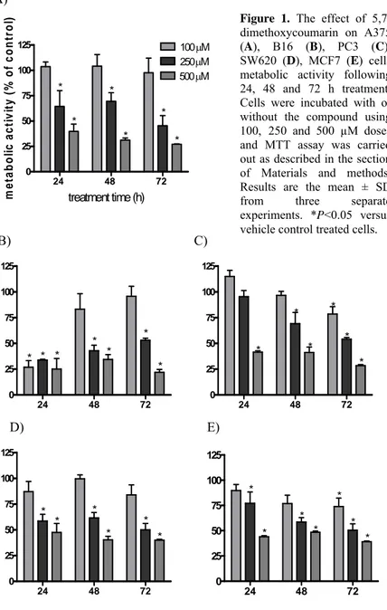
4.1 Reduction of metabolic activity of murine and human cell lines after treatment with 5,7-dimethoxycoumarin.
In the first set of experiments, the antineoplastic activity of 5,7-dimethoxycoumarin in various human melanoma and adenocarcinoma cell lines was measured using MTT assay. The compound significantly reduced metabolic activity, in dose-dependent manner up to 70% after 72 h treatment respect to the control. The concentration 100 μM did not show a significant growth inhibition activity, except for B16, PC3 and MCF7 lines. The concentrations 250 μM and 500 μM inhibited significatively cellular proliferation at each incubation time for every cell line (Figure 1). In Table 1, IC50 values, estimated after 72 h treatment, are reported and evidenced that
5,7-dimethoxycoumarin exhibited a clear effect not only on melanoma but also on adenocarcinoma cell lines.
A) 24 48 72 0 25 50 75 100 125 100μM 250 μM 500μM * * * * * * treatment time (h) m e ta bol ic a c ti v it y ( % of cont rol )
B) C) 24 48 72 0 25 50 75 100 125 * * * * * * * 24 48 72 0 25 50 75 100 125 * * * * * *
D) E) 24 48 72 0 25 50 75 100 125 * * * * * * 24 48 72 0 25 50 75 100 125 * * * * * * *
Figure 1. The effect of
5,7-dimethoxycoumarin on A375 (A), B16 (B), PC3 (C), SW620 (D), MCF7 (E) cells metabolic activity following 24, 48 and 72 h treatment. Cells were incubated with or without the compound using 100, 250 and 500 µM doses and MTT assay was carried out as described in the section of Materials and methods. Results are the mean ± SD from three separate experiments. *P<0.05 versus vehicle control treated cells.
4.2 Effect of 5,7-dimethoxycoumarin on growth and death of melanoma cell lines.
MTT assay did not reveal if the reduction of proliferation was related to growth arrest or cell death since both mechanisms could result in reduced cell numbers and an apparent loss of viability. To establish that, a Trypan blue exclusion test was carried out on melanoma cell lines. The results reported in Figure 2A show that 5,7-dimethoxycoumarin possessed an antiproliferative activity on B16 cells growth at all the concentrations tested. The compound inhibited the growth of B16 cells significatively, as compared with the control group, at 48 h and 72 h, accounting for 35 % to 47 %, 48 % to 68 % and 61 % to 72 %, at the doses of 100, 250 and 500 μM, respectively. The inhibitory effect was time-dose dependent.
In the same way, the treatment with 5,7-dimethoxycoumarin reduced significatively proliferation of A375 cells as well (Figure 2B), accounting for 0 % to 45 %, 17 % to 68 % and 41 % to 80 % of reduction at the doses of 100, 250 and 500 μM, respectively. The inhibition of cell growth was significant respect to the control in each treatment time, except for the concentration 100 μM at 24 h. The inhibitory effect was time-dose dependent.
A)
24 48 72 0 25 50 75 100 100μM 250μM 500μM * * * * * * * * treatment time (h) C e ll vi abi lit y ( % of cont ro l) B)
24 48 72 0 25 50 75 100 125 * * * * * * * * treatment time (h) C e ll v iab ilit y ( % o f c ont ro l)
Figure 2. (A) B16 and (B) A375 cell viability following 24, 48 and 72 h treatment with
5,7-dimethoxycoumarin. Cells were incubated with or without the compound using 100, 250 and 500 µM doses and Trypan blue exclusion test was carried out. Data are expressed as % of cell viability respect to vehicle control as described in the section of Materials and methods. Results are the mean ± SD from three separate experiments. *P<0.05 versus vehicle control treated cells.
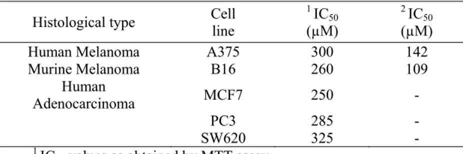
IC50 values at 72 h were obtained also through Trypan blue test and they
accounted for 109 μM and 142 μM in B16 and A375, respectively (Table 1). Interestingly, they were lower than those derived from MTT assay at the same time of treatment. That probably is a consequence of the minor
reliability of an indirect assay, such as MTT assay, respect to a direct one as Trypan blue test (Jabbar et al., 1989).
Table 1. IC50 of 5,7-dimethoxycoumarin in different cell lines following 72 h treatment. Cells were incubated with or without the compound using 100, 250 and 500 μM doses; then, MTT assay and Trypan blue exclusion test were carried out, as described in the section of Materials and methods. A grapf of viability versus drug concentration was used to calculate IC50 values for each cell line.
Histological type Cell line 1 IC50
(µM)
2 IC
50
(µM)
Human Melanoma A375 300 142
Murine Melanoma B16 260 109 Human Adenocarcinoma MCF7 250 - PC3 285 - SW620 325 - 1 IC
50 values as obtained by MTT assay.
2 IC
50 values as obtained by Trypan blue exclusion test.
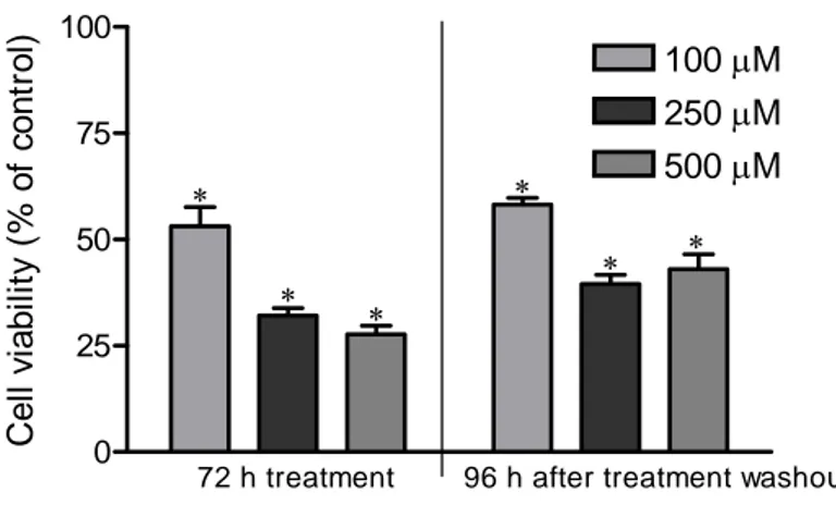
Moreover, the effect of the compound on murine melanoma cells was long lasting, since the same cells previously treated at doses of 100, 250, and 500 μM, washed, reseeded and cultured without treatment, showed a significant reduced performance of proliferation respect to untreated cells. After 96 h incubation at 37°C and 5% CO2, the growth reduction produced by previous
treatment was 42 %, 61 % and 57 % at the doses of 100, 250 and 500 μM, respectively. Statistical differences were observed between the values obtained with 100 μM and 250 μM doses, and with 100 and 500 μM doses (Figure 3). These results evidenced that the antiproliferative effect of the
compound was irreversible since proliferation did not return to control levels even at the end of 96 h recovery period.
The percentage of dead cells calculated as explained in Materials and Methods sections, after treatments for both cell lines was also evaluated; no significant cytotoxicity was observed at any experimental time and dose respect to the control (data not shown).
The 5,7-dimethoxycoumarin did not change significatively the growth of non-neoplastic cardiofibroblasts for none of the used concentrations (Figure 4).
0 25 50 75 100 * * *
72 h treatment 96 h after treatment washout
* * * 500μM 250μM 100μM C e ll v ia b ility ( % o f c o n tr o l)
Figure 3. The irreversible effect of 5,7-dimethoxycoumarin on B16 cell growth. Cells were
incubated with or without the compound using 100, 250 and 500 µM doses for 72 h; then, they were reseeded without treatment for 96 h. Data are expressed as % of cell viability respect to vehicle control as described in the section of Materials and methods. Results are the mean ± SD from three separate experiments. *P<0.05 versus vehicle control treated cells.
0 50 100 ctr 100μM 250μM 500μM 5,7-dimethoxycoumarin concentration c e ll via b ilit y (% o f c o n tr o l)
Figure 4. Cell viability of fibroblasts following 72 h treatment with 5,7-dimethoxycoumarin.
Cells were incubated with or without the compound using 100, 250 and 500 µM doses and Trypan blue exclusion test was carried out. Data are expressed as % of cell viability respect to vehicle control as described in the section of Materials and methods.
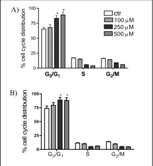
4.3 Block of cell cycle in G0/G1 phase after treatment of melanoma cells with 5,7-dimethoxycoumarin.
Since the strong inhibitory effect induced by 5,7-dimethoxycoumarin could be related to a decrease of DNA synthesis, we then investigated its effect on cell cycle events. After 24 h treatment, we demonstrated, by FACS analysis, that 5,7-dimethoxycoumarin induced a strong and dose dependent arrest of cell cycle progression in G0/G1 phase in B16 and A375 cell lines, when compared to control sample, with reduction of S and G2/M phases
population. In regard to B16 cells (Figure 5A), the accumulation in G0/G1 phase was significant at high dose and consistent with growth arrest when compared to the control. An accumulation of melanoma cells in G0/G1 phase was detected also after 48 and 72 h treatment, although the differences respect to the control were less significative than 24 h of incubation (data not shown). Similarly, in A375 cells, the 500 μM dose induced a G0/G1 arrest after 24 h (data not shown), which did not change at later treatment times; also 100 and 250 μM doses of 5,7-dimethoxycoumarin showed a significant increase in G0/G1 cell population after 48 h treatment (Figure 5B).
Figure 5. Effect of
5,7-dimethoxycoumarin on cell cycle progression in B16 cells after 24 h (A) and A375 cells after 48 h treatment (B). Cells were incubated with or without the compound using 100, 250 and 500 µM doses. After treatment, cells were stained with propidium iodide and flow cytometric analysis was performed as described in Materials and methods. The percentage of cell cycle distribution data for each treatment group are shown. Results are the mean ± SD from three separate experiments. *P<0.05 versus vehicle control treated cells.
B) A) G0/G1 S G2/M 0 25 50 75 100 ctr 100μM 250μM 500μM * * % cel l cycl e di st ri but io n G0/G1 S G2/M 0 25 50 75 100 * * % ce ll cy cl e d ist ri b u ti o n
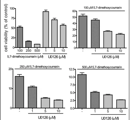
4.4 Effect of Mek 1/2 inhibitor on 5,7-dimethoxycoumarin antigrowth activity in B16 cell line.
In the previous paper, we detected that 5,7-dimethoxycoumarin inhibited the growth of melanoma cells. Now, to further investigate the role of MAPKs signalling pathway Ras/Raf/Mek/Erk in antineoplastic activity of 5,7-dimethoxycoumarin, we used the compound in combination with the Mek 1/2 inhibitor U0126. After 72 h treatment, U0126 reduced cell growth with a precentage of 8%, 29% and 43% for 1, 5 and 10 μM doses, respectively (Figure 6A). In the combination treatment, the inhibitor augmented growth inhibition induced by 5,7-dimethoxycoumarin, as shown in Figure 6 (B-D). When 100 μM dose was combined with increasing amounts of U0126, cell viability was reducEd respect to control from 52.1% to 45.4-22.3%; for 250 μM, from 16.2% to 10.8-4.1% and for higher dose, from 11% to 5.1-2.7%. These data suggest that Mek 1/2 inhibition can sensitize B16 cells to tested compound treatment and then, the combination therapy increased antiproliferative activity of 5,7-dimethoxycoumarin in synergic or additive mode.
100 250 500 1 5 10 0 25 50 75 100 U0126 (μM) 5,7-dimethoxycoumarin (μM) c el l v iabi lit y ( % of cont rol ) - 1 5 10 0 10 20 30 40 50 60 U0126 (μM) 100μM 5,7-dimethoxycoumarin - 1 5 10 0 10 20 U0126 (μM) 250μM 5,7-dimethoxycoumarin - 1 5 10 0.0 2.5 5.0 7.5 10.0 12.5 U0126 (μM) 500μM 5,7-dimethoxycoumarin
Figure 6. The effect of Mek 1/2 inhibitor U0126 on 5,7-dimethoxycoumarin activity in
murine melanoma cell line. B16 cells were incubated with the compound using 100, 250 and 500 µM doses or with U0126 using 1, 5 and 10 µM doses (A); and in combination treatment with 5,7-dimethoxycoumarin 100 (B), 250 (C) and 500 µM (D) and U0126 (1, 5 and 10 µM). After 72 h incubation, Trypan blue exclusion test was carried out. Data are expressed as % of cell viability respect to vehicle control as described in the section of Materials and methods. Results are the mean ± SD from three separate experiments. *P<0.05 versus vehicle control treated cells.
4.5 Morphological changes in melanoma cells after treatment with 5,7-dimethoxycoumarin.
Since the treatment with 5,7-dimethoxycoumarin showed low cytotoxicity, we hypothesised that growth inhibition was due to the induction of differentiation, a process that is generally coupled to a block in cellular proliferation (Umek et al., 1981; Zermati et al., 2000). To verify this hipothesis first we evaluated the morphology of treated melanoma cell lines under an inverted microscope. B16 cells analysis by digital images (Figure 7) showed a dose dependent decrease of cellular density in the culture treated with different doses of 5,7-dimethoxycoumarin. This result is in accordance with those obtained with Trypan blue test. Moreover, a clear morphological change was detected in cells. Infact the treatment induced the formation of dendrite-like projections which give a star-like shape to the cells respect to rounded untreated cells. This effect was more evident with the increase of treatment time and concentration and after 5,7-dimethoxycoumarin washout it did not recover the control morphology (data not shown). Similar results were obtained for A375 cells regarding cellular density; anyway, morphological changes were less evident respect to those obtained in B16 cells (data not shown).
A) B)
C) D)
Figure 7. Effect of 5,7-dimethoxycoumarin 100 µM (B), 250 µM (C) and 500 µM (D) respect
to control (A) on B16 cells growth and morphology after 72 h incubation. Cells were seeded and treated as described in the section of Materials and Methods; then digital images were obtained by observation with an inverted microscope at 10X.
4.6 Phosphorylation of ERK1/2 after treatment with 5,7-dimethoxycoumarin.
To unravel the potential mechanism underlying 5,7-dimethoxycoumarin-induced antigrowth effect, we examined the alterations in activation of a MAPK signalling pathway following treatment. The effects of
5,7-dimethoxycoumarin on Erk 1/2 phosphorylation in B16 cell line after 72 h treatment are shown in Figure 8: the treatment inhibited the activation of the protein in a dose-dependent manner resulting in a moderate to complete inhibition. Densitometric analysis allowed to calculate the effect of treatment, as compared to control untreated cells, in ERK phosphorylation: the compound reduced p-Erk 1/2 of 60%, 85% and 96% for 100, 250 and 500 μM, respectively. The compound was likewise effective on A375 cells bringing an inhibition, after 72 h treatment, of the Erk phosphorylation with a percentage of 70% in the case of higher dose (Figure 9).
1 2 3 4 p-Erk 1/2
α-tubulin
Figure 8. The effect of 5,7-dimethoxycoumarin on Erk 1/2 activity in B16 cells following
treatment 72 h. Cells were incubated with the compound using 100, 250 and 500 µM doses: lanes 2 (100 μM), 3 (250 μM), 4 (500 μM); or with the vehicle solvent: lane 1. After treatment, total cell lysates were prepared and subjected to SDS-PAGE followed by Western immunoblotting as described in the section of Materials and methods. Activation level of Erk 1/2 was analysed with its corresponding phospho-specific antibody.
p-Erk 1/2
α-tubulin
1 2
Figure 9. The effect of 5,7-dimethoxycoumarin on Erk 1/2 phosphorylation in A375
melanoma cells following treatment 72 h. Cells were incubated with (lane 2) or without (lane
1) the compound using 500 µM doses. After treatment, total cell lysates were prepared and
subjected to SDS-PAGE followed by Western immunoblotting as described in the section of Materials and methods. Activation level of Erk 1/2 was analysed with its corresponding phospho-specific antibody.
4.7 Phosphorylation of MEK 1/2 and inhibition of its kinase activity in melanoma cells after treatment with 5,7-dimethoxycoumarin.
In the next step, we sought to examine, following a 72 h treatment, the alterations in phosphorylation level of Mek 1/2, the kinase which phosphorylates Erk 1/2. The effects of 5,7-dimethoxycoumarin on the MAP kinase kinase activation, in B16 and A375 cells, are shown in Figure 10A and 10B, respectively: the treatment increased level of phospho-Mek 1/2 (p-Mek 1/2) in a moderate manner in both cell lines as compared with control samples. The augmentation of protein phosphorylation, estimated by densitometric analysis, was 16%, 32% and 85% for 100, 250 and 500 μM, respectively in B16 cells; it was 42%, 67% and 72% in A375 cell line. Considering promoting activity of 5,7-dimethoxycoumarin on Mek phosphorylation in melanoma cells, we were interested to understand the key
point in the phosphorylation cascade of this signalling pathway where the compound exerted, directly or indirectly, its antiproliferative activity. Up to now we described that treatment reduced Erk 1/2 phosphorylation, but it triggered a moderate augmentation in Mek 1/2 phosphorylation in both used cell lines. p-Mek 1/2 α-tubulin p-Mek 1/2 B α-tubulin A375 B16 A 1 2 3 4 5 6
Figure 10. The effect of 5,7-dimethoxycoumarin on Mek 1/2 phosphorylation in B16 (A) and
A375 (B) melanoma cells following treatment 72 h. Cells were incubated with the compound using 100, 250 and 500 µM doses: lanes 2 (100 μM), 4 (250 μM), 6 (500 μM); or with respective vehicle solvent: lanes 1, 3, 5. After treatment, total cell lysates were prepared and subjected to SDS-PAGE followed by Western immunoblotting as described in the section of Materials and methods. Activation level of Mek 1/2 was analysed with its corresponding phospho-specific antibody.
Then, the first question was regarding the effect of treatment on enzimatic activity of p-Mek, which was tested using Erk, which is activated upon dual phosphorylation by Mek, as its substrate in a kinase assay following immunoprecipitation of Mek 1/2. Immunoblot analyses results (Figure 11),
after incubation with a p-Erk 1/2 specific antibody, allowed to observe a significant decrease in p-Erk, demonstrating that 5,7-dimethoxycoumarin exerts its antiproliferative activity through inhibition of Mek 1/2 kinase activity; then, next reduction in Erk 1/2 triggered a terminal differentiation in treated melanoma cells, with melanin synthesis and dendricity.
1 2 3 4 p-Erk IP: anti-Mek Erk 1/2 p-Mek 1/2
Figure 11. The effect of 5,7-dimethoxycoumarin on in vitro kinase activity of p-Mek 1/2 in
B16 and A375 melanoma cells. B16 (1,2 )and A375 cells (3,4) were incubated for 72 h with the vehicle solvent (1,3) or with the compound using 500 μM dose (2,4). Mek 1/2 was immunoprecipitated with anti-Mek 1/2 antibody from equal protein aliquots of cell lysates and immune complexes were incubated with non activated ERK substrate solution for in vitro kinase assay. After Western blotting, phosphorylation level of Erk 1/2 was analysed with its corresponding phospho-specific antibody.
4.8 Alteration of phenotypic characteristics in melanoma cells after treatment with 5,7-dimethoxycoumarin.
Phenotypic alterations were observed through cytofluorimetric analysis of both treated melanoma cell lines. A change in light scatter properties of B16
and A375 cells treated with 5,7-dimethoxycoumarin was detected. In particular, a decrease of FSC (forward scatter) mean values respect to the solvent control was observed; at the same time, SSC (side scatter) mean values increased strongly following the treatment. This effect was more pronounced in B16 than A375 cells. In Figure 12 results relative to the treatment with 500 μM dose for 72 h are shown. Also phenotypic characterization of SW620 cells was carried out, but changes in FSC and SSC were not detected after treatment, using the same experimental conditions (data not shown).
A)
C) D)
B)
Figure 12. Light scatter of B16 (A-B) and A375 cells (C-D) incubated or not with
5,7-dimethoxycoumarin 500 µM for 72 h. In the scattergrams 5,7-5,7-dimethoxycoumarin-treated cancer cells (B-D) exhibited less forward scatter and much more side scatter than solvent-treated cells (A-C).