UNIVERSITÀ DEGLI STUDI DI SALERNO
Dipartimento di Farmacia
Dottorato di ricerca
in
Scienze Farmaceutiche
Ciclo XIV — Anno di discussione 2016
Coordinatore: Chiar.mo Prof. Gianluca Sbardella
Synthesis of novel ligands for the
stabilization of organometallic
complexes having potential antitumor
activity
settore scientifico disciplinare di afferenza:
CHIM/08
Dottorando Tutore
Dott. Chiar.ma Prof
1 Chapter I ... 1
Introduction ... 3
1.1 MRI Contrast Agents ... 6
1.2 Radiopharmaceuticals ... 10
1.3 Superoxide Dismutase Mimics ... 12
1.4 Platinum anticancer drugs ... 15
1.5 Ruthenium complexes ... 19
1.6 Metallocene and Titanocene complexes ... 22
1.6.1 Metoxy-aryl and alkyl titanocenes ... 26
1.7 NHC complexes in medicine. ... 33 1.7.1 Ag-NHC complexes ... 33 1.7.2 Au-NHC complexes ... 36 1.7.3 NHC-Cu complexes ... 41 1.8 Mammary carcinoma ... 44 1.8.1 Epidemiology ... 44 1.8.2 TNM classification ... 45 1.8.3 Molecular Classification ... 46 1.8.4 Therapy ... 47 1.9 Prostate cancer ... 53 1.9.1 Epidemiology ... 53 1.9.2 Classification ... 54 1.9.3 Therapy ... 54 1.10 References ... 57 2 Chapter II ... 68 2.1 Introduction ... 70
2.1.1 Group III metal complexes ... 70
2.2 Aims of the project ... 76
2.2.1 Evaluation of the drug-membrane interactions ... 77
2.3 Materials and Methods. ... 78
2.3.1 General procedure ... 78
2.3.2 Solvents ... 78
2.3.3 Reagents ... 78
2.3.4 Characterization techniques ... 78
2.3.5 Cell lines ... 79
2.3.6 Biological assay kit ... 79
2.3.7 Evaluation of the drug membrane interaction ... 79
2.3.8 Distillation of Cylopentadiene. ... 81
2.3.9 Synthesis of ligands ... 82
2.3.10 Synthesis of lithium salt ... 85
2.3.11 Synthesis of yttrium cyclopentadienyl complexes ... 86
2.3.12 Synthesis of scandium cyclopentadienyl complexes ... 90
2.3.13 Synthesis of neodymium cyclopentadienyl complexes ... 93
2.3.14 MTT assay. ... 97
2.4 Results and discussion ... 98
2.4.1 Synthesis of cyclopentadienyl ligands ... 98
2.4.2 Synthesis of group III cyclopentadienyl metal complexes103 2.4.3 Biological results ... 106
2.4.4 Evaluation of drug-membrane interaction ... 116
2.5 Conclusions ... 117
2.6 References: ... 118
3 Chapter III ... 122
3.1 Introduction ... 124
3.1.2 N-heterocyclic (NHC). Carbenes ... 128
3.2 Aim of the project ... 132
3.3 Materials and methods ... 134
3.3.1 General procedure ... 134
3.3.2 Solvents ... 134
3.3.3 Reagents ... 134
3.3.4 Characterization techniques ... 134
3.3.5 Synthesis of NHC-ligands ... 135
3.3.6 Synthesis of silver(I)-NHC complexes ... 138
3.3.7 Synthesis of gold(I)-NHC complexes ... 140
3.3.8 Synthesis of copper(I)-NHC complexes ... 142
3.3.9 Biological procedures ... 145
3.4 Results and Discussion ... 156
3.4.1 Synthesis of NHC ligands ... 156
3.4.2 Synthesis of Ag-NHC complexes. ... 159
3.4.3 Synthesis of Au(I)-NHC complexes. ... 161
3.4.4 Syntesis of Cu(I)-NHC complexes. ... 162
3.4.5 Biological results ... 163
3.5 Conclusions... 175
Abstract
Synthesis of novel ligands for the stabilization of organometallic complexes having potential antitumor activity
The design of new metal complexes as anticancer agents has received
considerable interest in recent years. Complexes of titanium (e.g.: titanocene dichloride), lanthanides complexes (e.g.: texaphyrrins lanthanide) and carbenic complexes of gold, silver and copper showed significant biological activity, and have progressed into clinical trials.
Thus, the target of this PhD project are the synthesis of new ligands and metal complexes. Firstly 5 cyclopentadienyl pro-ligands were synthesised:
6-phenylfulvene, 6-(p-metoxyphenyl)-fulvene e 6-(3',4'-dymethoxyphenyl)fulvene,
6-(3',5')-dymethoxyphenyl)fulvene and 6-(2',4')-dymethoxyphenyl)fulvene.
Then, the synthesis of 12 novel scandium, yttrium and neodymium complexes with these cyclopentadienyl ligands was carried out.
The complexes were tested on DU146 (Prostatic carcinoma) and MDA.MB213 (Breast cancer) to verify inhibition of cell-proliferation, using MTT test with standard procedures. All the complex showed a strong concentration-dependent ability of inhibiting the growth tumor cell, referring to antiblastic activity.
In the last years the synthesis of new carbenic ligands
(N-methyl-N’-[2-hydroxycyclopentan]-4,5-dychloroimydazole iodide,
N-methyl-N’-[2-hydroxycyclopentan]-4,5-dyphenylimydazole iodide,
N-methyl-N’-[(2-hydroxy-2-phenyl)ethyl]-imydazole iodide) and 8 new complexes of gold,
silver and copper was carried out.
The complexes were tested on MCF-7 (human mammary carcinoma expressing the estrogen receptor ERα/ER-positive), MDA-MB-231 (human mammary carcinoma not expressing the estrogen receptor/ER-negative), MCF-10 (breast glandular epithelium), using MTT test with standard
procedures. All the molecules showed a good inhibitory effect on the proliferation of two cancer cell lines. Many of them did not showed na any inhibitory effect on healthy cell. Instead, some compounds showed a only a mild effects only at very high concentrations.
Then, it was investigated if the antiproliferative activity of the complex AuL20 on MCF-7 was connected to the mechanisms of regulation of the cell cycle. Therefore, it was evaluated the level of expression of two proteins involved in the regulation of the cell cycle, p53 and p21 with immunoblotting, using β-actina as “loading control”. Results showed a marked modulation of the expression of p21 and p53, confirming that AuL20 can stop cell proliferation between the G1 and S phase.
Chapter I
Introduction
Biomedical inorganic chemistry has been increasingly developed in recent years becoming an important area of chemistry. The term “bioinorganic” includes a semantic contradiction that it seems to reflect a misconception going back to the beginning of modern science.
In 19th century chemistry was still divided into two groups: organic and inorganic chemistry. “Organic” chemistry included only substances isolated from organisms, while “inorganic” chemistry was referred to dead matter. In 1828 this distinction between inorganic and organic chemistry became meaningless when Friedrich Wöhler discovered the conversion of ammonium cyanate into urea. (1)
Nowadays organic chemistry is the chemistry of hydrocarbons and their derivatives. It consists of non-metallic hetero-elements such as N, O and S. “Biochemistry” becomes the new term to designate the chemistry of living organisms, independent from the origin of matter. For a long time biochemistry was concerned mainly with organic compounds. The improvement of trace analytical methods has also demonstrated the importance of some inorganic elements in biochemical processes and it has revealed a multitude of partially inorganic natural products such as:
Metalloenzymes (ca.40% of the known enzyme), especially oxidoreductase (Fe, Cu, Mn, Mo, Ni, V) and hydrolase (e.g. phosphatases: Zn, Mg, Ca, Fe).
Non enzymatic metalloproteins (e.g. hemoglobin: Fe)
Low-molecular-weight natural products (e.g. chlorophyll: Mg)
Coenzymes,( vitamin B12: Co)
Nucleic acids: (e.g. DNA n-(M+)n M= Na, K)
4
Antibiotics (e.g. ionophores: valinomycin/K)
Biominerals (e.g. bones, teeth, shells, coral, pearls: Ca. Si)
After the 1960 “bioinorganic” chemistry became an independent and highly interdisciplinary research area. This has been possible thanks to scientific contributions such as: the detection and characterization techniques in physics; the material and specific modifications based on site-directed mutagenesis from the biology; in the agricultural and nutritional sciences the effects of inorganic elements and their mutual interdependence; in pharmacology it comes from the interaction between drugs and endogenous or exogenous inorganic substances; from medicine the imaging and other diagnostic aids and chemotherapy; from the toxicology and environmental sciences the potential toxicity of inorganic compounds, depending on the concentration.(2)
Figure 1.1. Application of bioinorganic chemistry.
The Bioinorganic Chemistry can be apply in many scientific fields such as:
Anaerobic bacterial degradation in sewage plants or in sediments: Fe, Ni, Co;
Biomining (bacterial leaching, ≈15% of the global copper production): Cu, Au, Fe, U.
Environmental sector: agricultural trace elements problems (nitrogen fixation: Fe, Mo, V), pollution through metal species (Pb, Cd, Hg, As, Al, Cr), detoxification (e.g peroxidases(Fe, Mn, V)
Biomedical sector: radiodiagnostics (single, SPECT, PET; radiotherapy, Tc, I, Ga, In, Re), (MRI, x-ray, Gd, Ba, I), chemioterapy (Pt, Au, Li, B, Bi, As), biominerals, “inorganic” nutrients and noxious food components, drug development.
The previous list could be expanded, but certainly the more innovative field of the “bioinorganic” chemistry is biomedicine. All diagnostic radiology analyses such as SPECT (Single Proton Emition Computed Tomography), PET (Positron Emission Tomography) and other imaging techniques such as MRI (Magnetic Resonance Imaging), x-ray, use inorganic ions. For example Gd(III) complexes can be safely inject as contrast agents in MRI. In fact contrast agents toxicity can be easily controlled by their ligand design because paramagnetic ions can be targeted to specific organs. Other contrast agent used in in radiotherapy are also Tc, I, Ga, In, Re.
In the chemotherapy medicinal inorganic chemistry offers the possibility to discover novel drugs with new mechanisms of action. After the success of cisplatin scientists have tried to synthesize new orally administrable metal complexes with reduced toxicity and active against resistant tumors.(2)
6
1.1 MRI Contrast Agents
Nowadays a new powerful analysis in clinical diagnosis is MRI (Magnetic resonance imaging).(3) Diseases can be detected from differences in
1
H NMR resonances (mainly of H2O) between normal and abnormal tissues by
administration of contrast agents. These molecules are generally external paramagnetic agents. In fact, metal ions can also characterize according to their behaviour in a magnetic field based on atomic number, mass, isotopic abundance and oxidation state. Instead the radioactive elements can be differ by their half-life and energy of isotopic decay.
Most contrast agents contain Gd(III), Mn(II), or Fe(III) ions which have a large number of unpaired electrons (7, 5, and 5, respectively, high spin) and long electron spin relaxation times.(4,5)
Gadolinium, for example, is a mid-series lanthanide widely used in MRI contrast agents.(6) The electronic properties of Gd(III) is useful to enhance the clarity of MRI scans.
Generally non-physiological metals, such as the lanthanides, gallium, indium, technetium, and many others, hydrolyse, accumulate in tissues or are excreted too rapidly to be used in imaging in the absence of an appropriate ligand. A ligand is required not only to obviate accumulation of these metals but also to ensure a reasonable plasma half-life and enough time to obtain a high-contrast image. Gadolinium needs to be used with a ligand that has high thermodynamic stability and one vacant coordination position to bind the water molecule needed for MRI. The protein-binding moiety is chosen to target the chelated metal ion to the right tissue.(7)
Several Gd(III) complexes are now approved for clinical use and applied for the detection of abnormalities of the blood-brain barrier.(8)
Complexes containing DTPA (Magnevist) and DOTA ligands (Dotarem) are ionic, while BMA-DTPA (Omniscan) and HP-DOTA (Prohance) ones are neutral. Structures of Gd complexes approved for clinical uses are shown in figure 1.2. Gd N N N O O O O H N O O OH2 H N O O O O
OptimarkTM (Mallinckrodt Inc., MO. USA)
Gadoverselamide Gd-DTPA-BMEA) Gd N N N O O O O O OH2 O O O O O
MagnevistTM (Berlex, NJ, USA)
Gadopentetate dimeglumine Gd-DTPA 2-N N N N Gd O O OH2 OH O O O O
ProHanceTM (Bracco Diagnostics NJ, USA)
Gadoteridol Gd-HP-DO3A Gd N N N O O O O H N O OH2 H N O O O
OmniscanTM (GE Healthcare, NJ, USA) Gadodiamide Gd-DTPA-BMA N N N N Gd O O OH2 O O O O O O
1-DotaremTM (Guerbet, France)
Gadoterate meglumide Gd-DOTA 1-Gd N N N O O O O O OH2 O O O O O O
MultiHanceTM (Bracco Diagnostics, NJ, USA)
Gadobenatedimeglumine Gd-BOPTA
8
Their low osmolarity decreases the pain of the injections. All these agents are extracellular, and they diffuse rapidly into the interstitial space. The Gd(III) center is nine-coordinate in each complex and it contains one bound H2O ligand. Water exchange on Gd(III) is dissociative and steric crowding at the H2O site enhances the exchange rate.
Magnevist does not enter in cells and is excreted almost exclusively by the kidney. Introduction of a benzyloxymethyl substituent on the a-C atom of a terminal acetate of DTPA as in BOPTA produces a Gd(III) complex Gadobenate (MultiHance). It enters hepatocytes and is excreted in bile..(9,10) The coordination sphere of Gd(III) in Gadobenate is almost identical to that in Magnevist (nine-coordinate, distorted tricapped trigonal prism), and both complexes have similar stabilities and relaxivities.(10)
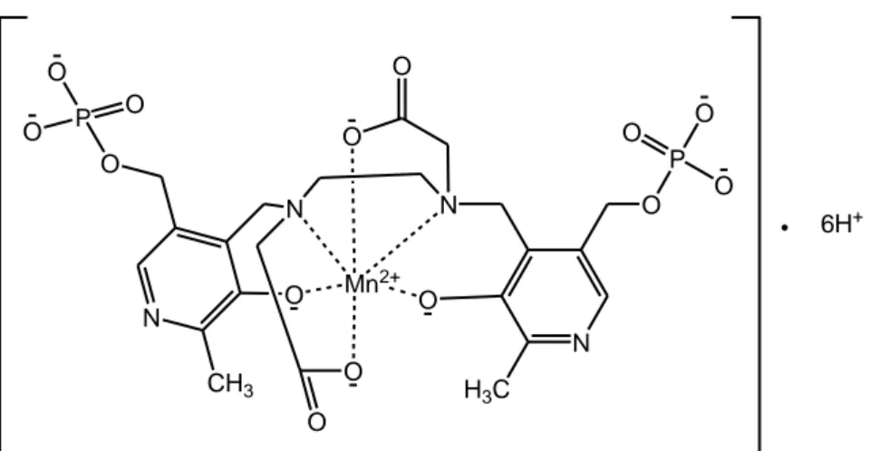
Mangafodipir (figure 1.3), known also with the brand name of Teslascan or as mangafodipir trisodium (11) is a contrast agent delivered intravenously to enhance the contrast during the MRI. It has two parts: paramagnetic manganese (II) ion and the chelating agent fodipir (dipyridoxyl diphosphate, DPDP). The distorted octahedral complex is used in clinical diagnosis to enhance contrast in the liver and to detect hepatocellular carcinomas).(12) The relaxivity of this complex is about 35% higher than Mn complexes of DTPA and DOTA, which also does not contain directly coordinated water (13)
Mn2+ O N O N O N H3C O P O O O O O N CH3 O P O O O O 6H+
10
1.2 Radiopharmaceuticals
99mTc and 201Tl, 111In, 67Ga, 51Co, 51Cr, 169Yb are high intensity γ-ray emitters,
89
Sr, 153Sm, and 186Re are β-emitters. Several of these radionuclides centers are used for diagnostic imaging and for therapy(14). Nuclear medicine has grown mainly to the availability of 99mTc radiopharmaceuticals; this isotope is used in over 80% of all diagnostic procedures.
The first generation of technetium radiopharmaceuticals was developed mainly thanks to these proprieties of the common complexes of 99mTc: absorption, distribution, metabolism and excretion. These studies led to 99mTc radiopharmaceuticals to examine the thyroid (99mTcO4-), the liver (99m
Tc-colloids), the bone (99mTc-phosphonates) and the kidney (99mTc-DTPA). (15, 16) The modern analytical tools, such as nuclear magnetic resonance spectroscopy, mass spectrometry and X-ray diffraction, helped researchers to understand the structure-activity relationships underlying the biological behavior of the 99mTc agents for their ability to determine the exact molecular structure of coordination compounds. Therefore, the precise design of new ligands and their 99mTc complexes led to discovery imaging agents for perfusion in the myocardium and brain. The cardiac imaging agents 99m Tc-MIBI (sestamibi, Cardiolite®) and 99mTc-tetrofosmin (Mvoview®) and the brain imaging agents 99mTc-HMPAO (exametazime, Ceretec®) and 99m Tc-EDC (bicisate, Neurolite®) are the results of the above strategy in the development of 99mTc complexes. (7)
Cardiolite® is used for myocardial perfusion imaging. Lipophilic cationic complexes imitate potassium ion and are taken up by the myocardium. Therefore the complex structure was designed(17) on the basis of this principle. The sequential metabolism of the six identical methoxy groups and of eight to hydroxyl groups in the liver leads to formation of 99mTc complexes with higher hydrophilicity which are not retained in myocardial tissues.(18).
While Ceretec® is an approved cerebral perfusion imaging agent for evaluation of stroke. It is taken up by the brain and is transformed into more hydrophilic species which are retained in the brain.
Current designs of imaging agents are based on the careful selection of the suitable biomolecules to function as effective vectors. It concerns in vivo delivery of radioactivity to more specific biological targets such as receptors and transporters.
This strategy implies that the method of marking must not induce any distortion in the active site of the biomolecule. Thus, these agents have required the development of sophisticated marking methods that go beyond the technologies used previously.
The introduction of the bifunctional chelating agents (BFCA), the new chemistries such as the Tc-tricarbonyl, Tc-nitrido, Tc-HYNIC and mixed ligands complexes have helped to achieve this objective. (7)
Monoclonal antibodies (mAbs) conjugated with radionuclides are used clinically for diagnosis of colorectal and ovarian cancer such as 111In satumomab pendetide, which contains the murine mAb B72.3 and is directed to TAG-72, an antigen expressed by many adenocarcinomas.(19) There are several other murine mAbs linked to 99mTc and 111 In in clinical trials..(20) Substantial progress has been made recently in the development of 99m Tc-based receptor-specific radiopharmaceuticals.(21) Encapsulation in fullerenes may also provide a novel method for the delivery of radionuclides to target sites.(22, 23)
12
1.3 Superoxide Dismutase Mimics
Oxidative damage is an inevitable side effect of cellular metabolism and it is the cause of systemic aging and genome instabilities such as telomere shortening, mitochondrial mutation, and chromosomal pathologies. Reactive Oxygen Species (ROS) are also one of the major causes of degenerative diseasesaffecting the elderly. (7)
The free radical superoxide, O2-, is a product of activated leukocytes
and endothelial cells. It is a mediator of ischemia-reperfusion, inflammatory and vascular diseases. It can react with NO to form damaging peroxynitrite, ONO2-.
The metalloenzyme superoxide dismutase (SOD) can destroy O2-:.
Cu-SOD and Zn-Cu-SOD are found in the cytoplasm of eukaryotic cells, while Mn-SOD is located in mitochondria (figure 1.4)
Mn+1 + O2- Mn+ + O2 (1)
Mn+ + O2- + 2H+ Mn+1 + H2O2 (2) Figure1.4. Superoxide dismutase reactions.
MnSOD mimics have been designed for laboratory synthesis by computer-assisted modelling.
Intramolecular electron transfer requires Mn(II) in the active site of the enzyme. Mechanistic studies confirmed that during catalysis the ion can reduce binding-energy barrier and help to bind ligands in a correct geometry. A series of active Mn(II) macrocycle compounds have been designed and two are now in clinical trials.(24)
Also Catalase mimics appear to require Mn(III) in the “active site”. A series of manganese complexes that are mimetic of both superoxide dismutase
and catalase have proven to be active in rodent models as synthetic catalytic scavengers of ROS (Figure).(25)
The use of these molecules in therapy is limited by their short plasma half-life because they endured clearance by the kidney and by their inability to penetrate cell membranes; in fact they have only an extracellular activity. Low molecular mass mimics of SOD are molecules with potential pharmaceutical interest.(26) For example, a variety of Mn and Fe-based on porphyrins and macrocyclic complexes exhibits SOD mimic activity.(27-30) MnII and MnIII macrocycles appear particularly promising.(31, 32) For example, SC-52608 (figure 1.5) is able to scavenge superoxide and to protect effectively myocardium from injury of ischemia and reperfusion.(33)
14 Mn N Cl N N N Cl H H N H H H SC-52608 Mn N N Cl N N N H H Cl H H X-40403 Mn N N Cl N N N H H Cl H H CH3 H3C M-40401 Mn N Cl N N N Cl H H N H H H SC-54417 Mn N Cl N N N Cl H H N H H H MnCAM Mn N N Cl N N N H H Cl H H SC-55858
Figure 1.5. Structures of manganese complexes tested as mimetics of
superoxide dismutase.
Instead, Manganese(III) 5,10,15,20-tetrakis(4-benzoic acid)-porphyrin (MnTBAP) can protect against neurodegeneration. It is studied for the treatment of brain diseases such as Parkinson’s and Alzheimer’s diseases.(34)
1.4 Platinum anticancer drugs
Chemotherapy is more widely used than surgery and radiotherapy in treatments for cancers.
Cisplatin (23, figure 1.6) or cis-diamminedichloroplatinum(II) (DDP) is considered the successful historical example of a metal-based drug since it was discovered by Rosenberg et al.(35) Platinum complexes are used in a lot of solid tumors such as lung tumor, colorectal tumor, ovarian tumor, head and neck cancer, genitourinary cancer and breast cancer (36, 37)
The glycolato complex (nedaplatin, 25) and oxalato complex (oxaliplatin, l-OHP, 26 , which contains R,R-1,2- diaminocyclohexane, DACH) are approved for clinical use respectively in Japan and in France. Particularly they are used in multi-drugs chemotherapy for treatment of advanced lung, colorectal, and ovarian cancers.(38, 39)
Pt NH3 Cl NH3 Cl 23 O O O O Pt H3N H3N 24 O Pt O O H3N H3N 25 N N Pt O O O O 26 H H H H
16
Platinum derivatives are alkylating agents. Cisplatin by diffusion enters in the cells where it is converted into the active form. Scientists have not determinate the active species with great precision. They believe that the active species is the monohydrate even if the most common species is the dehydrate form. The main function of the cisplatin is to bind itself to the DNA. In fact, although cisplatin is able to interact with many types of vital proteins for DNA replication and cell division, the main target remains the DNA.
Cisplatin, Carboplatin and oxalilplatino enter in the cells thanks to an active conveyor of the CU2+ - the CTR1 (copper transporter 1) - causing the degradation at the same time. The extrusion of these compounds occurs by active transport mediated by the conveyors of copper, ATP7A and ATP7B, and by the MRP system1 (multidrug resistance protein 1).(40)Inside of the cells the binders chloride of cisplatin are offset by water molecules producing strongly reactive molecules.Guanine N7 is the electron-richest site on DNA. It represents the most important binding site in the nucleic acids. The complexes of platinum activated react with the nitrogen in position 7 of guanine on DNA, causing the formation of guanine-guanine crosslinks. 1,2-GpG and 1,2-ApG crosslinks are the major adducts of platinum drugs with DNA. These ones inhibit the processes of replication and transcription and lead to breakage of single and double-stranded. Moreover they determine coding errors (miscoding) which are recognized by proteins of cell cycle control determining apoptosis. The properties of these adducts have been extensively characterized and reviewed.(41)
Platinum complexes possess considerable toxicity and numerous side effects. One of the main effects is the nephrotoxicity. Cisplatin may in fact cause renal damages: tubular degeneration, necrosis and mineralization of the epithelial tubular cells. Another effect is ototoxicity that arises especially in children. Ototoxicity leads to loss of hearing capacities. The initial symptom
includes chiming (ringing in your ears). These effects should stop at the end of the treatment, but some patients lost irreversibly the use of hearing over the high-frequency range (> 4KHz).
Other toxic manifestations such as nausea, vomiting, decrease in red blood cells, tingling in the hands and feet are also common of other antitumor drugs. The neurotoxicity of the drug manifests itself particularly in high doses, while decreases in platelets, facial swelling, dyspnea (shortness of breath), muscle cramps, blurred vision and loss of appetite are less common. Nausea and vomiting occur normally about an hour after the administration of the drug and they can last for several hours.
In addition as other drugs that form adducts with DNA, platinum complexes are associated with the onset of acute myelocytic leukemia even after 4 years from the treatment.(42)
Despite the use of these compounds in many multi-drug treatments there is a large interest in developing novel compounds that exhibit innovative characteristics in respect to the existing ones. The research activity is on time for the development of novel compounds. In addition to determining a minor systemic toxicity they should ensure a marked antineoplastic activity even for the tumor lines that have proved resistant. In order to achieve these objectives we began to design and to study metal complexes which possess in their structures metal centres different from platinum.
Pt–NHC complexes have been highlighted as a promising and original platform for building new cytotoxic drugs of the cisplatin series. (43-48)
Mixed NHC–amine Pt(II) complexes were prepared through modular two-step sequence leading to trans-configured square planar species by Marinetti et al.(47). Their efficiency against both cisplatin-sensitive (CEM and H460) and -resistant (A2780/DDP, CH1/DDP, and SK-OV-3) cell lines was demonstrated by in vitro experiment.All novel complexes exhibited cytotoxic
18
activities with IC50 at a micromolar range. IC50 of the measured complexes
against CEM T leukemia cells was in the 0.6–2.7 mM range, generally lower than that of cisplatin (3.0 mM) under the same experimental conditions. Some of the complexes outperformed cisplatin against H460 lung cancer cells, too. This study afforded a potent and innovative new chemical platform for cytotoxic drugs of the Pt(II) series based on NHC ligands.(47)
1.5 Ruthenium complexes
The first metal that arouses the interest of the scientific community with regard to the design of complex innovative anticancer was the ruthenium. Scientists believe that the ruthenium and platinum, sharing the same group on the periodic table of the elements, have the same antitumor effect that is a direct interaction with the DNA comparable with cisplatin.
However, subsequent studies have demonstrated that complexes of ruthenium(III) have actually distinct properties from those of the platinum. These compounds tend to accumulate more and more in neoplastic cells exploiting a transport system mediated by transferrin. In this way they have a certain degree of specificity while it is absent in the complexes of platinum.In fact, the ruthenium is able to mimic the iron by binding to the protein transferrin. The cells in rapid division, including tumor cells, require a greater amount of iron. This process translates into an up-regulation of receptors for transferrin on the cell membrane and a greater uptake of these complexes from the blood. In addition, before reaching the tumor mass, complexes of ruthenium(III) are inactive and their bio-activation is performed by reduction of the metal centre to ruthenium(II), favoured by the low levels of oxygen and by the high acidity of the tumor environment.(49)
Currently, two complexes of ruthenium(III)
trans-[RuCl4(Im)(DMSO)]ImH (NAMI-A) and trans-[RuCl4(Ind)2]IndH (KP1019)
are in clinical trial phase. Their structures are shown in figure 1.7.
Both complexes of Ru(III)+ are formed by a heterocyclic counter-ion, heterocyclic ligands and binders chloride. Despite their structural similarities, these complexes differ greatly for their antitumor activity profile.
20 N NH Ru N HN Cl Cl Cl Cl N H+ NH N HN Ru Cl Cl Cl Cl DMSO Na+ KP1019 NAMI-A
Figure 1.7. Complexes of Ruthenium studied in clinical trials.
Preclinical studies have shown that nami-A is able to inhibit the formation of metastases in different tumor animal models but it does not show cytotoxic direct effects. On the contrary KP1019 has shown antitumor effects directed against a wide range of xenografts tumor, through the induction of apoptosis..(50-55)The mechanisms of action proposed for the nami-A compound include: the interaction with the processes of regulation of the cell cycle (accumulation of cells in the G2/M phase); the alteration of the functions of endothelial cells required for the process of angiogenesis and the increase of the extracellular matrix around the blood vessels that irrigate the tumor and direct interaction with the nitrogenous bases of DNA. The mechanisms involved in the process of apoptosis induced by compound KP1019 include interference with the chain for the transport of the electrons, depolarization of the mitochondrial membrane and down-regulation of anti-apoptotic bcl-2
factor. The compound is also able to interact directly with the DNA, but this mechanism is not the main responsible of the antineoplastic effects of this compound. (56)
Thus, the activity of other Ru(II) complexes is currently being explored. For example, arenes are known to stabilize ruthenium in its oxidation state (2+), therefore the potential of Ru(II)–(6-arene) complexes as anticancer agents is under investigation (55,57,58). In literature we can found several studies about half-sandwich (6-arene)Ru(II) complexes with imidazole, sulfoxide, phosphane, chelating aminoacids, and diamine or diimine ligands. All these molecules was evaluated for cytotoxic activity. Both the size of the arene and the lability of the Ru–Cl bond have found to play a crucial role in determining the cytotoxicity of ruthenium(II) complexes of the type [(6-arene)RuCl(LL´)](PF6) with bidentate ligands LL´. Compounds with extended polycyclic arenes (e.g. tetrahydroanthracene) and LL´=ethylenediamine (en) are the most active of A2780 human ovarian cancer cells, whereas those with polar substituents on the arene such as COOCH3 (an
electron-withdrawing group) or with aromatic diimine ligands such as 2,20-bipyridine or 1,10- phenanthroline exhibit either poor or no activity.
22
1.6 Metallocene and Titanocene complexes
In recent years new anticancer drugs containing a metal different from platinum have been developed. The objectives of these research were the improving of the clinical efficacy, the reducing the toxicity and the overcoming of drug-resistance, all own limitation of platinum derivatives. Obviously the broadening of the spectrum of action of platinum complexes was a good reason for making research in this context.(59) The research has focused on transition metal complexes bearing as binders chlorides in cis or like labile ligands. Among the drugs the metallocene complexes of the type (C5H5)2MCl2 received many attentions because they represented a logical
extension of cis-platinum derivatives (51) . These complexes are constituted by a metal core, formed by metals transition such as Ti, Nb, Mo, etc. The coordination sites of the metal are occupied by two cyclopentadienyl rings (C5H5 or Cp) and by two labile linking, generally chlorides (Cl). See figure 1.8
M Cl Cl
R R
Figure 1.8. General structure of metallocene complexes
Köpf-Maier and coworkers(60) have investigated the antitumor activities of a whole series of metallocene dichloride complexes in vivo, after the variation of the transition metal. From this research, titanocene dichloride
(TDC) (figure 1.9) exhibited the most promising chemotherapeutic activity among all other metallocenes tested.(61.)
In fact, titanocene dichloride (Cp2TiCl2) showed medium
antiproliferative activity in vitro, but promising results in vivo.(62,63)
Really titanocene dichloride was the first metal-based chemotherapeutics to reach Phase I clinical trials from the development of cisplatin. However it showed promising in these preliminary studies, it did not progressed beyond Phase II due to its low efficacy vs. toxicity ratio (64,65). Unfortunately its efficiency in patients with metastatic renal cell carcinoma or metastatic breast cancer was too low to be pursued. (66,67)
The anticancer properties of cis-diethoxybis (1-phenylbutane-1,3-dionato) titanium(IV) [(bzac)2- Ti(OEt)2, known as Budotitane were first
reported in 1982.(68) It possesses activity towards animal tumors such as EAT and colon tumors.(69,70) Also the budotitane (figure 1.9), due to its good biological results in preliminary studies, reached Phase I of clinical trials, but it did not progress in Phase II due to formulation problems.(70) These difficulties have spurred the development of titanium complexes that display higher potency and hydrolytic stability.
24 Ti Cl Cl R R Ti O O OEt O OEt O H3C H3C
Budotitane Titanocene Dichloride
Figure 1.9. Structure of Titanocene dichloride (TDC) and Budotitane.
Titanocene dichloride does not bind with nucleotides and nitrogen bases, as the cis-platinum, but its mechanism and the biological action, as proposed by Sadler e co-workers on the basis of model studies, provides that titanium species could be complexed to phosphate and gives interaction with DNA.The titanium (IV) ion, by binding to specific sites of the iron (III), forms a strong complex with the transferrin, a protein of human plasma, and is thus supplied to tumor cells. (71)
The interesting results obtained with Cp2TiCl2 encouraged researches to
develop new complexes of this metal which might have higher hydrolytic stability and higher cytotoxic activity.11 In fact, in order to improve these properties of titanocene derivatives, polar side chains were attached to the Cp ligands. Titanocenes complexes, having polar substituents on cyclopentadienyl ring, such as alkoxy, amino or electron-withdrawing groups as carboxylic acid and esters, showed, in some cases, a very high activity in antitumoral tests.
(72-76)
reveal promising activity,(77-81) although, generally, higher activity was found in analogues where the two Cp units were not bound together. (82,83)
A lot of analogues of titanocene containing aromatic groups linked to the Cp have been synthesized.(84) One of the most interesting of this series, in the pharmacological field, is bis-[(p-methoxybenzyl)cyclopentadienyl]-titanium-dichloride (titanocene Y) shown in figure 1.10.
Its antiproliferative activity was studied in 36 human tumor cell lines(85) and in human tumors explanted.(86-89) The most significant results of these experiments in-vitro and ex-vivo had with the tumor cells of the prostate, cervix and kidney. Furthermore, titanocene Y was tested on breast cancer cell line MCF-7 revealing a promising medium-high cytotoxic activity with IC50
values of 76 M, quite close to those of the cis-platinum (37 M).(90)
Ti Cl Cl O O
Figure 1.10. Structure of Titanocene Y
Furthermore, replacing the two titanocene Y chlorides with an oxalate group by a simple reaction of anion exchange, it is obtained a product that has a greater hydrolytic stability, due to the chelating effect of oxalate group and it shows an activity 13-fold higher than the titanocene Y in-vitro studies on the cell line LLC-PK.(91) It is worth noting that this effect was not observed in all titanocene derivatives studies.(92)
26
1.6.1 Metoxy-aryl and alkyl titanocenes
Recently our studies reported the synthesis and cytotoxic activity of some titanocene complexes that they have substituents on cyclopentadienyl rings able to intra-molecularly coordinate the titanium cation, stabilizing the active species. The complexes showed very interesting antiproliferative activity on several cancer cell lines.
In particular we replaced the methoxy-aryl substituent of cyclopentadienes of titanocene Y with ethenyl-methoxy in the way of having the presence of a strongly coordinating group, able to stabilize the cationic species responsible for cytotoxic activity. We also verified, by substitution of chlorine atoms with dimethylamide, oxalate, or amino groups, the influence of other ligands leaving on the activity of the complexes.(93)
Some of the synthesized compounds showed a good cytotoxicity, in particular the complex bis-ethenylmethoxyl-cyclopentadienyl-titanium dichloride (T2) and ethenylmethoxyl-cyclopentadienyl titanium trichloride
(T1), see Fig. 1.11, on the cell line MCF-7 displayed IC50 values (84 and 57
µM, respectively) very close to those obtained with the cis-platinum and to that reported for the titanocene Y. Furthermore, the results of the hydrolysis of our titanocenes shown that the leaving groups (-Cl, -N(CH3)2, C2O4 or glycine)
influencing the rate of hydrolysis of the ciclopentadienyl groups and thus the stability of the complexes. (93)
Therefore, the presence of substituents, aryl methoxy group on cyclopentadienyl ring in titanocene Y or ethenyl-methoxy group in titanocene
T2 or in the half-titanocene T1, produces compounds having interesting
Ti Cl Cl O O T2 Ti Cl Cl Cl O T1
Figure 1.11. Structures of bis-cyclopentadienyl-ethenylmethoxyl-titanium dichloride
T2 and cyclopentadienyl-ethenylmethoxyltitanium trichloride T1
Although generalizations regarding structure-activity relationships are not yet clear, we could hypothesize that the neutral nucleophilic substituents of cyclopentadienyl (aryl methoxy or ethenyl-methoxy group) could intramolecularly coordinate to the titanium cation, thus preventing decomposition reactions. On the other hand, this hypothesis was suggested for analogous complexes able to give polymerization of propene or styrene having microstructures strongly influenced by the possible coordination of neutral substituent of cyclopentadienyl at the metal center.(94-96)
As mentioned above, in the literature several examples of titanocene-complexes showing cytotoxic activity are reported, the cyclopentadienyl-ethenylmethoxyl-titanium trichloride represents the first example of half-titanocene complex having this interesting cytotoxic activity.
Therefore, a series of novel half-titanocenes compounds were synthesized and characterized (see figure 1.12) by nuclear magnetic resonance (NMR), mass spectroscopy and elemental analysis.
28 Ti Cl Cl Cl OCH3
1b
Ti Cl Cl Cl OCH3 OCH32b
Ti Cl Cl Cl N(CH3)23b
Ti Cl Cl Cl4b
Ti Cl Cl Cl OCH3 H3CO5b
Ti Cl Cl Cl OCH3 H3CO H3CO6b
Figure 1.12. Structures of synthesized half-titanocenes.
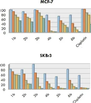
These complexes had different substituents on the Cp ligands, able to stabilize the titanium cation by intramolecular coordination. Preliminary cytotoxic studies of these titanium based compounds evaluated their growth regulatory effects in MCF7 and SkBr3 breast cancer cells. Several agents showed a moderate or a high antitumor activity. For example, 4b showed the strongest antiproliferative activity against MCF7 breast cancer cells, whereas 5b showed a very interesting activity against SkBr3 breast cancer cells (see figure 1.13).
In order to understand if a different group can influence the intramolecular coordination and consequently the stability of the complexes
and if the ether group is really essential for cytotoxic activity, several new-titanocene complexes (see figure 1.14) were synthesized and characterized having as ligands the 2-cyclopentadienyl-ethoxy-benzene [Cp-CH2CH2-O-Ph]
or the cyclopentadienyl-benzyl [Cp-CH2-Ph]. Then their cytotoxic activities on
human breast cancer cells MCF-7 and SkBr3 was evaluated.
Figure 1.13. Evaluation of growth responses to 10 µM of 1b-6b in MCF7 (a) and
30 Ti ClCl O O DC6 Ti gly gly O O DC6-gly
Ti
O
O
O
O
O
O
DC6-oxalateTi
Cl
Cl
ES2H
H
Ti
Cl
Cl
ES2-chelatedDC6 was synthesized to verify if a phenoxy group, instead of methoxy group present in T2, could better stabilize the titanium cation. In fact this
ligand may stabilize either with oxygen atom or with phenyl ring. DC6-oxalate and DC6-gly were synthesized to achieve different leaving groups, respect to chloride, on titanocene, which possibly could improve the activities of the complexes, by favorable pharmocokinetic properties.
ES2 was synthesized to prove if the ether group, which is present on titanocene Y, on T1 and T2, as well as on T2, was really necessary to have a
cytotoxic activity significantly higher than Cp2TiCl2, or if a more labile ligand,
having higher capability to π-donation can produce a molecule as much as pharmacologically interesting. ES2-chelated could have the same properties of ES2, but this ansa derivative, having the two cyclopentadienyl rings covalently linked, should be less stable and possibly to have lower activity than ES2.
Preliminary cytotoxic studies of ES2 compound, in order to evaluate their growth regulatory effects in MCF7 and SkBr3 breast cancer cells were carried out. The results were very interesting because this complex showed strong antiproliferative activity on both cell lines (see figure 1.15).
Figure 1.15: Evaluation of growth responses to 10 µM of ES-2 in MCF7 (a) and
32
Several titanium complexes were synthesized and tested for their cytotoxic activity against cancer cell lines. Most of these compounds showed significant anti-proliferative effects compared to cisplatin
As indicated by these researches the presence of a coordinating group, such as phenyl or ether one, is necessary to stabilize the active species, delay hydrolysis and generate a cytotoxic complex; the ether groups are important to improve the hydrolysis stability but are not necessary to have a significant cytotoxic activity; a more labile ligand, as the benzyl group, having greater ability of π–donation, produce complexes with interesting cytotoxic properties; a lower activity was found in the analogues where the two Cp units were bound by a two-carbon bridge.
Half-titanocene showed lower cytotoxic activity compared to their titanocene analogues; the labile ligands (Cl) do not have a strong impact on cytotoxic activity; a methyl group on the linker between Cp ring and the phenyl group decreases the cytotoxic activity; a small steric hindrance all around the complex is favoured; the pro-ligands show interesting cytotoxic activity but are not related to complexes cytotoxicity; further studies are needed to understand the exact mechanism through which these compounds exercise their action.
Therefore, Ti(IV) complexes are good lead compounds for pharmaceutical applications.
1.7 NHC complexes in medicine.
The chemotherapy, flanked to surgery and radiotherapy, is used for the treatment of cancer. Moreover there are numerous metals (Ag, Au, Cu) with binders carbenic N-heterocyclic groups that were studied in the pharmacological field.
1.7.1 Ag-NHC complexes
Silver has been since antiquity used as antimicrobial agent, it was earlier employed in the purification of drinking water, wine and vinegar, and also Hippocrates remarked its therapeutic properties.(97) To our days the silver salts are still used for example to treat chronic ulcers, burns extended, to prevent conjunctivitis in the new-born and in so many other infections.(98)
The activity of silver against Gram-positive and Gram-negative bacteria, fungi and yeast is due to Ag+ cations, which can interact with the cell membrane, interfere with the electron transport system of the cell and interact with thiol groups of the vital enzymes of bacteria. (99)
However the objective, in order to have a good therapeutic action antimicrobial, was to provide agents that released slowly and continuously the ion Ag+ in the affected area. In fact, the already known salts of silver nitrate or sulfadiazine silver, for example, (Figure 1.16), while having an antibacterial topical action, expose the subject to a relapse precisely because of the rapid release of the Ag+ and then have a rapid effect but not durable in combat infection.
Scientists tried therefore to synthesized stable complexes that could slowly release the ion in time. The choice of a suitable ligand, bonded to the metal, can make the difference.
34 H2N S N O O N N Ag+ A N O O O Ag+ B
Figure 1.16. Structures of Sulfadiazine (A) and silver nitrate (B).
The use of the complexes Ag-NHC that show a bond C-Ag relatively strong to generate a more stable complex compared to previous silver-based drugs is the subject of numerous research.(100)
The Ag-NHC can be synthesized by three synthetic approaches. The first provides for the treatment of the NHC with appropriate "sources" of silver at the temperature of liquid nitrogen. The second consists in treating compounds of silver as Ag2O, AgOAc or Ag2CO3 with the salt of the NHC at
room temperature or warm. The last method envisages the transfer reaction of step in basic conditions between the silver salt and salt NHC. The second approach is the most common and used because it is more convenient too. (101)
The Ag-NHC were also tested to evaluate their antitumor properties and has been highlighted cell death by apoptosis without developing necrosis. For these complexes the suggested action mechanism provides the depolarization of the membrane potential of mitochondria with consequent and probable release of mitochondrial proteins that provide to the apoptosis. Complexes did not cause overproduction of oxygen reactive species (ROS) and activation of caspase-3, while instead occurred translocation of apoptosis inductor factor and caspase-12 in the nucleus. These events promoted the DNA fragmentation and finally cell death. Therefore, complexes were not genotoxic because they did not change the distribution of the cell cycle.(102)
To further improve the release scientists encapsulated these silver complexes. This would also prevent the degradation at the level of the bloodstream in which there is present the chloride ion, which by interacting with NHC, it would cause the precipitation. For some complexes of Ag-NHC this encapsulation was made with poly (lactic-co-glycolic acid) combined with polyethylene glycol, and receptors of the folic acid were used to allow the delivery in a tumor site. Some of the various nanospheres prepared and loaded with complex Ag-NHC were then tested against MB157 (cell line breast cancer) and H460 (cell line of lung cancer), providing promising results.(103)
Good results are obtained with the saturated Ag-NHC and this has pushed to investigate the activity of unsaturated NHC and substituted in position 4 and 5 of the imidazole ring. In particular the effect of electron-attractor of chlorine atoms in position 4 and 5 determines a better stability of the relative complex Ag-NHC compared with a similar no-chlorinated.(104) This category of silver complexes derived from 4,5-dicloroimidazolo showed a certain degree of selectivity toward the ovarian cancer (OVCAR-3) and of the breast cancer (MB-157).
Scientists demonstrated in vivo activity of the complex 1, shown in figure 1.17, against a model of xenotransplantation ovarian cancer. Gautier Morel reported in the literature as the carbene N,N-diaryl-substituted, with its high lipophilicity, could be a good binder for metal complexes pharmacologically interesting indeed the cytotoxicity of the resultant complexes of silver was 40 times on 7 and HL60 and 7 times on MCF-7R is higher than that of cisplatin.
The good obtained results pushed research in this direction and it was found that in order to obtain a high degree of accumulation in tumor cells, the aromatic rings of the metal complex must be substituted in position 2, 3 and 4
36
as follows: 2-F, 3-F, 4-F, 4-OH or 4-OCH3. These substitutions have already
been made on the complex 1,2-diariletilenediammino platinum(II) for which these complexes can be considered to be derivatives of the latter.(105)
N N Cl Cl Ag O O N N Ag Cl N N Ag halide alkyl alkyl R R N N H R R R
Figure 1.17. Selected complexes of silver-NHC and 2,4,5 - triarylimidazoles.
1.7.2 Au-NHC complexes
The gold was, and still is, used in various biomedical applications such as for example the treatment of rheumatoid arthritis, cancer and as antimicrobial agent.(106-118)
The two lead structures from which it started to develop new chemotherapeutic agents based on gold are the auranofin and its analogue Et3PAuCl, shown in figure 1.18.
O AcO AcO AcO S OAc Au P Auranofin Au P Cl Et3PAuCl
Figure 1.18. Lead structures based on gold.
Studies into structure-activity relationship highlighted the importance of fosfinic portion while replacing the carbohydrate or of the halide in the respective molecules does not induce loss of activity. In recent years, however, the discovery of the NHC was exploited also in the field of biology. They have proved to be effective alternatives to phosphine precursors(119), thanks to the capacity σ-donor comparable to that of the phosphine ligands and the ease with which they can be carried out the structural modifications to the nitrogen atoms .
As well as the auranofin and its analogue Et3PAuCl induce apoptosis
through inhibition of the enzyme thioredoxin reductase (TrxR mitochondrial), in the same way the Au-NHC cause the death of sun cancer cells.(120) This would be justified by assuming a certain affinity between the complexes based on Au and involved enzymes. The system thioredoxin/thioredoxin reductase (Trx/TrxR) is involved in the regulation of processes intracellular redox. The scheme of action general of TrxR is reported in figure 1.19.
38
Tioredoxine, depending on the redox potential of its substrate, catalyses the reversible reactions that involve the formation or breaking of the disulphide bonds.
Thioredoxin is involved both in the defence against oxidative stress that in apoptosis; in particular it has been postulated to the formation of the complex protein in reduced form and kinase apoptosis signal 1 (ASK1) as responsible for the inhibition of apoptosis.(121)
NADPH + H+ TrxR NADP+
Trx-S2 Trx-(SH)2
Protein-(SH)2 Protein-S
2
Figure 1.19. Activities of reduttasica TrxR system/NADPH.
The TrxR has in the N-terminal portion a peptide sequence of the type cysteine-AA-AA-cysteine and the C-terminal portion of a sequence of the type selenocysteine-AA-AA-cysteine (where AA are amino acids different from cysteine). The presence of the atom of selenium justifies the affinity of the gold atom of the complex for this enzyme and in more considering that cancer cells show an increased metabolism, you may also explain the over-expression of this enzyme in this type of cell. Consequently the selective inhibition of this
type of enzyme is one of the parameters on which it bases for developing new chemotherapeutic drugs based on gold.(122) The inactivation of TrxR, due to the formation of a covalent bond with the selenium atom of the enzyme(123), determines an increase of thioredoxin in oxidized form that induces cell death as shown in figure 1.20.
Figure 1.20. The molecular mechanisms involved in the induction of the apoptotic
process caused by the complex of gold.
The lack of thioredoxin in reduced form involves less inhibition of ASK1 kinase for which there is the activation of the cascade of mitogen-activated protein kinase (MAPK), between which the c-Jun N-terminal kinases (JNK) and p38, which stimulates the apoptotic process. Added to this there is
40
the lesser capacity of TrxR to maintain in the reduced state proteins that play important roles in the cell as the Perossiredossina (Prx), favoring the accumulation of reactive oxygen species (ROS), in particular of hydrogen peroxide, and thus generating a strong oxidative stress. The oxidating environment created further contributes to the oxidation of thioredoxin and stimulates therefore indirectly the activation of ASK1 and the apoptotic process.(124)
However considering the great structural variety of binders it is not possible to define an univocal mechanism of action. In fact the gold compounds can trigger the apoptosis by various processes: mitochondrial damage that provides for the inhibition of thioredoxin reductase (TrxR) direct DNA damage, modification of the cell cycle, inhibition of the proteasome, modulation of specific kinase and other cellular processes.
An important factor for the targeting of malignant cells is represented by the possibility to adjust the lipophilicity of relevant complexes. In fact it has been shown an antimitocondrial effect which is manifested with swelling of these organelles, much faster because the greater the lipophilicity.(125)
The lipophilic cation delocalized (DLCs) can move quickly the lipid bilayer and focus on mitochondria guided by the increase of the membrane potential generated in turn from the respiratory chain. These DLCs is mainly concentrated in the mitochondria of cancer cells precisely as a result of the high potential of the membrane that is a characteristic of many tumor cells. Therefore the modulation of the lipophilicity might be an idea to generate a certain degree of selectivity toward the cancer cell than normal.
Furthermore the selectivity toward the tumor cell is an important parameter. In fact tumor cell shows an increased activity of TrxR, responsible of evasion of apoptosis. The inhibition of this enzyme can induce the cell apoptosis. The TrxR is considered an important target for this category of
drugs. Many compounds proved to be potent inhibitors of the purified TrxR enzyme, but few could inhibit it the cell.(126)
1.7.3 NHC-Cu complexes
The coordination compounds of Cu(I, II) have been investigated as potential antitumor agents only in the last few decades, particularly after the discovery of cisplatin, although copper has a long history of medical application.(127-132)
The complex Cu-NHC have been little studied from a biological point of view. The copper however, being important for the correct functionality of numerous enzymes and proteins and being involved in a series of physiological processes, should be less toxic than the other not essential metals. (133,134)
Recently a series of copper complexes was tested with regard to their own cytotoxicity and some have shown good antitumor activity both in vitro and in vivo. It has been demonstrated that the copper compounds may act through different mechanisms than those of cisplatin and the DNA would not represent the biological target main or however would not be the only molecular target for this class of molecules. In addition to the binding to DNA, the intercalation and the cleaving activity, these compounds can create damage cellular oxidative thanks to similar activities that show with superoxide dismutase and ROS generation after reaction with oxygen and intracellular peroxide. These are all events intracellular molecular that trigger the death of tumor cells through an apoptotic mechanism. Furthermore, for these copper compounds, an adjustment of the proteins antiapoptotic (Bcl-2 and Bcl-XL) has been highlighted on the tumor cells . This leads to the death of cancer cells through a programed cell death mechanism known as apoptotic paraptosi that
42
can be exploited for lead to death even those tumor cells with defects in the normal process of apoptosis as, for example, cells cisplatin resistant.(102)
The effect of complexes of copper-NHC reported in figure 1.21 was evaluated on the cell line MCF-7 (breast cancer).
All candidates, except the complex 14 that from a structural point of view is the most taken up, showed IC50 values lower than the micromolar and
that of Cisplatin which was used as the reference drug (Table 1).
N N CuCl 10 N N CuCl N N CuCl 11 12 N N CuCl 13 N N CuCl 14
Table 1. IC50 of the complexes Cu-NHC on cell line MCF-7 (μM).
Cisplatin 10 11 12 13 14
10,4µm 0,075µm 0.13µm 0,04µm 0,075µm 4,4µm
Due to greater activity the assembly 10 has been the object of further studies. In particular were evaluated the possible effects on cell cycle and it has been found a stop in step G1 at concentrations at least ten times lower than those of cisplatin (DDP).
Moreover, genotoxicity studies conducted using enveloped plasmids have demonstrated, as expected, that complexes of copper(I)-NHC, compared to those of palladium and silver, are the only ones capable of acting as nuclease.(104)
44
1.8 Mammary carcinoma
The mammary carcinoma is a malignant disease characterized by the uncontrolled proliferation of epithelial cells of the glandular tissue breast.
1.8.1 Epidemiology
Breast tumors represent, as a whole, the type of tumor most frequently diagnosed among women of all ages: 0-49 years (41%), 50-69 years (36%), ≥70 years (21%). It is the neoplasia more diagnosed in women in Italy (29% of all cancers diagnosed) and represents the first cause of oncological death. Risk factors for the onset of breast carcinoma is associated with several risk factors:
Age.
Previous thoracic radiotherapy.
Previous pathologies implants (dysplasia or cancer of the breast tissue).
Menarche early and late menopause.
First pregnancy at term in advanced age (> 30 years).
Failure to breast-feeding.
Lifestyle (obesity, poor physical activity, high consumption of alcohol, carbohydrates and saturated fats).
Hormone replacement therapy.
Familiarity and genetic factors: mutations of genes BRCA-1 and BRCA-2, p53, PTEN (disease of Cowden) and ATM (ataxia-telangiectasia).
Mutations of genes BRCA-1 (breast cancer susceptibility gene 1), located on chromosome 17, and BRCA-2 (breast cancer susceptibility gene 2), located on chromosome 13, are responsible of 2/3 of mammary carcinomas hereditary.(135)
The products of gene expression of BRCA1 and BRCA2 are two proteins involved in the reparative response mobile phone as a result of damage to the DNA. Mutations at the expense of these genes can determine an altered expression of these proteins or the expression of proteins with altered functionality, thus arranging the individual at the onset of neoplasias. The carcinomas related to mutations of genes BRCA-1 and BRCA-2 and familiar tend to occur at a younger age, with respect to the sporadic cases, although the increased risk related to the presence of these mutations persists throughout life.
1.8.2 TNM classification
The TNM system is the international system of classification of a tumor (Staging), which takes account of its evolution. This classification takes into account three parameters: the size of the original tumor (T), the involvement of the regional lymphonodes adjacent to the tumor (N) and the presence of metastases at distance (M).
Depending on the size of the original tumor has the classification
T0: no evidence of the original tumor.
T1: tumor up to 2 cm in the maximum size.
T2: tumor comprised between 2 cm and 5 cm in the maximum size.
T3: tumor more than 5 cm in the maximum size.
T4: tumor of any size with direct extension to the chest wall and/or to the skin.
46
As regards the lymph nodes, you define N0 a condition in which the regional lymph nodes are not affected, and with a growing abbreviation N1 to N3 the progressive involvement of a large number of lymph node stations:
N0: regional lymph nodes free of metastases.
N1: metastases in axillary lymph nodes ipsilateral mobile.
N2: metastases in axillary lymph nodes ipsilateral fixed.
N3: metastases in lymph nodes subclavean or supraclavicular ipsilateral.
The presence of metastases at distance is openly by the indication M1, while M0 indicates their absence.(136)
1.8.3 Molecular Classification
The biological characterization and molecular of carcinoma of the breast has offered in the last years innovative elements as regards aspects of prognosis and treatment.
The application of molecular biology methods has made it possible to classify the breast carcinomas in five different subtypes, depending upon the expression by the cancer cells, of specific receptors:
Luminal carcinomas: neoplasms characterized by the expression of Estrogen receptors (ER) and progesterone (PR), the absence of overexpression of the receptor HER2/neu and low proliferative index (reduced expression of the antigen Ki67).
Luminal carcinomas B (HER2 negative): neoplasms characterized by the expression of hormone receptors, absence of overexpression of the receptor HER2/neu and intense proliferative activity (high expression of the antigen Ki67).
Luminal carcinomas B (HER2 positive): neoplasms characterized by the expression of hormone receptors and by the overexpression of the receptor HER2/neu.
Carcinomas HER2 positive (not luminal): neoplasms which overexpress the receptor HER2/neu and that do not express the hormone receptors.
Carcinomas Basal-like: neoplasms characterized by failure of expression of hormone receptors and absence of overexpression of receptors HER2/neu.(137)
1.8.4 Therapy
Taking into account the fact that the mammary carcinoma can arise in different forms, the personalisation of therapy represents the biggest challenge . The choice of treatment must in fact take account of several factors which considerably influence the response to therapy, such as histological features, the molecular characteristics and the staging of the tumor. The main therapeutic strategies consist in loco-regional treatments, such as surgery and radiotherapy, and in systemic treatments of pharmacological type, such as chemotherapy, hormonal therapy and therapy with monoclonal antibodies (immunotherapy).
1.8.4.1 Surgery
In the field of surgery are distinguished destruction Surgery and conservative surgery. The destruction surgery comprises:
Radical Mastectomy: removal in the block of the breast and of the pectoral muscles greater and lesser with complete axillary dissection.
48
Modified radical mastectomy: preservation of the great pectoral muscle or both the pectoral muscles.
Simple Mastectomy: removal of the entire breast with the areola complex-nipple, preserving the pectoral muscles and the axillary lymph nodes.
Subcutaneous Mastectomy: removal of the mammary gland, preserving the areola and the nipple.
The interventions of conservative surgery include:
Removal of tumor nodule with a small portion of mammary tissue surrounding healthy.
Removal of the entire breast quadrant concerned by the neoplastic tissue. (138)
.
1.8.4.2 Radiotherapy
Radiation therapy is a therapeutic strategy that consists in the use of radiation at high energy to destroy neoplastic cells and prevent its growth.The main purpose of the preoperative radiotherapy is to reduce the volume of the tumor and to prevent the seepage into the surrounding structures. In this way the radiotherapy may make operable injury otherwise unresectable not or may allow the surgeon to put in place measures less destructive. The postoperative radiotherapy, or adjuvant, is adopted instead after treatments of conservative surgery or in order to reduce the risk of relapse.
1.8.4.3 Chemotherapy
For treatment of tumors of the breast are available different chemotherapeutic drugs to be administered alone or in combination (poly-chemotherapy).
Anthracyclines (doxorubicin and epirubicin): anticancer drugs belonging to the category of antibiotics cytotoxic.Their antineoplastic action is due to their intercalation between the nitrogenous bases of DNA and to the inhibition of topoisomerase type II.These mechanisms bring instability and DNA damage with consequent induction of the apoptotic process.
Taxanes (Paclitaxel and Docetaxel): drugs that exert their anti-neoplastic action going to stabilize microtubules, upon binding with the subunits of β-tubulin, and preventing depolymerisation. The alteration of the dynamic equilibrium tubulin-microtubules is responsible for an altered functionality of the mitotic spindle, causing the block of cellular division.
5-Fluorouracil: drug belonging to the class of antimetabolites analogues of pyrimidines. The antineoplastic effect is due to a threefold mechanism of action. The fluorodeoxyuridine monophosphate (FdUMP), the ribonucleotide which is generated by enzymatic route from this analog of the uracil, inhibits thymidylate synthase, enzyme responsible for the synthesis of thymidine, interfering with the synthesis of DNA. In addition, the fluorouracil inhibits the enzyme uracil phosphatase by preventing the use of the uracil in RNA synthesis and, lastly, is incorporated, for a small fraction, in the RNA, producing an abnormal RNA.


![Figure 2.4. Structures of lanthanide [N,N,N]-heteroscorpionate complexes with Ƞ 3 - -NNN-coordination of the heteroscorpionate ligand](https://thumb-eu.123doks.com/thumbv2/123dokorg/7218897.77190/84.892.382.537.224.452/figure-structures-lanthanide-heteroscorpionate-complexes-coordination-heteroscorpionate-ligand.webp)
