Neurobiology of Disease
Bim and Noxa Are Candidates to Mediate the Deleterious
Effect of the NF-
B Subunit RelA in Cerebral Ischemia
Ioana Inta,
1Stephan Paxian,
2Ira Maegele,
1Wen Zhang,
1Marina Pizzi,
3PierFranco Spano,
3Ilenia Sarnico,
3Sajjad Muhammad,
1Oliver Herrmann,
1Dragos Inta,
1Bernd Baumann,
4Hsiou-Chi Liou,
5Roland M. Schmid,
6and
Markus Schwaninger
11Department of Neurology, University of Heidelberg, 69120 Heidelberg, Germany,2Molecular Neurology Unit, Department of Neurology, University of Muenster, 48129 Muenster, Germany,3Division of Pharmacology and Experimental Therapeutics, Department of Biomedical Sciences and Biotechnologies, School of Medicine, University of Brescia, 25123 Brescia, Italy,4Department of Physiological Chemistry, University of Ulm, 89081 Ulm, Germany, 5Department of Medicine, Division of Immunology, Weill Medical College of Cornell University, New York, New York 10021, and6Department of Internal Medicine II, Technical University of Munich, 81675 Munich, Germany
The transcription factor nuclear factor
B (NF-B) is well known for its antiapoptotic action. However, in some disorders, such as
cerebral ischemia, a proapoptotic function of NF-
B has been demonstrated. To analyze which subunit of NF-B is functional in cerebral
ischemia, we induced focal cerebral ischemia in mice with a germline deletion of the
p52 or c-Rel gene or with a conditional deletion of
RelA in the brain. Only RelA deficiency reduced infarct size. Interestingly, expression of the proapoptotic BH3 (Bcl-2 homology domain
3)-only genes
Bim and Noxa in cerebral ischemia depended on RelA and the upstream kinase IKK (I
B kinase). RelA stimulated Bim and
Noxa gene transcription in primary cortical neurons and bound to the promoter of both genes. Thus, the deleterious function in cerebral
ischemia is specific for the NF-
B subunit RelA and may be mediated through Bim and Noxa.
Key words: Bim; Noxa; cerebral ischemia; RelA; c-Rel; p52
Introduction
Members of the Bcl-2 family are sentinels of neuronal cell
sur-vival. Several studies have documented the role of Bcl-2 proteins
in neuronal apoptosis after stroke (Graham et al., 2000; Cao et al.,
2002). Bcl-2 proteins share four Bcl-2 homology domains, BH1
to BH4 (Strasser, 2005). Only a single BH3 domain is
character-istic of a large subgroup of proapoptotic Bcl-2 family members,
which includes Bim and Noxa. At the mitochondrial membrane,
some BH3-only proteins directly activate the proapoptotic
pro-teins Bax and Bak to release cytochrome c and other death signals
that trigger the caspase cascade. The antiapoptotic Bcl-2 and
Bcl-X
Lblock Bax and Bak function. However, BH3-only proteins
reverse this block and release Bak and Bax to trigger apoptosis.
Thus, the ratio of proapoptotic and antiapoptotic Bcl-2 family
members determines the fate of the cell.
Gene transcription regulates the ratio of proapoptotic and
antiapoptotic Bcl-2 family members. DNA damage induces
apo-ptosis by stimulating expression of the BH3-only genes Noxa and
Puma through p53 (Oda et al., 2000; Yu and Zhang, 2003). Bim is
upregulated by the proapoptotic transcription factors FOXO and
E2F1 and by c-Jun N-terminal protein kinase (JNK)/c-Jun
signal-ing (Gilley et al., 2003; Kuan et al., 2003; Sunters et al., 2003;
Hershko and Ginsberg, 2004). However, antiapoptotic family
members are also regulated at the transcriptional level. The
in-duction of Bcl-2 and Bcl-X
Lby the antiapoptotic transcription
factor nuclear factor
B (NF-B) (Tamatani et al., 1999) is a well
known phenomenon.
NF-
B consists of the five subunits p50, p52, RelA, c-Rel, and
RelB. Surprisingly, in some paradigms, NF-
B promotes
apopto-sis (Kaltschmidt et al., 2000, 2002; Pizzi et al., 2002). In a mouse
model of stroke, NF-B was associated with neurodegeneration
because germline deletion of the gene for the NF-B subunit p50
reduced the infarct size (Schneider et al., 1999; Nurmi et al.,
2004). Furthermore, expression of an NF-B superrepressor was
neuroprotective (Xu et al., 2002; Zhang et al., 2005), and
inhibi-tion or deleinhibi-tion of the upstream kinase IB kinase (IKK)
pro-foundly reduced the infarct size (Herrmann et al., 2005). The
mechanisms that determine whether NF-B is proapoptotic or
antiapoptotic are not known. Possibly, the five subunits exert
distinct effects on cell survival. Indeed, functional diversity is
suggested by the unique phenotypes of knock-out mouse lines (Li
and Verma, 2002). Recent evidence has shown that RelA exerts a
proapoptotic and c-Rel an antiapoptotic effect in neurons (Pizzi
et al., 2002, 2005).
Received Feb. 13, 2006; revised Oct. 4, 2006; accepted Oct. 4, 2006.
We thank Philippe Bouillet (The Walter and Eliza Hall Institute of Medical Research, Melbourne, Australia) for the Bim–luciferase construct and Tadatsugu Taniguchi (University of Tokyo, Tokyo, Japan) for the Noxa–luciferase construct. BMS-345541 was kindly provided by James Burke (Bristol-Myers Squibb, Princeton, NJ). We received a kind contribution of nestin-cre mice from Soizic Bourteele and Oliver Planz (Friedrich-Loeffler-Institute, Tu¨bingen, Germany).
Correspondence should be addressed to Markus Schwaninger, Department of Neurology, University of Heidelberg, Im Neuenheimer Feld 400, 69120 Heidelberg, Germany. E-mail: [email protected].
I. Inta’s present address: Department of Pediatrics, University of Heidelberg, 69120 Heidelberg, Germany. W. Zhang’s present address: Department of Surgery, Tongji Hospital, Tongji Medical College, Huazhong Univer-sity of Science and Technology, Wuhan 430030, China.
DOI:10.1523/JNEUROSCI.3670-06.2006
To investigate the function of NF-B subunits other than p50
in vivo, we induced cerebral ischemia in mice with a germline
deficiency of p52 and c-Rel or with a conditional deletion of RelA
in the brain. Deficiency of RelA but not of p52 or c-Rel reduced
the infarct size. Moreover, RelA controlled the induction of the
BH3-only genes Bim and Noxa in cerebral ischemia, providing a
possible explanation for its deleterious effect. Our data suggest
that, in addition to its known effect on antiapoptotic members,
NF-B may fine-tune cell survival by regulating the transcription
of proapoptotic members of the Bcl-2 family.
Materials and Methods
Transgenic mice. RelAflox/floxmice will be described elsewhere (R. M.
Schmid, unpublished observations). LoxP sites were introduced between exons 6 and 7 and between exons 10 and 11 of the RelA gene. For controls, we used littermate RelAflox/floxmice that were negative for the nestin-Cre
balancer gene (Betz et al., 1996). After focal cerebral ischemia, nestin-Cre balancer mice and wild-type littermates had the same infarct size (data not shown). For Southern blot analysis, DNA isolated from the tails and various tissues of mice was digested with BglII and separated in 1% agarose gels. For hybridization of DNA, a 431 bp genomic probe was used, and bands were detected by PhosphorImager (GE Healthcare, Little Chalfont, UK). c-Rel⫺/⫺mice (Liou et al., 1999) were backcrossed for seven generations on a C57BL/6 background and p52⫺/⫺mice (Paxian et al., 2002) for⬎10 generations. Therefore, we used C57BL/6 mice as controls.
Cell culture. Cortical neurons were prepared from embryonic day 16
(E16) mice. For transfection, cells were plated on 24-well plates pre-coated with poly-D-lysine (50g/ml) at a density of 200,000 cells per well. For RNA preparation, 2,000,000 cells per well were plated on six-well plates. Cells were incubated in Neurobasal medium (Invitrogen, Karlsruhe, Germany) supplemented with B27 (Invitrogen),L-glutamine (0.5 mM), penicillin (100 IU/ml), and streptomycin (100g/ml). In these cultures,⬎95% of cells were positive for the neuronal marker NeuN (neuronal-specific nuclear protein). Cells were used after 10 d in vitro.
Models of cerebral ischemia. As an in vivo model of permanent focal
cerebral ischemia, a distal middle cerebral artery occlusion (MCAO) was performed. All mice were male and were anesthetized at the age of 3– 4 months by intraperitoneal injection of 150l of 2.5% Avertin (tribro-moethanol) per 10 g of body weight. A skin incision was made between the ear and the orbit on the left side. The parotid gland and the temporal muscle were removed by electrical coagulation. The stem of the MCA was exposed through a burr hole and occluded by microbipolar coagulation (Erbe, Tu¨bingen, Germany). Surgery was performed under a microscope (Hund, Wetzlar, Germany). A body temperature of 37°C was maintained in the mice by using a heating pad. After 48 h, mice were deeply reanes-thetized with Avertin and perfused intracardially with Ringer’s solution. The procedure for infarct measurement on cryosections and correc-tion for cerebral edema has been described previously (Herrmann et al., 2003). In a separate cohort of animals, the femoral artery was cannulated to measure arterial blood gases and mean arterial blood pressure. Arterial blood gases, glucose, and hemoglobin were measured 15 min before and 15 min into MCAO in a blood sample of 100l. For laser Doppler measurements, the probe (P415-205; Perimed, Piscataway, NJ) was placed 3 mm lateral and 6 mm posterior to the bregma. Relative perfu-sion units were determined (Periflux 4001; Perimed).
Drugs were administered 10 min before MCAO. Vehicle (0.9% saline) or BMS-345541 solution (both 2l) was injected into the lateral ventri-cle, using a 10l Hamilton syringe, 0.9 mm lateral, 0.1 mm posterior, and 3.1 mm deep relative to the bregma.
Oxygen glucose deprivation (OGD) was used as an in vitro model of ischemia. For OGD experiments, primary cortical neurons that had been in culture for 10 d were transferred into serum-free medium containing 5 mM2-deoxy-D-glucose (Merck, Darmstadt, Germany) for 1 h. Then, the cells were placed in an anaerobic chamber that was flushed for 10 min with a mix of 95% N2and 5% CO2. After incubation for the indicated
times, cells were removed from the anaerobic chamber and incubated under normal conditions for another 24 h. The control group was
cul-tured in parallel but did not receive 2-deoxy-D-glucose and was not flushed with N2/CO2. Then, RNA was extracted.
NF-B DNA binding assay. Mice were reanesthetized and perfused
with Ringer’s solution 4 h after MCAO. The ischemic core and periphery and contralateral cortices (each 18 mm2) were quickly dissected with a
sample corer (Fine Science Tools, Foster City, CA). Nuclear fractions were isolated using commercially available reagents (Nuclear Extract Kit; Active Motif, Rixensart, Belgium). DNA binding activity of NF-B sub-units p50, p65, c-Rel, p52, and RelB was monitored using a commercially available ELISA-based assay (TransFactor; BD Biosciences, Mountain View, CA) (Pizzi et al., 2005). Briefly, nuclear samples (3g) were incu-bated in an ELISA plate coated with oligonucleotides containing an NF-B consensus regulatory element sequence. The wells were washed and exposed to a primary antibody specific for each subunit of NF-B p50, p65, and c-Rel included in the assay kit. To detect p52 and RelB, wells were exposed to anti-RelB antibody (1:200, sc-226X; Santa Cruz Biotechnology, Heidelberg, Germany) and to anti-p52 (1:200; sc-848X; Santa Cruz Biotechnology). Binding of the primary antibody to protein was detected through a chromogenic reaction involving the enzymatic breakdown of 3,3⬘,5,5⬘-tetramethylbenzidine via a horseradish peroxi-dase (HRP)-conjugated secondary antibody. Reactions were quantified spectrophotometrically at a wavelength of 655 nm. For each run, a series of positive and negative controls was performed to ensure the detection specificity of NF-B DNA binding activity.
Immunoblots. Nuclear extracts (used for ELISA assay) or brain lysates
from control and RelACNSKOmice were resolved by 10% or 15%
SDS-PAGE. Prestained molecular size markers were used as molecular mass standards. The gels were electroblotted onto Hybond ECL nitrocellulose membrane (GE Healthcare). Immunodetection of the protein of interest was performed with the following antibodies: c-Rel, RelA, RelB, p52, extracellular signal-regulated protein kinase 2 (Erk2; Santa Cruz Biotech-nology), p50 (Abcam, Paris, France), Bim (Calbiochem, Schwalbach, Germany), phospho-IKK1 (Ser180)/IKK2 (Ser181; Cell Signaling Tech-nology, Beverly, MA), and actin (Sigma, Taufkirchen, Germany). The membranes were then probed with HRP-conjugated secondary anti-body. For detection, we used enhanced chemiluminescence (ECL; GE Healthcare). For RelA and p50 detection in brain lysates of RelACNSKOor
control mice, the ECL Advance Western Blotting Detection Kit (GE Healthcare) was used. Quantitative analysis was performed with the NIH ImageJ program.
Real-time reverse transcription-PCR. Mice were reanesthetized and
perfused with Ringer’s solution at 6, 15, or 24 h after MCAO. The
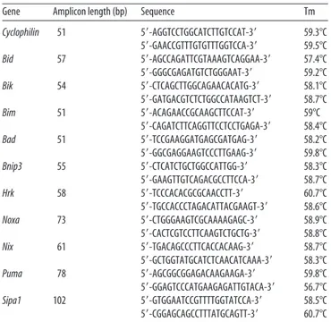
isch-Table 1. Primers used for real-time RT-PCR
Gene Amplicon length (bp) Sequence Tm
Cyclophilin 51 5⬘-AGGTCCTGGCATCTTGTCCAT-3⬘ 59.3°C 5⬘-GAACCGTTTGTGTTTGGTCCA-3⬘ 59.5°C Bid 57 5⬘-AGCCAGATTCGTAAAGTCAGGAA-3⬘ 57.4°C 5⬘-GGGCGAGATGTCTGGGAAT-3⬘ 59.2°C Bik 54 5⬘-CTCAGCTTGGCAGAACACATG-3⬘ 58.1°C 5⬘-GATGACGTCTCTGGCCATAAGTCT-3⬘ 58.7°C Bim 51 5⬘-ACAGAACCGCAAGCTTCCAT-3⬘ 59°C 5⬘-CAGATCTTCAGGTTCCTCCTGAGA-3⬘ 58.4°C Bad 51 5⬘-TCCGAAGGATGAGCGATGAG-3⬘ 58.2°C 5⬘-GGCGAGGAAGTCCCTTGAAG-3⬘ 59.8°C Bnip3 55 5⬘-CTCATCTGCTGGCCATTGG-3⬘ 58.3°C 5⬘-GAAGTTGTCAGACGCCTTCCA-3⬘ 58.7°C Hrk 58 5⬘-TCCCACACGCGCAACCTT-3⬘ 60.7°C 5⬘-TGCCACCCTAGACATTACGAAGT-3⬘ 58.6°C Noxa 73 5⬘-CTGGGAAGTCGCAAAAGAGC-3⬘ 58.9°C 5⬘-CACTCGTCCTTCAAGTCTGCTG-3⬘ 58.8°C Nix 61 5⬘-TGACAGCCCTTCACCACAAG-3⬘ 58.7°C 5⬘-GCTGGTATGCATCTCAACATCAAA-3⬘ 58.3°C Puma 78 5⬘-AGCGGCGGAGACAAGAAGA-3⬘ 59.8°C 5⬘-GGAGTCCCATGAAGAGATTGTACA-3⬘ 56.7°C Sipa1 102 5⬘-GTGGAATCCGTTTTGGTATCCA-3⬘ 58.5°C 5⬘-CGGAGCAGCCTTTATGCAGTT-3⬘ 60.7°C Tm, Melting temperature.
emic and contralateral cortices were quickly dissected and frozen on dry ice. Tissues were stored at⫺80°C. RNA from cortex or cultured cells was extracted with peqGOLD RNAPure (PEQLAB, Erlangen, Germany), according to the manufacturer’s instructions. RNA (10g, cortex; 7.5 g, cells) was transcribed with Moloney murine leukemia virus reverse tran-scriptase and random hexamers. Primers used for PCR amplification are described in Table 1. PCR was performed according to the follow-ing protocol: 10 min at 95°C, 15 s at 95°C, and 1 min at 60°C (40 cycles). Amplification was quantified with the Gene Amp 5700 sequence detector and the SYBR Green kit (PE Diagnos-tik, Weiterstadt, Germany). A linear concentra-tion–amplification curve was established by di-luting pooled samples. Quantified results for individual cDNAs were normalized to cyclo-philin. With this procedure, we can quantify results relative to a control group. The purity of the amplified products was checked by the dis-sociation curve and gel electrophoresis of se-lected samples.
Immunohistochemistry and terminal de-oxynucleotide transferase-mediated biotinylated UTP nick end labeling staining. For
immunohis-tochemical detection of active RelA, neurofilament-200, Bim, and Noxa, permanent MCAO was performed in wild-type mice. Two hours and 24 h after MCAO, mice were reanes-thetized and perfused with 4% paraformalde-hyde (PFA). Brains were incubated overnight in 4% PFA and then embedded in paraffin. Sec-tions (1m) were incubated overnight at 4°C with the following antibodies: active RelA (Mil-lipore, Billerica, MA), neurofilament-200 (Sigma), Bim (Stressgen Biotechnologies, Vic-toria, British Columbia, Canada), and Noxa (Imgenex, San Diego, CA). For detection, sec-ondary fluorescein-linked rabbit IgG anti-body (Vector Laboratories, Burlingame, CA) and rhodamine-conjugated anti-mouse IgG antibody (Jackson ImmunoResearch Laborato-ries, West Grove, PA) were used. To exclude the possibility of false labeling, we omitted primary and secondary antibodies.
For terminal deoxynucleotide transferase-mediated biotinylated UTP nick end labeling (TUNEL) staining, sections prestained with anti-Bim or anti-Noxa antibody were incu-bated with 50l of TUNEL reaction mix (In
Situ Cell Detection Kit, TMR red; Roche
Diag-nostics, Mannheim, Germany) for 1 h at 42°C. After washing three times, slides were mounted with medium containing 4 ⬘,6⬘-diamidino-2-phenylindole dihydrochloride (DAPI; Vectash-ield; Vector Laboratories).
Site-specific mutation and transfection. The
following plasmids have been described previ-ously: Noxa–luciferase (⫺183/⫹146) (Oda et al., 2000) and Bim–luciferase (⫺3600/⫹96) (Bouillet et al., 2001). NF-B binding sites were mutated in Noxa– and Bim–luciferase
plas-mids, using the QuikChange II Site-Directed Mutagenesis Kit (Strat-agene, La Jolla, CA) as recommended. To generate Noxa–mutated bind-ing sequence of theB promoter (mB)–luciferase and Bim–mB– luciferase plasmids, the following primers were used: CGT CAC ATG ACG TCA CCG AAG AAG TCA CGA TAA AAT GCG AGA GCC (Noxa–
mB1), GCA GCC CGA GTC TTG GAA GAG CCC CAG AGC CCA GAT TGG (Noxa–mB2), and GCC GTG GGG GGT AGG GCT TAT CTT CCG GCT TGC (Bim–mB). Mutated plasmids were confirmed by sequencing.
After 10 d in vitro, cortical neurons were transfected using
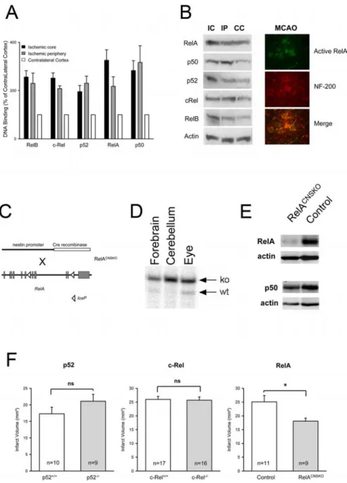
Lipo-Figure 1. Activation and function of NF-Bsubunitsincerebralischemia.A,AllfivesubunitsofNF-Bwereactivatedafter4h of MCAO. DNA binding in the ischemic and the corresponding contralateral cortex was measured with an ELISA-based assay. Values are means⫾ SEM (n ⫽ 6) and expressed as a percentage of the respective contralateral control. Core and periphery were dissected according to a standardized protocol (see Materials and Methods). B, Increased nuclear concentrations of the five subunits of NF-B after cerebral ischemia. Immunoblots of nuclear extracts of the ischemic core (IC), ischemic periphery (IP), and the contralateral cortex (CC) after 4 h of MCAO are shown (left). Nuclear translocation of active RelA (green) was detected in neurofilament-200-positive neurons (NF-200; red) 2 h after permanent MCAO by immunohistochemistry (right). C, Genetic strategy to generate RelACNSKOmice. The nestin-Cre balancer gene was introduced into mice that were homozygous for the floxed
RelA allele. Exons of the RelA gene are depicted as gray boxes. D, RelACNSKOmice were mosaics for the deleted RelA gene in several
organs. However, in cerebellum, forebrain, and eye, the mutated allele predominates, as shown by a Southern blot. ko, Knock-out; wt, wild type. E, RelA protein concentration was significantly reduced in the noninfarcted hemisphere of RelACNSKOmice
com-pared with controls ( p⬍0.05;n⫽13–15;two-tailedttest).Representativeimmunoblotsareshown.F,Conditionaldeletionof RelA in the brain reduced infarct volume, whereas deletion of p52 or c-Rel had no effect on the infarct size. The infarct volume was measured 48 h after permanent MCAO. *p⬍ 0.05 (two-tailed t test). ns, Not significant. Values are means ⫾ SEM. The sample size is noted in the boxes.
fectamine 2000 (Invitrogen), 0.2g per well of the Noxa or Bim plasmid, and 0.8g per well of the RcCMV-p65 or pBluescript plasmids as de-scribed previously (Potrovita et al., 2004). To normalize the transfection efficiency, 0.02g per well Renilla luciferase (phRLTK) control plasmid (Promega, Mannheim, Germany) was used. After 52 h, cells were harvested, and firefly and Renilla luciferase were measured using Dual Luciferase Reporter Assay (Promega).
Electrophoretic mobility shift assay. Cos cells
on 9 cm plates were transfected with RcCMV– p65 for 24 h. Cell extracts were prepared as de-scribed previously (Schreiber et al., 1989). Pro-tein and a double-stranded oligonucleotide from theB-enhancer (GAT CCA GAG GGG ACT TTC CGA GA) labeled with32P by a
Kle-now fill-in reaction were incubated at 4°C in the following buffer: 1 mMEDTA, 0.5g/l BSA,
10 mMTris-HCl, pH 7.5, 50 mMNaCl, 1 mM
DTT, and 5% glycerol (total volume, 20l). For competition, the following double-stranded oligonucleotides were used: mB, GAT CCA GAC CAT GGT ATC CGA GA; Bim, GAT CGG GGG TGG GGC TTA CCT TCC GGC; mBim, GAT CGG GGG TAG GGC TTA TCT TCC GGC; NoxaB2, GAT CGT CTT GGG GGA GCC CCA GAG CCC; and mNoxaB2, GAT CGT CTT GGA AGA GCC CCA GAG CCC. Protein–DNA complexes were resolved on a 6% nondenaturing polyacrylamide gel at 280 V.
Results
To find out which subunits of NF-B are
activated in cerebral ischemia, we
per-formed an ELISA-based analysis of DNA
binding of the NF-
B subunits. All five
subunits were activated in the ischemic
hemisphere, although the relative activation
between the core and the periphery of the
ischemic area differed between the subunits
(Fig. 1A). Similar results were obtained by
immunoblotting of the same nuclear
ex-tracts (Fig. 1B, left). Immunohistochemistry
with an antibody that specifically recognizes
active RelA (Kaltschmidt et al., 1995)
confirmed that RelA is activated in
neuro-filament-200-positive neurons (Fig. 1B,
right) (Herrmann et al., 2005).
Previous work has shown that the
sub-unit p50 contributes to ischemic brain
damage (Schneider et al., 1999; Nurmi et
al., 2004). Because germline deletion of
RelA causes embryonic death (Beg et al.,
1995), we used a conditional knock-out
approach to delete RelA in neurons and
glial cells. We deleted RelA in neural cells
(RelA
CNSKO) by crossing mice carrying
floxed RelA alleles with transgenic mice
ex-pressing the Cre recombinase under
con-trol of the nestin promoter (Betz et al.,
1996) (Fig. 1C). RelA
CNSKOmice were
healthy and viable. The deletion in the
Rel-A
CNSKOmice did not affect the expression
of the adjacent gene on the 3
⬘ side of RelA,
sipa, as evaluated by reverse transcription
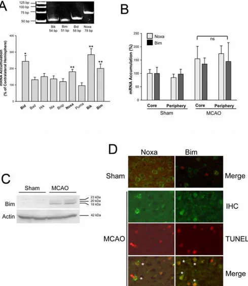
Figure 2. Expression of BH3-only genes after MCAO. A, mRNA accumulation in the ischemic cortex after 24 h of MCAO was quantified by real-time RT-PCR. Bottom, Values are means⫾ SEM (n ⫽ 12) and expressed relative to the contralateral nonisch-emic side. *p⬍0.02;**p⬍0.003(two-tailedttest).GelelectrophoresisverifiedthecorrectsizeofamplifiedPCRproducts(top).
B, mRNA accumulation of Bim and Noxa was not different between core and periphery of the infarcted area after 24 h of MCAO. ns,
Not significant. C, Immunoblotting showed upregulation of Bim 24 h after MCAO. The three bands correspond to BimEL (23 kDa), BimL (20 kDa), and BimS (18 kDa). D, Immunohistochemistry (IHC) demonstrated upregulation of Bim and Noxa after cerebral ischemia. Low levels of Noxa and Bim were detected in the cortex of sham-operated mice (top). Bim or Noxa (green) was expressed by TUNEL-positive cells (red) 24 h after MCAO (bottom). *, Double-positive cells.
Table 2. Physiological parameters 15 min before and 15 min after MCAO in RelACNSKOmice and littermate controls
Parameter
RelACNSKO Controls
Pre-MCAO Post-MCAO Pre-MCAO Post-MCAO MABP (mmHg) 70.0⫾ 2.1 61.4⫾ 3.3 69.1⫾ 3.8 61.6⫾ 3.1 Heart rate (per min) 433.3⫾ 9.1 468.6⫾ 10.4 403.4⫾ 9.7 452.4⫾ 10.1 Glucose (mg/dl) 185.1⫾ 9.9 224.5⫾ 17.0 189.6⫾ 9.8 245.3⫾ 21.2 Arterial pCO2(mmHg) 63.0⫾ 1.8 63.3⫾ 2.8 67.5⫾ 5.6 62.2⫾ 2.7 Arterial pO2(mmHg) 76.7⫾ 2.3 77.1⫾ 4.6 83.3⫾ 6.0 83.8⫾ 6.8 pH 7.15⫾ 0.02 7.11⫾ 0.02 7.17⫾ 0.03 7.14⫾ 0.03 Base excess ⫺8.8 ⫾ 0.8 ⫺10.7 ⫾ 0.9 ⫺8.2 ⫾ 1.3 ⫺10.5 ⫾ 0.9 Hb (g/L) 15.3⫾ 0.4 13.1⫾ 0.3 15.3⫾ 0.4 13.4⫾ 0.4 Laser Doppler (relative units) 86.7⫾ 4.3 14.0⫾ 0.7 93.8⫾ 4.1 11.5⫾ 1.6
Body weight (g) 32⫾ 2 32⫾ 3
None of the parameters differed significantly between the groups (t test). Values are means⫾ SEM; n ⫽ 5–8. MABP, Mean arterial blood pressure; Hb, hemoglobin concentration.
(RT)-PCR (sipa1 mRNA, 13.8
⫾ 3.0 relative units in control mice
vs 14.1
⫾ 8.1 relative units in RelA
CNSKOmice; n
⫽ 3–4). The
nestCre transgenic mouse line has been demonstrated to
in-duce a mosaic phenotype in several organs (Betz et al., 1996).
Indeed, Southern blot analysis showed that the RelA gene was
partially deleted in several tissues of RelA
CNSKOmice (data not
shown). However, this partial deletion had no effect on various
physiological parameters, which may affect brain damage in
MCAO (Table 2). In RelA
CNSKOmice, a marked deletion of the
RelA gene was observed in forebrain, cerebellum, and the eye
(Fig. 1 D). Because the nestin-Cre transgene targets mainly
neu-rons and astrocytes but not endothelial or microglial cells, a
com-plete loss of RelA in brain extracts is not expected. Immunoblots
of RelA in extracts of the nonischemic right hemisphere
demon-strated a significant reduction of RelA protein levels by 35% ( p
⬍
0.05; n
⫽ 13–15) (Fig. 1E). In the same extracts, there was a
nonsignificant trend toward lower protein concentrations of the
NF-B subunit p50 (Fig. 1E), which may be attributable to the
regulation of p50 expression by NF-B (Ten et al., 1992). The
infarct size after 48 h of MCAO was significantly reduced in
Rel-A
CNSKOmice, showing that RelA promotes ischemic damage
(Fig. 1 F, right). To investigate the functional consequences of
p52 activation, we used mice with a germline deficiency of the
subunit p52 (Paxian et al., 2002). These mice showed no
statisti-cally significant difference in infarct volumes compared with
wild-type controls (Fig. 1 F, left). The role of c-Rel was studied in
mice harboring a germline deletion of the c-Rel gene (Liou et al.,
1999). Germline deletion of c-Rel had no effect on the infarct
volume 48 h after onset of MCAO either (Fig. 1 F, middle).
Be-cause of poor health, RelB-deficient mice were not included in
this study (Weih et al., 1995).
To search for genes that may mediate the neurodegenerative
function of RelA, we measured the expression of various
BH3-only genes by real-time RT-PCR 24 h after permanent MCAO in
mice. Of nine BH3-only genes, Bid, Noxa, Bik, and Bim were
significantly upregulated after MCAO (Fig. 2 A). There was no
difference between the induction of Bim and Noxa mRNA in the
core and periphery of the ischemia (Fig. 2 B). Alternative splicing
generates three isoforms of Bim (BimEL, 23 kDa; BimL, 20 kDa;
and BimS, 18 kDa). Our real-time RT-PCR is specific for BimS,
which has the highest apoptotic potency of the isoforms.
Immu-noblots supported the induction of BimS and demonstrated that
the other isoforms were also enhanced by MCAO (Fig. 2C).
Moreover, immunohistochemistry confirmed the upregulation
of Bim and Noxa 24 h after permanent MCAO. Bim and Noxa
staining was partially colocalized with TUNEL, suggesting that
Bim and Noxa are implicated in apoptotic cell death after cerebral
ischemia (Fig. 2 D).
In cerebral ischemia, apoptotic cell death mainly affects
neu-rons. To approach the question of whether BH3-only genes are
induced in neurons, we exposed primary cortical mouse neurons
to camptothecin, a topoisomerase I blocker that models DNA
damage in cerebral ischemia. Camptothecin stimulated the
ex-pression of Noxa and Bim at 16 and 24 h of exposure, whereas the
expression of Bid and Bik was only enhanced after 24 h of
stimu-lation (Fig. 3A). A similar picture emerged after exposure of
cor-tical neurons to OGD, an in vitro model of cerebral ischemia.
Bim, Noxa, and Bik were significantly induced at the mRNA level
by 6 h of OGD (Fig. 3B). However, Bid expression was not
stim-ulated by OGD in cortical neurons. Thus, Bik, Noxa, and Bim are
induced by in vivo and in vitro models of cerebral ischemia.
To investigate the effect of RelA on the induction of BH3-only
genes, we measured mRNA levels of the four BH3-only genes that
were found to be upregulated in wild-type mice. The induction of
Bim and Noxa in wild-type mice was abolished in RelA
CNSKOmice, whereas the upregulation of Bik persisted in RelA
CNSKOmice (Fig. 4 A). Previous work has shown that NF-B is activated
in cerebral ischemia by the IKK complex. BMS-345541, a specific
IKK inhibitor (Burke et al., 2003), protected against ischemic
brain damage (Herrmann et al., 2005). IKK activity is reflected by
the phosphorylation state of IKK. Intracerebroventricular
injec-tion of BMS-345541 reduced IKK phosphorylainjec-tion 4.5 h after
MCAO in the periphery of the ischemic area, confirming that
BMS-345541 is an effective inhibitor of IKK in vivo (Fig. 4 B). To
explore the function of IKK in the induction of Bim and Noxa, we
injected mice intracerebroventricularly with BMS-345541 or
ve-hicle and measured Bim and Noxa mRNA at three time points
after MCAO. Bim and Noxa were first upregulated after 6 h of
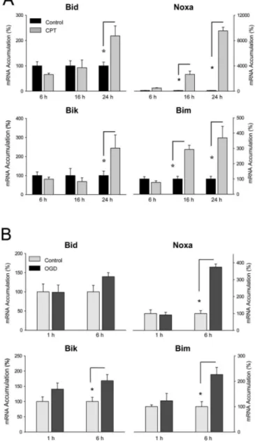
Figure 3. Induction of BH3-only genes in primary cortical neurons in in vitro models of cerebral ischemia. A, Camptothecin (CPT; 10M) exposure enhanced mRNA accumulation of
Bid, Bik, Noxa, and Bim in a time-dependent manner. Values are means⫾SEM(n⫽7–8)and expressed relative to the unstimulated control group. *p⬍ 0.006 [ANOVA; least significant difference (LSD) post hoc]. B, OGD stimulates mRNA accumulation of Bik, Noxa, and Bim in a time-dependent manner. Values are means⫾ SEM (n ⫽ 5–6) and expressed relative to the group not exposed to OGD. *p⬍ 0.01 (ANOVA; LSD post hoc).
MCAO; at this time, BMS-345541 inhibited the induction.
How-ever, BMS-345541 lost its effect after 24 h of MCAO (Fig. 4C).
These data suggest that Bim and Noxa are target genes of IKK and
RelA.
To further evaluate the possibility that NF-B stimulates gene
transcription of Noxa and Bim in neurons after cerebral
isch-emia, we transfected primary cortical neurons with reporter
fu-sion genes, in which the 5⬘-flanking sequences of the mouse Bim
(
⫺3600/⫹96) or the mouse Noxa gene (⫺183/⫹146) direct
lu-ciferase expression. Indeed, cotransfection of an expression
plas-mid for RelA stimulated Bim transcription approximately
four-fold (Fig. 5A) and Noxa transcription approximately twofour-fold
(Fig. 5B). A search for putative NF-B binding sites in the
5⬘-flanking sequence of the Bim and Noxa genes using the
TRANSFAC database found one potential binding site in the
promoter of the mouse Bim gene (⫺62/⫺53) and two close to the
transcription start site of the Noxa gene (
⫺49/⫺40; ⫹87/⫹96),
with the second site being almost palindromic. Mutation of the
NF-B binding site in the Bim promoter significantly reduced
luciferase expression (Fig. 5A). The residual effect of RelA after
mutation of the NF-B site suggests that the mutation may not
destroy RelA binding completely or there may be other sites in the
5
⬘-flanking sequence. Mutation of the first NF-B binding site
(B1) in the Noxa promoter had no effect, but mutation of the
second binding site (B2) blocked the stimulation by RelA (Fig.
5B). To further test whether RelA binds to the identified response
elements in the Bim and Noxa promoter
region, we performed electrophoretic shift
assays. The binding of RelA expressed in
Cos cells to a consensus NF-B binding
site was inhibited by wild-type Bim and
Noxa sites. The mutations that interfered
with RelA function also diminished
com-petition of RelA DNA binding (Fig. 5C).
These data support the notion that RelA
binds to the promoter of Bim and Noxa
and stimulates their transcription.
Discussion
Several investigators have reported on the
activation of NF-B in cerebral ischemia
(Mattson and Camandola, 2001). Here,
we specify this finding, showing that all
five subunits are activated. In view of this
broad activation of NF-B subunits, it was
interesting to compare their effects on
ischemic brain damage. Our data support
the concept that NF-
B subunits differ in
their functional significance. In our
para-digm of focal cerebral ischemia,
condi-tional deletion of RelA in the brain
re-duced the infarct size, whereas a germline
knock-out of the genes for the subunits
c-Rel and p52 had no effect. Notably, our
data do not exclude an effect of c-Rel and
p52 on parameters other than infarct size
or a potential compensation by redundant
mechanisms. Together with the previous
finding that p50
⫺/⫺mice have smaller
in-farcts (Schneider et al., 1999; Nurmi et al.,
2004), the reduced infarct size in RelA
C-NSKO
mice suggests that p50/RelA
het-erodimers or p50/p50 and RelA/RelA
ho-modimers promote ischemic brain damage. A slight reduction of
p50 levels in brains of RelA
CNSKOmice (Fig. 1 E) might have
contributed to protection in cerebral ischemia. The predominant
role of p50 and RelA is in accordance with their abundance in the
brain and in other tissues (Hoffmann et al., 2003; Kaltschmidt et
al., 2005). In the canonical NF-B pathway, p50 and RelA
activa-tion is triggered by the upstream IKK kinase complex with the
subunits IKK1, IKK2, and NEMO (NF-B essential modulator).
Indeed, IKK2 deletion or inhibition was shown to ameliorate
ischemic brain damage (Herrmann et al., 2005). Thus,
interfer-ence with the NF-B pathway at various levels reveals a
deleteri-ous role of NF-B in cerebral ischemia, which is in apparent
contrast to what has been found in other paradigms. There is
ample evidence that NF-B inhibits apoptosis in tumor cells,
hepatocytes, and many other cell types, including neurons (Pizzi
et al., 2002; Fridmacher et al., 2003; Tarabin and Schwaninger,
2004). In stroke models, however, protective effects of NF-
B,
which may be mediated by c-Rel (Pizzi et al., 2002), are overrun
by the deleterious action of p50 and RelA. In accordance with this
concept, deletion of c-Rel had no effect on infarct size (Fig. 1 F).
In contrast, the protective role of NF-
B seems to prevail in short
cerebral ischemia, which leads to ischemic preconditioning
(Blondeau et al., 2001).
NF-B promotes cell survival through induction of Bcl-2 and
Bcl-X
L(Lee et al., 1999; Tamatani et al., 1999; Mattson and
Ca-Figure 4. Induction of Bim and Noxa by MCAO depends on the IKK/NF-B signaling pathway. A, In RelACNSKOmice, Bim and
Noxa mRNA accumulation was not enhanced 24 h after MCAO. *p⬍ 0.004 compared with sham group.⫹p⬍ 0.02 compared
with wild-type mice with MCAO. n⫽3–4[ANOVA;leastsignificantdifference(LSD)posthoc].B,Phospho-IKK1/2intheperiphery (P) of the ischemic cortex was decreased in mice injected with 12g of BMS-345541 intracerebroventricularly as shown by immunoblot.Inthecore(C)oftheischemia,phospho-IKK1/2wasnotaffectedbyMCAO.Micewereanalyzed4.5hafterpermanentMCAO. Erk2 was used as control for protein loading. ns, Nonspecific band. C, The IKK inhibitor BMS-345541 transiently inhibited Bim and Noxa mRNA accumulation after MCAO. BMS-345541 was injected intracerebroventricularly immediately before MCAO. *p⬍ 0.05 compared with vehicle-treated mice with MCAO (ANOVA; LSD post hoc; n⫽4–6).ns,Notsignificant.Valuesaremeans⫾SEM.
mandola, 2001). Previous studies reported either an increase or a
decrease in Bcl-2 and Bcl-X
Lexpression in cerebral ischemia
(Krajewski et al., 1995; Gillardon et al., 1996; Chen et al., 1997). In
our stroke model, there was no change in Bcl-2 or Bcl-X
Lexpres-sion (data not shown). However, we found a reproducible
up-regulation of the proapoptotic BH3-only genes Bik, Bim, and
Noxa by cerebral ischemia. The induction of Bim by cerebral
ischemia has been reported previously (Shibata et al., 2002; Gao
et al., 2005).
Bim is localized in neurons (Shibata et al., 2002). Indeed, in
primary cortical neurons, in vitro OGD induced Bim and Noxa
expression. Interestingly, we found that conditional deletion of
RelA in neural cells or pharmacological IKK inhibition blocked
Bim and Noxa expression but had no effect on Bik induction by
cerebral ischemia. RelA seems to directly regulate transcription of
the Bim and Noxa gene by binding to the promoter because
mu-tation of the NF-B motif near the transcription start site of the
Bim and Noxa gene both interfered with the induction by RelA
and reduced the binding of RelA to the DNA. The sequence of the
NF-B site in the 5⬘-untranslated region of the Noxa gene
(GGG-GAGCCCC) matches the consensus binding site
GGGRN-NYYCC of NF-B exactly, where R represents purine, Y
repre-sents pyrimidine, and N reprerepre-sents any base (Ghosh et al., 1998).
The sequence in the Bim promoter (GGGGCTTACC) deviates by
a purine in position 8 from the consensus sequence. However, the
same variation occurs in the NF-B motif of the GM-CSF
(gran-ulocyte/macrophage colony-stimulating factor) gene, which is a
high-affinity binding site (Schreck and Baeuerle, 1990).
An indirect mechanism through which NF-B is able to
reg-ulate Noxa gene transcription was suggested by Aleyasin et al.
(2004). These authors found that, when DNA had been damaged,
NF-B stimulated Noxa and Puma gene transcription through
induction of p53 expression. Focal cerebral ischemia only
stimu-lated Noxa but not Puma mRNA accumulation, suggesting an
underlying mechanism other than p53. In addition to the direct
induction of Noxa and Bim by the IKK/NF-B pathway that we
propose here, indirect interactions with other signaling cascades
known to be involved in Bim expression are possible. IKK has
been shown to inhibit the transcription factor FOXO3a (Hu et al.,
2004), which stimulates Bim gene transcription (Gilley et al.,
2003; Sunters et al., 2003). Likewise, NF-B inhibits the effect of
JNK on gene transcription (De Smaele et al., 2001; Tang et al.,
2001; Park et al., 2004). JNK has been shown to control Bim
transcription in cerebral ischemia (Kuan et al., 2003; Okuno et
al., 2004). Thus, IKK/NF-B cross talk with FOXO3a or JNK may
counteract the induction of Bim transcription caused by the
di-rect stimulation. However, the role of such interactions must be
further investigated.
Of note, NF-B directs gene transcription of Bcl-2 family
members with opposite effects on mitochondrial permeability
that are either proapoptotic (Bax, Bim, and Noxa) or
antiapop-totic (Bcl-2 and Bcl-X
L). The relative control of these genes may
be the switch that determines the enigmatic role of NF-B for cell
survival. It is possible that the composition of the NF-B dimer or
the activation pattern of other transcription factors determines
whether the induction of proapoptotic or antiapoptotic Bcl-2
family members prevails.
References
Aleyasin H, Cregan SP, Iyirhiaro G, O’Hare MJ, Callaghan SM, Slack RS, Park DS (2004) Nuclear factor-B modulates the p53 response in neurons exposed to DNA damage. J Neurosci 24:2963–2973.
Beg A, Sha W, Bronson R, Ghosh S, Baltimore D (1995) Embryonic lethality and liver degeneration in mice lacking the RelA component of NF-kB. Nature 376:167–170.
Betz UA, Vosshenrich CA, Rajewsky K, Muller W (1996) Bypass of lethality with mosaic mice generated by Cre-loxP-mediated recombination. Curr Biol 6:1307–1316.
Blondeau N, Widmann C, Lazdunski M, Heurteaux C (2001) Activation of the nuclear factor-B is a key event in brain tolerance. J Neurosci 21:4668 – 4677.
Bouillet P, Zhang LC, Huang DC, Webb GC, Bottema CD, Shore P, Eyre HJ, Sutherland GR, Adams JM (2001) Gene structure alternative splicing, and chromosomal localization of pro-apoptotic Bcl-2 relative Bim. Mamm Genome 12:163–168.
Burke JR, Pattoli MA, Gregor KR, Brassil PJ, MacMaster JF, McIntyre KW, Yang X, Iotzova VS, Clarke W, Strnad J, Qiu Y, Zusi FC (2003) BMS-345541 is a highly selective inhibitor of I kappa B kinase that binds at an allosteric site of the enzyme and blocks NF-kappa B-dependent transcrip-tion in mice. J Biol Chem 278:1450 –1456.
Cao G, Pei W, Ge H, Liang Q, Luo Y, Sharp FR, Lu A, Ran R, Graham SH, Chen J (2002) In vivo delivery of a Bcl-xL fusion protein containing the TAT protein transduction domain protects against ischemic brain injury and neuronal apoptosis. J Neurosci 22:5423–5431.
Chen J, Graham SH, Nakayama M, Zhu RL, Jin K, Stetler RA, Simon RP (1997) Apoptosis repressor genes Bcl-2 and Bcl-x-long are expressed in the rat brain following global ischemia. J Cereb Blood Flow Metab 17:2–10.
De Smaele E, Zazzeroni F, Papa S, Nguyen DU, Jin R, Jones J, Cong R,
Figure 5. RelA binds to the Bim and Noxa promoter to stimulate transcription. A, Transient cotransfection of primary cortical neurons with an expression vector for RelA stimulated lucif-erase expression controlled by 3600 bp of the Bim promoter. Mutation of the NF-B site in the promoter significantly reduced luciferase expression. Values are means⫾ SEM.⫹p⬍ 0.0001
[n⫽ 8; ANOVA; least significant difference (LSD) post hoc]. B, RelA overexpression stimulated luciferase expression controlled by the Noxa promoter. Mutation of the NF-B binding site 2 (B2) but not of the binding site 1 (B1) blocked luciferase expression. *p ⬍ 0.01 (n ⫽ 8; ANOVA; LSD post hoc). The location of NF-B binding sites relative to other response elements is depicted at the top. wt, Wild type. C, Electrophoretic mobility shift assays showed that RelA binding to a consensus NF-Bbindingsitewasblockedbyanexcessoftheconsensussite(B), the NoxaB2, and the Bim NF-B binding sites, but not by the mutated sequences. RelA was expressed in Cos cells.
Franzoso G (2001) Induction of gadd45beta by NF-kappaB downregu-lates pro-apoptotic JNK signalling. Nature 414:308 –313.
Fridmacher V, Kaltschmidt B, Goudeau B, Ndiaye D, Rossi FM, Pfeiffer J, Kaltschmidt C, Israel A, Memet S (2003) Forebrain-specific neuronal inhibition of nuclear factor-B activity leads to loss of neuroprotection. J Neurosci 23:9403–9408.
Gao Y, Signore AP, Yin W, Cao G, Yin XM, Sun F, Luo Y, Graham SH, Chen J (2005) Neuroprotection against focal ischemic brain injury by inhibi-tion of c-Jun N-terminal kinase and attenuainhibi-tion of the mitochondrial apoptosis-signaling pathway. J Cereb Blood Flow Metab 25:694 –712. Ghosh S, May MJ, Kopp E (1998) NF-kB and Rel proteins: evolutionarily
conserved mediators of immune responses. Annu Rev Immunol 16:225–260.
Gillardon F, Lenz C, Waschke KF, Krajewski S, Reed JC, Zimmermann M, Kus-chinsky W (1996) Altered expression of bcl-2, bcl-x, bax, and c-Fos colocal-izes with DNA fragmentation and ischemic cell damage following middle cerebral artery occlusion in rats. Brain Res Mol Brain Res 40:254 –260. Gilley J, Coffer PJ, Ham J (2003) FOXO transcription factors directly
acti-vate bim gene expression and promote apoptosis in sympathetic neurons. J Cell Biol 162:613– 622.
Graham SH, Chen J, Clark RS (2000) Bcl-2 family gene products in cerebral ischemia and traumatic brain injury. J Neurotrauma 17:831– 841. Herrmann O, Tarabin V, Suzuki S, Attigah N, Prinz S, Schneider A, Coserea I,
Monyer H, Brombacher F, Schwaninger M (2003) Regulation of body temperature and neuroprotection by endogenous interleukin-6 in focal cerebral ischemia. J Cereb Blood Flow Metab 23:406 – 415.
Herrmann O, Baumann B, De Lorenzi R, Muhammad S, Zhang W, Kleesiek J, Malfertheiner M, Ko¨hrmann M, Potrovita I, Maegele I, Beyer C, Burke JR, Hasan MT, Bujard H, Wirth T, Pasparakis M, Schwaninger M (2005) IKK mediates ischemia-induced neuronal cell death. Nat Med 11:1322–1329. Hershko T, Ginsberg D (2004) Up-regulation of Bcl-2 homology 3
(BH3)-only proteins by E2F1 mediates apoptosis. J Biol Chem 279:8627– 8634. Hoffmann A, Leung TH, Baltimore D (2003) Genetic analysis of
NF-kap-paB/Rel transcription factors defines functional specificities. EMBO J 22:5530 –5539.
Hu MC, Lee DF, Xia W, Golfman LS, Ou-Yang F, Yang JY, Zou Y, Bao S, Hanada N, Saso H, Kobayashi R, Hung MC (2004) IkappaB kinase pro-motes tumorigenesis through inhibition of forkhead FOXO3a. Cell 117:225–237.
Kaltschmidt B, Kaltschmidt C, Hofmann TG, Hehner SP, Droge W, Schmitz ML (2000) The pro- or anti-apoptotic function of NF-kappaB is determined by the nature of the apoptotic stimulus. Eur J Biochem 267:3828 –3835. Kaltschmidt B, Heinrich M, Kaltschmidt C (2002) Stimulus-dependent
ac-tivation of NF-kappaB specifies apoptosis or neuroprotection in cerebel-lar granule cells. Neuromolecucerebel-lar Med 2:299 –309.
Kaltschmidt B, Widera D, Kaltschmidt C (2005) Signaling via NF-B in the nervous system. Biochim Biophys Acta 1745:287–299.
Kaltschmidt C, Kaltschmidt B, Henkel T, Stockinger H, Baeuerle PA (1995) Selective recognition of the activated form of transcription factor NF-kappa B by a monoclonal antibody. Biol Chem Hoppe-Seyler 376:9 –16. Krajewski S, Mai JK, Krajewska M, Sikorska M, Mossakowski MJ, Reed JC
(1995) Upregulation of bax protein levels in neurons following cerebral ischemia. J Neurosci 15:6364 – 6376.
Kuan CY, Whitmarsh AJ, Yang DD, Liao G, Schloemer AJ, Dong C, Bao J, Banasiak KJ, Haddad GG, Flavell RA, Davis RJ, Rakic P (2003) A critical role of neural-specific JNK3 for ischemic apoptosis. Proc Natl Acad Sci USA 100:15184 –15189.
Lee HH, Dadgostar H, Cheng Q, Shu J, Cheng G (1999) NF-kappaB-mediated up-regulation of Bcl-x and Bfl-1/A1 is required for CD40 sur-vival signaling in B lymphocytes. Proc Natl Acad Sci USA 96:9136 –9141. Li Q, Verma IM (2002) NF-kappaB regulation in the immune system. Nat
Rev Immunol 2:725–734.
Liou HC, Jin Z, Tumang J, Andjelic S, Smith KA, Liou ML (1999) c-Rel is crucial for lymphocyte proliferation but dispensable for T cell effector function. Int Immunol 11:361–371.
Mattson MP, Camandola S (2001) NF-kappaB in neuronal plasticity and neurodegenerative disorders. J Clin Invest 107:247–254.
Nurmi A, Lindsberg PJ, Koistinaho M, Zhang W, Juettler E, Karjalainen-Lindsberg ML, Weih F, Frank N, Schwaninger M, Koistinaho J (2004) Nuclear factor-kappaB contributes to infarction after permanent focal ischemia. Stroke 35:987–991.
Oda E, Ohki R, Murasawa H, Nemoto J, Shibue T, Yamashita T, Tokino T,
Taniguchi T, Tanaka N (2000) Noxa, a BH3-only member of the Bcl-2 family and candidate mediator of p53-induced apoptosis. Science 288:1053–1058.
Okuno S, Saito A, Hayashi T, Chan PH (2004) The c-Jun N-terminal pro-tein kinase signaling pathway mediates Bax activation and subsequent neuronal apoptosis through interaction with Bim after transient focal cerebral ischemia. J Neurosci 24:7879 –7887.
Park JM, Brady H, Ruocco MG, Sun H, Williams D, Lee SJ, Kato Jr T, Richards N, Chan K, Mercurio F, Karin M, Wasserman SA (2004) Targeting of TAK1 by the NF-kappa B protein Relish regulates the JNK-mediated immune response in Drosophila. Genes Dev 18:584 –594.
Paxian S, Merkle H, Riemann M, Wilda M, Adler G, Hameister H, Liptay S, Pfeffer K, Schmid RM (2002) Abnormal organogenesis of Peyer’s patches in mice deficient for NF-kappaB1, NF-kappaB2, and Bcl-3. Gas-troenterology 122:1853–1868.
Pizzi M, Goffi F, Boroni F, Benarese M, Perkins SE, Liou HC, Spano P (2002) Opposing roles for NF-kappa B/Rel factors p65 and c-Rel in the modula-tion of neuron survival elicited by glutamate and interleukin-1beta. J Biol Chem 277:20717–20723.
Pizzi M, Sarnico I, Boroni F, Benarese M, Steimberg N, Mazzoleni G, Dietz GP, Bahr M, Liou HC, Spano PF (2005) NF-kappaB factor c-Rel medi-ates neuroprotection elicited by mGlu5 receptor agonists against amyloid beta-peptide toxicity. Cell Death Differ 12:761–772.
Potrovita I, Zhang W, Burkly L, Hahm K, Lincecum J, Wang MZ, Maurer MH, Rossner M, Schneider A, Schwaninger M (2004) Tumor necrosis factor-like weak inducer of apoptosis-induced neurodegeneration. J Neu-rosci 24:8237– 8244.
Schneider A, Martin-Villalba A, Weih F, Vogel J, Wirth T, Schwaninger M (1999) NF-B is activated and promotes cell death in focal cerebral isch-emia. Nat Med 5:554 –559.
Schreck R, Baeuerle PA (1990) NF-kappa B as inducible transcriptional ac-tivator of the granulocyte-macrophage colony-stimulating factor gene. Mol Cell Biol 10:1281–1286.
Schreiber E, Matthias P, Mu¨ller MM, Schaffner W (1989) Rapid detection of octamer binding proteins with ‘mini-extracts’, prepared from small num-ber of cells. Nucleic Acids Res 17:6419.
Shibata M, Hattori H, Sasaki T, Gotoh J, Hamada J, Fukuuchi Y (2002) Temporal profiles of the subcellular localization of Bim, a BH3-only pro-tein, during middle cerebral artery occlusion in mice. J Cereb Blood Flow Metab 22:810 – 820.
Strasser A (2005) The role of BH3-only proteins in the immune system. Nat Rev Immunol 5:189 –200.
Sunters A, Fernandez de Mattos S, Stahl M, Brosens JJ, Zoumpoulidou G, Saunders CA, Coffer PJ, Medema RH, Coombes RC, Lam EW (2003) FoxO3a transcriptional regulation of Bim controls apoptosis in paclitaxel-treated breast cancer cell lines. J Biol Chem 278:49795– 49805. Tamatani M, Che YH, Matsuzaki H, Ogawa S, Okado H, Miyake S, Mizuno T, Tohyama M (1999) Tumor necrosis factor induces Bcl-2 and Bcl-x ex-pression through NFkappaB activation in primary hippocampal neurons. J Biol Chem 274:8531– 8538.
Tang G, Minemoto Y, Dibling B, Purcell NH, Li Z, Karin M, Lin A (2001) Inhibition of JNK activation through NF-kappaB target genes. Nature 414:313–317.
Tarabin V, Schwaninger M (2004) The role of NF-kappaB in 6-hydroxydopamine- and TNFalpha-induced apoptosis of PC12 cells. Naunyn Schmiedebergs Arch Pharmacol 369:563–569.
Ten RM, Paya CV, Israel N, Le Bail O, Mattei MG, Virelizier JL, Kourilsky P, Israel A (1992) The characterization of the promoter of the gene encod-ing the p50 subunit of NF-kappa B indicates that it participates in its own regulation. EMBO J 11:195–203.
Weih F, Carrasco D, Durham SK, Barton DS, Rizzo CA, Ryseck R-P, Lira SA, Bravo R (1995) Multiorgan inflammation and hematopoietic abnor-malities in mice with a targeted disruption of RelB, a member of the NF-kB/Rel family. Cell 80:331–340.
Xu L, Zhan Y, Wang Y, Feuerstein GZ, Wang X (2002) Recombinant adeno-viral expression of dominant negative IkappaBalpha protects brain from cerebral ischemic injury. Biochem Biophys Res Commun 299:14 –17. Yu J, Zhang L (2003) No PUMA, no death: implications for p53-dependent
apoptosis. Cancer Cell 4:248 –249.
Zhang W, Potrovita I, Tarabin V, Herrmann O, Beer V, Weih F, Schneider A, Schwaninger M (2005) Neuronal activation of NF-B contributes to cell death in cerebral ischemia. J Cereb Blood Flow Metab 25:30 – 40.