International PhD Program in Neuroscience
XXXI CYCLE
Protein Misfolding and Aggregation in
Neurodegeneration:
In Vitro And In Vivo Study Cases
PhD Thesis
Ramona Belfiore
Coordinator and Tutor:
P
ROF.
S
ALVATORES
ALOMONECo-Tutors:
P
ROF.
V
ITOD
EP
INTOP
ROF.
S
ALVATOREO
DDOBIOMETEC
DEPARTMENT OF BIOMEDICAL AND BIOTECHNOLOGICAL SCIENCES
2
TABLE OF CONTENTS
LIST OF ABBREVIATIONS
……….4
ABSTRACT
………...6
GENERAL INTRODUCTION
………..9
PROTEIN MISFOLDING AND AGGREGATION……….
9
NEURODEGENERATIVE DISEASES………..
10
AMYOTROPHIC LATERAL SCLEROSIS (ALS)………
11
PROTEIN MISFOLDING IN ALS………...
14
SOD1……….
14
Human SOD1………
15
SOD1 ALS-linked mutants ………...
16
MITOCHONDRIAL DISEASE IN ALS………...
17
VDAC...
19
SOD-VDAC interaction in ALS………....
22
ALZHEIMER’S DISEASE (AD)………
23
PROTEIN AGGREGATION AND ACCUMULATION IN AD………… ..
24
Amyloid protein……….
24
Aβ pathologic role……….
25
Tau protein……….
26
3
Mouse models of Aβ and tau……….
28
GLIAL REACTIVITY IN AD………...…
31
CHAPTER 1………...33
Hexokinase I N-Terminal Based Peptide prevents the VDAC1-SOD1
G93A interaction and re-establishes ALS cell viability
CHAPTER 2………...86
Temporal and Regional Progression of Alzheimer’s disease-like
pathology in 3xTg-AD mice
GENERAL DISCUSSION AND CONCLUSIONS……...140
REFERENCES
……...………...142
LIST OF PUBLICATIONS AND SCIENTIFIC CONFERENCES… 153
ACKNOWLEDGEMENTS………...………. 155
4
LIST OF ABBREVIATIONS
CNS Central nervous system
PNS Peripheral nervous system
FTD frontotemporal dementia
ALS Amyotrophic Lateral Sclerosis
GWAS Genome-wide association study
SMN1 Survival motor neuron 1
DPP6 Dipeptidyl Peptidase Like 6
VEGF Vascular endothelial growth factor
ROS Reactive oxygen species
SOD1 Superoxide dismutase 1
TARDBP Transactive response DNA-binding protein
FUS Fused in sarcoma
VCPV Valosin containing protein VAPB VAMP-associated protein type B
WT Wild type
ATP Adenosine triphosphate
VDAC Voltage-dependent Anion Channel
OMM Outer mitochondrial membrane
ADP Adenosine diphosphate
NADH Nicotinamide adenine dinucleotide
G6P Glucose-6-phosphate
AD Alzheimer’s disease
5
ApoE Apolipoprotein E
Aβ Amyloid-β
NFT Neurofibrillary tangles
APP Amyloid precursor protein
CTFα C-terminal fragment
BACE β-site APP cleaving enzyme
LTP Long-term potentiation
LTD Long-term depression
MAPT Microtubule associated protein tau
PHFs Paired helical filaments
TPK1 Thiamin pyrophosphokinase 1
GSK3-β Glycogen synthase kinase 3- β
6
ABSTRACT
Neurodegenerative diseases are nowadays increasing in incidence and widely distributed around the world. Many scientists are currently working on developing therapeutic tools to stop this rise and prevent the onset of disorders like Alzheimer's (AD), amyotrophic lateral sclerosis (ALS) and Parkinson's (PD). Despite those disorders show very different symptoms and morbidity, intracellular and extracellular protein misfolding and accumulation appears as a common pathological pathway. In the present thesis work I analyzed two cases of toxic protein deposition involved in ALS and AD onset using both in vitro and vivo techniques to study these toxic accumulations in cell culture, bacteria and animal models.
First, I looked at Superoxide Dismutase 1 (SOD1) mutant protein G93A (mutSOD1-G93A) whose neuronal deposit is associated to familial and sporadic Amyotrophic Lateral Sclerosis (ALS). Both ALS patients and transgenic mouse models show mitochondrial abnormalities such as swelling and vacuolization in spinal cords as pathological hallmarks. In this context, the mitochondrial porin VDAC1 (voltage dependent anion channel 1) has been proposed as a binding target of SOD1 mutant forms to mitochondria. To study how hVDAC1 interplays with mutSOD1-G93A we produced recombinants wtSOD1, mutSOD1-G93A and His-tagged recombinant VDAC1 protein. Interestingly, by affinity studies we found that VDAC1 protein specifically binds mutSOD1-G93A but not wtSOD1. Notably, it is known that the N-Terminal end of Hexokinase 1 (N-HK1) interacts with VDAC1: thus, we produced a synthetic peptide corresponding to the first 11 aa of human HK1 and tested its action as a potential interfering molecule between VDAC1- mutSOD1-G93A bond. In an electrophysiological study we observed that adding N-HK1
7
peptide promotes a significant instability of VDAC1 channel affecting the pore’s gating. Next, in a binding assay we verified the N-HK1 peptide interference in SOD1G93A-VDAC1 interaction. Both in a protein-protein interaction and in a protein–mitochondrial interaction we obtained a decrease of SOD1-VDAC1 binding in dependence of the increased NHK1 peptide concentration (85% in mitochondria). Summarizing our overall results, they show that in ALS condition, mut-SOD1-G93A binds VDAC1 and impairs HK1 binding and our results suggest for N-HK1 peptide a neuroprotective potential in ALS patients. Starting from these experiments it is possible to select one or more HK1 peptide having the highest chance of interfering with ALS-linked VDAC1/mutSOD1 toxic aggregates in in vivo models. The NHK-1 peptide is now patented.
The second part of my thesis work was focused on the Amyloid and tau protein accumulation in 3xTg-AD mice. In order to study neuropathology and cognitive deficits in Alzheimer’s disease (AD), several transgenic models of AD have been identified. Accumulation of amyloid-β and fibrillary tangles as well as impairments in working and learning memory are age-related hallmark of AD pathology. After almost 15 years from his creation, the 3xTg-AD mouse model is still one of the most reliable transgenic models. Despite the huge number of funding about this transgenic model in AD, it is still to be defined the detailed age-related progression of amyloid and tau pathology for each brain region. To produce a progressive characterization of Aβ and tau pathology in 3xTg-AD mice we aged female mice at 2, 6, 12 and 20 months of age. We tested mice in a behavioral assay named Morris Water Maze (MWM) and we used in vitro biochemical assays (ELISA, IHC, IF) to observe Aβ soluble and insoluble fraction as well as tau phosphorylation in both cortex and hippocampus of 3xTg-AD mice. Our data on MWM
8
demonstrate a progressive impairment in learning with a strongly significant difference between 3xTg-AD mice and controls (C57129 mice) from 6 months of age. Notably, we also found a progressive increase in both soluble and insoluble Aβ40 and Aβ42, an age dependent tau hyperphosphorylation at specific AD linked phospho-sites, and an intense glial reactivity. Overall, our data confirm that female 3xTg-AD mice consistently show AD-like pathology, therefore this transgenic mouse model can be used as an extremely powerful tool to investigate pathogenic mechanisms underlying Alzheimer’s disease.
9
GENERAL INTRODUCTION
PROTEIN MISFOLDING AND AGGREGATION
Most of the process involved in biological system’s activity are regulated by protein activation and protein-protein interaction (Westermarck et al., 2013). The correct functionality of these macromolecules, consisting in a linear sequence of amino acids, depends on their capacity to form three-dimensional structures and create more complexes interactions thanks to functional groups such as carboxylic acids, alcohols, thioethers etc (Belmont et al., 2001).
Inside the cells, proteins can self-assembly and, thanks to molecular chaperons, adopt the right folding (Ellis and Hartl, 1999). It is known that an incorrect protein folding can indeed lead to protein disfunction (gain or loss of function) and aggregation (Dobson, 2003). In many cases of conformational disorders, the pathological effect of protein misfolding is the gain of ability to alternatively interact under oligomeric forms, amorphous aggregates and fibrillar structures.
Those mis-functional aggregates can eventually create toxic intracellular and extracellular deposits that interfere with cell’s survival (Dobson, 2004). Causes and mechanisms responsible of protein’s misfolding can act at several levels of proteins synthesis: somatic mutations, epigenetic changes, transcriptional/translational mistakes (Bucciantini et al., 2002), unfunctional chaperon systems (ubiquitin–proteasome pathway, autophagy– lysosome pathway etc.) post transcriptional and trafficking errors are some of the most
10
represented events in misfolded protein-dependent diseases (Barral et al., 2004; Shinde and Inouye, 1993).
NEURODEGENERATIVE DISEASES
Neurodegeneration is a major cause of disease and source of an increasing worldwide morbidity and mortality (Erkkinen et al., 2018; Kompoliti et al., 2017). World impact, epidemiology and treatment of neurodegenerative diseases are extremely variegated although some of them such as Alzheimer’s disease, Amyotrophic Lateral Sclerosis and Parkinson’s disease do share key pathological events (Spires-Jones et al., 2017).
Notably, neurodegenerative diseases show synaptic dysfunction, neuroinflammation and sever neuronal loss (Kempuraj et al., 2016; Lepeta et al., 2016) which can involve selective regions of the central nervous system (CNS) and peripheral nervous system (PNS).
Interesting evidences specifically underline the existence of an overlap of clinical and pathological manifestation among different neurodegenerative disease: for instance, ALS patient sometimes show also frontotemporal dementia (FTD) and patients with FTD frequently show parkinsonian-like extrapyramidal symptoms (Rebekah M Ahmed et al 2016). Moreover, cognitive, behavioral, metabolic and primary motor’s changes have been observed to appear similar in many neurodegenerative disorders.(Camandola and Mattson; Levenson et al., 2014)
11
AMYOTROPHIC LATERAL SCLEROSIS (ALS)
Amyotrophic Lateral Sclerosis (ALS) is a neurodegenerative disease that selectively affects motoneurons of specific regions of the central nervous system (CNS) and leads to the death of patients in a few years from the onset (Grad et al., 2017; Wijesekera and Leigh, 2009). ALS was first described in 1869 by the French neurologist Jean-Martin Charcot, who first linked the progressive paralytic syndrome to the presence of lesions of the white and gray matter of the affected tissues (Goetz, 2000)
Today, this pathology has become the most common disorder of motor neurons with onset in adulthood: It shows an incidence equal to about 1 out of 100,000 individuals in the world, which decreases in some ethnic groups, such as the American Indians (Alonso et al., 2009; Grad et al., 2017; Ringholz et al., 2005), and increases dramatically in specific geographical regions such as Guam, Kii Peninsula in Japan and western New Guinea. In addition, incidence rates seem to increase with age, peaking between 70 and 80 years, and are higher in men than in women (Alonso et al., 2009). ALS is classified into two distinct forms: a sporadic form (sALS), the most widespread and without an apparent cause, and a hereditary-familial form (fALS), with autosomal dominant genetic transmission in most of the cases (Rowland and Shneider, 2001). Although today enormous progresses have been made in research, the onset of ALS, especially in sporadic forms, remains mysterious. Many advances, however, have been made on the understanding of familiar forms, many of which are today associated with known mutations. However, the lethal prognosis and the absence of treatments for ALS, suggest that much still needs to be done in the field of applied research.(Chiò et al., 2009; Gordon et al., 2013)
12
More than a century after Charcot's description, despite ALS etiology in most of the patients is still unknown, the genetic discoveries made on the familial form have greatly improved the understanding of the pathology. In fact, fALS concerns about 10-20% of cases and involves specific mutations in over 60% of cases.
Even in the sporadic form, association studies in genomics (GWAS) have been used to look for susceptibility factors that promote the death of motor neurons, without obtaining similar results.
For example, the abnormal number of copies of SMN1 appears to be a genetic risk factor (Corcia et al., 2002), as well as the DPP6 and VEGF genes, could be susceptible only in some geographical areas (Diekstra et al., 2012). Also, some environmental factors have been correlated with the onset of this pathology and among them the advancing age or exposure to tobacco smoke, although there is not enough evidence, as well as athleticism competitive sport or exposure to pesticides (Nelson et al., 2000; Scarmeas et al., 2002; Sutedja et al., 2007).
Specifically, a study conducted in Italy between 1970 and 2001 showed a risk equal to 6.5 times higher in professional soccer players than non-players (Chiò et al., 2005) and that repeated traumas can contribute to the onset of all neurodegenerative diseases (Beghi et al., 2010).
Not less important in the pathogenesis or the course of ALS appears to be the damage due to oxidative stress caused by the accumulation of reactive oxygen species (ROS); such damage, which may be a prelude to cell apoptosis and death of motor neurons (Bhat et al., 2015; Martin, 1999), would be due to different mechanisms including damage to crucial
13
molecules, the intracellular increase of free Ca2 + and the release of excitatory amino acids. Indeed, some researchers have reported in patients with sporadic and familial ALS, a high presence of typical oxidation products, such as malondialdehyde or oxidized membrane proteins, DNA and phospholipids.
Even if less widespread, familial forms of ALS are the most studied: it is not excluded, indeed, that the effects of specific mutations can promote mechanisms that are completely analogous to those that occur in sporadic forms. Over the years, the analysis of the mutations involved, showed a clear pathogenetic role of genes coding for proteins of fundamental importance for the cell.
Among these, the most common mutations involve the gene coding for SOD1 (Cu / Zn superoxide dismutase 1), TARDBP (transactive response DNA-binding protein of 43 kD), and FUS (fused in sarcoma) (Millecamps et al., 2010); although in a minor way, the genes encoding angiogenin, ataxin-2, ubiquilin-2, VCP (valosin containing protein) and VAPB (VAMP-associated protein type B) also seem to be associated with the disease; however, in many cases, the fALS phenotype is extremely similar. This suggests that the pathology may be due to different causes that converge in a single pathophysiological path (Ravits et al., 2013).
Despite the mechanisms that contribute to the death of motor neurons are not entirely clear, it is possible to state that, as in other neurodegenerative diseases, an important event may concern the aggregation of misfolded mutant proteins.
The incorrect protein folding, even partial, associated with these mutant proteins, can lead to the formation of toxic aggregates, which can influence the functioning of other cellular
14
organelles; moreover, it seems that the presence of misfolded proteins can cause a conformational change in wild type proteins in the vicinity: this could explain how a disease that starts at a focal point is then transmitted widely to all the affected tissue (Kanouchi et al., 2012)
PROTEIN MISFOLDING IN ALS:
SOD1
Superoxide dismutase (SOD) is a family of ubiquitous enzymes, which play a key role in the cell's defense mechanism against ROS. SOD proteins catalyze the dismutation of superoxide anion to hydrogen peroxide and molecular oxygen, thanks to the presence of one or more metal ions that play the role of cofactors.
The appearance of SOD in cells can be considered an evolutionary response to the presence of an oxidizing atmosphere, and therefore rich in ROS. Its mechanism has in fact originated in the first photosynthetic organisms and has evolved in the most complex organisms. Studies on his structural homologies and amino acid sequences permitted to determine the existence of two main SOD families: SOD containing Fe, Mn or Ni, and those containing Cu / Zn. (Youn et al., 1996).
In mammals, three distinct isoforms of SOD have been identified and characterized: SOD Cu / Zn, (isoforms 1 and 3), and MnSOD, (isoform 2). Although these isoforms perform similar functions, they show structural characteristics, chromosomal localization, metal cofactors and distinctly different cellular distribution (Parge et al., 1992). Researches on SOD gene sequences revealed that all three of the above genes consist of
15
five exons interrupted by 4 introns and that the promoters in SOD1 and SOD2 have a very rich region in GC, a TATA box and a CCAAT box. In the promoter region of SOD3, regions rich in GC were found both with two CCAAT boxes but not the classic TATA box (Folz and Crapo, 1994).
The most important isoform is undoubtedly the SOD1: indeed, it catalyzes about 95% of all the dismutation reactions that occur in the cell as it is present mainly in the cytosol; however, less abundant SOD1 has also been found in many organelles, such as lysosomes, peroxisomes, nucleus, and intermembrane space of mitochondria. Contrarily, SOD2 is kept strictly within the mitochondrial matrix, while the isoform 3 is present on the cell surface and in extracellular fluids such as lymph, synovial fluid and plasma.
Human SOD1
Human SOD isoform 1 is a homodimer enzyme, consisting of two identical subunits of 32 KDa. Each subunit is organized in a flattened barrel structure made by eight antiparallel β sheets and seven loops (Bordo et al., 1994). Furthermore, each subunit contains a Cu and a Zn ion joined by weak, non-covalent interactions. The secondary structure of Cu, Zn-SOD1 is made up of β antiparallel sheets and has two loops that make up the zinc binding site and the electrostatic channel through which superoxide is guided towards the active site. The Zn atom seems to exclusively have a structural role as it is needed to stabilize the active site: although it has been shown that its removal decreases the redox potential of Cu2 + and the catalytic property of the whole enzyme. The Zn ion is bound to the barrel structure thanks to three histidine (in positions 64, 72, 81) and to an aspartic
16
acid residue (84). The Cu ion, which represents the true catalytic site, is linked to four histidine imidazole (46, 48, 63, 120) to form a tetrahedral structure.
Inside the protein a positively charged channel is formed thanks to the presence of charged amino acid residues, which thanks to their arrangement allow the formation of an electrostatic field that facilitates the approach and the subsequent association of the superoxide radical with the metal. The reaction in which superoxide dismutase participates is the following (Santovito et al., 2006):
SOD-Cu2+ + O2- → SOD-Cu+ + O2
SOD-Cu+ + O2- + 2H+ → SOD-Cu2+ + H 2O2
SOD1 ALS-linked mutants
The involvement of SOD1 in ALS was discovered in 1991, when mutations on chromosome 21q22.1, the gene locus of SOD1, were associated with some forms of ALS; two years later, in 1993, Rosen described eleven mutations in the SOD1 gene. Today, mutations have become more than 150 (http://alsod.iop.kcl.ac.uk) and the SOD1-mediated pathology, referred to as ALS1 or type1, represents the first cause of fALS, thus covering about 25- 25% of cases (Santovito et al., 2006).
Nowadays, most of the characterized ALS-linked mutations on SOD1 gene are missense substitutions, distributed in all five exons. Furthermore, it has been shown that eight frameshift deletions and five insertions, all placed in exons 4 and 5, lead to an early truncation of the protein (Cudkowicz et al., 1997) and could be involved in the pathology. Every single mutation can be associated with specific characteristics and course. For example, one of the most frequent mutations, A4V (substitution of alanine in position 4
17
with valine), although it shows minor signs in the motor neuron, characterized an onset of the disease at a young age and a very rapid course, usually lasting less than 12 months (Juneja et al., 1997); the same aggressive phenotype is shared by other less common and completely different mutations, such as C6F, C6G, and G10V (Aksoy et al., 2003; Kim et al., 2003; Morita et al., 1996).
Despite over 20 years of research, the SOD1-mediated toxicity mechanism has not been clarified. Firstly, it was thought that the disease was caused by the loss of the antioxidant activity of SOD1 following mutation, with enormous consequences for the oxidation-reduction stress of the motoneuron. However, experimental evidence indicates that most of the SOD1 ALS-linked mutants are active and exhibit partial or total WT-like folding. Therefore, the toxicity mediated by SOD1 mutants must be ascribed to other unknown characteristics. One of the most studied hypotheses concerns, once again, the possibility that the misfolded proteins (even only partially, as in the case of the WT-like mutants) can form toxic aggregates for the cell. Furthermore, the aggregates of SOD1 can directly or indirectly promote a degeneration of cell organelles.
MITOCHONDRIAL DISEASE IN ALS
Mitochondria are cellular organelles that play a key role in bioenergetic metabolism as they are involved in the synthesis of ATP, regulation of the redox state of the cell, osmotic regulation, pH control, cytosolic calcium homeostasis and cellular signaling (Rutter and Rizzuto, 2000). Mitochondria form watertight compartments in two lipid membranes: the inner membrane and the outer membrane. The inner membrane houses the mitochondrial
18
respiratory chain and provides a very effective barrier to ionic flow. It also looks towards the mitochondrial matrix, which contains the components of the tricarboxylic acid and beta oxidation cycle (Reddy, 2007). The highly porous outer membrane allows substances with a low molecular weight to move between the cytosol and the intermembrane space.
Mitochondrial dysfunction has been observed in the spinal cord of patients with ALS and transgenic mouse models of ALS, and seems to be due to morphologic anomalies of the organelle as swelling and vacuolization. These characteristics have undoubtedly shown the involvement of the mitochondrion in the ALS and they appear as events following molecular interactions.
Furthermore, some studies have highlighted the relationship between mitochondrial dysfunction and accumulation of SOD1 mutants. These studies were performed in transgenic mice harboring SOD1 G93A point mutation. The G93A mutant represents one of the most prevalent mutations in patients with familial ALS; it is defined WT-like because of his property to act like the wild protein and has also been indicated as an excellent representative model for the study of SOD1 ALS-linked mutants (Higgins et al., 2002). In these works, it has been shown that G93A-SOD1 co-localizes with the outer membrane of mitochondria (Kawamata and Manfredi, 2008) and this could cause a perturbation of the physiological regulation of communication and exchange between mitochondria and cytosol, also explaining the reason for this altered morphology mitochondrial. However, it is not entirely clear why the G93A mutant is selectively recruited into the mitochondria of affected tissues (Liu et al., 2004). In summary, this study allowed to hypothesize that the association of SOD1 mutant aggregates may be an early event in the pathogenesis of ALS, causing damage to the mitochondrial membrane with
19
consequent loss of mitochondrial membrane potential, swelling and vacuolation of the organelle (Wong et al., 1995). Besides alteration of mitochondrial morphology, the damage caused by accumulation of SOD1 mutant, can produce interruption of the mitochondrial respiratory chain activity (Jung et al., 2002), reduction of mitochondrial Ca2 + and buffering capacity of the mitochondria (Carrì et al., 1997). In support of this, it has been observed that the dysregulation of the electron transport chain complexes is present both in SOD1-G93A transgenic mice and in human patients with ALS. SOD1 mutations also appear to influence not only the mitochondria function, morphology and bioenergetics, but also the axonal transport of mitochondria is interrupted by SOD1 mutants in ALS (Bowling et al., 1993).
Further confirmation is given by the study by Israelson et al, who discovered that SOD1 G93A mutants, but also other forms such as G85R, accumulate on the cytoplasmic face of the outer mitochondrial membrane by binding the voltage-dependent anionic channel (VDAC) (Israelson et al., 2010)
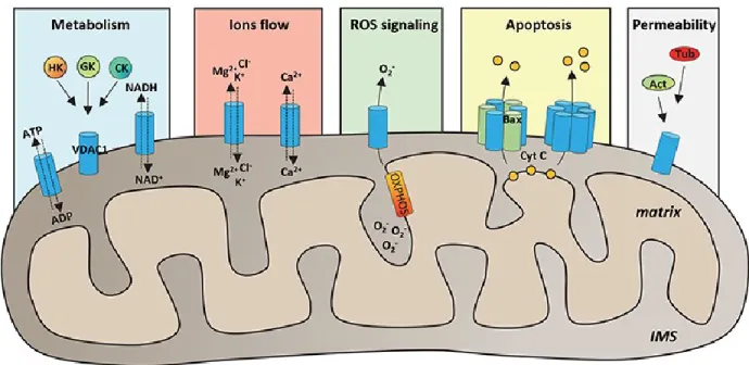
VDAC
The family of mitochondrial pores VDAC (Voltage-dependent Anion Channel) represents the most abundant protein class of outer mitochondrial membrane (OMM) among all the eukaryotes. The VDAC have a molecular weight of about 32 kDa and form aqueous pores that can work as a molecular filter for solutes from 3000 up to 5000 (Sampson et al., 1997; Schein et al., 1976); The VDAC was first studied around 40 years ago, when Schein et al detected the pore-forming activity in an extract of Paramecium tetraurelia mitochondria
20
(Sampson et al., 1998). Mammalians have three different isoforms known as VDAC1, 2 and 3. Although the structure of the genes is widely preserved the expression of the three isoforms varies depending on the tissue: for instance, isoforms VDAC1 and 2 are expressed mainly in heart, skeletal muscle, liver and brain whereas VDAC3 is expressed in testicles, liver, ovary, adrenal, lung, spleen and renal muscles. Over all, out of the three isoforms, VDAC1 is the most expressed and the most studied isoform, followed by VDAC2 and VDAC3 (Craigen and Graham, 2008; Yamamoto et al., 2006).
VDAC1 is composed by 19 β-strands, arranged to form a barrel, and by an α-helix at the N-terminal end facing the inside of the channel. The N-terminal is thought to be involved in maintaining the correct pore conformation and for the voltage dependent gating, which turns out to be the main characteristic of the VDAC.
Electrophysiological studies have shown that, VDAC1 can vary its conductance according to the applied voltage. At low voltage values, ranging from + 10 to -10 mV, VDAC1 shows a high conductance, and is in an open state; when the voltage reaches values higher than ± 30 mV, it adopts a closed conformation with inferior conductance (Mannella, 1997). Therefore, it is believed that the voltage of the mitochondrial membrane can influence the state of opening of the channel and thus the function of the mitochondria.
In line with his role in bidirectional traffic, VDAC1 allows the passage through the OMM of substrates such as pyruvate, malate, succinate, ions, ATP/ADP And NADH (Shoshan-Barmatz et al., 2010). Consequently, VDAC1 down-regulation results in the reduction of the exchange of metabolites between mitochondria and cytosol, which makes VDAC1 essential to produce energy and to the cell growth. Similarly, alterations of the mitochondrial function are related to the closure of the VDAC1, which limits the normal
21
flow of metabolites from inside and outside the mitochondria (Holmuhamedov and Lemasters, 2009; Vander Heiden et al., 2000). Due to its location, VDAC1 can interact with the proteins that mediate and regulate the integration of mitochondrial functions with other cellular activities: for instance, VDAC1 interacts with Hexokinase and Creatine kinase to convert ATP just generated into high energy reserve forms such as glucose-6-phosphate (G6P) and creatine glucose-6-phosphate, respectively in brain and muscles. The over-expression of VDAC1 in some cancer cells may be correlated to its multifunctional activities, as required by the demanding cells raised amount of energy (Shoshan-Barmatz et al., 2017). The set of functions of VDAC1 in the mitochondria are summarized in the figure 1.
22
SOD-VDAC interaction in ALS
In their work, Israelson et al. show a series of experimental evidence to indicate the link between SOD1 and VDAC1 's ALS-linked mutants as the main cause of mitochondrial dysfunction (Le Verche and Przedborski, 2010). In this study, in fact, it was seen that VDAC1, but not VDAC2, coimmuno-precipitates with the catalytically active mutants SOD1 G93A, and with those inactive, H46R and G85R in affected tissues and extracted from the transgenic mice; Conversely, the wild-type protein is not able to aggregate and adhere to the mitochondria. In addition, despite the brain tissue not showing pathology, contains abundant amounts of VDAC1 and SOD1 mutant, no evidence of an interaction between the two proteins was found, demonstrating that the interaction is tissue-specific. The authors suggest that this regional specificity could be related to the higher content in the brain of protein interacting with VDAC1, principally Hexokinase, which is less expressed in the spinal cord (Le Verche and Przedborski, 2010). Hexokinase can next compete with mutant SOD1 and drastically reduce the bond to VDAC1. Furthermore, electrophysiological studies have shown that mutants SOD1 proteins G93A e G85R, differently from the WT SOD1, reduce the ionic conductance of VDAC1. These functional data suggest that, in the presence of mutant SOD1, VDAC1 can be partially closed, and that this conformational state has a negative impact on normal mitochondrial functions.
From this study, it emerged an exciting new hypothesis: a mitochondrial channelopathy as basis of neurodegeneration that occurs in motor neurons affected by ALS. Since mutants of SOD1 ALS-linked do accumulate on the OMM, as a cause of alteration of mitochondrial functions this event could contributing to motor neuron’s degeneration in familial form of ALS.
23 ALZHEIMER’ S DISEASE
Alzheimer’s disease (AD) is the most common neurodegenerative disorder worldwide. Estimates indicate that more than 26 million people are living with AD and by 2050 the number of people with this disorder may reach 100 million (2016). Nowadays no effective cures have been found for AD patients that generally dye in 3 to 10 years after diagnosis. Aging is the major risk factor for the development of Alzheimer’s disease especially in the 95% of cases that are classified as sporadic and of unknown causes (Bekris et al., 2010). Sporadic form of AD normally occurs late in life and has it been shown that 33-50% of people older than 85 have AD (Bekris et al., 2010). Thought the years the average age of population is becoming higher and consequently the number of new AD cases is estimated to increase very quickly (Bird, 2008).
The remaining 5% of AD cases are familial form of AD (FAD) and are commonly triggered by mutations in one of three genes, presenilin 1 and 2 and amyloid precursor protein (APP, Querfurth and LaFerla, 2010). The presence of one of those mutation generally unsure the future insurgence of AD although several other mutations can be responsible of FAD. Notably, the presence of a specific form of ApoE gene (ApoE4) represent a major risk factor of AD and in many cases anticipate the age of onset in patents that have predisposition to the disease: this often leads to an earlier and higher severity of the pathology and so of the prognosis. Moreover, it’s possible to distinguish early onset and late onset AD cases and both can occur in a familial or sporadic form of the disease.
Clinically, one of the first symptoms of AD is the deficits in episodic memory (Welsh et al., 1992). As the disease progresses, other cognitive domains are involved and deficits in
24
language, executive functions, and learning tasks appear evident. Finally, patients become bedridden and perish due to other comorbidities. Two hallmarks of neuropathology in AD are plaques, mainly composed of amyloid-β (Aβ) peptide, and neurofibrillary tangles (NFT), mainly formed by hyperphosphorylated tau (Selkoe, 2001). The events triggering AD pathology and the molecular mechanisms linking aging to AD are not known.
PROTEIN AGGREGATION AND ACCUMULATION IN AD
Amyloid protein
A is as small peptide generated by proteolytic reaction from an integral membrane glycoprotein, the amyloid precursor protein (APP). In a physiological scenario, APP protein can be processed by two different pathways: non-amyloidogenic and amyloidogenic pathway (O’Brien and Wong, 2011). The non-amyloidogenic pathway requires a cleavage from -secretase, which produce a soluble N-terminal fragment (sAPPα) -released in the extracellular space- and a C-terminal fragment (CTFα) -cleaved to a membrane-bound C-terminal fragment and a soluble N-terminal fragment (p3) by a γ-secretase (Branca et al., 2014). Differently, the amyloidogenic pathway starts with the cleavage of APP at residue 99 by the β-site APP cleaving enzyme (BACE, Vassar et al., 1999) and the release of sAPPβ to the extramembrane space.
Next, a -secretase cleaves the intramembrane C99-terminal predominantly generating extracellular Aβ peptides and intracellular C-terminal domain. The -secretase cleavage most frequently happens at two different positions, generating either Aβ40 or Aβ42. The former is more common while the latter is more toxic (Chow et al., 2010).
25
In normal condition, at the extracellular space, Aβ binds to different isoforms ApoE protein: ApoE 2 and 3 leads more likely to his transportation to degradation pathways, whereas the bond to ApoE 4 seems to favorite the accumulation and aggregation of Aβ peptides (Kanekiyo et al., 2014).
Mutations in the APP gene and presenilins gene have been identified and several of these mutations lead to early-onset autosomal-dominant familial Alzheimer’s disease (eFAD), likely by increasing total Aβ production or by selectively increasing of the longer more amyloidogenic Aβ42 species (Ryan and Rossor, 2010). Unbalance on BACE1 expression, Aβ-EpoE interaction and neprilysin levels have been shown to be positively correlated with plaques deposition and severity of the disease (Tarasoff-Conway et al., 2015).
Aβ pathological role
It has been shown as Aβ peptide is able to create different conformational states with different fibrillary structures in in vitro experiments (Cheon et al., 2016). This capacity in vitro is confirmed also on experiments with Aβ purified from human brains: the structure of Aβ fibrils was different according to the AD phenotype of different patients (Condello et al., 2018).
When Aβ conformers of sporadic and familial AD and brain-derived plaques were inoculated in transgenic mice, AD pathology on those mice changed in speed, propagation, and plaque phenotype: this yield to suppose that Aβ conformation is responsible of different state of propagation and correlates with the cognitive impairment (Sengupta et al., 2016). These observations also correlate with the data suggesting that Aβ role in
26
synaptic toxicity: Aβ peptides are able to inhibit long-term potentiation (LTP) and reduce synaptic plasticity (Li et al., 2011) possibly causing the reduction of cognitive ability in AD. Moreover, the inhibition of LTP by Aβ produce the induction of hippocampal long-term depression (LTD) causing changes in changes in postsynaptic responsiveness or excitability (Bliss and Cooke, 2011).
Tau protein
The MAPT (microtubule associated protein tau) is a phosphoprotein mostly present in axons to stabilize the microtubule structure (Drubin and Kirschner, 1986) . Tau is a major component of NFTs characterizing AD brains and their accumulation is related to neuronal loss and cognitive deficits (Mandelkow and Mandelkow, 2012).
In human, tau gene is located on chromosome 17 and is composed by 15 exons: his alternative splicing at exons 2, 3 and 10 in the human brain leads to existence of six isoforms (Liu and Gong, 2008). According to both the number of imperfect tandem repeated sequences and number of inserts at N-terminal we distinguish zero, one, or two inserts of 29 amino acids at the N-terminal part (exon 2 and 3 - 0N, 1N and 2N), and three or four repeat-regions at the C-terminal part (exon 10-3R, 4R) (Kadavath et al., 2015).
No mutations of tau gene have been found in AD although mutant tau protein is present in several disease such as FTD and Pick’s disease as prove that changes in tau protein are linked to neurodegeneration and cognitive impairment (Iqbal et al., 2010). In AD brains, tau is phosphorylated to a higher degree at physiological sites and is also phosphorylated in additional sites that are defined specific of AD pathology.
27
Pathologically phosphorylated and hyperphosphorylated tau detach from microtubules and accumulates as paired helical filaments (PHFs) and NFTs (Alonso et al., 2006). Those pathological sites include pSer422, pThr212/pSer214 (AT100), and pThr231/pSer235 (AT180, Xia et al., 2015).
Thus, despite there is not genetic association between tau and AD onset, extracellular accumulation of tau surrounded by reactive microglial does significantly drive AD pathology especially in early states of the disease.
Interaction of A and tau in AD
As the amyloid cascade hypothesis says, A deposit may be the first responsible of triggering AD contributing to tau accumulation as NFT and causing neuronal death (Rajmohan and Reddy, 2017)). Despite many evidences support this hypothesis, the molecular mechanisms linking the two pathological accumulation is still unknown
.
When Lewis et al. (2001) crossed two independent transgenic lines, one overexpressing mutant APP and the other overexpressing mutant tau, the double transgenic mice developed A pathology at the same age as the single APP transgenic mice but showed enhanced tau pathology. This led the authors to conclude that either APP or A influences tau pathology in vivo. Many in vitro studies have proposed tau as downstream mediator of A toxicity in primary neuronal cultures: for instance, Takashima and colleagues reported a role for a GSK3β homologous as TPK1 since treatment of primary hippocampal cultures with A causes an increase of his activity and correlates with the A-induced neurotoxicity (Takashima, 2006).28
As A-treated neurons were also positive for Alz50, a conformational specific tau antibody, (Yankner et al., 1989) and TPK1 is linked to phosphorylation of tau (TAKASHIMA et al.), this observation suggests that A can induce tau pathology via TPK1. The involvement of GSK3β in A-induced toxicity has been confirmed by other studies (Hooper et al., 2008; Llorens-Marítin et al., 2014; Takashima, 2006) that show that A can enhance the activity of different kinases that phosphorylate tau.
By use of tau knock-out mice it has been proved that A toxicity needs the presence of tau protein: when treated with A, hippocampal neurons from wild-type mice or from mice expressing human tau showed neuronal degeneration in 96h while neurons lacking tau did not showed degenerations (Rapoport et al., 2002).
It was also observed that A can induce tau formation of filaments via activation of caspase 3 protein: the latter is able to cleave tau at his C-terminal, producing a more toxic form of tau (delta-tau) which is also found in NFT (Gamblin et al., 2003; Noble et al., 2013) and is increased in as the cognitive impairment in AD.
Finally, some observations have been done on the role of both calcium and mitochondria on Aβ-tau-mediated spine-loss and cognitive deficits: Aβ induced-dendritic spine loss correlates positively with presence of missorted tau, increased calcium levels and impaired mitochondrial distribution(Spires-Jones and Hyman, 2014; Zempel et al., 2010).
Mouse models of Aβ and tau
The use of mouse model represents one of the best tools of research in AD despite none of them recapitulate the full scenario of the disease. After the APP gene was isolated,
29
several groups have attempted to generate transgenic models of AD (Higgins et al., 2002; Mucke et al., 1994; Quon et al., 1991).
The first successful model of A pathology was the PDAPP mice where levels of the human APP were more than 10-fold over endogenous mouse APP levels (Elder et al., 2010). At mid-age these mice develop A deposits which increase as the mice age and are distributed among hippocampus, corpus callosum and cerebral cortex. The PDAPP mice also develop behavioral deficit, neuritic plaques, synaptic loss and gliosis (Jacobsen et al., 2006; Schaeffer et al., 2011).
Another widely used mouse-model, the Tg2576 mice overexpressing APP, develop A deposits by 9-10 months as well memory impairments in an age-related manner (Dineley et al., 2002; Westerman et al., 2002). Most of the APP animal models develop A pathology especially represented by accumulation of the longer and toxic form (A42): the presence of the A42 is indeed negatively correlated with cognition levels much more than A40 peptide (Murphy and LeVine, 2010).
Differently of what happen in human brain though mice model exclusively expressing mutant APP don’t show NFT deposition. The double transgenic APP/PS1 mice show an earlier onset of Aβ pathology which indicate the importance of mutations at PS genes which is still is not sufficient to generate NFT in those mice (Hall and Roberson, 2012). AD pathology is not genetically referred to tau mutations, although other disease like FTDP-17 are characterized by NFT deposition due to a mutation known as tauP301L (Ramsden et al., 2005).
30
Many groups of researchers have been working on generating mice overexpressing the human mutant tau gene to better study NFT formation and their involvement in AD (Lewis et al., 2001). Gotz and collaborators in 2001 overexpressed the human tau P301L using the Thy1.2 promoter successfully observing the formation of NFT in hippocampus and neocortex. However, those mice don’t show changes in cognition indicating that tau mutation and NTF accumulation is not sufficient to generate cognitive neither A pathology: at that point was clear that both tau and A pathology are needed to recreate AD-like pathology.
To this end, both groups Gotz et al and Lewis et al., with different approaches, generated transgenic mice with APP ant tau mutations: although as these mice show intense motor deficits and die prematurely was not possible to use them in behavioral tasks. To obtain a model with both plaques and tangles Oddo and colleagues generated the 3xTg-AD mice: these transgenic mice harbor three mutant human genes, APPSwe, Tau P301L, and PSN1 M146V. Extracellular plaques and NFT tangles deposition appear in early stages and increase as the mice age: by 12 months of age those mice show a full- AD like pathology comprehensive of synaptic dysfunction, LTP deficits and cognitive impairments. These changes are associated with selective neuroinflammation and progressive cognitive impairments have also been widely reported. These mice are being used by over 100 investigators throughout the world, which has led to the generation of multiple independent colonies.
Converging evidence indicates that the phenotype of 3xTg-AD mice has shifter over the years and contradicting reports about onset of pathology or cognitive deficits are apparent in the literature (Hebda-Bauer et al., 2013; Montacute et al., 2017; Oh et al., 2010).
31
Here I sought to stage the current progression of AD-like pathology in 3xTg-AD mice. The data obtained will facilitate the design of preclinical studies in which these are used to test new therapeutic approaches.
GLIAL REACTIVITY IN AD
One of the hallmark of AD pathology is represented by astrogliosis and microgliosis (Serrano-Pozo et al., 2011). The CNS is indeed involved in a series of inflammatory events that in a normal scenario are physiologically balanced to protect the neuronal component from external or internal pathological stimuli (Kyritsis et al., 2014).
Astrocytes in AD brains show a wide reactivity which manifests as increased expression of glial fibrillary acidic protein (GFAP) protein and are mostly distributed around amyloid plaques as their main role is neuroprotection and delimitation of damaging deposits(Sofroniew and Vinters, 2010). They also contribute to ATP, glutamate and GABA trafficking, calcium homeostasis, regulation and release of inflammatory cytokines and grow factors (Verkhratsky et al., 2016). Indeed, elevated levels of proinflammatory cytokines as TNFα, LPS, IFNγ and IL-1β appear in human brains and several mice models of Alzheimer’s disease (Rubio-Perez and Morillas-Ruiz, 2012).
In the same way of the astrocytes, microglial cells in CNS are activated by toxic triggers and have been shown to surround both amyloid plaques and NFT in AD brains (Solito and Sastre, 2012). Intrigant questions regard the function of gliosis in neurodegeneration and point the attention to a very delicate balance between protective and toxic roles of astrocytes and microglia activation.
32
Specific founding show that an initial reactivity is necessary for protection against amyloid deposits and in a pathological scenario, as the brain age, an insufficient response from glial cells leads to an overload of amyloid triggers (Grolla et al., 2013; Liddelow and Barres, 2017; Sokolowski and Mandell, 2011).
On the other side, an excessive inflammatory response can be itself the cause of acceleration of the progression of neuronal loss in AD pathology (Clayton et al., 2017). For instance, studies have shown that microglia and astroglia activate each other in a continuous loop that bring to increased APP and BACE1 expression and Aβ production in astrocytes (Heneka et al., 2005; Zhao et al., 2011).
This suggest that the balance between proinflammatory and anti-inflammatory response plays a main role in aging related disease like AD to avoid neuronal death and behavioral deficits (Sochocka et al., 2017).
33
34
Scientific Reports | 6:34802 | DOI: 10.1038/srep34802
Hexokinase I N-terminal based peptide prevents the VDAC1-SOD1
G93A interaction and re-establishes ALS cell viability
Andrea Magrì1,2,3, Ramona Belfiore2,3, Simona Reina1,2,3, Marianna Flora Tomasello4, Maria Carmela Di Rosa1,2, Francesca Guarino2,3, Loredana Leggio1,2, Vito De Pinto2,3 & Angela Messina1,2
1 Department of Biological, Geological and Environmental Sciences, Section of
Biochemistry and Molecular Biology, University of Catania, Italy.
2 National Institute of Biostructures and Biosystems (INBB), Italy.
3 Department of Biomedical and Biotechnological Sciences, University of Catania,
Italy.
4 CNR Institute of Biostructures and Bioimaging, Catania, Italy.
Abstract
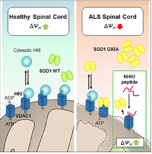
Superoxide Dismutase 1 mutants associate with 20–25% of familial Amyotrophic Lateral Sclerosis (ALS) cases, producing toxic aggregates in mitochondria, notably in spinal cord. The Voltage Dependent Anion Channel isoform 1 (VDAC1), in the outer mitochondrial membrane, is a docking site for SOD1 G93A mutant in ALS mice and the physiological receptor of Hexokinase I (HK1), which is poorly expressed in mouse spinal cord. Our results demonstrate that HK1 competes with SOD1 G93A for binding VDAC1, suggesting that in ALS spinal cord the available HK1-binding sites could be used by SOD1 mutants for docking mitochondria, producing thus organelle dysfunction. We tested this model by studying the action of a HK1-N-terminal based peptide (NHK1). This NHK1 peptide specifically interacts with VDAC1, inhibits the SOD1 G93A binding to mitochondria and restores the viability of ALS model NSC34 cells. Overall, our results suggest that NHK1 peptide could be developed as a therapeutic tool in ALS, predicting an effective role also in other proteinopathies.
35
Introduction
Amyotrophic Lateral Sclerosis (ALS) is a fatal neurodegenerative disease characterized by the progressive degeneration of both upper and lower motor neurons [1] (MNs). Over 160 missense mutations in the Superoxide Dismutase 1 (SOD1) gene account for 20–25% of familial ALS cases [2], causing MNs death by accumulation of mutant SOD1 (mutSOD1) insoluble toxic aggregates [3]. Interestingly, mutSOD1 aggregates associate with the mitochondrial cytoplasmic side, especially in spinal cord MNs, producing mitochondrial failure [4,5]. Despite it is well known that mitochondria play a central role in bioenergetics metabolism, oxidative stress, apoptosis and axonal transport, the intimate underlying mechanism linking mitochondrial dysfunction in MNs of ALS patients or mice to mutSOD1 still remains elusive. Moreover, it is not yet well understood why MNs are more susceptible to the disease in comparison to other tissues. A previous report showed that, only in the ALS rat spinal cord, mutSOD1 bind directly to the Voltage Dependent Anion Channel isoform 1 (VDAC1), reducing its channel activity [6]. VDAC1 is considered the master regulator of the mitochondria thanks to its crucial action of gate for metabolic and energetic substrates of the organelle [7,8]. Moreover, VDAC1 is the physiological receptor of Hexokinases [9] (HKs). HKs catalyze the glucose phosphorylation and, by binding to VDAC1, they gain a preferential access to newly synthesized ATP. Furthermore, mitochondrial-bound HKs protect the cell from apoptosis, since they diminish VDAC1 propensity to interact with pro-apoptotic protein Bax [10,11]. Interestingly, reduced levels of HK1 were detected in spinal cord, compared to the brain or to other tissues [12]. Therefore, high levels of mutSOD1 binding to VDAC1 correlate with low levels of HK1 in spinal cord. Based on these evidences, we have hypothesized
36
that in ALS a reduction of HK1 concentration increases VDAC1 propensity to interact with mutSOD1, producing thus mitochondrial dysfunction and cell death.
In this work, we demonstrate the intrinsic ability of SOD1 G93A, but not SOD1 wild type (SOD1 WT), to interact with VDAC1 and to compete with HK1 for binding VDAC1. We also show that a synthetic peptide, corresponding to the HK1 N-terminal region (NHK1 peptide) is able to interact with VDAC1, in in vitro and in cellulo, modifying its channel conductance. In addition, the NHK1 peptide inhibits the VDAC1-SOD1 G93A interaction in mitochondria purified from NSC34, a mouse motor neuron-like hybrid cell line. Moreover, the expression of the NHK1 peptide in NSC34 cell stably expressing SOD1 G93A, a recognized ALS cell model, recovers the mitochondrial malfunctioning linked to mutSOD1, and largely contrasts the cell death. Overall, our data suggest that VDAC1 and HK1 play a key role in the bioenergetics metabolism of the MNs and could be considered as a promising therapeutic target in ALS.
Results
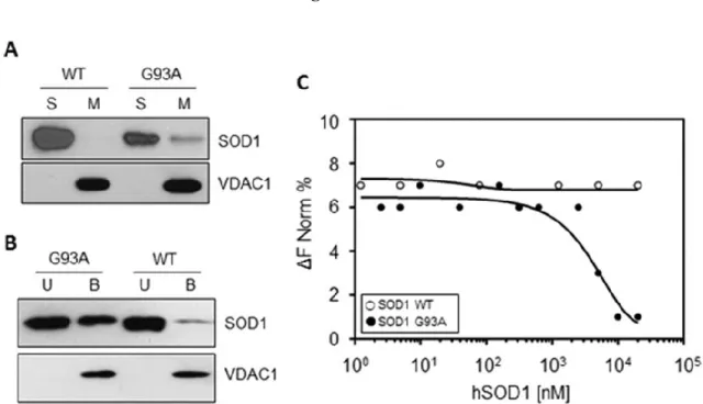
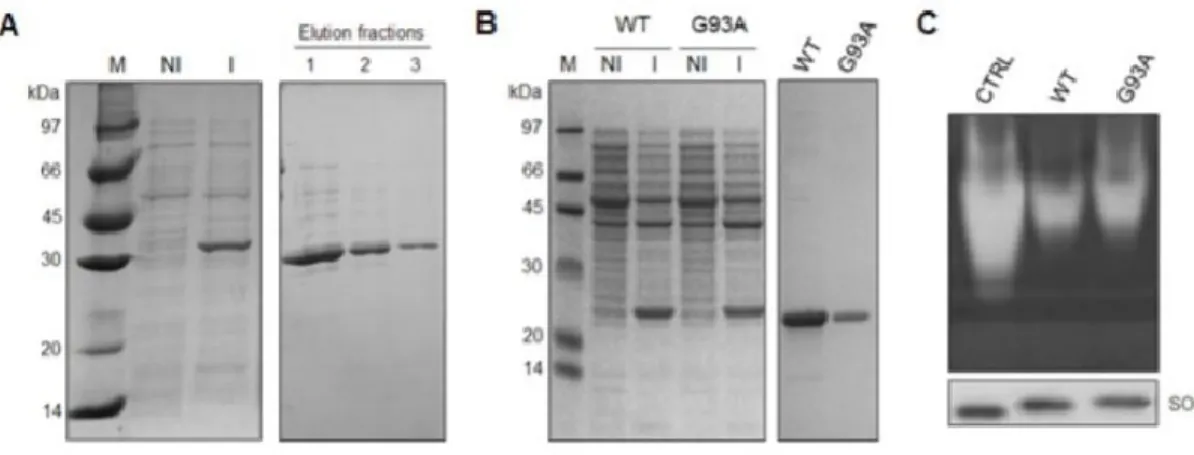
SOD1 G93A, at variance with SOD1 WT, binds VDAC1 with high affinity. The
interaction between VDAC1 and SOD1 G93A in ALS model rat was reported [6] and later questioned [13]. To validate our experimental plan, recombinant human VDAC1, SOD1 WT and SOD1 G93A were expressed, purified and refolded (Fig. S1), and the affinity of SOD1 proteins for mitochondria or VDAC1 was checked by means of several approaches. Intact isolated mitochondria from the motor neuron-like NSC34 cells were incubated with SOD1 WT or G93A, and mitochondrial membranes were precipitated by centrifugation. Then, the SOD1 concentration was revealed by western blot in mitochondrial membranes
37
or in the supernatant fraction, using VDAC1 as a loading control. Results in Fig.1A show that, while SOD1 WT was found exclusively in the supernatant, a fraction of SOD1 G93A co-precipitated in the mitochondrial pellet. According to the literature [3,6], this data indicates that the recombinant SOD1 G93A, but not SOD1 WT, binds the mitochondria surface. The affinity of SOD1 proteins for VDAC1 was then studied by using an in vitro binding assay. Purified and refolded VDAC1 was immobilized on Ni-NTA magnetic beads and incubated with SOD1 proteins. Then, VDAC1-binding complexes were isolated by the application of a magnetic field. Figure 1B shows that SOD1 G93A was found distributed between VDAC1-bound and -unbound fraction, while SOD1 WT was almost exclusively in the unbound fraction. The VDAC1-SOD1 interaction was quantitatively assayed by Microscale Thermophoresis (MST) analysis. MST measures any variation in the thermal migration of a fluorescently labeled binding partner; changes of fluorescence in a heated spot of the protein solution is a function of increasing interacting protein concentration and can be exploited to calculate the binding affinity coefficient (Kd). The fluorescent-labeled VDAC1 was incubated with increasing concentrations of SOD1 proteins and the changes in fluorescence monitored. Again, as shown in Fig.1C, while no fluorescence change was visible in the presence of SOD1 WT, the incubation with growing concentrations of SOD1 G93A produced fluorescence decrease, indicating that SOD1 G93A specifically interacts with VDAC1. Depletion curve was used to calculate the Kd, which was estimated 4,81 μM.
Overall, the results showed here indicate that the mutant SOD1 G93A specifically interacts with the cytosolic surface of purified mitochondria and with the purified VDAC1
38
with high affinity. The SOD1 WT is instead unable to bind mitochondria and VDAC1, confirming the data in the literature [6].
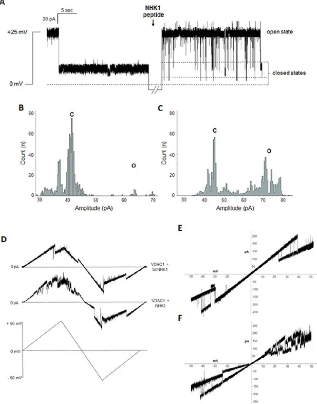
SOD1 G93A modulates VDAC1 channel activity. A most relevant VDAC feature is the
voltage dependence [14]. VDAC1 is characterized by a typical conductance of 4 nS in 1 M KCl, at low positive or negative voltages (± 10 mV). In these conditions, the channel stays stably in an open and high-conducting state. However, raising the voltage, already at ± 20–30 mV, VDAC1 switches rapidly to a closed and low-conducting state, where it can remain for quite a long time [14–16]. The ability of SOD1 proteins to interfere with VDAC1 activity was analyzed in terms of conductance perturbation. Purified VDAC1 was reconstituted into a planar phospholipid bilayer (PLB) and its channel conductance was monitored before and after addition of SOD1 proteins, on cis or trans side of the membrane. Fig. 2A shows a typical record of ion current through a single VDAC1 channel, in 1 M KCl and an applied potential of + 25 mV. In these experimental conditions, the addition of SOD1 WT did not modify the VDAC1 closed state. Conversely, the addition of SOD1 G93A, on the cis side of the membrane, promoted VDAC1 channel instability: the conductance switched from the stable low-conductance state to several high-conducting states, indicating a specific interaction between the two proteins. Moreover, the effect of SOD1 proteins on the voltage-dependence of VDAC1 was monitored by triangular voltage ramps from 0 to ± 50 mV. The upper curve in Fig.2C shows the typical VDAC1 voltage response. The current linearly follows the voltage applied up to about ± 25 mV, where VDAC1 decreases its channel conductance through step-like transitions, remaining in low-conducting states. A perturbation of voltage-dependence is exclusively visible when SOD1 G93A interacted with VDAC1 (lower
39
curve, Fig.2C), while no modification in the usual VDAC1 pattern was noticeable upon addition of SOD1 WT (middle curve, Fig.2C). In conclusion, VDAC1 loses its ability to linearly respond to the voltages applied in the presence of the mutant SOD1 G93A.
SOD1 G93A competes with Hexokinase I (HK1) towards binding-site(s) on VDAC1.
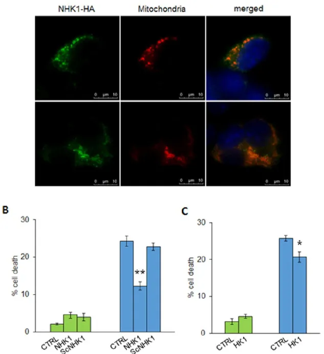
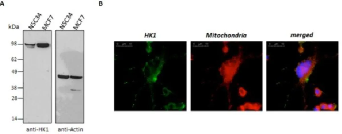
It is well known that HKs bind to VDAC1 in physiological conditions [9]. We hypothesized that the available area for interaction with soluble proteins is a limited, exposed portion of the transmembrane pore VDAC1. This hypothesis suggests that there could be a competition between different proteins towards the same, or close, site(s) of the VDAC1. For this reason, we repeated the VDAC1-SOD1 G93A binding assay, adding increasing concentrations of HK1. We found that SOD1 G93A binds VDAC1 (Fig.3A): the addition of HK1 strongly decreased (about 40%, Fig.3B) the SOD1 G93A bound to immobilized-VDAC1 (Fig.3A), despite the concentration of SOD1 G93A was overwhelming the stoichiometry of VDAC1 binding site(s). The reduction corresponds to the increase of HK1 in VDAC1-bound fraction, and it is proportional to the HK1 concentrations added in the assay (Fig.3A). Therefore, in such experimental conditions, HK1 is able to impair the VDAC1-SOD1 G93A interaction, suggesting a competition of the two proteins for the same binding site(s). The interference of HK1 in the VDAC1-SOD1 G93A interaction was further investigated in NSC34 cells stably transfected with inducible human SOD1 G93A (NSC34-SOD1G93A), a recognized ALS cell model, and compared with SOD1 WT (NSC34-SOD1WT) expressing cells [17]. We previously controlled that the motor-neuron NSC34 cell line contain a low level of total HK1 in comparison to other cells, as shown in Fig. S2. HK1 mostly localizes to mitochondria (Fig. S2). NSC34-SOD1G93A or NSC34-SOD1WT cells were then transiently
40
transfected with increasing concentrations of constructs encoding HK1-GFP and the measured fluorescence was related to the mitochondria-bound HK1-GFP. The expression of HK1-GFP was also controlled in NSC34 cells (see Fig.S3), where HK1-GFP localization was also analyzed by fluorescence microscopy. As expected, in NSC34-SOD1WT cells, most of HK1-GFP localized to mitochondria, as demonstrated by the typical punctuated staining (Fig.3C). However, in NSC34-SOD1G93A cells the HK1-GFP signal became diffused, indicating a partial shift towards the cytosol (Fig.3C). A quantification of the mitochondria-related fluorescent signal was obtained after a limited permeabilization with digitonin18 of NSC34 cells expressing SOD1 WT or G93A. In this experiment, the cytosolic GFP fluorescence was allowed to leave the cell and the fluorescence retained inside the cell was assumed to be due to the mitochondria-associated HK1-GFP. Indeed, the permeabilization of NSC34-SOD1WT or G93A stable cell lines, transfected also with GFP, promoted the loss of the signal, in any tested condition. Conversely, the signal was strongly retained (60–70%) in NSC34-SOD1WT expressing also HK1-GFP (Fig.3D), indicating that HK1-GFP binds to mitochondria, in proportion to the added concentration. Interestingly, in NSC34-SOD1G93A, HK1-GFP in the cell was reduced (30–40% of the control). Therefore, the HK1-GFP ability to bind mitochondria was clearly reduced in the presence of SOD1 G93A, but not of SOD1 WT (Fig.3D). Considering that HK1 binds mitochondria exclusively docking VDAC1, this result supports our hypothesis that HK1 and SOD1 G93A compete for the same VDAC1 binding site/s.
A N-terminal HK1-based peptide interacts with VDAC1 and modulates its channel conductance. It is well known that the N-terminal end of HK1 is responsible of the
41
enzyme interaction with VDAC1 and modulates its channel activity [19–21]. To evaluate the ability of HK1 N-terminus to interfere with the binding between VDAC1-SOD1 G93A, we produced a synthetic NHK1 peptide, corresponding to the first 11 amino acids of human HK1. The electrophysiological behavior of human VDAC1, reconstituted into a PLB, was monitored before and after addition of NHK1 on the cis or trans side of the membrane. Under standard experimental condition (1 M KCl and an applied potential of + 25 mV) the VDAC1 channel was mainly in low-conducting closed states. The addition of NHK1 on one side of the membrane induced several fast and reversible events of VDAC1 low-conducting closed states (reported in Fig.4A). The effect of NHK1 on the channel conductance is even more evident by plotting the amplitude values obtained in the current traces as a function of the number of events. While for VDAC1 alone the amplitude values appear as a main peak corresponding to the closed states (Fig.4B), the presence of NHK1 caused a different distribution of events: an additional, different peak corresponding to the open state was visible, together with the closed states peak (Fig.4C). No influence on the electrophysiological activity was instead detected upon addition of a scramble peptide (ScNHK1) used as control (data not shown). The effect of NHK1 on the voltage-dependence of VDAC1 was monitored by triangular voltage ramps. Again, while no difference was found in presence of ScNHK1 (upper curve, Fig.4D), the presence of NHK1 strongly affected the channel voltage-dependence of VDAC1 (lower curve, Fig.4D). In this experiment, indeed, VDAC1 completely loose, very precociously (already at ± 15 mV applied), its ability to linearly respond to the applied voltage: a continuous switch from open to closed states was observed, especially at positive potentials. Similarly, the current vs. voltage (I–V) plot shows that in the presence of only VDAC1,
42
I–V plot is linear in the voltage range ± 10–25 mV, and the current transitions (slope), corresponding to decreased conductance states, appeared outside this range (Fig.4E). Upon NHK1 incubation, VDAC1 current transitions at positive voltages are very noisy and pass through different sub-conductance states (Fig.4F). Notably, at negative membrane potentials, regions of decreased slope appeared already at −10 mV, suggesting that NHK1 peptide raises VDAC1 sensitivity to negative voltage applied. No similar result, instead, was observed in the presence of ScNHK1 (data not shown). The electrophysiological analysis strongly indicates the ability of NHK1 peptide to interact specifically with VDAC1 and to modulate its channel conductance and voltage-dependence.
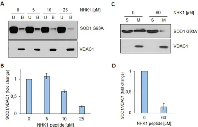
NHKI peptide highly impairs the interaction between SOD1 G93A and VDAC1. To
evaluate the NHK1 ability to interfere with VDAC1-SOD1 G93A interaction, a binding assay was performed. Increasing concentrations of NHK1 were added to immobilized-VDAC1, before SOD1 G93A addition. Results in Fig.5A clearly show that, in the presence of 10 and 25 μ M of NHK1 peptide, the amount of SOD1 G93A in VDAC1-bound fraction was reduced by the 40% and 80%, respectively (Fig.5B). No similar effect was observed by repeating the assay in presence of ScNHK1. This result indicates that, in vitro, NHK1 strongly impairs the interaction between VDAC1 and SOD1 G93A in a more effective way than the whole HK1 protein. In fact, a 20-fold smaller concentration of peptide was used to obtain a result similar to that obtained by using the whole HK1 in the previous experiment (see Fig.3A). The ability of NHK1 to inhibit SOD1 G93A from binding the mitochondrial surface was investigated using intact mitochondria purified from NSC34 cells. Mitochondria were incubated with SOD1 G93A, in the presence or absence of 60
43
μM NHK1 peptide. Figure 5C shows that SOD1 G93A was distributed between the supernatant and mitochondrial fractions, in the absence of peptide. Following incubation with NHK1, a dramatic decrease of 85% SOD1 G93A bound to mitochondria was seen (Fig.5D). Again, no effect was found by incubating mitochondria with the ScNHK1 peptide. These results prove that NHK1 peptide strongly hinders the SOD1 G93A binding to purified VDAC1 or mitochondria.
NHK1 peptide restores the NSC34-SOD1G93A cells viability counteracting the mitochondrial dysfunction. In order to analyze the effect of NHK1 peptide in our
ALS-like in cellulo system, the subcellular distribution of NHK1 peptide was investigated. NSC34 cells were transiently transfected with a plasmid encoding for a HA-tagged NHK1 peptide and for a mitochondrial-targeted Red Fluorescent Protein (mtDsRED) [18]. Immunofluorescence assays in Fig.6A revealed that NHK1 peptide mainly co-localized with mtDsRED signal, indicating its ability to bind mitochondria in NSC34 cells. Then, the NHK1 peptide, as well as the whole HK1 as control, was expressed in NSC34-SOD1G93A or SOD1WT to evaluate its ability to counteract the toxicity mediated by mutSOD1. Literature reports indicate that SOD1 mutant expression in NSC34 cell promote a significative loss of cell viability, accordingly with the specific mutation [22]. Our results show that, upon expression of SOD1 G93A, NSC34 cells loose about 20% of cell survival (Fig.6B). However, this toxic effect was counteracted by the NHK1 expression: an improvement of cell viability, (about 50% of the control, Fig.6B) was observed; furthermore, a similar but lower effect was also found upon overexpression of the whole HK1 (Fig.6C). Therefore, supplementation of NHK1 peptide (or HK1) to NSC34-SOD1G93A cells promotes a recovery of the cell viability. To evaluate the NHK1
44
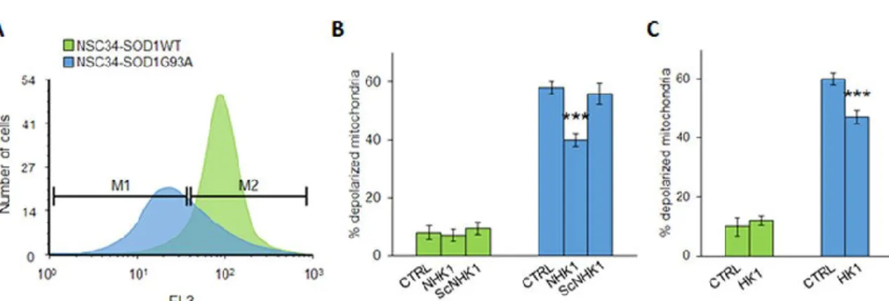
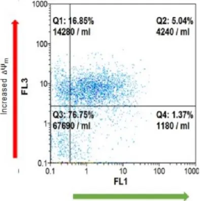
influence on mitochondrial functionality, mitochondrial membrane potential variation (ΔΨ m) was assayed in the presence of NHK1 peptide or HK1. ΔΨ m is related to the ATP production by oxidative phosphorylation; therefore, it is considered an indication of good mitochondrial and cellular health [23,24]. Using mitochondria-targeted fluorescent probes, we estimated in flow cytometry the rate of mitochondrial depolarization. SOD1G93A cells shown a high level of depolarized mitochondria compared to SOD1WT. Indeed, as showed in Fig.7A, the emission peak of fluorescence for NSC34-SOD1G93A cells was significantly lower compared to the NSC34-SOD1WT peak. The uptake of the probe into mitochondria is ΔΨ m-dependent: thus, this result means that SOD1 G93A expression strongly affects the cell energetic metabolism. A quantification of fluorescent-negative NSC34-SOD1G93A cells, corresponding to the depolarized mitochondria rate, was performed as previously reported [24], and resulted in a dramatic increase of about 60%, compared to NSC34-SOD1WT (Fig.7B). In this dramatic situation, the NHK1 expression, but not ScNHK1, resulted in a partial recovery of the physiological ΔΨ m, since depolarized mitochondria were reduced by 20% in NSC34-SOD1G93A cells (Fig.7B, S4).
Similarly to NHK1 peptide, the expression of the whole HK1 reduced the depolarized mitochondria of about 15% (Fig.7C). In conclusion, the NHK1 peptide, and minimally the whole HK1, is able to partially recover the mitochondrial functionality and, consequently, the cell vitality in the ALS model NSC34 cells.
45
Discussion
A previous report showed that mutSOD1 interact with VDAC1 in spinal cord mitochondria from ALS model rat6. In our work we preliminary confirmed that the mutant SOD1 G93A, but not the SOD1 WT, is able to interact with VDAC1 immobilized on magnetic beads. Furthermore, we determined, by MST analysis, the binding affinity of SOD1 G93A with VDAC1 and the effect of SOD1 G93A on VDAC1 conductance. Our electrophysiological data showed that, at the voltages applied which stably close VDAC1, SOD1 G93A, but not the SOD1 WT, promotes a prolonged instability of VDAC1 conductance. On the other side, Israelson and coworkers demonstrated that addition of SOD1 G93A to PLB-reconstituted VDAC1, at the voltages applied which stably maintain VDAC1 in an open state, promotes a partial closure of VDAC1 channels [6]. Therefore, both results strongly support a direct effect of mutated SOD1 G93A on VDAC1 conductance. From the literature and from these convincing results we hypothesized that VDAC1 could be the specific docking site on the OMM for the SOD1 G93A, and possibly for all SOD1 mutants. The influence of the interaction in the gating features of VDAC1 could indeed explain the mechanism of impairment of the bioenergetics metabolism and the oxidative stress of the ALS MNs [6]. In this perspective, the physiological interactions involving VDAC1 in spinal cord MNs could be altered in ALS, giving thus an explanation for the specific susceptibility to the disease showed by this tissue. Another recent report showed that SOD1 G93A, but unexpectedly also SOD1 WT, preferentially bound to Bcl2, rather than VDAC1, in ALS mitochondria [13]. The binding promoted a conformational change of Bcl2 that, in turn, altered its physiological interaction with VDAC1, producing mitochondrial dysfunction [13]. This means that SOD1 G93A might bind VDAC1 or