3
Table of Contents:
Abstract
5
Riassunto
8
1 Introduction
11
1.1 Parkinson’s Disease 11 1.1.1 Etiology 11 1.1.2 Pathophysiology 11 1.1.3 Therapy 14 1.2 Experimental models of PD 17 1.2.1 Animal models 18 1.2.1.1 Neurotoxin-Induced PD Models 18 1.2.1.2 Transgenic PD Models 18 1.2.2 In vitro models 20 1.3 Genetic bases of PD 23 1.3.1 LRRK2 23 1.3.2 G2019S LRRK2 mutation 25 1.4 DA neurons vulnerability 271.4.1 Dopamine D2 and D3 Receptors (D2R)/(D3R) 27
1.4.2 Nicotinic Acetylcholine Receptors (nAChRs) 29
1.4.3 D3R-nAChR Heteromerization 30
1.5 Neuroinflammation 33
2 Aim of the project
37
3 Materials and methods
39
3.1 Animals 39
3.2 Primary cultures of midbrain neurons and treatments 39
3.3 Morphological analyses of mouse and human DA neurons 39
3.4 HEK293 cultures, transfection and treatments 40
3.5 Neuronal differentiation from human iPSC lines 41
3.6 Western blot 42
3.7 Immunofluorescence analyses 42
3.8 Endogenous DA release 42
4
3.10 RT-PCR 43
3.11 Differentiation of iPSC-derived astrocytes and treatments 44
3.12 Astrocytes immunofluorescence analyses 45
3.13 Human astrocytes calcium imaging analyses 45
3.14 Neurons/astrocytes co-cultures experiments 45
3.15 Statistical Analysis 46
4 Results
47
4.1 The D3R-nAChR heteromer 47
4.1.1 Role of D3R in the D3R-nAchR heteromer 47
4.1.2 D3R-nAChR heteromer signaling properties 50
4.2 Neuronal differentiation from human induced pluripotent stem cells 55
4.2.1 Morphological and functional analysis of DA neurons derived from
LRRK2-G2019S iPSC 55
4.2.2 DA neurons from LRRK2 G2019S are resistant to the neurotrophic
effects of nicotine and D3R preferring agonists 59
4.2.3 Altered membrane localization of D3R and nAChR in DA neurons
derived from LRRK2-PD iPSC 61
4.2.4 Normalizing LRRK2 activity rescues D3R-nAChR heteromer formation and localization at the plasma membrane and restores the neurotrophic
effects of ropinirole and nicotine. 64
4.3 Astrocyte differentiation from human induced pluripotent stem cells 68
4.3.1 Differentiation and characterization of human iPSC-derived astrocytes 68 4.3.2 Differentiation and characterization of iPSC-derived astrocytes from
LRRK2 G2019S PD 72
4.3.3 Functional characterization of iPSC-derived astrocytes from
LRRK2 G2019S PD patients 74
4.3.4 Dopamine D2 receptor expression and localization in iPSC-derived astrocytes 75
5 Discussion
80
6 References
89
5
Abstract
Dopamine (DA) plays a fundamental role in the regulation of different physiological functions including motor activity, cognition, learning, reward mechanisms and working memory. The functional role of DA is such that the progressive degeneration of DA neurons underlies the development of Parkinson’s disease (PD). The effects of DA are mediated by five receptors (D1-D5) that differ for structure, pharmacology and transduction signalling (Missale et al., 1998). One of the characteristics of DA receptors is their propensity to directly interact with other receptor subtypes to form heteromers with peculiar transductional, functional, and pharmacological properties (Bono et al., 2020). We recently reported that in mouse dopaminergic (DA) neurons, the dopamine D3 receptor (D3R) and the nicotinic ion channels (nAChR) interact to form a heteromeric complex (D3R-nAChR heteromer) that exerts neurotrophic effects when activated by nicotine, leading to neurons with enlarged cell bodies and increased dendrites arborization. We also found that the D3R-nAChR heteromer is present in human DA neurons derived from induced pluripotent stem cells (iPSC) and that nicotine, by activating this complex, protects DA neurons against neuronal injury. The direct interaction between the D3R and the beta2 subunit of nAChR was defined by using bioluminescence resonance energy transfer assays in transfected cells and proximity ligation assay in both mouse and human DA neurons (Bontempi et al., 2017; Bono et al,2019). However, the biochemical properties of the D3R-nAChR heteromer have been never investigated yet. In this study, mouse DA neurons and human iPSC-derived DA neurons were used for investigating the pharmacological and signalling properties of the D3R-nAChR complex. In particular, mouse and human DA neurons have been treated with both nicotine and the D3R agonist quinpirole for different times and analyzed for morphological effects and biochemical analyses; the activation of the ERK/MAPK cascade involved in neuronal long-term modifications, has been mainly analyzed. Morphological and biochemical experiments have been also performed by incubating cells with nicotine or quinpirole in the presence of specific interfering peptides able to disrupt the D3R-nAChR heteromer. Together, the results obtained in this study further elucidate the molecular and biochemical mechanisms underlying the neuroprotective effects of nicotine and quinpirole induced by the activation of the D3R-nAChR heteromer. In particular, our results indicate that nicotine and quinpirole-induced morphological effects in mesencephalic dopaminergic neurons involve nAChR along with dopamine D3R-mediated recruitment of ERK / PI3K signalling.
Parkinson’s disease (PD) is a common neurodegenerative disorder characterized by the loss of DA neurons in the pars compacta of the substantia nigra. Although the majority of cases are sporadic, a
6 proportion of PD are linked to genetic mutations. In particular, the G2019S mutation in the LRRK2 gene, that encode for a large multidomain protein involved in multiple cellular processes (Goldwurm et al., 2005), is the most frequently observed. One of the various functions associated with LRRK2 activity is the regulation of vesicle trafficking (Sanna et al., 2012; Cirnaru et al., 2014; Rassu et al., 2017).
We have provided evidence that the heteromeric complex D3R-nAChR is crucially involved in morphological plasticity and neurons homeostasis, thus indicating that abnormal D3R-nAChR function may be associated with the vulnerability of DA neurons. Along this line, through the use of iPSC technology we studied the impact of the properties of the heteromeric complex D3R-nAChR in PD patient-derived DA neurons with LRRK2 G2019S mutation.
The results show that DA neurons derived from PD patients with LRRK2 mutation are resistant to the neurotrophic effects of nicotine and D3R agonist, show a reduced expression of both the dopamine D3 receptors (D3R) and the nicotinic acetylcholine receptors (nAChR) and the formation of the D3R-nAChR heteromer. Interestingly, D3R and D3R-nAChR as well as the corresponding heteromer membrane localization were rescued by inhibiting the abnormally increased kinase activity.
Moreover, analysis of synaptic function demonstrated a remarkable dysregulation of receptor mechanisms controlling dopamine release, suggesting that G2019S mutation significantly affects the expression and trafficking and function of key receptors controlling DA function.
Since its crucial role in preserving DA neurons homeostasis, the D3R-nAChR heteromer may represent a novel target for drugs designed for supporting DA neurons plasticity and survival against toxic damages in various pathologies, such as Parkinson’s disease.
To study the mechanisms of neurodegeneration in PD, researchers have focused their attention primarily on the affected DA neurons. However, astrocytes play a significant role in the vulnerability of these neurons (Lee et al., 2019). However, due to the difficulty in obtaining human astrocytes, their role in these pathologies is still poorly characterized. On this line, we have developed a protocol to differentiate iPSC derived from healthy control, patients with LRRK2 G2019S mutation (LRRK2-PD) and their respective corrected isogenic lines (LRRK2-ISO) in astrocytes to investigate their possible involvement in PD. Through immunofluorescence and functional assays, we have shown that our protocol consistently produces an enriched and functional population of mature astrocytes. A full set of neurotransmitter receptors are expressed in astrocytes, including dopamine D2 receptors (D2R), shown to act as key negative regulator of neuroinflammation. We found that astrocytes derived from iPSC of PD patients carrying the LRRK2 G2019S mutation are characterized by altered D2R localization
7 at the plasma membrane suggesting that in these patients, astrocyte D2R defects may represent a relevant molecular event leading PD progression. Targeting astrocytes thus represent an attractive alternative approach to develop new therapies to treat neurological disorder.
8
RIASSUNTO
La dopamina (DA) svolge un ruolo fondamentale nella regolazione di diverse funzioni fisiologiche tra cui l'attività motoria, l'apprendimento, i meccanismi di ricompensa e la memoria. La progressiva degenerazione dei neuroni dopaminergici (DA) è alla base dello sviluppo del morbo di Parkinson (PD). Gli effetti della DA sono mediati da cinque recettori (D1-D5) che differiscono per struttura, proprietà farmacologiche e di trasduzione del segnale (Missale et al., 1998). Una delle caratteristiche dei recettori DA è la loro propensione a interagire direttamente con altri sottotipi di recettori per formare eteromeri con peculiari proprietà trasduzionali, funzionali e farmacologiche (Bono et al., 2020). Recentemente abbiamo riportato che nei neuroni DA del topo, il recettore della dopamina D3 (D3R) e i canali ionici nicotinici (nAChR) interagiscono tra loro per formare un complesso eteromerico (D3R-nAChR) che quando stimolato dalla nicotina esercita effetti neurotrofici sui neuroni, e questo si traduce in un’aumentata arborizzazione dei dendriti e un ingrandimento dei corpi cellulari. Abbiamo anche scoperto che l'eteromero D3R-nAChR è presente nei neuroni DA umani derivati da cellule staminali pluripotenti indotte (iPSC) e che la nicotina protegge i neuroni DA dalle lesioni neuronali tramite l’attivazione di questo complesso. L'interazione diretta tra la subunità D3R e la subunità beta2 del nAChR è stata definita utilizzando saggi di trasferimento di energia di risonanza di bioluminescenza (BRET) in cellule trasfettate e test di legatura di prossimità (PLA) in neuroni DA sia murini che umani (Bontempi et al., 2017; Bono et al., 2019). Le proprietà biochimiche dell'eteromero D3R-nAChR non sono state ancora studiate. Quindi in questo lavoro di tesi sono stati utilizzati i neuroni DA di topo e i neuroni DA derivati da iPSC umani per indagare le proprietà farmacologiche e biochimiche del complesso D3R-nAChR. In particolare questi neuroni sono stati trattati sia con nicotina che con il quinpirolo un’agonista D3R, per tempi diversi e analizzati per gli effetti morfologici e biochimici; in particolare è stata analizzata l'attivazione della cascata delle ERK/MAPK coinvolta nelle modificazioni neuronali a lungo termine. Sono stati inoltre condotti esperimenti morfologici e biochimici incubando le cellule con nicotina o quinpirolo in presenza di specifici peptidi interferenti in grado di disgregare l'eteromero D3R-nAChR. I risultati da noi ottenuti chiariscono ulteriormente i meccanismi molecolari e biochimici alla base degli effetti neuroprotettivi della nicotina e del quinpirolo indotti dall'attivazione dell'eteromero D3R-nAChR e indicano che gli effetti morfologici indotti dalla nicotina e dal quinpirolo nei neuroni DA mesencefalici coinvolgono il recettore nAChR insieme al reclutamento mediato dal D3R della via di segnalazione ERK / PI3K.
9 La malattia di Parkinson (PD) è una malattia neurodegenerativa caratterizzata dalla perdita di neuroni DA nella pars compacta della substantia nigra. Sebbene nella maggior parte dei casi sia sporadica, una piccola porzione del PD è legata a mutazioni genetiche. In particolare, la mutazione più frequentemente osservata è la G2019S nel gene LRRK2, che codifica per una proteina multidominio coinvolta in vari processi cellulari (Goldwurm et al., 2005) tra cui la regolazione del trafficking vescicolare (Sanna et al., 2012; Cirnaru et al., 2014; Rassu et al., 2017). Abbiamo fornito prove che indicano che il complesso eteromerico D3R-nAChR è coinvolto in modo cruciale nella plasticità morfologica e nell'omeostasi dei neuroni, indicando così che una funzione anomala del complesso D3R-nAChR può essere associata a una maggior vulnerabilità dei neuroni DA. Su questa linea, attraverso l'uso della tecnologia iPSC abbiamo studiato l'impatto delle proprietà del complesso eteromerico D3R-nAChR nei neuroni DA derivati da pazienti PD con mutazione LRRK2 G2019S. I nostri risultati mostrano che i neuroni DA derivati da pazienti parkinsoniani con mutazione LRRK2 sono resistenti agli effetti neurotrofici della nicotina e dell'agonista D3R, mostrando un'espressione alterata sia dei recettori dopaminergici D3 (D3R) che dei recettori nicotinici dell'acetilcolina (nAChR) e di conseguenza anche della formazione dell'eteromero D3R-nAChR. È interessante notare che inibendo l’aumentata attività chinasica di LRRK2, la localizzazione recettoriale in membrana del D3R e del recettore nAChR, così come del complesso D3R-nAChR, è stata ripristinata. Inoltre, l'analisi della funzione sinaptica ha dimostrato una notevole disregolazione dei meccanismi recettoriali che controllano il rilascio di dopamina, suggerendo che la mutazione G2019S influenza significativamente l'espressione, il traffico e la funzione dei recettori chiave che controllano la funzione dopaminergica. Dato il suo ruolo cruciale nel preservare l'omeostasi dei neuroni DA, l'eterodimero D3R-nAChR può rappresentare un nuovo bersaglio per farmaci progettati per supportare la plasticità dei neuroni DA e la sopravvivenza contro i danni tossici in varie patologie, come il morbo di Parkinson. Per studiare i meccanismi di neurodegenerazione nella malattia di Parkinson, i ricercatori hanno concentrato la loro attenzione principalmente sui neuroni DA. Tuttavia, gli astrociti svolgono un ruolo significativo nella vulnerabilità di questi neuroni (Lee et al., 2019) ma a causa della difficoltà nell'ottenere astrociti umani, il loro ruolo in queste patologie è ancora poco caratterizzato. In questa direzione, abbiamo sviluppato un protocollo per differenziare iPSC derivati da controlli sani, da pazienti con mutazione LRRK2 G2019S (LRRK2-PD) e le rispettive linee isogeniche corrette (LRRK2-ISO) in astrociti per indagare il loro possibile coinvolgimento nel PD. Attraverso tecniche di immunofluorescenza e analisi funzionali, abbiamo dimostrato che il nostro protocollo produce costantemente una popolazione funzionale di astrociti maturi. Inoltre, negli astrociti è espresso un set completo di recettori dei
10 neurotrasmettitori, inclusi i recettori della dopamina D2 (D2R) e dopamina D3 (D3R), che hanno dimostrato di agire come regolatori chiave della neuroinfiammazione. Abbiamo scoperto che gli astrociti derivati da iPSC di pazienti parkinsoniani con la mutazione LRRK2 G2019S sono caratterizzati da una localizzazione alterata del recettore D2R sulla membrana plasmatica, suggerendo che in questi pazienti i difetti nella localizzazione del D2R possono rappresentare un evento molecolare rilevante che porta alla progressione del PD. Il targeting degli astrociti può rappresentare un approccio alternativo attraente per lo sviluppo di nuove terapie per il trattamento dei disturbi neurologici.
11
1 INTRODUCTION
1.1 Parkinson’s Disease
1.1.1 Etiology
Parkinson disease (PD) is the second most common neurodegenerative disorder characterized by severe motor symptoms, including static tremor, bradykinesia, postural imbalance and muscle rigidity. PD affects more than six million people globally, predominantly those over the age of 65 years. The frequency of the disorder is about 1.3 cases per 100000 people younger than 45 years of age, 3100 per 100 000 in those aged 75–85 years, 4300 per 100 000 in those older than 85 years (De Lau, 2006). Similar to other neurodegenerative disease, ageing is the major risk factor, although 10% of people have young-onset PD, defined as a diagnosis between 21 and 50 years of age (Schrag,2006). For later-onset PD, patients are usually diagnosed over 70 years of age (Pagano et al., 2016). The exact cause of PD is still unknown. Both environmental and genetic factors are recognized to affect disease risk and progression. Only approximately 10% of PD cases can be directly attributed to genetic factors (Trinh, 2013). There is also likely a strong contribution of environmental factors in PD. Regarding environmental factors, neuroleptic drugs, exposure to pesticides, smoking, caffeine intake, and manganese toxicity are all reported to increase the risk of developing parkinsonism (Kieburtz, 2013). PD is twice more common in men than in women in most populations. A protective effect of female sex hormones, a sex-associated genetic mechanism or sex-specific differences in exposure to environmental risk factors might explain this male preponderance (Poewe et al., 2017).
1.1.2 Pathophysiology
Recent evidence, highlighting that axonal and synaptic pathologies are among the cardinal features of PD, suggests that synaptic derangement may play a crucial role in the pathogenic processes leading to PD. The cardinal motor symptoms of PD results from the progressive loss of dopaminergic (DA) neurons in the substantia nigra (SN) pars compacta (SNpc) in the midbrain. SNpc neurons form the nigrostriatal DA pathway; thus, loss of SNpc neurons leads to the striatal dopamine deficiency responsible for the major symptoms of PD (Fig.1). Degeneration of DA neurons results in a threshold decrease of approximately 80% dopamine in the striatum, leading to the emergence of motor
12 symptoms (Blesa, 2014). Nerve cell loss in the substantia nigra correlates with the severity and the duration of motor dysfunction (Ma et al., 1997). This leads to a neurochemical misbalance of the basal ganglia circuits caused by dopamine (DA) loss leading to severe progressive motor disability, reduced quality of life and expectancy. Gross macroscopic atrophy of the brain is not a feature of Parkinson disease; rather neuronal degeneration occurs in only certain types of neurons within particular brain regions. In early-stage disease, loss of DA neurons is restricted to the ventrolateral substantia nigra with relative sparing of other midbrain DA neurons, but becomes more widespread by end-stage disease. The loss of these DA neurons even early in the disease suggests that the degeneration in this region starts before the onset of motor symptoms, which is supported by several recent clinic-pathological studies (Damier et al.,1999; Dijkstra et al., 2014).
The other pathological hallmarks include the formation of Lewy bodies (LB) and Lewy neurites (LN) consisting of neuronal and axonal aggregates of misfolded α-synuclein (α-syn), resulting in dopaminergic denervation of the corpus striatum (Fig.1). α-syn is a presynaptic nerve terminal protein of 140 amino acids. Native α -syn appears unfolded and represents approximately 1% of cytosolic protein in the brain. Results from α -syn knockout mice suggest a role for α–syn as a presynaptic activity-dependent negative regulator of dopamine release. Knock-in and overexpression of α -syn mutants seem to be especially toxic to DA neurons (Cuervo et al., 2004). These outcomes support the conjecture that α–syn plays an important role in the nigrostriatal dopamine pathway. Proteome studies have shown that LB consists of more than 300 proteins, of which approximately 90 have been confirmed by immunohistochemistry in various post-mortem studies and are associated with α-syn, protein degradation systems, molecular chaperones or axonal damage (Wakabayashi et al., 2013). The relevance of α-syn deposition in PD pathophysiology has been corroborated by the discovery that mutations or multiplications in the α-syn locus (SNCA) are responsible for the onset of some familial forms of PD (Singleton et al.,2003; Ibanez et al., 2004).
13 Fig.1. Neuropathology of Parkinson’s disease (Dauer 2003)
In 2003, Braak et al. postulated the hypothesis of an associated staging system for PD based on a specific pattern of LB spreading studies (Braak 2003, Braak 2003) (Fig.2). These studies were followed by the dual-hit hypothesis, postulating that sporadic PD starts in two places: the neurons of the nasal cavity and the neurons in the gut (Hawkes et al.,2009). The fusion of these theories is now known as Braak’s hypothesis of PD. It has been described that LB deposits show a distinctive topographical distribution pattern (Braak 2003, Braak 2003). In the earliest phase (Stage 1), the cerebral areas involved are the dorsal motor nucleus of the vagus and anterior olfactory structures. Then, (Stage 2) the deposition in brainstem nuclei and adjacent structures, such as caudal Raphe nuclei and the locus coeruleus, starts. Only in the midstage 3 and 4, LB appear in substantia nigra pars compacta in dopaminergic neurons and this phase coincides with the onset of clinical motor signs. Lately, in the final stages (4, 5 and 6) the pathology extends to cortical areas. Remarkably, LB are not restricted to the central nervous system (CNS), but have also been found in peripheral tissue as well as vagus nerve (Beach et al.,2010) the submucosal plexus in the gut (Braak et al.,2006) cardiac sympathetic ganglia and superior cervical ganglion (Del Tredici et al., 2010).
14 Fig.2.Braak's stadiation (Braak et al., 2006)
From these places, the pathology is hypothesized to spread according to a specific pattern, via the olfactory tract and the vagal nerve, respectively, toward and within the CNS.
1.1.3 Therapy
Parkinson’s disease is still an incurable progressive disease, but treatment substantially improves quality of life and functional capacity. DA precursor levodopa (L-DOPA) has remained the gold standard for Parkinson disease and over time, all patients with Parkinson disease will require treatment with this agent (LeWitt, 2016). This drug is particularly effective in the early stages of the disease and achieves therapeutic dose levels in a relatively short time, but its chronic assumption is associated with the onset of severe adverse effects such as motor complications, like fluctuations in motor response and dyskinesias, and hallucinations. After five or seven years, patients treated with L-DOPA progressively lose their pharmacological response starting to display side effects such as wearing on off and dyskinesias, whose resolution request add-on therapies introduction. This includes drugs enhancing synaptic DA levels by reducing its catabolism, such monoamine oxidase-B (MAO-B) inhibitors (selegiline) and catechol O-methyl transferase (COMT) inhibitors (tolcapone,
15 entecapone, opicapone) to be used in association with L-DOPA (Simola et al.,2010) (Fig.3). Inhibition of COMT in the periphery will further enhance bioavailability and the half-life of L‑ DOPA, which is of particular benefit in patients who have developed motor fluctuations of the wearing-off type (Muller, 2015). Oxidation via monoamine oxidase type B (MAO-B) in glial cells is a major clearance mechanism for synaptically released dopamine, next to presynaptic reuptake via the dopamine transporter, inhibition of MAOB prolongs and increases synaptic dopamine concentrations (Schapira,2011).
The actions of dopamine on striatal medium spiny neurons are mediated via two classes of dopamine receptors. Dopaminomimetics with direct activity to dopamine receptors (dopamine receptor agonist) mainly target the D2 receptor family and were first introduced into Parkinson disease therapy in the 1970s with the ergot alkaloid bromocriptine and have since become an important medical therapy for motor symptoms (Fox et al., 2011). An important advantage of dopamine agonists is their longer half-life than L DOPA, which makes them attractive candidates as adjunct therapies in patients with motor fluctuations. Drawbacks include their potential to induce drowsiness and impulse dyscontrol the latter being possibly associated with their preferential activity at D3 receptors that are located in the ventral striatum, which causes excessive stimulation of the brain reward systems (Connolly, 2014). However, in parallel with disease progression DA agonists lose their efficacy and must be replaced by the canonical L-DOPA therapy.
Fig.3 Dopaminergic drug targets in Parkinson disease (Poewe, 2017).
Despite the remarkable effect of dopaminergic therapy on the symptoms of Parkinson disease, there is a clear need for therapies that target other pharmacological systems. (Jankovic, 2012).
16 The adverse effects of the pharmacological therapies for PD have prompted an outstanding interest in the surgical treatment of PD. These surgical approaches have expanded from early lesion procedures to targeted electrical stimulation of specific areas in the basal ganglia circuitry able to rebalance the activity of the basal ganglia. Thalamic Deep brain stimulation (DBS) is based on the finding that high-frequency (100–200 Hz) electrical stimulation of specific brain targets can mimic the effect of a lesion without the need for destroying brain tissue and involves the implantation of an electrode in brain tissue (Bronstein et al., 2011). In general, ideal candidates have idiopathic PD with an excellent L-DOPA response but motor complications due to long-term medical treatment (Bronstein,2011). An important field of study in PD therapeutic strategies include gene therapy to deliver target genes in specific brain regions using viral vector-based technologies, thus limiting off-target effects. Gene therapy can be used to treat diseases by the introduction of therapeutic genes or by replacing, silencing, or correcting faulty genes. Many different approaches exist, but the primary strategy is the use of engineered non-replicating viral vectors; predominantly various serotypes of recombinant adeno-associated virus (AAV) or lentivirus. The disadvantages of these approaches are related to the fact that gene delivery is irreversible and the amount of gene inserted in target cells cannot be controlled or could be inserted in wrong sites causing in turn tumors. So far, several possible targets for genetic treatment of PD have been identified. These targets can be classified as either disease modifying or non-disease modifying. The non-disease modifying treatments are aimed at ailing parkinsonism symptoms by attempting to normalize aberrant firing in the basal ganglia by expression of either dopaminergic or GABAergic enzymes. These treatments are symptomatic and do not alter the underlying pathophysiological process. Disease modifying strategies revolve around stopping PD-mediated cell death and/or regenerating lost neurons. The most investigated approach has been nigral overexpression of growth factors found to have neuroprotective properties (Axelsen, 2018). Optogenetics is a rapidly emerging technology using targeted expression of light-sensitive ion channels (opsins) or G-protein coupled receptors which enables precise control of neural activity of transfected specific cell populations (Diester, 2011). Chemogenetics relies on G-protein coupled receptors (GPCR) are found in abundance all over the body and contributes to a great number of physiological processes. Researchers have succeeded in engineering GPCRs only responsive to otherwise inert substances (Jorgensen, 2017).
Gene therapy with optogenetics and chemogenetics could prove a viable alternative option for treating symptoms of PD as they provide a more specific intervention as compared to DBS or ablation. Genome editing with the CRISPR-CAS9 technology might be another future form of
17 personalized gene therapy for known mutations leading to PD (Singh et al., 2017). It remains to be seen what will be the time window in the lifespan of individual patients for editing disease genes in this manner (Yang et al., 2016).
However, despite recent advances in the symptomatic treatment of PD, currently there are no interventions that stop the progression of this disease. Together, these facts highlight the great need to find new potential drug targets and strategies for the treatment of PD.
1.2 Experimental models of PD
One of the major reasons for the lack of understanding of the underlying pathophysiology of PD is the absence of reliable experimental models that recapitulate all features of the human disease (Gunaseeli et al.,2010). Since brain biopsies are impossible, the analysis of this pathology is based on post mortem studies. However, post-mortem studies do not provide information on the causes and stages of the disease since they represent only the final phase of the disease and may have suffered alterations following the various pharmacological treatments. An ideal disease model recapitulating the respective pathophysiology and clinical manifestations accurately, must be highly reproducible should be easy to maintain and easily available in adequate number. The current models reproduce the two most salient changes found in the brains of patients with PD:
the degeneration of DA neurons and the existence of protein aggregates consisting mainly of α-synuclein. Choosing a specific cellular model implies the need to focus on one aspect of the disease while overlook others. Animal models, particularly genetic ones, have often been used to reproduce the effects of mutations observed in patients. These models allow a more complete and integrated vision of the pathology but at the same time are affected by the discrepancies related to the diversity of species and often do not share features found in human pathology. Current animal models lack many essential human characteristics and conventional in vitro models, in turn, are limited in their capacity to provide information regarding many functional and systemic responses (Nikolakopoulou et al., 2020). Recently human induced pluripotent stem cells (iPSC) became an attractive alternative for the study of human diseases, particularly those with genetic causes. Given their inherent self-renewing properties and the potential to differentiate into any phenotypic cell, iPSC are considered a promising model for drug discovery. More notably, the iPSC cells from the patients serve as a true in vitro model of that particular disease and reflect the same pathological features as in in vivo
18 conditions. Furthermore, because iPSC can be derived from the relevant patients themselves, they could enable personalized disease modeling that would be a central part of precision medicine (Gunaseeli et al.,2010).
1.2.1 Animal models
1.2.1.1 Neurotoxin-Induced PD Models
The investigation of pathogenic and pathophysiological mechanisms of Parkinson's disease relies on experimental models reproducing, in the animal, the pathological and behavioral features of the disease (Noyce et al., 2012). Animal models play a key role in devising novel pharmacological approaches to therapy, in developing new treatment strategies and in understanding the nature of the pathogenic processes involved in neuronal loss.
An ideal animal model of PD should display a progressive degeneration of nigrostriatal DA neurons and show the presence of LB-like protein inclusion as well as other typical disease-related alterations such as oxidative stress and mitochondrial dysfunctions
Common toxin-induced animal models include stereotaxic injections into the SNpc or, preferably, into the medial forebrain bundle that conveys the efferent fibres from nigral cell bodies to the striatum (Blandini, 2012) of 6-hydroxydopamine (6-OHDA), 1-methyl-4-phenyl-1,2,5,tetrahydropyridine (MPTP) and α-syn preformed fibrils (PFFs) (Thakur et al., 2017). The 6-hydroxydopamine (6-OHDA) remains the most widely used tool to induce a nigrostriatal lesion in the rat. 6-OHDA is a synthetic neurotoxic compound selectively destroying DA and noradrenergic neurons in nigrostriatal pathway. As an analog of dopamine, 6-OHDA can be taken up into DA neurons by dopamine transporters (DATs) (Carvey et al.,2005). MPTP primarily causes damage to the nigrostriatal DA pathway with a profound loss of DA in the striatum and SNc (Dauer, 2003).
PFFs derived from the aggregation of recombinant syn monomers can initiate the formation of syn pathogenic inclusions in neurons from mice. In both mice and rat PFF models, introduction of α-syn inclusions in the striatum caused neuronal dysfunction, mitochondrial damage, and eventually retrograde degeneration of nigrostriatal DA neurons (Duffy et al.,2018).
1.2.1.2 Transgenic PD Models
Increasing evidence suggests that genetics plays a major role in the etiology of PD, as some genes are linked to the development of rare forms of PD. Although the majority of cases are sporadic, the recent discovery that mutations or multiplications in a number of gene loci cause familial forms of PD
19 has prompted the development of new genetic-based animal models to investigate the biological basis of PD (Dawson et al., 2010). The most diffused mutations causing familial PD are located on the leucine-rich repeat kinase 2 (LRRK2) locus. Nonetheless mutations and multiplications of α-syn gene (SNCA) as well as Parkin, DJ-1 phosphatase and tensin homolog—PTEN-induced novel kinase 1 (PINK1) mutations have been correlated to early onset PD likewise many others susceptibility genes. (Bardien et al.,2011). LRRK2 mice models display mild or not functional disruption of the nigrostriatal DA neurons of the SNc and no LB-like pathology. PINK1 KO mice have an age-dependent, moderate reduction in striatal DA levels accompanied by low locomotor activity, (Gispert et al.,2009). KO models of DJ-1 mice with a targeted deletion of exon 2 or insertion of a premature stop codon in exon 1 show decreased locomotor activity, a reduction in the release of evoked DA in the striatum but no loss of SNc DA neurons and no change of the DA levels (Goldberg et al.,2005).
Three missense mutations of α-syn, encoding the substitutions A30P, A53T and E46K have been identified in familial PD (Vekrellisetal et al.,2011; Schapira et al.,2014). Furthermore, the duplication or triplication of α-syn is sufficient to cause PD,suggesting that the level of α-syn expression is a critical determinant of PD progression. Transgenic mice expressing the A53T or A30P mutation show abnormal motor behavior and granular or filamentous protein aggregates, but fails to induce neuron de generation (Tofaris et al., 2006).
Despite all the benefits biomedical research on animals can bring, several studies show negative or inconsistent results. Animal models occasionally fail to reproduce the complexity of human behavioral disorders and fail to recreate various aspects of human pathology (Kalueff et al. 2007). Moreover, not all the results obtained on animals can be translated directly to humans.
Various reasons can be evoked. First, despite the great similarities, there are differences between a given animal species and humans. The availability of an experimental model mimicking all the major pathological and phenotypic features of PD is a crucial need that remains to be addressed (Bezard et al., 2013). Such a tool would be instrumental for a full understanding of PD pathogenesis, which would lead to the identification of disease‐modifying therapies. Various toxic and transgenic models of PD are currently available, all with significant advantages and disadvantages. If we consider toxic models, substantial nigrostriatal degeneration is generally obtained, with good replication of PD motor symptoms although no consistent LB‐like formation is detected. On the other hand, transgenic models offer insights into selected molecular aspects of PD pathogenesis, particularly for the familial forms. However, the absence of consistent neuronal damage in the nigrostriatal pathway remains a major limitation for these models (Blandini, 2012). Thus, until a ‘perfect’ model is developed, any
20 suitable research strategy will rely on the accurate selection of the model to be used based on the specific research needs.
1.2.2 In vitro models
The neuronal dysfunctions, at the cellular and molecular level, that occur in the early non-symptomatic phases of the disease and which precede neuronal death are not yet clear. This is a crucial issue as a limited understanding of pathogenic mechanisms drastically precludes the development of drugs that counteract the progression of the disease. One of the main limitations to our understanding of the early events that occur in DA neurons during the development of PD is the lack of adequate experimental models that recapitulate the key pathological features of the disease. The mechanisms involved in the progression of PD are still unknown.
Cellular models have several advantages (Lopes et al., 2017): are highly proliferative; are high reproducibility, can be easily genetically manipulated, some of them, present all the dopaminergic machinery, for example enzymes for dopamine metabolism and synaptogenesis (Lopes et al., 2017); many of them have human origins and therefore human genetic background.
However, the use of these models is limited by relevant disadvantages: they often require further differentiation steps to have the morphological and/or physiological characteristics of DA neurons; they have oncogenic origin; therefore, their main feature is the high proliferation which is in clear contrast with neuronal feature (Herrup and Yang, 2007; Lopes et al., 2017).
The introduction of human induced pluripotent stem cells (iPSC) technology is an excellent alternative to study disease mechanisms and can be used to develop patient-specific cell lines (Fig.4). Therefore, iPSC allow to overcome some of the obstacles, given either by the use of animal models, ethical issues and the limited availability of post-mortem human brain tissue. More in detail, iPSC give the opportunity to have disease-relevant neuronal subtypes that retain the specific genetic background of the patient (Ferrari et al., 2020). Human induced pluripotent stem cells (iPSC) are generated from somatic cells by reprogramming. The somatic cells reprogramming technology was pioneered by Shinya Yamanaka in 2006 and showed that introduction of four transcription factors (OCT4, SOX2, KLF4 and c-MYC) could convert somatic cells into PSCs (Takahashi, 2006) and then differentiate in any cellular phenotype. iPSC represent a means to derive physiologically relevant cell types from patients and healthy controls, offering promise for the testing of individualized medicine approaches and could provide a promising model for fundamental research and drug screening (Fig.4). Somatic cells from PD patients with SNCA, LRRK2, PINK1, GBA and Parkin mutations have
21 been successfully induced to iPSC and differentiated in DA neurons and disease-related pathologic phenotypes have been identified in these cells.
The use of genetic PD iPSC offer the promise of addressing the contribution of individual genetic factors and functional relevance of underlying molecular pathways in the development of PD (Kang et al.,2016). Furthermore, patient iPSC-derived neuronal cells offer a direct insight into the early-stage and progressive pathologic alternations in disease, further recapitulating the molecular pathogenesis of disease (Ke et al., 2019). Moreover, the quality of iPSC-based studies depends on the availability of appropriate controls any phenotypes observed in a patient’s iPSC-derived cells should only be interpreted via comparison with control cells. Use of gene-targeting strategies based on CRISPR/Cas9 can be an excellent tool for modeling some monogenic diseases or to study the contribution of single or several gene variants to a certain pathology. In this sense, iPSC derived from PD patients carrying a certain mutation can be used not only to model the disease but through the correction of this specific mutation and generation of isogenic cell lines to be used as a control (Musunuru, 2013). This ‘isogenic’ approach offers the unique possibility to analyze the effects of a single mutation within the same genetic background (Weykopf et al., 2019). The discovery and development of iPSC technology, especially when combined with gene-editing strategies, represents a novel and more reliable platform helpful for neurodegenerative disease modeling, a system that can be employed as well for performing screenings to identify new therapies able to counteract course of these pathologies (Garcia-Leon et al.,2019).
In our laboratory by using a modified version of dual-SMAD protocol developed by Kriks et al (2011), in human iPSC derived from healthy control, we have successfully reproduced the process of midbrain neurogenesis, efficiently generating a neuronal population enriched in TH/DAT- positive DA neurons (~40% of cells) and expressing the correct set of genes and proteins typical of DA neurons. There are presents other neuronal populations such as GABAergic (~ 23%) and glutamatergic (~ 28%) neurons. (Bono et al., 2018). Moreover, our DA neurons release DA in a regulated way under basal conditions and after stimulation with either a non-specific synaptic vesicle releaser such as potassium (K+) or nicotine. Thus, based on both mRNA expression and the functional interaction between nAChR and D2-like receptor in controlling DA release, it is indicated that DA neurons generated from human iPSC reach the full maturation. Increasing evidence suggests that DA and D2-like receptors are required for neural differentiation during both development and neurogenesis in adult brain (Kim et al., 2006; Yoon et al., 2011; Belinsky et al.,2013). The possibility to monitor DA neurons development during their in vitro differentiation from iPSC represents a powerful tool for investigating the role of
22 D2-like receptors in these processes, the mRNAs encoding for D2R, D3R, and D4R were detected in the undifferentiated iPSC, suggesting their susceptibility to DA already in the pluripotent state (Bono et al., 2018). This demonstrates that iPSC technology represents a new strategy for the modeling of neurodegenerative diseases, and allows investigating the impact of mutations on the homeostasis of DA neurons and could offer new opportunities for the identification of new drug targets.
We have recently developed a protocol for the differentiation of human astrocyte starting from iPSC, based on Yan et al., (2013) protocol, which allows to obtain mature astrocytes. from iPSC after 40 days. IPSC cells derived from a healthy subject were cultured in specific medium until the fully differentiated astrocytic phenotype was achieved (Filippini et al.,2020). In particular, by immunofluorescence techniques we then identified the mature astrocytes, positive for the GFAP protein (a typical marker for mature astrocytes), corresponding about 40-45% of all the cells present in culture. In this thesis we will use the innovative human iPSC-technology model for mimicking PD phenotype in vitro that, at present, likely represents the experimental model for recognizing pre-degenerative neuronal dysfunctions. In particular, iPSC derived from two PD patients carrying LRRK2 mutation, their respective isogenic controls in which the mutation has been corrected by gene editing techniques (Reinhardt et al., 2013) and healthy control were differentiated in DA neurons and astrocytes to investigate their possible involvement in PD.
23
1.3 Genetic bases of PD
There is an increasing list of mutations related to the onset ofPD. They account for more than 50% of early-onset cases and about 2–3% of late-onset forms of PD. Findings of gene mutations linked to the onset of familial PD cases have confirmed their critical roles in the progress of PD.
The mutations of main seven gene associated to PD were α-synuclein, LRRK2, PINK1, Parkin, DJ-1, VPS35 and GBA1 (Fig.5).
Fig. 5:Pathological changes in cells caused by mutant PD-related genes (Lebedeva 2018).
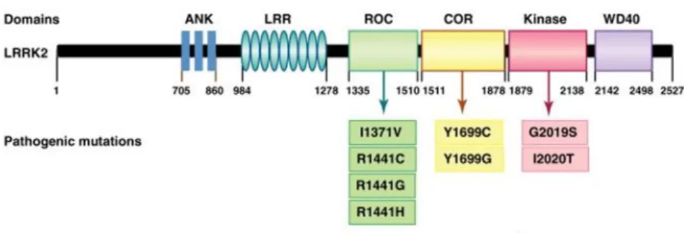
1.3.1 LRRK2
Leucine-rich repeat kinase 2 (LRRK2/PARK8 gene) is a 2527-amino-acid-long protein with a catalytic core region composed of the ROCO protein family signature ROC-COR bidomain followed by a kinase domain (Fig.6). The ROC domain is named for Ras of complex proteins as it has some homology to small GTPases including Ras. The COR domain is characteristic of the ROCO protein family and is so named because it is C-terminal of ROC. The kinase domain is generally similar to other Ser/Thrtype protein kinases, although it is only close to the LRRK2 homolog. The catalytic core is flanked N-terminally bya leucine-rich repeat domain (LRR) and C-N-terminally by a WD40 repeat domain which is deemed essential for protein folding, thus controlling LRRK2 function and kinase activity. In its N-terminal region, LRRK2 displays a large number of unusual repeat sequences (Marin,2006). Mutations of LRRK2 represent the highest risk of familial PD, causing autosomal dominant PD. There are several
24
missense mutations concentrated in the catalytic center of the protein, including R1441C/G/H in the ROC domain, Y1699C in the COR region and G2019S and I2020T in the kinase domain (Greggio, 2009).The dominant, pathogenic mutations described up to date, occur within the enzymatic core of LRRK2 suggesting that modification of LRRK2 activity greatly affects PD onset and progression.
The similarity in PD phenotype and age of onset between homozygous and heterozygous mutation carriers suggests that pathogenic mutations might act by conferring a toxic function on LRRK2 (Tsika, 2012).
Fig. 6: LRRK2 domains (Dae, 2012)
LRRK2 predominantly exists as a dimer under native conditions, a state that appears to be stabilized by multiple domain-domain interactions. It’s possible to suppose to a model of a large protein with a central catalytic GTPase/kinase region surrounded by protein–protein and perhaps protein– membrane interaction motifs, forming homo and possibly heterodimers (Greggio et al.,2008).
The precise physiological role of this protein is unknown but presence of multiple functional domains suggesting involvement in wide variety of functions but the presence of both a ROC/GTPase and a kinase domain suggests that it may play a role in intracellular signaling (Wallings et al., 2015). The presence of two enzymatic domains suggests the potential for an intrinsic regulatory mechanism of LRRK2 activity with parallels to the classic Ras/Raf signal cascade. While GTPase activity does not appear to critically require a functional kinase domain (Smith et al.,2006; Ito et al.,2007), autophosphorylation within the GTPase domain may serve a regulatory function (Kamikawaji et al.,2009; Webber et al., 2011), which implies a complex yet poorly understood bidirectional relationship between the GTPase and kinase domains of LRRK2.
25
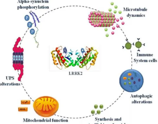
Within cells, LRRK2 associates with various intracellular membranes and vesicular structures including endosomes, lysosomes, multivesicular bodies, the mitochondrial outer membrane, lipid rafts, microtubule-associated vesicles, the Golgi complex, and the endoplasmic reticulum (Cookson, 2010). This distribution could reflect a functional role in multiple pathways (Fig.7).
Fig. 7: LRRK2 implication in cellular mechanisms
1.3.2 G2019S LRRK2 mutation
The G2019S and the nearby I2020T mutation are located at the N-terminal portion of the activation loop in kinase domain. The G2019S mutation substitutes a serine for a highly conserved glycine located in subdomain VII of the kinase domain (West et al.,2007) the mutation is situated within this segment of the activation loop and it has been speculated that the glycine residue here imparts conformational flexibility (Shan et al.,2009); therefore, replacement of the glycine with a serine residue may alter LRRK2 dynamics. The penetrance of the G2019S-LRRK2 mutation appears to have a clear age-dependent effect and varies from around 50% at age 50, to ~ 74% at age 79 (Healy et al.,2008); although some patients do not manifest any clinical features even in their 80 (Paisan-Ruiz
26 et al.,2013). This mutation increases kinase activity by increasing the catalytic rate of the enzyme; it does not enhance substrate affinity (Thomas, 2007). The G2019S mutation facilitates substrate access, thereby leading to a toxic increase in kinase activity. G2019S is the only mutation that consistently shows increased kinase activity (Greggio, 2009) changes in LRRK2 kinase activity appear to be toxic, and seem to induce degeneration of dopamine neurons.
Cellular toxicity, in both the absence and presence of oxidative stress, and the formation of inclusion bodies were observed when overexpressing G2019S-LRRK2 in cell lines and primary neuronal cultures (Heo et al., 2010). These results, and the fact that genetic inactivation of LRRK2 kinase activity showed a protective effect against such a toxic phenotype, suggest that an alteration in LRRK2 kinase activity is potentially involved in the neurotoxic and pathogenic mechanisms of LRRK2-PD. LRRK2 not only inhibits neuronal survival but also impairs dopamine signal. Accumulating evidence suggests that synaptic dysfunction might be an early event in neurodegeneration. LRRK2 and Rab5b, a regulator of endocytic vesicle trafficking (de Hoop et al., 1994; Fischer von Mollard et al., 1994), colocalize and interact on synaptic vesicles (SVs) providing evidence for a role of LRRK2 in SV trafficking (Shin et al.,2008) (Fig.7). In neurons, the vesicle trafficking controls fundamental physiological functions such as neurotransmitter or protein release and uptake, localization of membrane receptors, changes in plasma membrane composition and, not least, organelle biogenesis. In synaptic terminals, LRRK2 silencing leads to a redistribution of vesicles, alters recycling dynamics and increase vesicle trafficking (Piccoli et al.,2011), suggesting that LRRK2 is implicated in the regulation of receptor trafficking.
LRRK2 G2019S mutation impairs dopamine receptor D1 internalization, leading to an alteration in signal transduction (Rassu et al.,2017). In addition, LRRK2 G2019S mutation enhances the kinase activity and results in the impairment of synaptic vesicle trafficking selectively in ventral midbrain neurons, including dopaminergic neurons (Pan et al., 2017). Furthermore, a significant accumulation of D2R was observed in the Golgi apparatus associated with a significant increase in the amount of total protein, suggesting that the LRRK2-G2019S mutation may be involved in altering the localization and / or degradation pathways of D2R (Rassu et al., 2017).
In regard to dopamine signaling, LRRK2 mutations have been shown to affect activity dependent DA neurotransmission and catecholamine release (Tong et al., 2009). This manifests as reduced levels of extracellular striatal dopamine and reduced levels of dopamine metabolites. Cultured dopamine neurons from LRRK2 transgenic mice display markedly reduced neurite complexity (Ramonet et al., 2011) and reduced striatal DAT levels have also been reported. This change, suggests impaired dopamine re-uptake could contribute to altered dopamine levels in LRRK2 transgenic mice.
27
Therefore, studying LRRK2 pathobiology may also provide clues into the pathogenic mechanisms underlying the sporadic disease. Even if it is widely accepted that LRRK2 mutations are crucial for PD pathogenesis, their precise role in the etiology of PD needs to be further investigated. In particular, the biochemical and molecular neuronal dysfunctions occurring in the pre- symptomatic early stages of the disease that precede neuronal death need to be investigated. This is a crucial issue since a limited understanding of pathogenic mechanisms drastically precludes the development of drugs counteracting the progression of the disease.
1.4 DA neurons vulnerability
The cardinal motor symptoms of PD are caused by the death of DA neurons which are mainly situated in the midbrain. Dopamine is critical for the control of the brain functions, such as motor activity, attention, reward mechanisms and cognition. The effects of DA are mediated by five different receptors, that belong to the G protein-coupled receptor (GPCR) family and are divided into D1-like (D1 and D5) and D2-like (D2, D3, D4) subtypes. This classification is generally based on the original biochemical observations showing that dopamine is able to modulate adenylyl cyclase (AC) activity as a result their activation decreases the cAMP accumulation modulating the activity of PKA and its effectors (Forn et al., 1974; Greengard et al.,1999). Interestingly, increasing evidence suggests that DA receptors can diversify and amplify their repertoire of signaling by forming homo- and hetero-dimers, a property typically shared by the GPCR family that greatly increases their heterogeneity (Carli et al., 2018).
The prerequisite for the study of pre-degenerative defects of PD is to know the physiological properties of human DA neurons, among them, the dopamine D2 and D3 receptors (D2R / D3R) (Collo et al.,2008; Bono et al.,2018), the nicotinic acetylcholine receptors (Collo et al., 2013; Bontempi et al., 2017) have been reported to provide trophic support to DA neurons in mouse cell models.
1.4.1 Dopamine D2 and D3 Receptors (D2R)/(D3R)
The highest levels of D2R are found in the striatum, the nucleus accumbens, and the olfactory tubercle. D2R are also expressed at significant levels in the substantia nigra, ventral segmenta area, hypothalamus, cortical areas, septum, amygdala, and hippocampus (Missale et al., 1998; Gerfen, 2000).
28 In the substantia nigra, D2R are localized both on dopaminergic neurons, but also on neurons targeted by dopaminergic afferences. In addition to having a dual localization, D2R are a heterogeneous population formed by two molecularly distinct isoforms, named D2S (S=short) and D2L (L=long) generated by alternative splicing of the same gene (Gingrich, 1993). These two alternatively spliced isoforms differ in the presence of an additional 29 amino acids in the third intracellular loop. These variants of the D2R have distinct anatomical, physiological, signaling, and pharmacological properties. D2S has been shown to be mostly expressed presynaptically and to be mostly involved in autoreceptor functions, whereas D2L seems to be predominantly a postsynaptic isoform (Usiello et al., 2000; De Mei et al., 2009).
D2 dopamine receptors seem to be the predominant type of autoreceptors that are involved in the presynaptic regulation of the firing rate, synthesis of dopamine and release of dopamine. Activation of presynaptic D2-class autoreceptors generally causes a decrease in dopamine release that results in decreased locomotor activity, whereas activation of postsynaptic receptors stimulates locomotion. (Wolf and Roth, 1990; Missale et al., 1998). Recent data indicate that D2R also expressed in microglia and astrocytes which are a key negative regulator for neuroinflammation and controlling innate immunity in the central nervous system. (Zhang an Barres 2010; Shao et al.,2013).
The D3R is expressed on DA neurons, both at the somatodendritic level and at synaptic terminals, in the substantia nigra (SN) and ventral tegmental area (VTA), as well as in the ventral striatum in the limbic areas and in the islands of Calleja and cerebellum (Missale et al., 1998). The D3R seem to exert a moderate inhibitory action on locomotion through the involvement of postsynaptic receptor populations (Joseph et al., 2002; Sokoloff et al., 2006).
At the molecular level, D2R/D3R are coupled to the G alphai/o family of G proteins leading to inhibition of adenylyl cyclase (AC) (Missale et al., 1998). There is now increasing evidence that D2R/D3R are also critical to supporting DA neuron homeostasis (Fiorentini et al.,2015) and are crucially involved in DA neurons development, morphological plasticity and protection, suggesting that an abnormal D2R/D3R function could increase DA neurons vulnerability (Bono el al.,2018). It was recently reported that the D2-like receptor agonists quinpirole and 7-OH-DPAT exert neurotrophic effects on DA neurons in primary mouse mesencephalic cultures, as shown by increased maximal dendrite length, number of primary dendrites and soma area (Collo et al., 2008). These effects required the activation of the PI3K-Erk1/2 and PI-3K-Akt-mTOR pathways (Collo et al.,2012, 2013). Stimulation of D3R promotes neurotrophic effects and plays a crucial role in triggering key intracellular events with neuroprotective potential (Bono et al., 2018, 2019) and also the activation of
29 D2R / D3R could promotes the proliferation of neuronal progenitor cells (Kim et al., 2006; Bono et al., 2018). Taken together, these data suggest that the D3R may be crucially involved in the control of DA neurotrophism during development (Diaz et al.,1997; Bono et al., 2018), a property also shared with the D2R (Kim et al., 2006; Yoon et al.,2011), and in counteracting early pathological events that may subsequently result in neurodegeneration.
Interestingly, in animal models of transgenic mice carrying the LRRK2 mutation, impairment in the expression and function of D2R leading to neuronal synaptic dysfunction has been observed (Tong et al., 2009) and also recent data indicate that D2R also expressed in microglia and astrocytes which are a key negative regulator for neuroinflammation and controlling innate immunity in the central nervous system. (Zhang et al., 2010; Shao et al.,2013).
Therefore, it might be useful to investigate the impact of the LRRK2 G2019S mutation on D2R /D3R activity and homeostasis of DA neurons. D2R / D3R expression or defective function may represent an early and pre-degenerative event in patients carrying the LRRK2 mutation with various consequences that likely contribute to making DA neurons more vulnerable.
1.4.2 Nicotinic Acetylcholine Receptors (nAChRs)
The activity of DA neurons is regulated by an integrated interplay between different proteins and receptor systems including the nAChR. nAChRs are a heterogeneous family of ligand-gated ion channels, composed by various α(α2-α7) and β(β2-β4) subunits, which are activated by nicotine (Quick et al., 2011). nAChR, are characterized by an especially high permeability to Na+ and K+, resulting in cell excitation. This excitation may activate voltage gated calcium channels allowing calcium influx into the cell. One of the primary functions of nAChR is to modulate synaptic transmission and synaptic plasticity triggered by other neurotransmitters, resulting in alterations in affective behavior, attention and cognition (Benowitz, 2009).
It is well known that stimulation of nAChR increases DA neuron firing and DA release (De Kloet et al.,2015). Nicotine promotes the morphological remodeling of DA neurons and regulates various genes controlling neuronal morphogenesis (Doura et al., 2010; Collo et al., 2013). Different nAChR subtypes are co-localized with D2-like receptors in DA nerve terminals (Exley and Cragg 2008; Zoli et al.,2015). Epidemiological studies have identified a negative correlation between smoking and the development of neurodegenerative disorders such as Parkinson's disease. These findings have been attributed to the ability of nicotine to act as a neuroprotective agent (Picciotto, 2008).
30 In the study by Mappin-Kasirer et al., (2020) that involved 30,000 British male doctors, a protective effect of tobacco on PD risk was observed.
There is evidence that nAChR located on DA neurons also provide neurotrophic and neuroprotective support to DA neurons, an effect requiring functional D3R and suggesting the existence of a positive crosstalk between these receptor systems (Bontempi et al., 2017; Bono et al., 2018, 2019).
1.4.3 D3R-nAChR Heteromerization
GPCR have been classically thought to exist as monomeric entities. However, the current view of GPCR organization assumes, in fact, that these receptors may form heteromers by direct interaction with members of the same receptor family and with structurally and functionally divergent families of receptors (Angers et al., 2002; Ferre’ et al., 2009). Sometimes heterodimerization is an absolute requirement for the formation of functional receptors may give rise to novel receptors units with unique pharmacological, signaling and trafficking properties (Borroto-Escuela et al., 2014).
Heteromerization often affects the ligand binding properties of interacting receptors and alters the potency of agonists in generating intracellular signals. In addition, heteromerization may generate new binding sites, an observation that may have a significant impact on drug discovery, providing the framework to develop drugs specifically targeting a given heterodimeric pair (Milligan, 2008). By influencing G- protein specificity, heteromerization may be responsible for alterations in the signaling pathways activated by a given receptor. Interacting receptors may also exhibit a specific G-protein coupling and activate peculiar transduction pathways (Gaitonde and González-Maeso, 2017). Heteromerization may also represent a novel mechanism modulating agonist-mediated trafficking, by either decreasing or enhancing receptor desensitization and internalization. Different intermolecular interactions may contribute to receptor dimerization. Either transmembrane and intracellular domains are likely to play a central role in receptor- receptor interactions by the formation of hydrophobic or non-covalent electrostatic bonds respectively (Guo et al., 2003; Lopez-Gimenez et al., 2007; Mancia et al., 2008). DA receptor heteromers have been the most studied complexes so far. It’s known that the D3R may form heterodimers with other DA receptor subtypes, such as the D1R and the D2R (Scarselli et al., 2001; Fiorentini et al., 2008).
As previously mentioned, the D3R and nAChRs participate to the control of DA neuron firing and plasticity. In particular, the D3R seems to reduce DA release and regulates the mechanisms of structural plasticity; nAChRs activated by acetylcholine or nicotine facilitate the switch from tonic to burst firing mode, increase the release of DA and modulate neuronal plasticity (Picciotto et al., 2008).
31 These observations thus underlie the important role of the nicotinic system in the control of DA transmission in both physiological and pathological conditions. Since nAChR and both D2R and D3R are extensively co-localized in both DA neuron cell bodies and nerve terminals (Zoli et al., 2002), the possibility that functional interactions may occur between DA receptors and nAChR has been investigated and clearly demonstrated (Grilli et al., 2009; Bontempi et al., 2017). It is likely that D2R, D3R and nAChR might be associated into different heteromeric complexes that may exert strong control over DA release and DA neuron viability (Bontempi et al., 2017) (Fig.8). Nicotine, in fact, besides controlling DA release, supports DA neuron regeneration and modulates the expression of different genes regulating neuronal morphogenesis (Quik et al., 2006; Doura et al., 2010; Bono et al., 2019).
Has been reported that nicotine provides neurotrophic support to DA neurons by increasing their dendritic arborization and soma size (Bontempi et al.,2017; Bono et al., 2018). This effect is mediated by the α4β2 nAChR subtype and depends on functional D3R, since it was blocked by D3R preferential antagonists and was absent in DA neurons from D3R-KO mice (Collo et al., 2013) suggesting the existence of a positive crosstalk between D3R and nAChR in promoting DA neurotrophism.
Along this line, by using Bioluminescence Resonance Energy Transfer (BRET), we have shown that the D3R directly interacts with the β2 subunit of nAChR to form a heteromeric complex. Interestingly, disruption of D3R-β2 interaction by a cell-permeable interfering peptide abolished the effects of nicotine on DA neuron morphology (Bontempi et al., 2017), suggesting that the D3R-nAChR heteromer represents the molecular unit triggering nicotine-mediated neurotrophic effects.
The Ras-ERK and PI3K-mTORC1 pathways represent key mechanisms for cells to regulate cell survival, proliferation, and motility. In addition to their independent signaling programs that provide compensatory mechanisms, the pathways extensively cross-talk to positively and negatively regulate each other (Mendoza et al.,2011). The PI3K/ERK1/2 signaling pathway has been shown to sustain D2R/D3R-mediated morphological changes in primary rodent neuronal cultures (Alonso et al., 2004; Kumar et al., 2005; Collo et al., 2008). In mouse DA neurons, the engagement of the PI3K-ERK1/2 signaling, associated with the D3R, is required for nicotine-induced structural plasticity (Collo et al.,2008) suggesting that the phosphorylation and activation of ERK1/2 may be the intracellular event associated with the heteromer stimulation and strictly related to neurotrophic effects. The nicotine- elicited activation of MEK-ERK and PI3K-Akt- mediates structural plasticity, because the MEK inhibitor PD98059 or the PI3K inhibitor LY294002, blocked nicotine-induced dendrite growth and soma size increase. These findings indicate that nicotine-induced structural plasticity at mesencephalic
32 dopaminergic neurons involves α4β2 nAChRs together with dopamine D3R-mediated recruitment of ERK/Akt signaling (Collo et al., 2013). Moreover, it was reported that stimulation of D3R in mouse primary midbrain DA neurons results in the phosphorylation of ERK1/2 pathway, an effect mediated by PI3K. It has also been demonstrated that in iPSC-derived DA neurons, D3R stimulation by quinpirole transiently activated the ERK1/2 pathway. In addition, a chronic treatment with quinpirole led to a significant remodeling of both dendritic arborization and soma size of TH-positive DA neurons, an effect likely dependent on the ERK cascade (Bono et al.,2018).
Moreover, by using the proximity ligation assay (PLA), we identified the D3R-nAChR heteromer in cultured DA neurons and mouse mesencephalic brain sections (Bontempi et al., 2017), as well as in iPSC-derived DA neurons (Bono et al.,2019). The D3R-nAChR heteromer is also involved in neuroprotection of DA neurons. By using glucose deprivation (GD)-induced neurotoxicity, we reported that both D3R agonists and nicotine modulate alpha-synuclein (alpha-syn) accumulation and protect DA neurons against neuronal injury (Bellucci et al., 2008; Bono et al.,2018). More recently, it was reported that nicotine inhibits alpha-syn accumulation in iPSC-derived DA neurons, an effect that was specifically blocked by D3R antagonists (Bono et al.,2018) and was lost in the presence of the specific interfering peptide (Bono et al., 2019), suggesting that, beside the induction of neurotrophic effects, the D3R-nAChR heteromer is the molecular unit involved in neuroprotection and inhibition of alpha-syn accumulation.
These observations suggest that alterations in the assembly and function of this receptor complex, may result in early dysfunctions contributing to the specific vulnerability of DA neurons. The D3R-nAChR heteromer may thus represent a novel target for drugs designed for supporting DA neurons plasticity and survival against toxic damages in various pathologies, including PD.
33 Fig. 8: Representation of the heteromerization between the D3R and the α4β2 nAChR in DA neurons (Bono et al.,
2020).
1.5 Neuroinflammation
Glial cells outnumber neurons in the brain and play important roles in the neuroinflammation that accompanies brain damage in neurodegenerative diseases. In PD, dopaminergic neuronal loss is accompanied by inflammatory changes in microglia, astrocytes and innate immune cells. Acute neuroinflammatory response has beneficial effects in the central nervous system (CNS) and prompts repair of damaged tissues; however, when chronically sustained and dysregulated, inflammation can lead to significant tissue and cellular damage (Qin et al., 2007).
In general, neuroinflammation is closely associated with neuronal damage and cell death through many biological mechanisms, such as elevated oxidative stress, glial (astrocyte and microglia) cell activation, excitotoxicity, proapoptotic mechanisms, and mitochondrial dysfunction (Niranjan, 2014). Under neuroinflammatory conditions, activated glial cells release pro-inflammatory and neurotoxic factors that induce neuronal damage and neurodegeneration (Harry and Kraft 2008). Evidence implicating cytokines in nigrostriatal pathway degeneration and from post-mortem analyses indicated that the levels of several cytokines including TNF-α and interleukin 1-beta (IL-1β) are significantly elevated in the area of substantia nigra (Pieper et al., 2008). In addition to these, IL-1α and IL-6 are also involved in the pathophysiology of PD and play a very important role in dopaminergic