Clinical Study
Medication-Related Osteonecrosis of the Jaws: Considerations
on a New Antiresorptive Therapy (Denosumab) and Treatment
Outcome after a 13-Year Experience
Gianfranco Favia,
1Angela Tempesta,
1Luisa Limongelli,
1Vito Crincoli,
1and Eugenio Maiorano
21Department of Interdisciplinary Medicine, Complex Operating Unit of Odontostomatology, “Aldo Moro” University,
Piazza G. Cesare 11, 70124 Bari, Italy
2Department of Emergency and Organ Transplantation, Operating Unit of Pathological Anatomy, “Aldo Moro” University,
Piazza G. Cesare 11, 70124 Bari, Italy
Correspondence should be addressed to Angela Tempesta; [email protected] Received 25 March 2016; Revised 8 September 2016; Accepted 25 September 2016
Academic Editor: Saso Ivanovski
Copyright © 2016 Gianfranco Favia et al. This is an open access article distributed under the Creative Commons Attribution License, which permits unrestricted use, distribution, and reproduction in any medium, provided the original work is properly cited.
Medication-related osteonecrosis of the jaw (MRONJ) is a serious complication in patients receiving antiresorptive therapies for bone neoplastic localizations and osteoporosis. The aim of this study was to evaluate the clinicopathological features of MRONJ in a cohort of patients treated by new antiresorptive drugs (denosumab) and the corresponding outcome after 13-year maximum follow-up. Overall, 244 patients affected by MRONJ were treated from 2003 to 2015. After clinical and radiological examinations, all lesions were staged according to a dimensional staging system and then surgically treated. All the denosumab-related lesions were classified as stage II or III, thus requiring a more or less invasive surgical approach, despite the results of many recent studies, which suggested a conservative medical approach with early resolution for MRONJ in patients on denosumab. In the current series, 86.9% of treated lesions showed complete clinical and radiological healing, while 13.1% recurred; all recurrences were detected in patients who could not interrupt chemotherapy, steroids, and/or antiresorptive drugs administration due to their general conditions. In conclusion, all oral specialists should be aware of the MRONJ risk among patients taking new antiresorptive drugs; moreover, our protocol based on surgical treatment guided by dimensional staging could be considered effective in view of the low recurrence rate.
1. Introduction
Osteonecrosis of the jaw (ONJ) is a well-known complication of antiresorptive or antiangiogenic therapy for the manage-ment of osteoporosis and other cancer-related conditions, including hypercalcemia of malignancy and bone metastases of solid tumours such as breast, prostate, and lung cancer, and for the management of lytic lesions in the setting of multiple myeloma [1]. Such lesion was initially termed bisphosphonate-related osteonecrosis of the jaw (BRONJ) as it usually followed the administration of different types of bisphosphonates (BPs). Subsequently, with the advent of new classes of antiresorptive or antiangiogenic medications,
such as denosumab, sunitinib, bevacizumab, and ipilimumab, giving rise to similar complications [2–4], the American Association of Oral and Maxillofacial Surgeons (AAOMS) in 2014 recommended the change in the nomenclature into medication-related osteonecrosis of the jaw (MRONJ) [5].
Moreover, AAOMS has modified the MRONJ definition to distinguish this condition from other bone anomalies and, accordingly, patients may be considered to harbour MRONJ if all the following characteristics are present: current or pre-vious treatment with antiresorptive or antiangiogenic agents, exposed necrotic bone or bone that can be probed through an intraoral or extraoral fistula in the maxillofacial region that has persisted for longer than 8 weeks, no history of radiation
Volume 2016, Article ID 1801676, 9 pages http://dx.doi.org/10.1155/2016/1801676
findings, such as osteosclerosis and periosteal hyperplasia, and nonspecific symptoms, such as pain
Medical therapy and clinical-radiological follow-up
Stage I
Bone exposure and/or radiographic evidence of necrotic bone, or persisting alveolar sockets< 2 cm in the major diameter, with or without pain
Medical therapy, surgical debridement, and low-level laser therapy (LLLT)
Stage II
Bone exposure and/or radiographic evidence of necrotic bone between 2 and 4 cm in maximum diameter, with pain responsive to NSAIDs and possible abscesses
Medical therapy and small open-access surgery with piezosurgery of bone margins
Stage III
Bone exposure and/or radiographic evidence of necrotic bone> 4 cm in the maximum diameter, with strong pain, responsive or not to NSAIDs, abscesses, orocutaneous fistulas, and/or maxillary sinus and mandibular nerve involvement
Medical therapy and wide open-access surgery, with extensive maxillary (Caldwell-Luc technique) or mandibular resection, and piezosurgery of bone margins
therapy to the jaws, or obvious metastatic localization to the jaws [5].
BPs are nonmetabolized analogues of pyrophosphate, which prevent osteoclast-mediated bone resorption [6]. In the past, they represented the first-line antiresorptive agents for the management of postmenopausal osteoporosis and for the treatment of bone metastases of solid tumours [7]. More recently, additional molecules have been introduced, such as denosumab, a subcutaneously dosed monoclonal antibody against the receptor activator of nuclear factor-𝜅B ligand (RANKL), which inhibits osteoclast functions and associated bone resorption.
Patients on denosumab for metastatic bone disease receive 120 mg subcutaneously every 4 weeks, while 60 mg subcutaneously every 6 months is used for the management of patients with osteoporosis/osteopenia. The half-life of denosumab is 26 days, while the half-life of BPs ranges from 10 to 12 years. Moreover, differently from BPs, denosumab does not seem to accumulate in the bone, thus leading many authors to maintain that denosumab-related MRONJ are less aggressive and require a more conservative therapeutic approach [8–10].
MRONJ often follows oral surgery or traumatic injuries; however, MRONJ was also detected in patients who had not undergone any invasive dental procedures during the treatment with BPs or denosumab [11].
There are still controversies about the treatment of MRONJ with regard to drug discontinuation, medical ther-apy, surgery, or other therapies [12]. Also, a large variety of treatment modalities have been proposed, including con-servative medical management, mainly repeated cycles of antibiotics, distinct types of surgery (surgical debridement or marginal resection), or other noninvasive therapies, such as hyperbaric oxygen, ozone therapy, and low-level laser therapy for biostimulation [13].
The AAOMS has proposed a staging system to select the best treatment strategy [5]: patients with stages I and II MRONJ should be treated using a conservative approach to
prevent the progression of the lesions and limit complica-tions related to chronic infection. Such treatments include conservative debridement of bone sequestra, local irrigation with povidone-iodine, daily rinsing with 0.12% chlorhexidine mouthwash, antibiotic therapy, and pain control [5]. In cases of stage III lesions, marginal resection or surgical debride-ment is always indicated and the goal of surgery should be to eliminate necrotic bone, acting as foreign material and thus increasing the risk of infection [1].
Franco et al. in 2014 [14] proposed a dimensional staging of MRONJ to more appropriately assess the therapeutic strategy (Table 1).
The aim of this study was to report all cases of MRONJ, treated from 2003 to 2015, in the Odontostomatology Unit of the University of Bari, with specific attention to those occurring in denosumab-treated patients, and to highlight the results of our treatment strategies after a 13-year experi-ence.
2. Materials and Methods
Patients with a diagnosis of MRONJ, referred to the Odon-tostomatology Unit of the University of Bari, Italy, from 2003 to 2015, currently or previously on therapy with BPs and/or other antiresorptive drugs for neoplastic diseases or osteoporosis, with presence of oral exposed necrotic bone or bone that can be probed through a fistula that has persisted for longer than 8 weeks, were included in this study; patients who had received radiation therapy in the oral and maxillofacial area were excluded, leading to a 244-case study cohort.
A detailed medical history was recorded for each patient, with specific regard to the primary disease and related therapies (BPs, antiresorptive and/or antiangiogenic drugs), dose and duration of treatment, other comorbidities, and related drugs administration. Subsequently, signs of bone necrosis and clinical symptoms were evaluated, together with the site and size of the lesions and possible triggering
(a) (b)
(c) (d)
(e) (f)
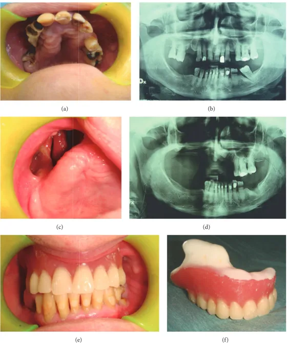
Figure 1: At clinical examination, presence of necrotic bone exposure (a) classified as stage III MRONJ depending on the dimensional staging system on the right maxilla in a female 67-year-old patient affected by breast cancer, who underwent denosumab administration 11 times. Rx OPT shows bone support reduction on the right maxilla. Patient referred the spontaneous loss of 4 teeth (1.7, 1.6, 1.3, and 1.2) after she underwent the orthopantomography, probably due to the progressive bone necrosis and resorption (b). After 16-month follow-up, clinical (c) and radiological (d) healing of the patient who underwent a removable prosthetic restoration (e-f).
events. After radiographic evaluation, including orthopan-tomography and Enhanced Multislices Spiral Computed Tomography with 3D reconstruction, all lesions were staged (Figures 1 and 2) according to both the AAOMS [5] criteria and the dimensional staging [14], the latter being used for the subsequent choice of the surgical approach. Upon consultation with the treating physician, patients suspended any BPs/antiresorptive/antiangiogenic administration not less than 3 months before surgical treatment; chemotherapy and corticosteroids administration were suspended 3–5 days before the surgical procedure and until wound healing, to
reduce the recurrence risk. Patients were also administered at least 3 cycles of antibiotic therapy before treatment, consisting in a combination of ceftriaxone (1 g/i.m. daily) and metronidazole (500 mg/per os twice a day) for 8 days with 10 days of interruption after each cycle. The surgical treatment was based on the dimensional staging: surgical debridement for stage I lesions, small open-access surgery with piezosurgery of bone margins for stage II, and wide open-access surgery with bone extensive resection (Figure 2) and piezosurgery of bone margins for stage III lesions [14]; a gel compound with hyaluronic acid and aminoacids was
(a) (b)
(c) (d)
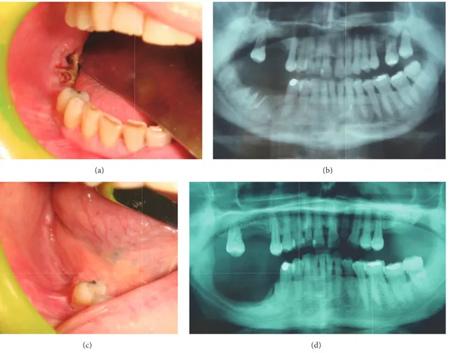
Figure 2: At clinical examination, presence of postextractive necrotic bone exposure on the right mandible (a) of a female 51-year-old patient affected by breast cancer who underwent denosumab administration 9 times. Rx OPT confirms the presence of an area of bone alteration (b) which was diagnosed as stage III MRONJ accordingly with the dimensional staging systems and was surgically treated. After 12-month follow-up, clinical (c) and radiological (d) healing of the treated lesion without signs of recurrence is evident.
put into the residual bone cavity and on the suture stitches to reduce the wound healing time. All surgical specimens were then sent for histopathological examination.
Each patient underwent an accurate clinical follow-up on a weekly basis in the first month and then a clinicora-diological follow-up at 1, 3, 6, and 12 months after surgery (Figures 1 and 2). If deemed necessary, patients restarted BPs/antiresorptive/antiangiogenic treatments not less than 1 month after surgery.
After one-year follow-up, lesions were considered suc-cessfully treated if they completely healed based on clinical or radiological features, or if the lesions could be downstaged according to the AAOMS criteria (Figures 1 and 2); MRONJ occurring at the same site during the clinicoradiological follow-up were considered “recurrences.”
This study was performed in accordance with the prin-ciples of the Declaration of Helsinki and has been approved by our institution’s ethical committee (study number 4599, Prot. 1528/C.E.); the patients released informed consent on diagnostic and therapeutic procedures and possible use of the biologic samples for research purposes.
3. Results
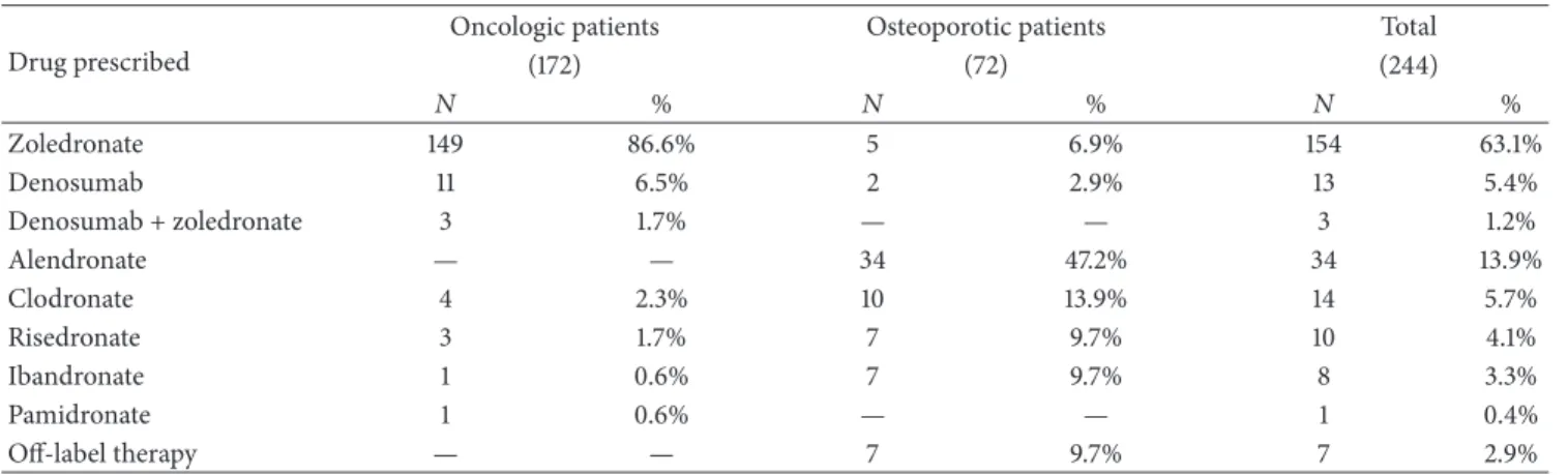
Overall, 244 patients affected by MRONJ (184 females, 60 males) were treated at the Odontostomatology Unit of the University of Bari from 2003 to 2015. Among them, 72 (29.5%) were osteoporotic patients, while 172 (70.5%) were oncologic patients suffering from different malignancies: 75 patients were affected by breast cancer, 53 by multiple myeloma, 23 by prostatic cancer, 6 by lung cancer, 6 by leukaemia/lymphoma, 5 by renal cancer, and 4 by thyroid cancer. Patients had been given different types of antiresorptive drugs, zole-dronate being the most frequently used, followed by alen-dronate, denosumab, cloalen-dronate, risealen-dronate, ibanalen-dronate, and pamidronate, as illustrated in Table 2.
Moreover, parenteral administration of antiresorptive drugs was referred by 187 (76.6%) patients and oral admin-istration by 57 (23.4%). Also, 54 patients (45 oncologic, 9 osteoporotic) presented multiple MRONJ lesions and, therefore, a total of 322 lesions were treated, 207 in the mandible and 115 in the upper jaw. Tables 3 and 4 illustrate the correlations between the staging of the lesions (according
Table 2: Patients’ antiresorptive therapy.
Drug prescribed
Oncologic patients Osteoporotic patients Total
(172) (72) (244) 𝑁 % 𝑁 % 𝑁 % Zoledronate 149 86.6% 5 6.9% 154 63.1% Denosumab 11 6.5% 2 2.9% 13 5.4% Denosumab + zoledronate 3 1.7% — — 3 1.2% Alendronate — — 34 47.2% 34 13.9% Clodronate 4 2.3% 10 13.9% 14 5.7% Risedronate 3 1.7% 7 9.7% 10 4.1% Ibandronate 1 0.6% 7 9.7% 8 3.3% Pamidronate 1 0.6% — — 1 0.4% Off-label therapy — — 7 9.7% 7 2.9%
Table 3: MRONJ staging in oncologic patients.
Antiresorptive drug AAOMS staging system [5] Dimensional staging system [14]
Stage 0 Stage I Stage II Stage III Stage 0 Stage I Stage II Stage III
Zoledronate 1 13 126 76 1 22 66 127 Denosumab 4 7 3 8 Zoledronate + denosumab 2 1 1 2 Clodronate 4 2 1 1 Risedronate 2 1 1 2 Ibandronate 1 1 Pamidronate 1 1
Table 4: MRONJ staging in osteoporotic patients.
Antiresorptive drug AAOMS staging system [5] Dimensional staging system [14]
Stage 0 Stage I Stage II Stage III Stage 0 Stage I Stage II Stage III
Zoledronate 3 2 4 1 Denosumab 2 2 Alendronate 3 31 5 10 15 14 Clodronate 9 2 3 4 4 Risedronate 1 6 4 1 5 5 Ibandronate 7 1 5 3 Off-label 4 3 5 2
to both the AAOMS criteria [5] and the dimensional staging system [14]) and the different antiresorptive agents used.
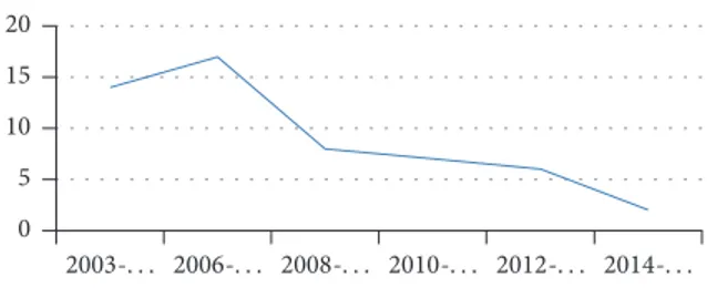
We also assessed the distribution of the 45 patients affected by multiple MRONJ according to the year of presen-tation and the results are synthetically reported in Figure 3.
The histopathological analysis of the surgical samples highlighted typical features of MRONJ, as already reported [15]: extensive bone necrosis, without residual osteocytes/ osteoblasts, large and empty Haversian canals, abundant in-flammatory cell infiltration, and several basophilic bacterial colonies interspersed with necrotic debris. Also, nonnecrotic tissues containing larger amounts of bone, fewer and smaller Haversian canals, larger and thicker osteons, osteocytes of larger diameter, relative paucity of osteoclast-like cells of rel-atively smaller size, and reduction of the marrow spaces were
detected (Figure 4). Histopathological examination demon-strated complete overlap of the morphological alterations occurring in the lesions of patients treated by denosumab, in comparison with those treated by BPs.
During follow-up (mean: 16 months, range: 10–37) 280 treated lesions (86.9%) showed complete clinical and radio-logical healing, while 35 lesions (13.1%) recurred. Among the recurrences, 1 case was detected in a patient on denosumab (6% of the denosumab lesions) and the remaining were in patients on BPs (15% of the BPs lesions); 6 patients died of complications related to their oncologic disease. All recur-rences were observed in patients who could not interrupt chemotherapy, corticosteroids, and/or antiresorptive drugs administration due to their general conditions.
Patients with multiple lesions 0 5 2010-. . . 2014-. . . 2006-. . . 2008-. . . 2012-. . . 2003-. . .
Figure 3: Progressively decreasing number of patients with multiple MRONJ lesions from 2003 to 2015.
A
C B
Figure 4: Histopathological features of MRONJ in a patient treated by denosumab. Abundant inflammatory cell infiltration and basophilic bacterial colonies (A) interspersed with necrotic debris are evident. Also, nonnecrotic tissues containing larger amounts of lamellar bone (B), fewer and smaller Haversian canals (C), and larger and thicker osteons are detectable. Haematoxylin-Eosin, 10x.
4. Discussion
Antiresorptive agents that target osteoclasts, thereby inhibit-ing bone resorption and subsequent bone loss, currently are considered the cornerstone for the treatment and prevention of bone metastases of solid tumours and osteoporosis [16, 17]. In the past, intravenous and oral BPs were considered the gold standard for the treatment of such conditions; more recently, other molecules have been proposed, such as the fully human monoclonal antibody denosumab, which was approved by the FDA in 2010 for the treatment and prevention of osteoporosis and bone metastases in oncologic patients.
Although BPs are nonmetabolized analogues of pyro-phosphate, while denosumab is a monoclonal antibody against the receptor activator of nuclear factor-𝜅B ligand (RANKL), both share the same mechanism of action, con-sisting in the inhibition of osteoclast functions and associated bone resorption.
Several clinical trials showed that denosumab is more effective than BPs for the prevention of skeletal-related events in patients with metastatic bone disease and in reducing the risk of bone fractures in osteoporotic patients [18–22]. Moreover, denosumab has a half-life of several weeks and is not eliminated via the kidneys, differently from BPs, which are primarily excreted renally, bound to hydroxyapatite, and
for patients more prone to develop BPs-related complications, such as MRONJ, or those with compromised renal function. Cases of denosumab-related MRONJ have been reported during randomized clinical trials for the treatment of patients with metastatic bone disease [18–20, 24–28], the majority of them being associated with known risk factors, consisting in invasive dental procedures and poor oral hygiene [24], and the duration of treatment [29].
The current study is based upon the analysis of 244 patients affected by 322 MRONJ lesions, treated from 2003 to 2015, who had undergone different antiresorptive drugs administration, either parenterally (76.6%) or per os (23.4%), with zoledronate being the most frequently administered (63.1%), especially in oncologic patients.
Tables 3 and 4 show the distribution of the studied cases according to the type of antiresorptive drug and the severity of lesions staged according to AAOMS [5] and to Franco et al. [14]. Although statistical analyses were unsuitable, due to the limited number of patients treated by denosumab, some preliminary considerations may be drawn. In our experience all the denosumab-related lesions were classified as stage II (12.5%) or III (87.5%) with either staging system, thus requiring surgical treatments, which were more invasive for stage III lesions. Furthermore, while 34 recurrences were detected in 228 patients on BPs (14.95% recurrence rate), such complication occurred only in 1 patient of 16 on denosumab (6.25 recurrence rate).
Such data, though requiring powerful statistical confir-mation on larger case series, seem to indicate that MRONJ lesions arising in patients on denosumab are more frequently detected in stages II and III, according to both staging systems. Furthermore, we have no knowledge of previous reports dealing with the recurrence rates of treated MRONJ lesions in patients on either BPs or denosumab; nevertheless, the results of the current study indicate that such adverse event is quite rare (15%) in patients on BPs and even rarer (6%) in those on denosumab. Such low recurrence rates also support the effectiveness of the staging system proposed by our group [14] for the selection of the most appropriate treatment based on the extension of the lesions.
Although the first MRONJ case was reported over a decade ago, the pathophysiology of this disease has not been fully elucidated. Altered bone remodeling, osteoclastic or angiogenesis inhibition [5], and relative reduction of blood vessels in MRONJ patients have been reported as possible pathogenetic factors [30]. Favia et al. suggested that reduced angiogenesis following BPs therapy is not mandatory for ischemic-necrotic changes to occur, the latter possibly taking place as a consequence of the expansion of the nonosteonic bone compartment, due to the reduced osteoclastic effects without concurrent and adequate increased blood supply [15]. In the current study we were able to detect similar morphological changes in the tissues from the lesions of both patients with BPs-related and denosumab-related MRONJ; on these premises, we may hypothesize that the above
mechanism may be pathogenetically involved in the origin of denosumab-driven osteonecrosis of the jaws.
Recent studies comparing the incidence of MRONJ in cancer patients treated by denosumab or zoledronate showed a slightly increased incidence in the former group (1.7– 1.8% versus 1.3%) [24, 31]. Moreover, the risk of developing MRONJ also was significantly increased among patients who received any antiresorptive therapy for metastatic bone disease, in comparison with patients affected by osteoporo-sis/osteopenia [32], thus suggesting that concomitant or additional treatments, such as chemotherapy, or increased doses of antiresorptive agents may be responsible for such increased risk. Though lacking statistical confirmation, the results of this study provide additional evidence to such hypothesis and also show that the same may also apply to patients undergoing denosumab therapy.
It is known that denosumab has a shorter (26-day) half-life in comparison with BPs (10–12 years) and that it does not accumulate into bone and becomes inert within 6 months after the last administration. Several studies showed that patients on denosumab develop less severe MRONJ lesions than those on BPS and that drug discontinuation, either before an invasive dental procedure or after their development, is effective in preventing such lesions [5, 33] and may show faster resolution of MRONJ, compared to patients on zoledronate [24]. Also, Stopeck et al. maintained that MRONJ occurring in patients on denosumab more frequently were mild or moderate in severity and could be treated conservatively by mouth rinses and antibiotics or, occasionally, by limited surgical treatments (i.e., sequestrec-tomy, debridement, and curettage) [18], while other studies reported spontaneous resolution of MRONJ 7, 11, 15, and 18 months after the discontinuation of denosumab [25, 26, 34].
The results of the current study are not in complete agreement with the above reported data: no patient on denosumab was found in stage 0 or I, all of them were treated by more or less invasive surgery, and some of them also developed MRONJ recurrence after treatment, though at lower rates than patients on BPs. More importantly, the lack of relevant morphological differences of MRONJ lesions occurring in patients on denosumab, in comparison with those on BPs, apparently gives support to a common pathogenetic mechanism responsible for osteonecrosis and subsequent superinfection. If these data will receive further support, denosumab-related MRONJ will deserve the same attention as that reserved to BPs-treated patients, at least in terms of clinical management and surgical treatment of larger lesions, staged according to the model we proposed. Also, prevention or early identification of MRONJ in patients being treated by denosumab should rely upon the same tools adopted for those treated by BPs, including patients’ and practitioners’ consciousness of denosumab-related adverse events and the efficacy of dental hygiene and dietary sup-plements (e.g., calcium and vitamin D) for their prevention. Also, recurrent denosumab-related MRONJ only occurred in cancer patients and, more frequently, in those who could not discontinue concomitant treatments (antineoplastic, steroids, etc.), pointing at the need of prolonged follow-up for these patients.
Finally, we have showed (Figure 3) consistently decreased occurrence of multiple MRONJ lesion over the last 12 years; this favorable trend may be attributable to the positive effects of prevention and should encourage further dissemination of data on MRONJ among general practitioners and general dentists.
5. Conclusions
Since the first reports in 2003, thousands of cases of MRONJ have been published in the English literature, but the pathogenesis of MRONJ, its treatment options, and patients’ outcome still remain complex and multifactorial and the continuous introduction of new antiresorptive drugs, such as denosumab, makes the situation even worse. In our experi-ence, patients treated with denosumab presented generally with stages II-III lesions, thus requiring surgical treatments and not only conservative management, as reported in many previous studies. In view of the limitations of this study, mainly due to the restricted number of patients on denosumab who developed MRONJ, attentive description of additional series of cases is required to possibly confirm our preliminary findings. Such new studies should possibly confirm the effective role of prevention to at least detect lesion at a smaller size and reduce recurrence rates and of the proposed staging system to drive the choice between surgical versus nonsurgical treatment options.
Ethical Approval
This study was performed in accordance with the principles of the Declaration of Helsinki and has been approved by the authors’ institution ethical committee (study number 4599, Prot. 1528/C.E.).
Consent
The patients released informed consent on diagnostic and therapeutic procedures and for the use of such data and possible use of biologic samples for research purposes and scientific publication.
Disclosure
Data from the patients included in this study were treated anonymously.
Competing Interests
The authors declare that there are no competing interests regarding the publication of this paper.
References
[1] R. E. Marx, Y. Sawatari, M. Fortin, and V. Broumand, “Bis-phosphonate-induced exposed bone (osteonecrosis/osteopet-rosis) of the jaws: risk factors, recognition, prevention, and
Oral Pathology and Oral Radiology, vol. 113, no. 3, pp. e1–e3,
2012.
[3] S. Otto, S. Baumann, M. Ehrenfeld, and C. Pautke, “Successful surgical management of osteonecrosis of the jaw due to RANK-ligand inhibitor treatment using fluorescence guided bone resection,” Journal of Cranio-Maxillofacial Surgery, vol. 41, no. 7, pp. 694–698, 2013.
[4] S. E. C. Pichardo, S. C. C. Kuypers, and J. P. R. van Merkesteyn, “Denosumab osteonecrosis of the mandible: a new entity? A case report,” Journal of Cranio-Maxillofacial Surgery, vol. 41, no. 4, pp. e65–e69, 2013.
[5] S. L. Ruggiero, T. B. Dodson, J. Fantasia et al., “American association of oral and maxillofacial surgeons position paper on medication-related osteonecrosis of the jaw—2014 update,”
Journal of Oral and Maxillofacial Surgery, vol. 72, no. 10, pp.
1938–1956, 2014.
[6] G. Ficarra, F. Beninati, I. Rubino et al., “Osteonecrosis of the jaws in periodontal patients with a history of bisphosphonates treatment,” Journal of Clinical Periodontology, vol. 32, no. 11, pp. 1123–1128, 2005.
[7] G. H. Nancollas, R. Tang, R. J. Phipps et al., “Novel insights into actions of bisphosphonates on bone: differences in interactions with hydroxyapatite,” Bone, vol. 38, no. 5, pp. 617–627, 2006. [8] A. Stopeck, M. Rader, D. Henry et al., “Cost-effectiveness of
denosumab vs zoledronic acid for prevention of skeletal-related events in patients with solid tumors and bone metastases in the United States,” Journal of Medical Economics, vol. 15, no. 4, pp. 712–723, 2012.
[9] R. Baron, S. Ferrari, and R. G. G. Russell, “Denosumab and bisphosphonates: different mechanisms of action and effects,”
Bone, vol. 48, no. 4, pp. 677–692, 2011.
[10] J. Uyanne, C. C. Calhoun, and A. D. Le, “Antiresorptive drug-related osteonecrosis of the jaw,” Dental Clinics of North
America, vol. 58, no. 2, pp. 369–384, 2014.
[11] L. A. Assael, “New foundations in understanding osteonecrosis of the jaws,” Journal of Oral and Maxillofacial Surgery, vol. 62, no. 2, pp. 125–126, 2004.
[12] G. Ficarra and F. Beninati, “Bisphosphonate-related osteonecro-sis of the jaws: an update on clinical, pathological and manage-ment aspects,” Head and Neck Pathology, vol. 1, no. 2, pp. 132– 140, 2007.
[13] P. Vescovi and S. Nammour, “Bisphosphonate-Related Osteone-crosis of the Jaw (BRONJ) therapy. A critical review,” Minerva
Stomatologica, vol. 59, no. 4, pp. 181–213, 2010.
[14] S. Franco, S. Miccoli, L. Limongelli et al., “New dimen-sional staging of bisphosphonate-related osteonecrosis of the jaw allowing a guided surgical treatment protocol: long-term follow-up of 266 lesions in neoplastic and osteoporotic patients from the University of Bari,” International Journal of Dentistry, vol. 2014, Article ID 935657, 10 pages, 2014.
[15] G. Favia, G. P. Pilolli, and E. Maiorano, “Histologic and histo-morphometric features of bisphosphonate-related osteonecro-sis of the jaws: an analyosteonecro-sis of 31 cases with confocal laser scanning microscopy,” Bone, vol. 45, no. 3, pp. 406–413, 2009. [16] M. Kajizono, H. Sada, Y. Sugiura et al., “Incidence and risk
fac-tors of osteonecrosis of the jaw in advanced cancer patients after treatment with zoledronic acid or denosumab: a retrospective
the American Society of Clinical Oncology,” Journal of Clinical
Oncology, vol. 28, no. 36, pp. 5327–5347, 2010.
[18] A. T. Stopeck, A. Lipton, J.-J. Body et al., “Denosumab compared with zoledronic acid for the treatment of bone metastases in patients with advanced breast cancer: a randomized, double-blind study,” Journal of Clinical Oncology, vol. 28, no. 35, pp. 5132–5139, 2010.
[19] D. Henry, S. Vadhan-Raj, V. Hirsh et al., “Delaying skeletal-related events in a randomized phase 3 study of denosumab versus zoledronic acid in patients with advanced cancer: an analysis of data from patients with solid tumors,” Supportive
Care in Cancer, vol. 22, no. 3, pp. 679–687, 2014.
[20] K. Fizazi, M. Carducci, M. Smith et al., “Denosumab versus zoledronic acid for treatment of bone metastases in men with castration-resistant prostate cancer: a randomised, double-blind study,” The Lancet, vol. 377, no. 9768, pp. 813–822, 2011. [21] A. Lipton, K. Fizazi, A. T. Stopeck et al., “Superiority of
deno-sumab to zoledronic acid for prevention of skeletal-related events: a combined analysis of 3 pivotal, randomised, phase 3 trials,” European Journal of Cancer, vol. 48, no. 16, pp. 3082– 3092, 2012.
[22] L. Sun and S. Yu, “Efficacy and safety of denosumab versus zoledronic acid in patients with bone metastases: a systematic review and meta-analysis,” American Journal of Clinical
Oncol-ogy, vol. 36, no. 4, pp. 399–403, 2013.
[23] R. G. G. Russell, N. B. Watts, F. H. Ebetino, and M. J. Rogers, “Mechanisms of action of bisphosphonates: similarities and differences and their potential influence on clinical efficacy,”
Osteoporosis International, vol. 19, no. 6, pp. 733–759, 2008.
[24] F. Saad, J. E. Brown, C. Van Poznak et al., “Incidence, risk factors, and outcomes of osteonecrosis of the jaw: integrated analysis from three blinded active-controlled phase III trials in cancer patients with bone metastases,” Annals of Oncology, vol. 23, no. 5, pp. 1341–1347, 2012.
[25] J. Malan, K. Ettinger, E. Naumann, and O. R. Beirne, “The relationship of denosumab pharmacology and osteonecrosis of the jaws,” Oral Surgery, Oral Medicine, Oral Pathology and Oral
Radiology, vol. 114, no. 6, pp. 671–676, 2012.
[26] N. Ohga, Y. Yamazaki, K. Tsuboi, and Y. Kitagawa, “Healing of osteonecrosis of the jaw (ONJ) after discontinuation of deno-sumab in a patient with bone metastases of colorectal cancer: a case report and hypothesis,” Quintessence International, vol. 46, no. 7, pp. 621–626, 2015.
[27] A. T. Stopeck, K. Fizazi, J.-J. Body et al., “Safety of long-term denosumab therapy: results from the open label extension phase of two phase 3 studies in patients with metastatic breast and prostate cancer,” Supportive Care in Cancer, vol. 24, no. 1, pp. 447–455, 2016.
[28] P. Diz, J. L. L´opez-Cedr´un, J. Arenaz, and C. Scully, “Deno-sumab-related osteonecrosis of the jaw,” Journal of the American
Dental Association, vol. 143, no. 9, pp. 981–984, 2012.
[29] M. R. Smith, F. Saad, S. Oudard et al., “Denosumab and bone metastasis-free survival in men with nonmetastatic castration-resistant prostate cancer: exploratory analyses by baseline prostate-specific antigen doubling time,” Journal of Clinical
[30] C. A. Migliorati, M. M. Schubert, D. E. Peterson, and L. M. Seneda, “Bisphosphonate-associated osteonecrosis of mandibu-lar and maxilmandibu-lary bone: an emerging oral complication of supportive cancer therapy,” Cancer, vol. 104, no. 1, pp. 83–93, 2005.
[31] W.-X. Qi, L.-N. Tang, A.-N. He, Y. Yao, and Z. Shen, “Risk of osteonecrosis of the jaw in cancer patients receiving deno-sumab: a meta-analysis of seven randomized controlled trials,”
International Journal of Clinical Oncology, vol. 19, no. 2, pp. 403–
410, 2014.
[32] T. B. Dodson, “The frequency of medication-related osteone-crosis of the jaw and its associated risk factors,” Oral and
Maxillofacial Surgery Clinics of North America, vol. 27, no. 4, pp.
509–516, 2015.
[33] D. D. Damm and D. M. Jones, “Bisphosphonate-related osteonecrosis of the jaws: a potential alternative to drug holi-days,” General Dentistry, vol. 61, no. 5, pp. 33–38, 2013. [34] K. H. Taylor, L. S. Middlefell, and K. D. Mizen, “Osteonecrosis of
the jaws induced by anti-RANK ligand therapy,” British Journal
of Oral and Maxillofacial Surgery, vol. 48, no. 3, pp. 221–223,
Submit your manuscripts at
http://www.hindawi.com
Hindawi Publishing Corporation
http://www.hindawi.com Volume 2014
Oral Oncology
Journal ofDentistry
International Journal ofHindawi Publishing Corporation
http://www.hindawi.com Volume 2014
Hindawi Publishing Corporation
http://www.hindawi.com Volume 2014
International Journal of
Biomaterials
Hindawi Publishing Corporation
http://www.hindawi.com Volume 2014
BioMed
Research International
Hindawi Publishing Corporation
http://www.hindawi.com Volume 2014
Case Reports in
Dentistry
Hindawi Publishing Corporation
http://www.hindawi.com Volume 2014
Oral Implants
Journal ofHindawi Publishing Corporation
http://www.hindawi.com Volume 2014
Anesthesiology Research and Practice
Hindawi Publishing Corporation
http://www.hindawi.com Volume 2014 Radiology
Research and Practice
Environmental and Public Health
Journal of
Hindawi Publishing Corporation
http://www.hindawi.com Volume 2014
The Scientific
World Journal
Hindawi Publishing Corporationhttp://www.hindawi.com Volume 2014
Hindawi Publishing Corporation
http://www.hindawi.com Volume 2014
Dental Surgery
Journal ofDrug Delivery
Journal ofHindawi Publishing Corporation
http://www.hindawi.com Volume 2014
Hindawi Publishing Corporation
http://www.hindawi.com Volume 2014
Oral Diseases
Journal ofHindawi Publishing Corporation
http://www.hindawi.com Volume 2014
Computational and Mathematical Methods in Medicine
Scientifica
Hindawi Publishing Corporationhttp://www.hindawi.com Volume 2014
Pain
Research and Treatment Hindawi Publishing Corporationhttp://www.hindawi.com Volume 2014 Hindawi Publishing Corporation
http://www.hindawi.com Volume 2014
Endocrinology
International Journal ofHindawi Publishing Corporation
http://www.hindawi.com Volume 2014
Hindawi Publishing Corporation
http://www.hindawi.com Volume 2014