Published Online
April 9, 2010 DOI:10.1016/SO140-6736(09)62073-0
Faculty of Medicine, University of Brescia, Brescia, Italy (F Scolari MD); Azienda Ospedaliera Spedali Civili di Brescia, Seconda Divisione di Nefrologia e Dialisi, Presidio di Montichiari, Italy (F Scolari); and Division of Nephrology, Departments of Medicine and Community Health Sciences, University of Calgary, AB, Canada (P Ravani MD) Correspondence to: Dr Francesco Scolari, Division of Nephrology, Ospedale di Montichiari (Brescia) Via Ciotti 154, 25018 Montichiari (Brescia), Italy
Atheroembolic renal disease
Francesco Scolari, Pietro Ravani
Atheroembolic renal disease develops when atheromatous aortic plaques rupture, releasing cholesterol crystals into the small renal arteries. Embolisation often aff ects other organs, such as the skin, gastrointestinal system, and brain. Although the disease can develop spontaneously, it usually develops after vascular surgery, catheterisation, or anticoagulation. The systemic nature of atheroembolism makes diagnosis diffi cult. The classic triad of a precipitating event, acute or subacute renal failure, and skin lesions, are strongly suggestive of the disorder. Eosinophilia further supports the diagnosis, usually confi rmed by biopsy of an aff ected organ or by the fundoscopic fi nding of cholesterol crystals in the retinal circulation. Renal and patient prognosis are poor. Treatment is mostly preventive, based on avoidance of further precipitating factors, and symptomatic, aimed to the optimum treatment of hypertension and cardiac and renal failure. Statins, which stabilise atherosclerotic plaques, should be off ered to all patients. Steroids might have a role in acute or subacute progressive forms with systemic infl ammation.
Introduction
Atheroembolic renal disease, sometimes referred to as renal cholesterol crystal embolisation, is a form of renal failure that is secondary to occlusion of renal arteries, arterioles, and glomerular capillaries with cholesterol crystals originating from atheromatous plaques of the aorta and other major arteries. Atheromatous material can be dislodged spontaneously or after intravascular trauma or anticoagulation. Typically, embolisation aff ects the kidneys, skin, gastrointestinal system, and brain. For this reason, atheroembolic renal disease is regarded as part of a multisystemic disorder.1–5 Athero embolic disease, like
thromboembolism, can complicate severe atherosclerosis. However, thrombo embolism develops when a thrombus overlying an ulcerated plaque embolises and lodges in medium or large arteries, causing localised ischaemia.
Panum6 fi rst described atheroembolism in 1862, in the
autopsy report of the Danish sculptor, Thorwaldsen, who died from a heart attack. In a coronary artery, a ruptured atheroma was identifi ed, with atheromatous material fi lling the lumen distally. In English records, the fi rst report of atheroembolismwas made in 1926 by Benson,7
who reviewed three cases of coronary embolisation. In 1945, Flory1 proved the embolic origin of cholesterol
crystals from eroded aortic atheromatous plaques. 40 years
later, Fine and co-workers4 reviewed 221 cases of cholesterol
crystal embolisation, underlining the low rate of antemortem clinical diagnosis. In the past two decades, atheroembolic renal disease has changed from being a pathological curiosity to a clinical syndrome. Some studies8–12 provided an accurate assessment of the main
characteristics of the disorder, including risk factors, causal events, clinical and laboratory fi ndings, and renal and patient outcomes. Thus, this disease can now be thought of as a recognisable cause of renal disease, with diagnosis before death possible in most cases.
Epidemiology
Atheroembolic renal disease complicates widespread atherosclerosis in adults older than 60 years.8–12 The
disorder predominantly aff ects men and is rare in black people, possibly because of decreased recognition of the cutaneous lesions.13 Incidence is unknown. In clinical
series, prevalence varies, probably because of sampling bias. In unselected autopsy series,14–17 the frequency of
atheroemboli fi ndings is low, ranging from 0·31% to 2·40%. However, in autopsy studies2,16–19 done in elderly
patients and those who died after aortic surgery or aortography, researchers have reported an increased frequency, from 12% to 77%. In two large renal biopsy studies,20,21 frequency of 1% was reported. However, in
people older than age 60 years, the prevalence was 4·0–6·5%.22,23 In clinical practice, Mayo and Swartz9 have
estimated that 5–10% of all cases of acute renal failure could be due to atheroembolism.9 Belenfant and
colleagues10 reported that 3% of patients admitted to a
tertiary renal intensive care unit were diagnosed with atheroembolic renal disease.10
Pathophysiology
Presence of diff use aortic atherosclerosis is essential for development of atheroembolic renal disease.1–3 The
usual risk factors for atherosclerosis (age older than 60 years, male sex, diabetes, hypertension, and cigarette smoking) are also major risk factors for atheroembolic renal disease11,24,25 (panel 1). The atherosclerotic plaque,
which is the main pathological entity of atherosclerosis,
Search strategy and selection criteria
We searched Medline (January, 1950–July, 2009), the Cochrane Library (January, 1993–July, 2009), and Embase (January, 1966–July, 2009) with the search terms “atheromatous embolism”, “athero-embolism”, “atheroembolism”, “cholesterol crystals”, “cholesterol embolism”, “cholesterol embolization”, or “cholesterol emboli”. We also searched for these terms combined with “clinical trials”, “prevention”, “pathogenesis”, “pathophysiology”, “diagnosis”, and “epidemiology”. We had no language restrictions. We focused on original research, systematic reviews, and reviews or editorials. We also searched the reference lists of selected articles identifi ed by the search strategy. The date of the last search was July 31, 2009.
is characterised by a fi brous cap overlying a core containing necrotic cellular debris, foam cells, and lipids, including cholesterol crystals. Haemodynamic stress, infl ammation, and haemorrhage can destabilise the plaque, which becomes friable and prone to erosion and rupture.26 The disease develops when the fi brous
cap is denuded and portions of its core components break off , reaching the arterial bloodstream.1–5 Plaque
disruption caused by shear stress or intrinsic mechanisms leads to spontaneous atheroembolism— the most common form in the 1980s.4
Nowadays, atheroembolic renal disease tends to be iatrogenic, accounting for as many as 76–77% of cases in recent series,10–12,27,28 suggestive of the increased use of
vascular procedures, anticoagulation, or thrombolysis4,11,24
(table 1). Thurlbeck and Castleman2 fi rst recognised
embolisation as a complication of vascular surgery causing plaque disruption during vessel incisions, cannulation, manual manipulation, or clamping. Embolisation can develop with any vascular procedure, such as abdominal aortic aneurysm resection and endovascular surgery, and aortoiliac, aortofemoral, and coronary artery bypass surgery.11,24 Angiography is the
most common iatrogenic cause, accounting for up to 80% of cases.8,10,27 The disorder has been reported after
aortography, renal, mesenteric, or coronary angiography, and percutaneous transluminal angioplasty with and without stenting. Mechanical trauma has a key role.
Guidewires and catheters can scrape aortic walls and disrupt atherosclerotic plaques.
Coronary angiography is the most common procedure causing embolism.24,27 However, the risk associated with
coronary angiography might be underestimated in some study designs. Large retrospective studies reported incidence rates of one to two new cases per 1000 procedure-years.15,29,30 Conversely, incidence of the
disease after coronary angiography was several times higher than this rate, at 18–24 new cases per 1000 procedure-years in two prospective studies.31,32
Failure to search systematically for the disease in retrospective studies might be one possible explanation for this discrepancy. Since use of angiographic and endovascular procedures has increased exponentially during the past decades, and these procedures are now liberally extended to patients older than 60 yearsand with increasingly severe disease, the frequency of atheroembolic renal disease could reasonably be expected to rise in patients at risk.
Atheroembolic renal disease is an uncommon complication that is reported in patients given anti– coagulants (warfarin, heparin, and low-molecular-weight heparin) or after thrombolytic therapy.3–5,8–12 These
therapies can undermine the stabilising protective eff ects of thrombi on ulcerated plaques.33–36 In studies of patients
with biopsy-proven atheroembolic renal disease, anticoagulation was regarded as a precipitating factor in 13–76% of cases.4,5,8–12 However, in the absence of
preceding invasive vascular procedures, anticoagulation was the sole triggering factor in only 7% of patients.
Once in the bloodstream, emboli lodge in small arteries of 150–200 mm in diameter.1–4 In studies20,21,37–40 in people
and animals, results suggest that emboli produce a microcrystalline angiitis, characterised by an endothelial infl ammatory reaction developing in a stepwise pattern. Shortly after lodging, polymorpho–nuclear leukocytes and esosinophils infi ltrate the aff ected arterioles, preceding mononuclear-cell infi ltration and giant-cell formation. Thrombus formation then takes place, with endothelial proliferation and intimal fi brosis, leading to arterial obstruction. This fi nal late phase occurs within 2–4 weeks. These processes generally result in ischaemia, sometimes in infarction, and rarely in necrosis. Because the crystals are insoluble in body fl uids and not removable by phagocytosis, they persist indefi nitely.
Histologically, cholesterol crystal emboli are identifi ed in the lumen of arcuate and interlobular arteries as biconvex, needle-shaped, and empty clefts, referred to as ghost cells, because they dissolve during specimen processing. Rarely, small crystals lodge in the aff erent arterioles and glomerular capillaries. The tissue damage caused by these crystal emboli is usually patchy. Glomerular and interstitial changes are mainly ischaemic, with a varying extent of glomerular obsolescence and interstitial fi brosis. In the early phases, areas of acute tubular necrosis can be identifi ed. Cholesterol crystal
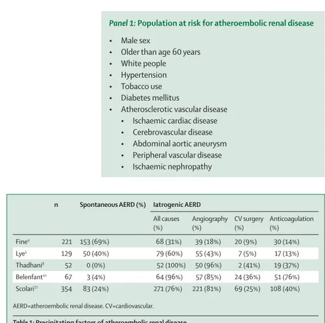
Panel 1: Population at risk for atheroembolic renal disease
• Male sex
• Older than age 60 years • White people • Hypertension • Tobacco use • Diabetes mellitus
• Atherosclerotic vascular disease • Ischaemic cardiac disease • Cerebrovascular disease • Abdominal aortic aneurysm • Peripheral vascular disease • Ischaemic nephropathy
n Spontaneous AERD (%) Iatrogenic AERD
All causes (%) Angiography (%) CV surgery (%) Anticoagulation (%) Fine4 221 153 (69%) 68 (31%) 39 (18%) 20 (9%) 30 (14%) Lye5 129 50 (40%) 79 (60%) 55 (43%) 7 (5%) 17 (13%) Thadhani8 52 0 (0%) 52 (100%) 50 (96%) 2 (41%) 19 (37%) Belenfant10 67 3 (4%) 64 (96%) 57 (85%) 24 (36%) 51 (76%) Scolari27 354 83 (24%) 271 (76%) 221 (81%) 69 (25%) 108 (40%) AERD=atheroembolic renal disease. CV=cardiovascular.
emboli are also identifi ed in the skin, gastrointestinal tract, and muscle11,20,21 (fi gures 1 and 2).
Clinical features
Clinical presentation of atheroembolic renal disease depends on the location and size of the embolising plaque, lodging sites, intensity and recurrence of the showering process, and pre-existing atherosclerotic disease. The ascending aorta and proximal aortic arch are the major sources of emboli to the cerebral and retinal arteries. Emboli to the visceral and renal arteries and to arteries of the legs and feet come from the descending thoracic and abdominal aorta.1–4 Subclinical
or mild episodes can go completely unrecognised. However, massive embolisation can lead to a fulminant clinical picture, with multi-system involvement. 8–12
(panel 2, table 2).
Kidney function can be aff ected acutely, subacutely, or in a chronic but slow, progressive way.8–12,24,25 A pronounced
renal impairment with acute onset, arising within 1 week of a clear causal event, aff ects 20–30% of patients.24,27 This
abrupt renal impairment is due to massive migration of crystals, and is associated with evidence of embolisation elsewhere, aff ecting gastrointestinal and cutaneous systems in most cases. The most common form of atheroembolic renal disease is a subacute presentation, with progressive renal failure developing in a stepwise way during several weeks after an inciting event. Frock and co-workers41 reported that the mean interval between
the arteriographic procedure and diagnosis of disease was 5·3 weeks.41 This long delay between the triggering
event and the onset of renal insuffi ciency suggests a
causal role of recurrent emboli showers and infl ammatory endothelial reaction. Another presentation is a chronic, slowly progressive renal impairment, often ascribed to nephroangiosclerosis or ischaemic nephropathy, which frequently co-cluster with cholesterol emboli. This mild form, with low-grade, clinically silent crystal migration is frequently underdiagnosed because extrarenal signs are absent and renal biopsy samples are not taken.42
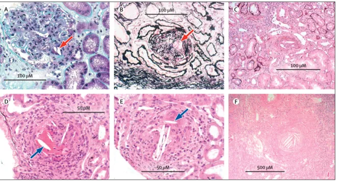
Figure 1: Renal cholesterol crystal emboli
Intraglomerular cholesterol crystals (arrows, A–B); cholesterol crystals in a renal arteriole (C); crystals in an arcuate artery with a pseudovasculitis infl ammatory reaction (arrow, D); crystals in an arcuate artery, and encasement of a crystal by a giant cell (arrow, E); and organised occlusive crystals in a renal arteriole (F).
A D E F B C D B E 100 μM 50 μM 50 μM 500 μM 100 μM 100 μM
Figure 2: Cholesterol crystal emboli in derma arterioles
Low-power view of cholesterol crystals (arrow, A); and high-power showing clusters of cholesterol crystals (B–D).
A B C D 500 μM 50 μM 100 μM 100 μM
The clinical course of renal failure can be variable. Dialysis is needed in 28–61% of patients with acute or subacute disease, with 20–30% partly recovering kidney function after a variable period of dialytic support.8–12
Recovery can be due to reversal of infl ammation and resolution of concurrent acute tubular necrosis in
ischaemic areas. Finally, atheroembolic renal disease can be associated with severe, uncontrolled hypertension. Renal infarction is a rare fi nding.20,21
Skin lesions are the most common extrarenal manifestations. Incidence of skin lesions is about 35%.43
However, in most recent studies,8–12,44–46 the frequency
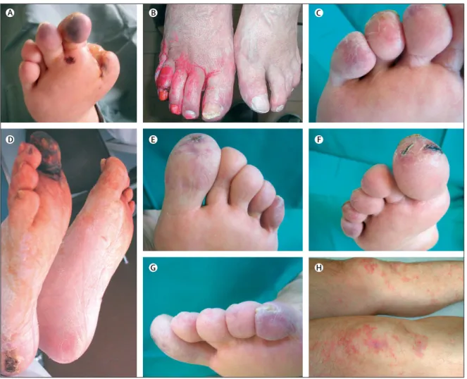
ranged from 75% to 96%. The classic skin lesions of this disease are blue toe syndrome and livedo reticularis. Aff ected toes are blue, cyanotic, painful, and cool to touch. The lesion might progress to ulceration, digital infarcts, or gangrene needing amputation. Patients often have palpable distal pulses. Livedo reticularis is a net-like, mottled red-blue discolouration usually on the lower legs and feet but sometimes on the buttocks and trunk. Blue toes and livedo reticularis are most evident on physical examination when the patient is upright. Rarely, the skin lesions are erythematous painful nodules or palpable purpura, requiring exclusion of leukocytoclastic vasculitis44–46 (fi gure 3).
The gastrointestinal system is the third most often aff ected system (18–48% of patients), leading to substantial morbidity.8–12 Gastrointestinal tract involvement can take
many forms. Patients can develop symptoms such as occult or frank blood loss and abdominal pain. Bleeding originates from any site, including the stomach, and results from superfi cial mucosal ulcerations or infarcts. Abdominal pain is caused by ischaemia of the bowel wall, potentially creating malabsorption and diarrhoea. Frank bowel infarction and perforation usually arise in the setting of catastrophic multi-organ disease. The liver, gallbladder, and pancreas are less commonly aff ected.47–51 Gastrointestinal eff ects are often
overlooked. Diagnosis can be established by pathological examination, because the endoscopic appearance is non-specifi c.52 Patient outcomes in the presence of
gastro-intestinal disease are very poor.24,27
The true incidence of neurological embolisation is unknown and diffi cult to estimate, because the defi nitive diagnosis depends on evidence of embolisation to other organs. In the largest studies, neurological manifestations occurred in 4–23% of the patients with atheroembolic renal disease.8–12,27 Mental confusion, transient ischaemic
attack, focal neurological defi cits, and amaurosis fugax are common fi ndings.53–55 Retinal emboli can aff ect
6–25% of patients,56 representing a key to diagnosis
(fi gure 4).8–12 Rarely, spinal cord infarction with leg
paralysis develops.57–60
Myositis and splenic infarcts are rare manifestations.61
Emboli deriving from the aortic root or the proximal segments of the coronary arteries are often associated with angina and sudden cardiac death.62,63 Pulmonary
involvement proven by biopsy sample, characterised by diff use alveolar haemorrhage mimicking systemic vasculitis, has been described in a few patients. The pathogenetic mechanisms of pulmonary haemorrhage remain poorly understood—a local infl ammatory reaction elicited by emboli might have a role.64–67 In autopsy studies,
subclinical involvement of adrenals, testes, prostate,
Panel 2: Clinical manifestations of atheroembolic renal
disease
Kidney
• Acute, subacute, and chronic renal failure • Severe uncontrolled hypertension • Renal infarction
Skin
• Livedo reticularis • Blue toe syndrome • Ulceration and gangrene • Purpura
Gastrointestinal system
• Abdominal pain • Gastrointestinal bleeding
• Bowel ischaemia, infarction, and obstruction • Pancreatitis, cholecystitis, and abnormal liver tests • Splenic infarcts
Heart
• Myocardial ischaemia • Myocardial infarction
Central nervous system
• Transient ischaemic attacks • Amaurosis fugax
• Altered mental status • Cerebral infarction • Spinal cord infarction
Eye
• Retinal emboli (Hollenhorst plaques)
Systemic signs • Fever • Weight loss • Malaise • Myalgia • Anorexia
n Skin lesions GI tract CNS Retinal emboli Eosinophilia
Fine4 221 75 (35%) 21 (10%) 0 14 (6%) 22 (73%)
Lye5 129 55 (43%) 16 (10%) 15 (12%) 12 (10%) 57 (71%)
Thadhani8 52 26 (50%) 15 (29%) 12 (23%) 13 (25%) 11 (22%)
Belenfant10 67 60 (90%) 22 (33%) 3 (4%) 15 (22%) 39 (59%)
Scolari27 354 266 (75%) 43 (12%) 35 (10%) 24 (7%) 238 (67%)
GI=gastrointestinal. CNS=central nervous system.
thyroid, and virtually any organ has been reported4,17
(fi gure 5). Finally, several non-specifi c fi ndings can accompany the course of atheroembolic renal disease, such as fever, weight loss, myalgias, and headache, suggesting the systemic nature of the disease.4,8–12,20,21
Laboratory fi ndings
Laboratory test fi ndings are non-specifi c, such as anaemia, leukocytosis, thrombocytopenia, and raised concentrations of infl ammatory markers (ESR or C-reactive protein). Results of urinalysis are typically benign, with few cells and a minimum amount of proteinuria—a fi nding consistent with ischaemic damage.4,4,8–12 Nephrotic-range
proteinuria has been described11,68,69 in a few patients with
superimposed biopsy-proven focal glomerulosclerosis or diabetic nephropathy. Eosinophilia is an abnormal laboratory fi nding that frequently occurs during the acute phase of the disease, thereby showing immunological activation at the surface of the exposed emboli.8–12 In a
previous review, Kasinath and co-workers70 described
eosinophilia in 80% of cases of atheroembolic renal disease. Eosinophilia was typically transient, ranging from
6% to 18% in a recent study.70 67% of 354 patients had an
eosinophil count higher than 500 cells per μL, confi rming that eosinophilia can help establish the diagnosis.27
Other features can include hypocomplementaemia,71
which has been reported in 39% of patients in one study.5
However, this fi nding was not consistent with results of other studies.8–12 Abnormal laboratory fi ndings can
implicate specifi c organs: increased concentrations of hepatic enzymes suggest liver disease, and raised amylase and lipase concentrations pancreatic disease; raised creatinine phosphokinase concentration are suggestive of myositis, and increased serum aspartate amino-transferase and lactate dehydrogenase concentrations are suggestive of renal infarction.4,10,11 Finally, a few studies9,27,72
reported proportions of cholesterolaemia in patients with atheroembolic renal disease, with total cholesterol higher than 5·2 mmol/L ranging from 23% to 27%, and 64%.
Diagnosis
Because atheroembolisation is ubiquitous, atheroembolic renal disease can mimic several diff erent clinical syndromes.21,73 Knowledge of the risk factors and
A B C
D E F
G H
Figure 3: Skin lesions in atheroembolic renal disease
recognition of the variable clinical presentations can heighten the likelihood of making a premortem diagnosis.8–12,27 The typical patient at risk is a man older
than age 60 years with known atherosclerotic disease presenting with a clinical triad of a precipitating event, acute or subacute renal failure, and typical skin fi ndings. Presence of eosinophilia should raise the level of suspicion. Renal biopsy is regarded as the defi nitive
method for diagnosis. Renal samples yield a positive diagnosis in more than 75% of patients with acute or subacute renal failure.74 However, taking a renal biopsy
sample is not always feasible, especially in sick patients. Taking a biopsy sample of skin lesions, a straightforward, and relatively non-invasive procedure, has a high diagnostic yield approaching 92%.41 Feet and lower legs
are also the best sites for biopsy.74,75 Occasionally,
histological confi rmation can be made from biopsy samples from less likely target organs, such as gastrointestinal tissues.24
Tissue biopsy sampling is not necessary when cholesterol crystals are seen in the retinal vessels (Hollenhorst plaques), which can be identifi ed in 10–25% of cases.8–12 For this reason, fundoscopy examination
should never be omitted. Diagnosis by tissue sample is not needed in the presence of the classic triad. For patients with the classic triad, atheroembolic renal disease can be diagnosed solely on a clinical basis.8–12,27 Thus, renal biopsy
sampling can be avoided in some patients. Conversely,
A C E B D F 50 μM 50 μM 50 μM 50 μM 100 μM 100 μM
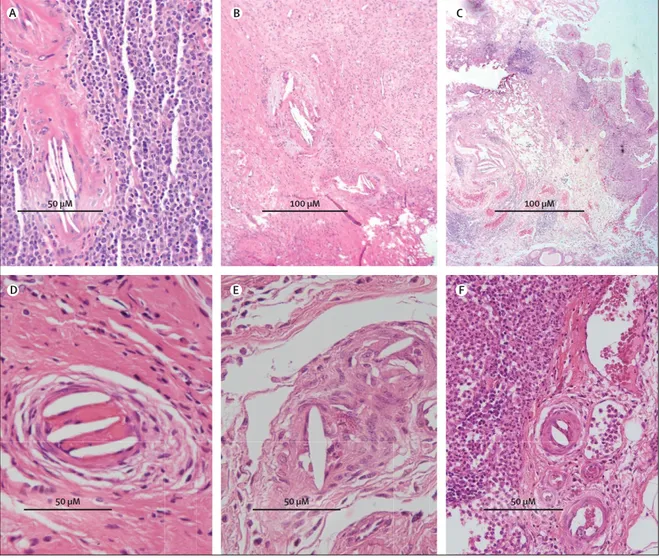
Figure 5: Cholesterol crystal in extrarenal sites
Spleen (A); prostate (B); bladder (C); stomach (D); ileum (E); and lymph node (F).
Figure 4: Retinal cholesterol crystal emboli
renal biopsy is crucial for diagnosis of cases with chronic, smouldering forms of this disease, which are frequently spontaneous and usually develop in the absence of evident extrarenal signs11,24,27 (panel 3 and table 3).
Diff erential diagnoses are contrast nephropathy, small-vessel vasculitis, drug-induced interstitial nephritis, and subacute bacterial endocarditis. In contrast nephropathy, the rise in plasma creatinine concentrations happens immediately after the radiographic study, and the course is diff erent—creatinine concentration peaks within a few days after exposure and returns to or near baseline after 10–14 days.11,20,21,24 Extrarenal emboli, if present,
enable diagnosis of atheroembolic renal disease. The disease can be distinguished from systemic vasculitis by urinalysis, typically characterised by the absence of proteins and blood cells in atheroembolic renal disease as opposed to the nephritic sediment of vasculitis, and by an autoimmune screening including antineutrophil cytoplasmic antibodies testing.11,24 The temporal relation
between use of an off ending drug and occurrence of fever, skin rash, eosinophilia, and acute renal failure strongly suggests acute tubulointerstitial nephritis, a diagnosis that should be confi rmed by renal biopsy. Presence of subacute bacterial endocarditis needs to be ruled out, especially if hypocomplementaemia is present. Signs of systemic infection and echocardiographic fi ndings exclude atheroembolic renal disease.
Embolic disease in renal allografts
Atheroembolic renal disease in renal allografts is rare, with a frequency ranging from 0·39% to 0·47%.76,77 So far,
only 45 cases have been reported—too few to defi ne the natural history of the disorder.76–85 Such disease is probably
underestimated because of the use of small needles for taking biopsy samples and sampling error. Atheroemboli causing injury to the renal allograft can arise from either the donor or the recipient vessels. Two distinct clinical presentations have been described. The fi rst is an early atheroembolic renal disease, with emboli frequently released from the donor’s arteries before or during organ harvesting. More rarely, early embolisation originates from the recipient’s atheromatous vessels during the anastomosis. The early form is usually associated with primary non-function, and the embolic disease is confi ned to the allograft. The second form is a late clinical presentation, which can arise years after transplantation in stable grafts. In this case, emboli originate from the recipient’s vessels. The disease is usually associated with recipitating factors, and in some cases shows features of a systemic disorder.
Renal outcomes of early and late atheroembolic disease seem to diff er. Graft loss is frequently associated with early disease, in which the emboli originate from the donor. Prognosis can be especially poor in the setting of primary non-function. Conversely, late manifestations usually have a good outcome, with graft recovery in most cases.11,76–78 The reason for this diff erence could be
attributable to extensive embolisation in an atherosclerotic donor during organ procurement. Because the tendency to accept donors and recipients older than age 60 years and to use marginal donors with advanced atherosclerosis has increased, atheroembolic renal disease in renal allografts will probably be encountered more often than previously. To reduce the risk of atheroemboli, an accurate assessment of organ donors should be done. At the time of organ procurement, manipulation of the aorta should be kept to a minimum, mobilising the kidneys without cross-clamping the aorta, as done for living-related donors. Finally, the number of invasive vascular work-up procedures should be kept to a minimum.11,77
Treatment
For patients with atheroembolic renal disease, the aim of treatment is to restrict the extent of ischaemic damage and prevent recurrent embolisation. No defi nitive treatment has been established. Therapeutic measures are mostly preventive and supportive.8–12,24–27 Mainstays of
Panel 3: Diagnostic criteria for atheroembolic renal disease
1 Patient at risk
• Men older than 60 years • Longstanding hypertension • Tobacco use
• Diff use atherosclerotic disease
2 Presence of classic triad
• Exposure to precipitating factor • Acute or subacute renal failure
• Peripheral signs of embolisations (eg, blue toe syndrome)
3 Gastrointestinal or neurological eff ects and eosinophilia
• Should raise the level of suspicion
4 Histological confi rmation
• Pathological specimens obtained from the kidney, skin, or muscle
• Skin biopsy sample
• Simple, low-invasive procedure with high diagnostic yield • Tissue biopsy sample not necessary in presence of retinal
emboli
• Tissue sample not needed in presence of classic triad • Renal biopsy is crucial to diagnosis of chronic forms of
atheroembolic renal disease
n Biopsy sample Clinical Postmortem
Kidney Skin Muscle GI tract Other
Fine4 221 15 (7%) 18 (8%) 18 (8%) 5 (2%) 13 (6%) 0 152 (69%)
Thadhani8 52 16 (31%) 9 (17%) 11 (21%) 0 0 5 (9%) 11 (22%)
Belenfant10 69 24 (36%) 15 (22%) 0 12 (18%) 0 16 (24%)* 0
Scolari27 354 63 (18%) 13 (37%) 0 4 (1%) 0 14 (42%)* 7 (2%)
*Clinical diagnosis was confi rmed by retinal emboli in some patients. GI=gastrointestinal.
preventive treatment are based on restriction of exposure to precipitating factors, such as withdrawal of anticoagulant therapy after carefully considering the pros and cons of these drugs, and avoidance of any additional radiological or aortic surgery procedure. Medical intervention in established cases is mostly for symptom management, and should aim to provide optimum treatment of associated hypertension and cardiac and renal failure. Cardiac dysfunction and hypertension should be aggressively treated. In some cases, renal replacement therapy is needed for uraemia and fl uid balance control in patients that are unresponsive to high-dose diuretic therapy.10–12,24,27,74 The optimum type of dialysis is not known.
Both peritoneal dialysis and haemodialysis have been used.10,11,86,87 Peritoneal dialysis might improve the chances
of renal recovery because it does not need anticoagulation. In the absence of intestinal ischaemia and malnutrition, peritoneal dialysis should be the preferredtreatment modality. However, haemodialysis without or with only minimalanticoagulation is not contraindicated. In most severe cases, during the acute phase, adequate nutritional support can be provided by parenteral nutrition.10
No clinical trials of treatment for patients with atheroembolic renal disease have been done. However, results of some studies of steroids and statins suggest that they might have a potential role. Possible benefi ts of steroids have been reported in small series.88–90 Belenfant
and co-workers10 used low-dose steroid (0·3 mg/kg) in
18 patients with relapsing disease who had improved symptoms and nutritional intake.10 Results of other series72
have shown benefi cial eff ects with high doses of steroids. Conversely, other reports4 have shown little or marginal
benefi t from steroid treatment. In a prospective study27 of
354 patients with atheroembolic renal disease, steroids were not associated with improved renal or patient outcomes. Thus, steroid use is still controversial, although it could have a role in patients with multi-system involvement, recurrent and progressive disease, and systemic infl ammation.
Interest has grown in the potential protective role of statins. Occasional cases of atheroembolic renal disease have responded to statins.91 Ina prognostic study,12 patients
assigned statin therapy had a lowered risk of development of endstage renal disease. A large prospective study27
confi rmed this fi nding. Statins had a benefi cial eff ect even when statin therapy was started after diagnosis of atheroembolic renal disase. The protective eff ect of statins could be attributable to plaque stabilisation and regression through lipid-lowering and anti-infl ammatory mech-anisms. Plaque stabilisation might result in a lowered risk for further embolisation. However, the observational nature of these data precludes establishment of causality. Despite these limitations, use of statins could be justifi able (panel 4). Finally, there are isolated reports of successful therapy in small numbers of patients given iloprost, pentoxifylline, and LDL apheresis. These approaches have yet to be tested in controlled studies. Other drugs such as antiplatelet agents and low-molecular weight dextran do not provide any benefi t.11,24
Surgical removal of the source of emboli could off er a defi nitive treatment option. However, surgical treatment is often not feasible and is associated with substantial morbidity. A clear embolic source is diffi cult to identify. Additionally, patients are frequently too weak for surgery. Finally, the necessary aortic clamping during surgery would induce major risk for recurrence. A surgical approach was assessed by Keen and colleagues92 in a
prospective study of 100 patients. They concluded that surgery can be an option when the emboli source is located in the infrarenal aorta. If supra-renal aorta is implicated, morbidity and mortality is increased because of the risk for visceral and renal atheroembolisation. Thus, surgical intervention should be regarded as a rescue therapy, and restricted to life-threatening situations. To eliminate the embolic source, percutaneous transluminal angioplasty and stent placement have been undertaken in a few patients with aortoiliac and femoral artery sources of embolisation.93,94 These endovascular procedures could
also be associated with a high risk of further embolisation,
Panel 4: Treatment of atheroembolic renal disease
• Major goal
• Restrict extent of the ischaemic damage • Prevent recurrent embolisation • No defi nitive treatment has been established • Therapeutic modalities are mostly preventive and
supportive
• Preventive treatment aims to avoid further precipitating factors
• Withdrawal of anticoagulant therapy
• Avoid any new radiological or aortic surgery procedure • Medical intervention
• Mostly symptomatic
• Aggressive treatment of associated hypertension and cardiac and renal failure
• Optimum type of dialysis (peritoneal dialysis vs haemodialysis) unknown
• Adequate nutritional support when needed • Steroids in patients with multisystem involvement
and recurrent, progressive disease
• Statins should be off ered to all patients with atheroembolic renal disease
n Renal failure needing dialysis
Recovery of dialysis-dependent renal failure
Maintainence dialysis (end of follow-up) 1-year mortality Fine4 221 62 (28%) 13 (21%) 0 179 (81%) Lye5 129 52 (40%) 13 (26%) 0 83 (64%) Thadhani8 52 23 (44%) 7 (32%) 0 45 (87%) Belenfant10 67 41 (61%) 16 (39%) 23 (35%) 9 (13%) Scolari27 354 11 (33%) 33 (28%) 88 (25%) 60 (17%)
possibly occurring as a consequence of vessel injury caused by catheters and guide wires within the aorta (type 1 embolisation) or by active plaque disruption during balloon dilationor stenting (type 2 embolisation).95
Defi nitive recommendations for use of this approach cannot yet be made.
Primary prevention of atheroembolic renal disease is important, restricting indications for angiography and surgical procedures in atherosclerotic patients. Non-invasive diagnostic methods, suchas MR angiography or computer-assisted tomographicangiography, could in part obviate the risk for catheter-induced embolisation. During endovascular procedures, use of proper and cautious techniques, including a so-called no touch technique avoiding direct trauma of the catheter tip to the atheromatous vessel wall might reduce risk of embolisation. Finally, the potentialapplication of embolic protection devices, which removes atheromatous debris, could help keep risk of embolisation to a minimum.
Outcomes
Renal survival
Few outcome studies have been done in patients with atheroembolic renal disease. Renal prognosis is poor. Lye and co-workers5 reported that 40% of 129 patients needed
dialysis, of whom only 21% recovered suffi cient renal function to stop dialysis. In a series, Thadhani and colleagues8 reported that 44% of 52 patients underwent
dialysis. Belenfant and co-workers10 reported that dialysis
was needed in 61% of 67 patients, with 13 patients regaining suffi cient renal function to be free of dialysis. In a prospective study,12 37% of 95 patients needed dialysis,
14 recovered suffi cient renal function to stop dialysis, and 23 remained dialysis dependent (24%; table 4). The most important predictors of endstage renal disease needing permanent dialysis therapy were pre-existing chronic renal insufficiency and longstanding hypertension. Another major fi nding was a protective benefi t of statins—patients given statin treatment had a signifi cantly lower risk for development of end-stage renal disease than did those not given statins.12
Patient survival
Atheroembolic renal disease is generally associated with high mortality. An 81% mortality rate was reported by Fine and co-workers at 1 year.4 Lye and colleagues5 reported
a 64% mortality rate at 1 year. The major cause of death was cardiovascular. A favourable clinical outcome, with a 1-year mortality rate of 13%, was reported by Belenfant,10
with an aggressive therapeutic approach. The protocol was based on avoidance of anticoagulation and aortic manipulating procedures, good control of hypertension and heart failure, dialytic therapy, and adequate nutritional support. Benefi ts of similar supportive care were confi rmed in a prospective study27 of 354 patients, with an
83% 1-year patient-survival rate (table 4). Predictors of increased risk for death were diabetes and heart failure,
baseline levels of renal function, acute or subacute renal failure, and development of extrarenal manifestations, especially aff ecting the gastrointestinal tract (relative risk [RR] 2·57, 95% CI 1·69–3·93). Finally, previous statin use was associated with improved patient survival (RR 0·44, 0·28–0·67). Notably, the same predictors were associated with an increased risk for endstage renal disease, suggesting a common underlying mechanism for renal and cardiovascular events.27
Future research
Atheroembolic renal disease has become a recognisable cause of renal failure. However, its incidence in patients at risk remains unknown, and diagnosis depends on how much experience centres have had with this disease. Ideally, a systematic search for signs and symptoms of the dis ease in populations undergoing aortic procedures would provide information to answer this important question. In terms of prognosis of established cases, avoid ance of known precipitating factors and individualised supportive measures could substantially improve clinical out come.96,97 However, the
disorder greatly aff ects patient and renal survival, and further research is urgently needed to alter its clinical course. Three diff erent areas of clinical investigation need to be explored.
The fi rst is the potential benefi t of specifi c therapies, such as steroids and statins. A reactive infl ammation around the crystals is suggested by eosinophilia, a cytokine-mediated occurrence,98 and has a role in causing
or worsening ischaemia. However, anti-infl ammatory benefi ts might be counterbalanced by harmful eff ects of steroids on the cardiovascular system. For this reason, benefi ts and harms of steroid treatment should be tested in clinical trials. The protective role of statins12,27 should
also be confi rmed by intervention studies. Atheroembolic renal disease alone represents an indication for standard statin therapy. Intensive statin therapy seems to provide more cardiovascular protection than does standard therapy in patients with coronary disease,99,100 and whether
this eff ect applies to patients with atheroembolic renal disease needs to be tested.
The second area of research is the modality of renal replacement therapy in patients needing dialysis. Clinical trials are necessary to test whether standard haemodialysis needing anticoagulation is associated with worse patient and renal outcomes than is peritoneal dialysis. Last, the prognostic eff ect of antiembolic devices during endovascular procedures on the evolution of atheroembolic renal disease is unknown. This approach is the standard of care during carotid artery stenting because it reduces procedure-related embolic strokes.101 Such protection
devices have also been used during renal artery stenting with promising results.102 Insuffi cient information is
available to test the pros and cons of a broadened use to reduce short-term and long-term atheroembolic com-plications of endovascular procedures.
Contributors
Both authors contributed equally to the design and writing of this Seminar.
Confl icts of interest
We declare that we have no confl icts of interest.
Acknowledgments
We thank R Tardanico (University of Brescia, Italy) for providing fi gure 1, F Facchetti (Brescia, Italy) for fi gure 2, C Rollino (Turin, Italy) for fi gure 4A, A Marciello (Turin, Italy) for fi gure 4B, R Tardanico for fi gure 5, and B J Barrett (Memorial University of Newfoundland, Canada) for his critical comments on this report and H Benediktsson (University of Calgary, Canada) for his assistance with the histology section preparation.
References
1 Flory CM. Arterial occlusions produced by emboli from eroded aortic atheromatous plaques. Am J Pathol 1945; 21: 549–65.
2 Thurlbeck WM, Castleman B. Atheromatous emboli to the kidneys after aortic surgery. N Engl J Med 1957; 257: 442–47.
3 Kassirer JP. Atheroembolic renal disease. N Engl J Med 1969;
280: 812–18.
4 Fine MJ, Kapoor W, Falanga V. Cholesterol crystal
embolization: a review of 221 cases in the English literature. Angiology 1987; 38: 769–84.
5 Lye WC, Cheah JS, Sinniah R. Renal cholesterol embolic disease. Case report and review of the literature. Am J Nephrol 1993;
13: 489–93.
6 Panum PL. Experimentell Beitrage zur Lehre von der Embolie. Virchows Arch 1862; 25: 308–10.
7 Benson RL. The present status of coronary arterial disease. Arch Pathol Lab Med 1926; 2: 876–916.
8 Thadhani RI, Camargo CA Jr, Xavier RJ, Fang LS, Bazari H. Atheroembolic renal failure after invasive procedures. Natural history based on 52 histologically proven cases. Medicine 1995; 74: 350–58. 9 Mayo RR, Swartz RD. Redefi ning the incidence of clinically detectable
atheroembolism. Am J Med 1996; 100: 524–29.
10 Belenfant X, Meyrier A, Jacquot C. Supportive treatment improves survival in multivisceral cholesterol crystal embolism.
Am J Kidney Dis 1999; 33: 840–50.
11 Scolari F, Tardanico R, Zani R, et al. Cholesterol crystal embolism: A recognizable cause of renal disease. Am J Kidney Dis 2000;
36: 1089–109.
12 Scolari F, Ravani P, Pola A, et al. Predictors of renal and patient outcomes in atheroembolic renal disease: a prospective study. J Am Soc Nephrol 2003; 14: 1584–90.
13 Saklayen MG. Atheroembolic renal disease: preferential occurrence in whites only. Am J Nephrol 1989; 9: 87–88.
14 Cross S. How common is cholesterol embolism? J Clin Pathol 1991;
44: 859–61.
15 Drost H, Buis B, Haan D, Hillers JA. Cholesterol embolism as a complication of left heart catheterisation. Report of seven cases. Br Heart J 1984; 52: 339–42.
16 Kealy WF. Atheroembolism. J Clin Pathol 1978; 31: 984–89. 17 Moolenaar W, Lamers CB. Cholesterol crystal embolization in the
Netherlands. Arch Intern Med 1996; 156: 653–57.
18 Ramirez G, O’Neill WM, Lambert R, Bloomer HA. Cholesterol embolization: a complication of angiography. Arch Intern Med 1978;
138: 1430–32.
19 Gore I, Collins D. Spontaneous atheromatous embolization. Review of the literature and a report of 16 additional cases. Am J Clin Pathol 1960; 33: 416–26.
20 Jones DB, Iannaccone PM. Atheromatous emboli in renal biopsies. An ultrastructural study. Am J Pathol 1975; 78: 261–76.
21 Lie JT. Cholesterol atheromatous embolism. The great masquerader revisited. Pathol Annu 1992; 2: 17–50.
22 Preston RA, Stemmer CL, Materson BJ, Perez-Stable E, Pardo V. Renal biopsy in patients 65 years of age or older. An analysis of the results of 334 biopsies. J Am Geriatr Soc 1990; 38: 669–74. 23 Stone W, Fogo A. Hypertension and renal disease in the elderly.
Boston, MA: Blackwell, 1992.
24 Meyrier A. Cholesterol crystal embolism: diagnosis and treatment. Kidney Int 2006; 69: 1308–12.
25 Modi K, Rao V. Atheroembolic renal disease. J Am Soc Nephrol 2001;
12: 1781–87.
26 Arroyo LH, Lee RT. Mechanisms of plaque rupture: mechanical and biologic interactions. Cardiovasc Res 1999; 41: 369–75.
27 Scolari F, Ravani P, Gaggi R, et al. The challenge of diagnosing atheroembolic renal disease: clinical features and prognostic factors. Circulation 2007; 116: 298–304.
28 Mittal BV, Alexander MP, Rennke HG, Singh AK. Atheroembolic renal disease: a silent masquerader. Kidney Int 2008; 73: 126–30. 29 Johnson L, Esente P, Giambartolomei A, et al. Peripheral vascular
complications of coronary angioplasty by the femoral and brachial techniques. Cathet Cardiovasc Diagn 1994; 31: 165–72.
30 Nasser T, Mohler E third, Wilensky R, Hathaway D. Peripheral vascular complications following coronary interventional procedures. Clin Cardiol 1995; 18: 609–14.
31 Fukumoto Y, Tsutsui H, Tsuchihashi M, Masumoto A, Takeshita A. The incidence and risk factors of cholesterol embolization syndrome, a complication of cardiac catheterization: a prospective study. J Am Coll Cardiol 2003; 42: 211–16.
32 Saklayen MG, Gupta S, Suryaprasad A, Azmeh W. Incidence of atheroembolic renal failure after coronary angiography. A prospective study. Angiology 1997; 48: 609–13.
33 Rudnick MR, Berns JS, Cohen RM, Goldfarb S. Nephrotoxic risks of renal angiography: contrast media-associated nephrotoxicity and atheroembolism—a critical review. Am J Kidney Dis 1994;
24: 713–27.
34 Feder W, Auerbach R. “Purple toes”: an uncommon sequela of oral coumarin drug therapy. Ann Intern Med 1961; 55: 911–17. 35 Moldveen-Geronimus M, Merriam JC Jr. Cholesterol embolization.
From pathological curiosity to clinical entity. Circulation 1967;
35: 946–53.
36 Hitti WA, Wali RK, Weinman EJ, Drachenberg C, Briglia A. Cholesterol embolization syndrome induced by thrombolytic therapy. Am J Cardiovasc Drugs 2008; 8: 27–34.
37 Eliot RS, Kanjuh VI, Edwards JE. Atheromatous Embolism. Circulation 1964; 30: 611–18.
38 Snyder HE, Shapiro JL. A correlative study of atheromatous embolism in human beings and experimental animals. Surgery 1961; 49: 195–204.
39 Gore I, McCombs HL, Lindquist RL. Observations on the fate of cholesterol emboli. J Atheroscler Res 1964; 4: 527–35.
40 Warren BA, Vales O. The ultrastructure of the stages of atheroembolic occlusion of renal arteries. Br J Exp Pathol 1973;
54: 469–78.
41 Frock J, Bierman M, Hammeke M, Reyes A. Atheroembolic renal disease: experience with 22 patients. Nebr Med J 1994; 79: 317–21. 42 Zucchelli P, Zuccalà A. The diagnostic dilemma of hypertensive nephrosclerosis: the nephrologist’s view. Am J Kidney Dis 1993;
21: 87–91.
43 Falanga V, Fine MJ, Kapoor WN. The cutaneous manifestations of cholesterol crystal embolization. Arch Dermatol 1986; 122: 1194–98. 44 Liew YP, Bartholomew JR. Atheromatous embolization.
Vasc Med 2005; 10: 309–26.
45 Donohue KG, Saap L, Falanga V. Cholesterol crystal embolization: an atherosclerotic disease with frequent and varied cutaneous manifestations. J Eur Acad Dermatol Venereol 2003; 17: 504–11. 46 Jucgla A, Moreso F, Muniesa C, Moreno A, Vidaller A. Cholesterol
embolism: still an unrecognized entity with a high mortality rate. J Am Acad Dermatol 2006; 55: 786–93.
47 Ben-Horin S, Bardan E, Barshack I, Zaks N, Livneh A. Cholesterol crystal embolization to the digestive system: characterization of a common, yet overlooked presentation of atheroembolism. Am J Gastroenterol 2003; 98: 1471–79.
48 Moolenaar W, Lamers CB. Cholesterol crystal embolization and the digestive system. Scand J Gastroenterol Suppl 1991; 188: 69–72. 49 Moolenaar W, Lamers CB. Cholesterol crystal embolisation to the
alimentary tract. Gut 1996; 38: 196–200.
50 Moolenaar W, Lamers C. Gastrointestinal blood loss due to cholesterol crystal embolization. J Clin Gastroenterol 1995;
21: 220–23.
51 Moolenaar W, Lamers C. Cholesterol crystal embolization to liver, gallbladder, and pancreas. Dig Dis Sci 1996; 41: 1819–22. 52 Paraf F, Jacquot C, Bloch F, de Montpreville V, Bruneval P.
Cholesterol crystal embolization demonstrated on GI biopsy. Am J Gastroenterol 2001; 96: 3301–04.
53 Beal M, Williams RS, Richardson EP Jr, Fisher CM. Cholesterol embolism as a cause of transient ischemic attacks and cerebral infarction. Neurology 1981; 31: 860–65.
54 Price DL, Harris J. Cholesterol emboli in cerebral arteries as a complication of retrograde aortic perfusion during cardiac surgery. Neurology 1970; 20: 1209–14.
55 Masuda J, Yutani C, Ogata J, Kuriyama Y, Yamaguchi T. Atheromatous embolism in the brain: a clinicopathologic analysis of 15 autopsy cases. Neurology 1994; 44: 1231–37.
56 Hollenhorst RW. Signifi cance of bright plaques in the retinal arterioles. JAMA 1961; 178: 23–29.
57 Desnuelle C, Lanteri-Minet M, Hofman P, Butori C, Bedoucha P, Chatel M. Cholesterol emboli with neurologic manifestation in the spinal cord. Rev Neurol 1992; 148: 715–18.
58 Reich MP. Paraplegia following resection of an abdominal aortic aneurysm. Report of a case of atheromatous embolization to the anterior spinal artery. Vasc Surg 1968; 2: 230–34.
59 Blankenship J, Mickel S. Spinal cord infarction resulting from cardiac catheterization. Am J Med 1989; 87: 239–40.
60 Slavin R, Gonzalez-Vitale J, Marin O. Atheromatous emboli to the lumbosacral spinal cord. Stroke 1975; 6: 411–15.
61 Robinson R, Pemberton M, Goddard MJ. Myositis due to cholesterol emboli. Postgrad Med J 1993; 69: 947–49.
62 Teja K, Crampton RS. Intramural coronary arteritis from cholesterol emboli: a rare cause of unstable angina preceding sudden death. Am Heart J 1985; 110: 168–70.
63 Keon WJ, Heggtveit HA, Leduc J. Perioperative myocardial infarction caused by atheroembolism. J Thorac Cardiovasc Surg 1982; 84: 849–55. 64 Case records of the Massachusetts General Hospital. Weekly
Clinicopathological Exercises. Case 11–1996. A 69-year-old man with progressive renal failure and the abrupt onset of dyspnea. N Engl J Med 1996; 334: 973–79.
65 Sabatine M, Oelberg D, Mark EJ, Kanarek D. Pulmonary cholesterol crystal embolization. Chest 1997; 112: 1687–92.
66 Vacher-Coponat H, Pache X, Dussol B, Berland Y. Pulmonary-renal syndrome responding to corticosteroids: consider cholesterol embolization. Nephrol Dial Transplant 1997; 12: 1977–79. 67 Walton TJ, Samani NJ, Andrews R. Systemic cholesterol crystal
embolisation with pulmonary involvement: a fatal combination after coronary angiography. Postgrad Med J 2002; 78: 288–89.
68 Haqqie S, Urizar R, Singh J. Nephrotic-range proteinuria in renal atheroembolic disease: report of four cases. Am J Kidney Dis 1996;
28: 493–501.
69 Greenberg A, Bastacky S, Iqbal A, Borochovitz D, Johnson J. Focal segmental glomerulosclerosis associated with nephrotic syndrome in cholesterol atheroembolism: clinicopathological correlations. Am J Kidney Dis 1997; 29: 334–44.
70 Kasinath B, Corwin H, Bidani A, Korbet S, Schwartz M, Lewis E. Eosinophilia in the diagnosis of atheroembolic renal disease. Am J Nephrol 1987; 7: 173–77.
71 Cosio F, Zager R, Sharma H. Atheroembolic renal disease causes hypocomplementaemia. Lancet 1985; 2: 118–21.
72 Dahlberg P, Frecentese D, Cogbill T. Cholesterol embolism: experience with 22 histologically proven cases. Surgery 1989;
105: 737–46.
73 Scoble JE, O’Donnell PJ. Renal atheroembolic disease: the Cinderella of nephrology? Nephrol Dial Transplant 1996; 11: 1516–17.
74 Smyth JS, Scoble JE. Atheroembolism. Curr Treat Options Cardiovasc Med 2002; 4: 255–65.
75 Anderson WR, Richards AM. Evaluation of lower extremity muscle biopsies in the diagnosis of atheroembolism. Arch Pathol 1968;
86: 535–41.
76 Ripple M, Charney D, Nadasdy T. Cholesterol embolization in renal allografts. Transplantation 2000; 69: 2221–25.
77 Lai C, Randhawa P. Cholesterol embolization in renal allografts: a clinicopathologic study of 12 cases. Am J Surg Pathol 2007;
31: 536–45.
78 de Takats D, Pollock L, O’Donnell P, Snowden S, Bewick M, Scoble JE. Is cholesterol embolic disease an unrecognized cause of renal graft dysfunction? Nephrol Dial Transplant 1996; 11: 1325–27.
79 Jennings W, Smith J, Corry R. Atheromatous embolization as a cause of increasing creatinine levels in a renal transplant patient. Transplant Proc 1990; 22: 279.
80 Scolari F, Tardanico R, Pola A, et al. Cholesterol crystal embolic disease in renal allografts. J Nephrol 2003; 16: 139–43. 81 Shappell H, Nylander W, VanBuren D, Fogo A. Adult man with
primary allograft nonfunction. Am J Kidney Dis 2000; 35: 997–1001. 82 Singh I, Killen P, Leichtman A. Cholesterol emboli presenting as
acute allograft dysfunction after renal transplantation. J Am Soc Nephrol 1995; 6: 165–70.
83 Ackoundou-N’Guessan C, Bismuth J, Canet S, Iborra F, Mourad G. Partial recovery of delayed graft function due to cholesterol emboli after renal transplantation. Saudi J Kidney Dis Transpl 2008;
19: 631–35.
84 Schonermarck U, Guba M, Weiss M, Illner WD, Arbogast H, Bosch T. Cholesterol atheroembolic disease in kidney allografts—case report and review of the literature. Clin Nephrol 2006; 66: 386–90. 85 Ott U, Gerth J, Grone HJ, Grone E, Wolf G. Cholesterol embolization
in a renal graft. Clin Transplant 2008; 22: 677–80.
86 Siemons L, van den Heuvel P, Parizel G, Buyssens N, De Broe M, Cuykens J. Peritoneal dialysis in acute renal failure due to cholesterol embolization: two cases of recovery of renal function and extended survival. Clin Nephrol 1987; 28: 205–08.
87 Theriault J, Agharazzi M, Dumont M, Pichette V, Ouimet D, Leblanc M. Atheroembolic renal failure requiring dialysis: potential for renal recovery? A review of 43 cases. Nephron Clin Pract 2003; 94: 11–18.
88 Yucel A, Kart-Koseoglu H, Demirhan B, Ozdemir F. Cholesterol crystal embolization mimicking vasculitis: success with corticosteroid and cyclophosphamide therapy in two cases. Rheumatol Int 2006;
26: 454–60.
89 Nakayama M, Nagata M, Hirano T, et al. Low-dose prednisolone ameliorates acute renal failure caused by cholesterol crystal embolism. Clin Nephrol 2006; 66: 232–39.
90 Mann S, Sos TA. Treatment of atheroembolization with corticosteroids. Am J Hypertens 2001; 14: 831–34.
91 Woolfson RG, Lachmann H. Improvement in renal cholesterol emboli syndrome after simvastatin. Lancet 1998; 351: 1331–32. 92 Keen R, McCarthy W, Shireman P, et al. Surgical management
of atheroembolization. J Vasc Surg 1995; 21: 773–80.
93 Matchett W, McFarland D, Eidt J, Moursi M. Blue toe syndrome: treatment with intra-arterial stents and review of therapies. J Vasc Interv Radiol 2000; 11: 585–92.
94 Carroccio A, Olin JW, Ellozy SH, et al. The role of aortic stent grafting in the treatment of atheromatous embolization syndrome: results after a mean of 15 months follow-up. J Vasc Surg 2004; 40: 424–29. 95 Rocha-Singh KJ, Eisenhauer AC, Textor SC, et al. Atherosclerotic
peripheral vascular disease symposium II: intervention for renal artery disease. Circulation 2008; 118: 2873–78.
96 Polu KR, Wolf M. Clinical problem-solving. Needle in a haystack. N Engl J Med 2006; 354: 68–73.
97 Scoble JE. Is nihilism in the treatment of atheroembolic disease at an end? Am J Kidney Dis 1999; 33: 975–76.
98 Cogan E, Schandene L, Papadopoulos T, Crusiaux A, Goldman M. Interleukin-5 production by T lymphocytes in atheroembolic disease with hypereosinophilia. J Allergy Clin Immunol 1995; 96: 427–29. 99 LaRosa J, Grundy S, Waters D, et al. Intensive lipid lowering with
atorvastatin in patients with stable coronary disease. N Engl J Med 2005; 352: 1425–35.
100 Pedersen TR, Faergeman O, Kastelein JJ, et al. High-dose atorvastatin vs usual-dose simvastatin for secondary prevention after myocardial infarction: the IDEAL study: a randomised controlled trial. JAMA 2005; 294: 2437–45.
101 Reimers B, Corvaja N, Moshiri S, et al. Cerebral protection with fi lter devices during carotid artery stenting. Circulation 2001; 104: 12–15. 102 Henry M, Henry I, Klonaris C, et al. Renal angioplasty and stenting
under protection: the way for the future? Catheter Cardiovasc Interv 2003; 60: 299–312.