Nef protein induces differential effects in CD8
þ
cells from
HIV-1-infected patients
F. Silvestris, G. Camarda, A. Del Prete, M. Tucci and F. Dammacco
Department of Biomedical Sciences and Human Oncology, University of Bari, ItalyAbstract Background The Nef protein of HIV-1 is suspected to play a role in the depletion of uninfected CD4þlymphocytes that leads to AIDS. By contrast its effect on CD8þcells, whose functions are also deregulated during HIV-1 infection, is presently unclear. Here we describe a number of derangements inducedin vitro by Nef in CD8þcells from HIV-1-infected patients.
Design Peripheral lymphocytes from 16 HIV-1þ subjects and 9 uninfected individuals were cultivated on a Nef-transfected mouse fibroblast layer exposing the carboxyl-terminal region of the viral protein on cell membrane. The cultures were then measured for both apoptosis and proliferation by subdiploid DNA content and Ki67 expression, respectively, whereas the molecular analysis of purified CD8þcells investigated the Fas-L mRNA levels in Nef-treated CTLs. In addition, we evaluated the Nef-induced variation in the extent of CD8þ/HLA-DRþsubset, which includes non cytotoxic cells secreting T-cell antiviral factor (CAF) and a soluble factor inhibiting the HIV-1 replication.
Results The viral protein induced in peripheral blood lymphocytes (PBL) a moderate tendency to proliferate, as measured by the increment of Ki67 antigen, particularly on the CD8þsubset of HIV-1 infected individuals (
P< 0·05). This profile was particularly evident in cultures from patients with severe CD4þ lymphopenia and paralleled an apparent expansion of the CD8þ/CD57þ suppressor cell subset. Molecular analysis of purified CD8þcells revealed a defective expression of Fas-L mRNA in Nef-cultured CTLs, whereas the viral protein exerted a down modulatory effect on the CD8þ/HLA-DRþ subset (P< 0·05), thus suggesting a potential inhibition of CAF.
Conclusions These results support a potential role of Nef in the progression of HIV-1 infection as a number of cellular functions are affected in the CD8þsubset. In particular, the defective functions of CD8þ cells induced by the viral protein could contribute, at least partly, to the escape of HIV-1 from the immune control of these cells.
Keywords CD8þcells, cytolysis, Fas–L, HIV, Nef. Eur J Clin Invest 1999; 29 (11): 980–991
Introduction
Both the human (HIV-1) and the simian (SIV) immuno-deficiency viruses are complex retroviruses whose genomes comprise many regulatory genes, namelytat, rev, vif, vpu, nef, vpr and vpx [1,2]. Among them, vif, vpu, vpr and nef are not needed for virus replicationin vitro and are there-fore termed ‘nonessential’ [3].
Thenef gene is located at the 30ofenv gene and partially
overlaps the U3 region of the 30 LTR. The product of
HIV-1nef is a 27–30 kDa protein whose myristoylation at its N-terminus [4] permits its linkage to the cytoplasmic leaflet of the plasma membrane. However, perturbation of the lipid bilayers by the myristoylated protein also leads the carboxyl-terminal portion of Nef to protrude on the cell membrane [5]. Even so, infected lymphocytes usually express Nef in both their cytosol and nuclei and may also solubilize the protein by the cell membrane [6–8].
The biological role of Nef on HIV-1 replication is controversial. In contrast to itsin vitro inefficacy [9–12], recent studies have shown that the intact nef gene is pivotal in maintaining a high virus load in rhesus monkeys infected with the pathogenic SIVmac239 clone, and for
Correspondence to: Prof. Franco Silvestris MD, DIMO, University of Bari, Section of Internal Medicine and Human Oncology, P.zza Giulio Cesare, 11–70124 Bari, Italy. Tel.: þ39 80 5478771; fax: þ39 80 5478820; e-mail:[email protected] Received 16 March 1999; accepted 30 June 1999
Nef protein down-regulates CD8þcells 981
the development of AIDS [13]. In addition, the wild-type nef gene from HIV-1 significantly increases the viral load in the SCID-Hu mouse model (a severe combined immuno-deficient mouse transplanted with human fetal liver and thymus) [14], whereas its mutated form showing a struc-tural defect in 30 LTR sequences has been tentatively
associated with long-term non-progression of the disease in HIV-1 infected subjects [15]. These observations sug-gested thatnef gene and its product could be involved in the lymphocyte depletion that leads to AIDS. Demonstration of the suppressive effect of Nef on T-cell activation as well as the inhibition of CD4þcell growth [16] and the down-regulation of both TCR-induced IL-2 production [17], and activation of transcriptional factors NF-kB and AP-1 [18] has lent support to this interpretation. In addition, Th1 cytokine secretion is specifically suppressed in T cells incu-bated with Nef protein, whereas Th2 production is scarcely affected [19].
Nef may directly exert a cytotoxic effect on CD4þcells by inducing their apoptosis [20,21], just asenv [22] and tat [23] promote this in T cells and contribute to the devel-opment of CD4þ lymphopenia. This hypothesis is sup-ported by suppression of the in vitro proliferation of CD4þcells and the fact that membrane expression of the C–terminus domain of Nef in infected lymphocytes cocultured with unprimed T-cell lines activates their suici-dal death [20,21,24]. Further investigations have suggested that the Nef-induced down-regulation of CD4 molecules correlates with the intracellular levels of Nef [25], whereas its apoptogenic effect on T and B cells, macrophages and neutrophils during HIV-1 infection is apparently unrelated to the CD95/Fas pathway [26,27]. Conversely, soluble Nef has been shown to induce considerable cellular activation as a result of its superantigen effect on MHC class II molecules [28,29].
This work studied the effect of Nef on peripheral lym-phocytes from HIV-1 infected individuals at different stages. CD4þcells were clearly affected and underwent a variable degree of suppression, whereas CD8þcells showed an apparent increase in their proliferative rate, especially the CD8þ/CD57þcells which exhibit a prevalently sup-pressor phenotype. In addition, Nef inhibited both the expression of Fas-L on cytotoxic CD8þcells and the pro-liferation of HLA-DRþlymphocytes, namely a population that suppresses HIV-1.
Methods
Plasmid construction
Full-lengthnef was amplified from pNL4–3 (kindly pro-vided by Dr M. Federico, Laboratory of Virology, Istituto Superiore di Sanita`, Rome, Italy) by polymerase chain reaction (PCR) using the primer pair covering the ATG and the ‘stop codon’ of the nativenef sequence [4]. The amplification employed 25 cycles at 948C for 45
product was then purified and cloned into pTracer eukary-otic expression vector (Invitrogen, Pero, Italy) to create pTracer-nef.
Cell lines and transfection procedures
In preparing the feeder layer expressing Nef for human lymphocyte cultures, we adopted a xenogeneic adherent cell line to avoid molecular interactions between syngeneic human cells potentially perturbing the cellular response. Therefore, we used the NIH-3T3 cells, a fibroblast line established from NIH Swiss mouse embryo cultures (ATCC, Rockville, Maryland, USA), which have been successfully employed in transfection studies exploring the response of human NK cells [30]. The 3T3 cells were grown in DMEM (at high glucose rate) medium supple-mented with 10% FCS andL-glutamine and subsequently
transfected with 5 mg of pTracer-nef by Lipofectamine (Gibco-BRL, Life Technologies Italia, San Giuliano Milanese, Italy) according to the manufacturer’s instruc-tions. After appropriate selection in G418 at 1 mg mL¹1, single clones were isolated and further analysed by RT-PCR to assess the quantitative expression of nef. We then selected nine clones showing high levels of nef RT-PCR product as evaluated by direct O.D. measurement after extraction from agarose gel (ConcertTMGel Extrac-tion Kit, Gibco BRL, Milan, Italy). In addiExtrac-tion, the mem-brane expression of Nef was estimated by flow cytometry with the anti-Nef monoclonal antibody (MoAb) AE6 (obtained from ‘AIDS Research and Reference Reagent Program’, Division of AIDS, NIAD, NIH, Bethesda MA, USA). Four clones (3T3–2, ¹3, ¹5, and ¹7) were positive for high membrane expression of Nef as demon-strated by a relative fluorescence intensity higher than one decade. Comparative experiments in preliminary tests in which peripheral blood lymphocytes (PBL) were incubated with each of the Nef-positive transfectants showed a major suppressive effect on CD8þ subsets by the 3T3–7 transfectant. As the protein was expressed on the membrane of more than 95% of cells in this clone, we used the 3T3–7 transformant as adherent layer to culture lymphocytes.
PBL purification and coculturing with Nef-transfected cells
The study included 16 HIV-1 infected patients at different disease stages and 9 healthy donors. Seven patients were arbitrarily considered as severely lymphopenic (CD4þcells #300 mL¹1
). PBL were purified from heparinized blood using Ficoll Hypaque gradient centrifugation and subse-quent removal of adherent cells by incubation for 45 min at 378C. The cells were then washed and cultured at 1×10⁶mL¹1 in 6-well plates containing the adherent 3T3–7 at 50–60% of confluency in RPMI-1640 plus
harvested, washed and used for further analysis. To pro-perly evaluate the effect of Nef, cellular analyses were compared in all instances to parallel cultures from each PBL sample of both patients and controls using non-transfected 3T3 cells as substrate.
Measurement of proliferation and apoptosis in cells stimulated by Nef
Proliferation was assessed by double fluorescence measure-ment of the expression of Ki67 antigen, in association with subset phenotyping with specific MoAbs to CD4, CD8, and CD16 antigens (Becton-Dickinson, Mountain View, CA, USA). Double fluorescence was also used to measure the size of the CD8þ/CD57þsubset in response to Nef. To evaluate apoptosis, cells were harvested and permeabilized by 70% ethanol for 1 h at þ48C prior to staining with propidium iodide at 50 mg mL¹1 in PBS to detect subdiploid DNA [31,32]. The extent of apoptosis was then expressed as the percentage of cells with sub-diploid DNA. These analyses were conducted in a FACS-can (Becton-Dickinson) using the CellQuest program.
Preparation of purified CD8þcell suspension and phenotype analysis
The effect of Nef on T-cell subsets was examined by purifying CD8þsubsets from each PBL sample preincu-bated with nef-transfected cells and the relative control by immunomagnetic isolation using Dynabeads M-450 (Unipath, Milan, Italy). The full procedure recommended by the manufacturer was employed, providing a final enrichment of 92·3% of CD8þcells. Double fluorescence analysis then evaluated the expression of HLA-DR anti-gens, which define the activated phenotype of CD8þcells as response to the incubation with Nef.
Molecular analyses
In view of the proliferative response of CD8þcells to Nef, we measured the expression of Fas-L mRNA in purified CD8þ populations, because Fas-L is a prevalent marker of cytotoxic cells (CTL) within this subset. Thus, mRNA was isolated from 1×10⁶CD8þcells in both unstimulated and Nef-stimulated cultures from 11 patients by the guani-dium thiocyanate-cesium chloride procedure (Invitrogen, Celbio, Pero, Italy), and transcribed into first-strand cDNA with the Boehringer-Mannheim (Milan, Italy) cDNA kit. The Fas-L wild-type-specific primers were designed in relation to the known gene structure as follows: 50-GCC CAA GCT TGA AGC AGC CCT-30
(FW), and 50-TGC TGT GTG CAT CTG GTC GGT
AGA-30 (RV) related to the exon-2 of the gene. Both
primers and cDNA from each CD8þ preparation were added to the PCR mixture (Perkin-Elmer, Cetus, Norwalk, Connecticut, USA) with subsequent amplification in a
thermal cycler for 35 cycles (1 min 948C, 1 min 65 8C, 1 min 728C) and the Fas-L PCR product was visualized on a 1·5 agarose gel with ethidium bromide. Fas-L mRNA was estimated by evaluating the relative bands as ‘trace quantity value’ (o.d.×mm of each band) by Quantity-One 4·3 software in the Fluor-S gel analyzer (Bio-Rad Labs, Hercules, CA, USA).
Statistical analyses
Mean values of cellular subsets and phenotype expression between groups were compared by Student’s t-test and, in several instances, by the Wilcoxon test as a nonpara-metric method.
Results
Nef induces a variable proliferation on PBL
Since Nef has been consistently reported to suppress T cells [16,20,21], we first assessed the extent of apoptosis in PBL cultures from both HIV-1-infected patients and healthy controls after 60 h of incubation with the nef-3T3–7 transfectant. An apparent inhibition of apoptosis was noted in both groups of cultures. Nef induced a variable though significant decrease in the relative extent of the mean subdiploid DNA peaks (M16 SD): 14·2 6 5% and 10·76 3% in Nef-treated cultures, as compared to 32·46 4% and 19·6 6 3% in untreated cultures from patients with severe (CD4þ# 300 mL¹1) and moderate (> 300 mL¹1
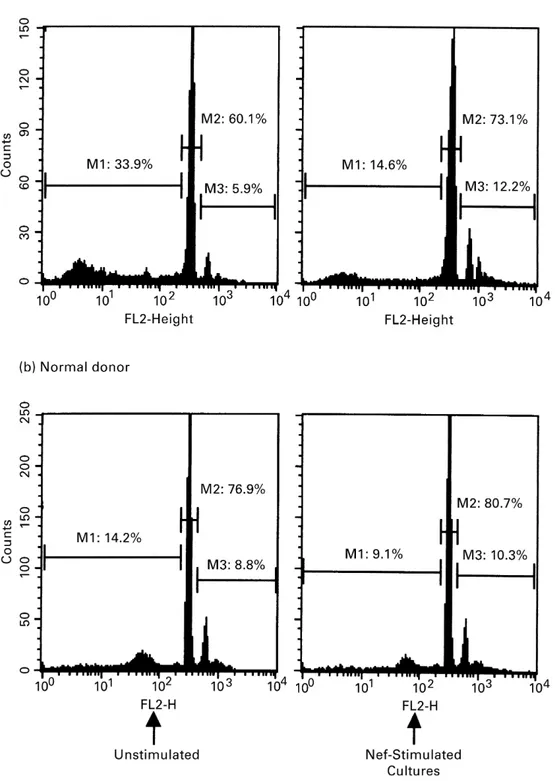
) lymphopenia (P< 0·02 in both instances). The control cultures from uninfected donors showed a similar, though lower decline of apoptosis: mean M1 8·76 3% in Nef-stimulated vs. 14·6 6 3 in untreated cultures. However, this variation was not different in a statistical mode (P> 0·2). These data are in agreement, and were in apparent concordance with reference values from our previous work [32], though the prolonged xeno-antigen stimulation by 3T3 cells in increasing the PBL apoptosis independently of Nef cannot be excluded. Repre-sentative profiles of this down-modulated apoptosis in a patient with severe CD4þ lymphopenia (Pt. # 1) and a control subject are shown in Fig. 1. In both instances, the subdiploid DNA cell population was quantitatively reduced in cells harvested after incubation with the 3T3– 7 clone (right panels). Morphological features of cell activation and proliferation, namely the expansion of both size and forward scatters, were also evident in both the euploid and the hyperdiploid cell populations gated in M2 and M3, respectively. These individual variations of fluorescence intensity were not statistically evaluated, though they were clearly detected in cells cultured in the presence of Nef and occurred in most preparations.
Further experiments were addressed to identify the cellular subset(s) with a prominent tendency to proliferate. Double fluorescence flow cytometry was used to measure Ki67 expression in CD16þ, CD4þ and CD8þ subsets
Nef protein down-regulates CD8þcells 983
Figure 1 Variation of ploidy in PBL from a severely
lymphopenic HIV-1 patient and a normal donor after 60 h of incubation in the presence of myristoylated Nef protein of HIV-1 exposed by the membrane of transfected 3T3–7 fibroblasts (right panel). This treatment usually reduced M1 peak corresponding to the extent of the subdiploid DNA content in
apoptotic cells as compared to the control culture with untransfected fibroblasts (left panel). By contrast, M2 (euploid cells) and M3 (hyperdiploid cells) populations were proportionally increased. The pattern is representative of most cultures from patients where inhibition of apoptosis reflected the relative tendency to proliferate.
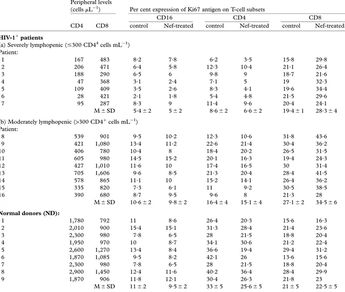
following a 60-h-culture with Nef. Table 1 shows the distribution of relative values of peripheral CD4þ and CD8þ cell counts in association with the effect of Nef in groups of cultures from patients and controls. Nef generally had little effect on the proliferation of CD16þ and CD4þ cells, because their mean values within each group showed a decrease, thus suggesting an apparent suppression. On comparing single values within each subset, however, it was found that patients with severe lymphopenia were variably affected in relative CD4þcell proliferation. Similarly, a proliferative decrease was prevalent in the uninfected controls and the mean values of suppression were significant (P< 0·02), thus confirming the susceptibility of uninfected cells to Nef-induced apoptosis [17,21]. In contrast, a clear-cut trend to proliferation was detected
in the CD8þ subset. The increase of positive cells was higher in patients than in the controls and the difference between mean values was significant (P< 0·05 in both instances) in each group. We also recorded a moderate
Table 1 Peripheral levels of CD4þand CD8þcells and variation of Ki67 antigen expression on specific T-cell subsets after 60-cultures of PBL in the presence of Nef expressed by the membrane of 3T3-7 transfected fibroblasts as layer (Nef-treated) compared to control cultures using untransfected cells (control). Significant variations included an increase of the Ki67 expression in CD8þsubset from both groups of patients with severe as well as moderate CD4þlymphopenia (P< 0.05 in both instances). In addition, a slight though significant decrease of CD4þsubset was also observed in cultures from control donors (P< 0.02)
Peripheral levels
(cells mL¹1) Per cent expression of Ki67 antigen on T-cell subsets
CD16 CD4 CD8
CD4 CD8 control Nef-treated control Nef-treated control Nef-treated
HIV-1þpatients
(a) Severely lymphopenic (#300 CD44
cells mL¹1) Patient: 1 167 483 8.2 7.8 6.2 3.5 15.8 29.8 2 206 471 6.4 5.8 12.3 10.4 21.1 26.4 3 188 290 6.5 6 9.8 9 18.7 21.6 4 47 368 3.1 2.4 7.1 5 19 32.3 5 109 409 3.5 2.6 8.3 4.1 19.6 34.4 6 28 421 2.1 1.8 5.4 4.8 21.5 29.6 7 95 287 8.3 9 11.4 9.6 20.4 24.1 M6 SD 5.46 2 56 2 8.66 2 6.66 2 19.46 1 28.36 4 (b) Moderately lymphopenic (>300 CD4þ cells mL¹1) Patient: 8 539 901 9.5 10.2 12.3 10.6 31.8 43.6 9 421 1,080 13.4 11.2 22.6 21.4 30.4 36.2 10 406 780 10.4 8 18.4 20.2 26.5 31.5 11 605 980 14.5 15.2 20.1 16.3 19.4 24.3 12 427 1,010 11.6 10 17.4 16.5 30 31.4 13 705 1,606 9.6 8.5 21.3 20.4 28.4 41.5 14 578 865 11.1 10 15.2 14.1 26.4 36.2 15 335 820 7.3 6.1 11 9.2 30.5 38.5 16 390 680 8.7 9.5 9.6 8 21.3 28 M6 SD 10.66 2 9.86 2 16.46 4 15.16 4 27.16 2 34.56 6 Normal donors (ND): 1 1,780 792 11 8.6 26.4 20.3 15.6 16.3 2 2,010 900 15.4 15.1 31.3 28.4 21.4 23.6 3 2,300 980 7.8 6.5 28 21.5 18.8 20.4 4 1,950 970 10 8.7 34.1 30.6 21.2 22.4 5 2,600 1,270 13.4 8.4 36.6 19.4 29.4 31.2 6 1,870 1,085 9.5 8.2 42.1 26 13.6 15.6 7 2,300 980 7.8 6.5 28 21.5 18.8 20.4 8 2,900 1,450 12.4 11.6 40.2 36.4 28.4 29.9 9 1,870 906 11.8 12.1 30.4 26.3 21.8 23 M6 SD 116 2 9.56 2 336 5 25.66 5 216 5 22.56 5
Figure 2 Cytofluorimetric evaluation of changes in the size of
CD8þ/CD57þsubset in HIV-1þpatients with and without severe lymphopenia (peripheral CD4þcells< 300 mL¹1) and in a normal subject. The fluorescence profiles are related to single subjects and show representative patterns for each group of cultures. Incubation of PBL in the presence of Nef (right panels) induced a major subset growth in cultures from lymphopenic patients, as in section (a), and a poor increase in those from nonlymphopenic patients (b) and healthy controls (c). Left panels refer to control cultures prepared with untransfected fibroblasts.
though not significant tendency to proliferation (P> 0·1) in cultures from healthy controls.
A major proliferative effect is detectable in CD8þ/CD57þcells
Additional phenotyping tests identified the CD8þsubsets sensitive to Nef. The effect of Nef on the CD8þ/CD57þ subset, which displays a suppressor phenotype, was investi-gated by measuring the extent of the double positive CD8þ/ CD57þ subset on harvested PBLs from all cultures. An apparent increase in the percentage of these cells was observed in most Nef-stimulated patient cultures from patients showing the proliferative trend by Ki67 expression. Major elevations of CD8þ/CD57þcells (100% of increase) occurred prevalently in Nef-stimulated cultures from the severely CD4þ lymphopenic patients (
P< 0·01). In fact, the mean value of 16·86 4% of CD8þ/CD57þ cells in cultures from this group rose to 35·16 6% after incubation with Nef. Cell cultures from nonlymphopenic patients also showed a slight increment of mean levels (24·66 3% vs. 20·36 7% in unstimulated cultures), whereas no evident variations were observed in the control cultures from the uninfected donors (10·96 2% vs. 9·8 6 3% of CD8þ/CD57þ cells in unstimulated cultures). Figure 2 compares representative profiles from a severely lympho-penic (patient no. 4), a nonlympholympho-penic patient (patient no. 15) and a control donor (normal donor no. 1): the CD8þ/CD57þ increase was doubled in the first patient, whereas it was small in the second. The pattern relative to the control shows that Nef has little effect on CD8þ/ CD57þpopulation in cultures from healthy donors. These data suggested that Nef stimulation results in greater expansion of suppressor CD8þcells when CD4þcells are severely depleted as in the advanced stages of HIV-1 infection.
Nef down-regulates Fas-L expression in cytotoxic CD8þcells from HIV-1-infected patients
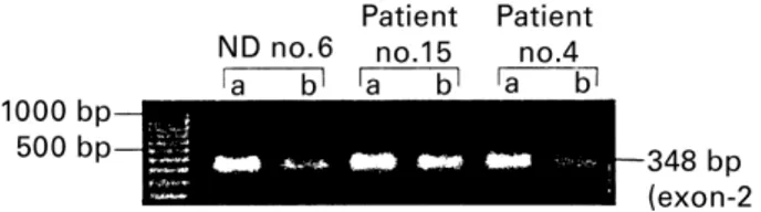
We next looked to assess whether or not Nef was able to affect the CD8þ cells displaying the cytolytic phenotype (CTL). Fas-L expression was then explored in these cells by measuring the relative mRNA levels of purified CD8þ cells from most patient cultures, five patients with severe and six with moderate lymphopenia. In addition, CD8þ cells were harvested from the cultures of five healthy controls. We observed an apparent diminution of the Fas-L bands in Nef-exposed CD8þcells from all culture samples. Figure 3 illustrates this pattern in a representative subject from each group, namely from patients no. 4 and 15, and normal donor no. 6. The quantitative evaluation by the Fluor-S analyzer confirmed a general decrease of Fas-L expression after treatment with Nef (lanes b). We found a difference in the Fas-L mRNA trace quantity values, which were apparently higher in patients with severe CD4þ lymphopenia (0·112 of mean reduction
value as compared to 0·087 o.d.×mm of the other group of patients). However, these variations were not statistically evaluated in comparing groups of samples because of the high intragroup variability.
Suppressor anti-HIV-1 CD8þlymphocytes are inhibited by Nef
Lastly, the role of Nef on noncytotoxic HIV-1-specific suppressor CD8þ lymphocytes in cultures from HIV-1-infected subjects was examined. This anti-HIV-1 response appears to be associated primarily with activated CD8þ cells expressing both HLA-DR and CD28 antigens and acts through a soluble inhibitory factor termed CAF (CD8þT-cell antiviral factor) [33]. Therefore, our analysis investigated the effect of Nef on this specific subset by measuring in double fluorescence the expression of HLA-DR molecules in purified CD8þ cells from four patients with severe and 5 with moderate lymphopenia. Nef down-regulated HLA-DR in most cultures. Patients with the lowest CD4þlevels showed a significant suppres-sion of their CD8þ/HLA-DRþ population in response to Nef: the mean number declined from 32·96 8 to 15·16 7 (P < 0·02). A similar effect was also observed in single patients with moderate CD4þ lymphopenia. However, although the decrement of the mean value of HLA-DRþ cells ranged from 37·66 4% in untreated cultures to 32·16 6% in the presence of Nef, it was not statistically significant (P> 0·2).
Figure 4 reports the cytofluorimetric pattern of two patients from each group and gives an example of the suppressive effect of Nef on these cells, which was higher than 50% in the CD8þpopulation of both patients with severe CD4þ lymphopenia (section a). Similarly, a lower suppression of proliferation was demonstrable in both patients with moderate lymphopenia (section b), whose average value was about 5%, and reflected the results
Figure 3 Comparative PCR amplification of Fas-L mRNA in
CD8þcells purified from cultures stimulated by Nef (lane b) with respect to unstimulated cultures (lane a). The treatment down-regulated Fas-L mRNA in all instances, particularly in patients with severe CD4þlymphopenia (patient no. 4 is representative of this group).
Figure 4 Representative patterns of cytofluorimetric assays
related to quantitative variations of activated (HLA-DRþ) CD8þ cells in cultures from patients with severe (a) and moderate (b) lymphopenia in response to 60 h of incubation with Nef. This treatment induced a substantial decrease in this subset, especially in patients with advanced disease.
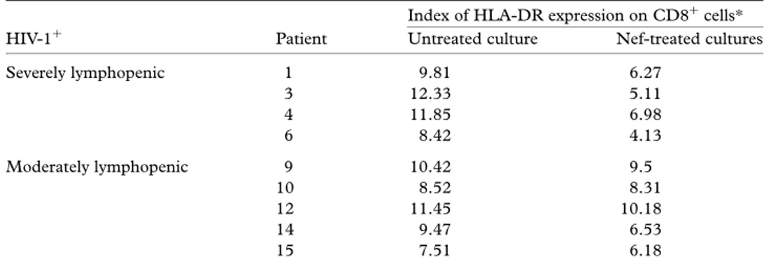
of other patients with similar CD4þ cell levels. Lastly, Table 2 includes the variations of mean values in fluores-cence intensity of DR expression following treatment with Nef. In most instances we observed a variable suppression of DR antigens in CD8þcells incubated with Nef. However, such variations of relative fluorescence intensity in DR expression were not calculated in statis-tical terms because of the small number of samples. These results suggested that activated anti-HIV-1 suppressor CD8þcells could be directly inhibited by Nef in culture.
Discussion
Our study on the effect of Nef on CD8þcells during HIV-1 infection has shown that, in addition to its cytolytic effect on other T cells, it stimulates the growth of CD8þ/CD57þ subsetin vitro, and down-modulates both the expression of Fas-L, a death factor of CTLs, and the proliferation of CD8þ cells activated to HIV-1; namely the CD8þ/HLA-DRþ subset. These immune dysfunctions of CD8þ cells are significant in patients with severe lymphopenia and probably contribute to the progression of HIV-1 infection to AIDS. Recent work has provided new insights into the function of Nef during HIV-1 disease. Originally seen as a negative regulator for virus replication [9] it is now being shown to play a positive role in replicating HIV-1 in primary T cells and T-cell lines [34–36]. It also down-regulates cell surface levels of CD4 molecules [37,38] and induces cytolysis of CD4þ cells in either its soluble [21] or its myristoylated form [20,24]. Although the myristoylated Nef peptide is anchored on the internal sheet of the plasma membrane by the N-terminal glycine, the myris-toylation procedure induces a disordering effect on the lipid bilayers, resulting in nonlamellar phases of the whole membrane layer and extrusion of the other terminal por-tion of the viral protein [39] [40]. Exhibipor-tion of the carboxy-terminal domain of Nef on the HIV-1-infected T-cell surface is critical in provoking cell death by cytotoxi-city in uninfected CD4þ lymphocytes [5,24], whereas
interaction of Nef with specific cellular kinases has sug-gested that it interferes with signalling pathways that promote T-cell activation [41].
Our present study emphasizes that, in contrast with the down-regulation of CD16þand CD4þcells, the myristoy-lated form of Nef from nef-3T3–7 transfectant induces a number of functional defects within the CD8þ subset that may promote the escape of both HIV-1 and infected cells from immune control. However, as it has been repor-ted that soluble Nef may affect proliferation of CD4þcells [21], further cellular suppression by soluble Nef mole-cules possibly released in our cultures by either trans-fected fibroblasts or intrans-fected cells cannot be ruled out. The first derangement induced by Nef concerns the increase of CD8þ/CD57þ cells. This subset is pheno-typically distinct because of its prevalent suppression of B-cell differentiation, proliferation and Ig secretion [42,43]. Although no specific antiviral cytotoxicity has been attributed to this population [44], its oligoclonal expansion occurs in several clinical conditions including cytomegalovirus infection [45], Crohn’s disease [46], common variable immunodeficiency [47], and HIV-1 infection [48]. In this disease as well as in the immune deficiency of bone marrow transplantation recipients [49], suppression was related to a 20-kDa, heat-stable suppres-sor factor [50] that inhibits lectin-driven proliferation and cytolysis [51]. In our study, we observed that a broad expression of Ki67 antigen was presented by CD8þcells cultured in the presence of Nef and paralleled the expan-sion of the CD57þ cell subset, in particular in patients with severe CD4þlymphopenia. By contrast, mostly stable or weakly increased values of CD57þcells were detected in cultures from moderately lymphopenic patients and the controls. We interpreted the differential proliferative response to Nef as a result of the divergent levels of CD4þ cellsin vivo. As patients with AIDS or severe lymphopenia have a minimal expression of the myristoylated protein on infected CD4þcells as a result of critical lymphocyte depletion, exposure of their PBL to the high virus antigen load as provided bynef-3T3–7 cells could have resulted in a major stimulation and growth of the CD57þpopulation.
Table 2 Variation of relative fluorescence intensity of HLA-DR antigen expression in CD8þcells from HIV-1þpatients after their culture in the presence of Nef
Index of HLA-DR expression on CD8þcells* HIV-1þ Patient Untreated culture Nef-treated cultures
Severely lymphopenic 1 9.81 6.27 3 12.33 5.11 4 11.85 6.98 6 8.42 4.13 Moderately lymphopenic 9 10.42 9.5 10 8.52 8.31 12 11.45 10.18 14 9.47 6.53 15 7.51 6.18
*The HLA-DR expression index was defined as the ratio of mean fluorescence channels (specific MoAb/isotype-matched control MoAb).
Nef protein down-regulates CD8þcells 989
Our study also points to the down-regulation of Fas-L expression in cytotoxic CD8þ cells treated with Nef. Fas-L is the functional coreceptor of Fas and induces apoptosis in Fasþ target cells by oligomerization of the receptor. This mechanism is used by most effector cells and Fas-L is considered a major phenotypic marker of CTLs. The apparent down-modulation of mRNA in treated cells in our study was in line with the relative increase of the CD57þsubset, especially in patients with severe lymphopenia. As Nef-induced cytolysis of unin-fected lymphocytes is independent of Fas/Fas-L system [26,27], this finding supports the view that the viral protein does not act through the Fas/Fas-L pathway. On the other hand, similarly defective Fas-L mRNA expression has been described in advanced HIV-1 infection [52–54], suggesting that the antigenic charge of soluble Nef may partly contribute to Fas-L down-regulation, though defec-tive CTL function in this disease could be the result of the increased suppression by CD57þ cells [45,48]. Our study, in fact, documented CD57þexpansion in advanced infection following exposure to Nef, though its suppression of Fas-L via other mechanisms cannot be excluded.
Another interesting finding was the detrimental effect of Nef on HIV-1 suppressor CD8þlymphocytes resulting in a defective expression of HLA-DR molecules. These data are in apparent contrast with the increase of Ki67þ cells within the same subset. However, previous studies have pointed out a possible discrepancy between both cell cycle and activation antigen expression in CD4þand CD8þ T cells in patients receiving the HAART (highly active antiretroviral treatment) therapy, especially during the chronic HIV-1 infection [55] [56]. In particular, though in the presence of a proliferative trend involving the total T-cell population, activation of CD8þ T cells measured by both CD38 and HLA-DR expression was significantly affected in this subset [57]. Our results show-ing a similar inhibition of HLA-DR molecules in response to Nef in patients with advanced disease are in line with these observations. On the other hand, CD8þcells expres-sing both HLA-DR and CD28 markers are thought to suppress HIV-1 replication by CAF in CD4þlymphocytes and macrophages [33]. We observed an evident decline of this population in cultures treated with Nef. Suppression of HIV-1 replication by CAF in naturally or acutely infec-ted CD4þ cells has been described as dose-dependent and correlated with clinical state and CD4þcell levels in peripheral blood [58]. Although we did not measure the extent of CAF secretion in cultures, the depletion of HLA-DRþ cells after incubation with Nef suggests a parallel reduction in the release of the suppressive factor, particularly in patients with advanced disease.
Our data on the effect of Nef on CD8þcells support recent studies describing its multifactorial role in the depletion of immune cells during HIV-1 infection. In addition to its cytolysis of uninfected CD4þlymphocytes, Nef may down-regulate the MHC class I on infected cells to promote their escape from cytolysis of functional
non cytotoxic anti-HIV-1 CD8þ cells are ways of escap-ing immune surveillance. Additional work is required to investigate the molecular defects of such Nef-related deregulations of CD8þ cells, though they may be the multiple outcome of a single deviation primarily induced by either soluble or myristoylated Nef. For instance, altera-tion in the secrealtera-tion of CD8þ cell cytokines could be a primary event leading to differentiated dysfunctions within the subset. Preliminary data from our laboratory support this hypothesis, in that CD8þ/CD57þ cells treated with Nef showed high mRNA expression of several suppressive cytokines, including IFN-g and IL-4, which could signifi-cantly influence both Fas-L expression in CTLs and CAF secretion in activated CD8þcells.
Acknowledgement
This work was supported by the National AIDS Research Project (1998) of the Italian Ministry of Health, ISS, Rome (Grant no. 59403·40).
References
1 Subbramanian RA, Cohen EA. Molecular biology of the human immunodeficiency virus accessory protein.J Virol 1994; 68: 6831–5.
2 Cullen BR. Regulation of HIV gene expression.AIDS 1995;
9 (Suppl. A): 19–32.
3 Trono D. HIV accessory proteins: leading roles for the supporting cast.Cell 1995; 82: 1898–923.
4 Allan JS, Coligan JE, Lee THet al. A new HTLV-III/LAV encoded antigen detected by antibodies from AIDS patients. Science 1985; 20: 810–3.
5 Curtain CC, Lowe MG, Arunagiri CK, Mobley PW, MacReadie IG, Azad AA. Cytotoxix activity of the amino-terminal region of HIV type I Nef protein.AIDS Res Hum Retroviruses 1997; 13: 1213–20.
6 Franchini G, Robert-Guroff M, Ghrayeb J, Chang NT, Wong-Staal F. Cytoplasmic localization of the HTLV-III 30
orf protein in cultured T cells.Virology 1986; 155: 593–9. 7 Guy B, Rivie`re Y, Dott K, Regnault A, Kieny MP. Mutational
analysis of the HIV Nef protein.Virology 1990; 176: 413–25. 8 Kienzle N, Bachmann M, Muller WEG, Muller-Lantzsch N. Expression and cellular localization of the Nef protein from human immunodeficiency virus-1 in stably transfected B-cells.Arch Virol 1992; 124: 123–32.
9 Ahmad N, Venkatesan S. Nef protein of HIV-1 is a transcriptional repressor of HIV-1 LTR.Science 1988; 241: 1481–85.
10 Luciw PA, Cheng-Mayer C, Levy JA. Mutational analysis of the human immunodeficiency virus: the orf-B region down-modulates virus replication.Proc Natl Acad Sci USA 1897; 84: 1434–8.
11 Niederman TMJ, Thielan BJ, Ratner L. Human immunodeficiency virus type 1 negative factor is a
12 Terwilliger E, Sodroski JG, Rosen CA, Haseltine WA. Effects of mutations within the 30 orf open reading frame
of human T-cell lymphotropic virus type III (HTLV-III/LAV) on replication and cytopathogenicity.J Virol 1986; 60: 754–60.
13 Kestler HW, Ringler DJ, Mori Ket al. Importance of the Nef gene for maintenance of high virus loads and development of AIDS.Cell 1991; 65: 651–62.
14 Jamieson BD, Aldrovandi GM, Planelles Vet al. Requirement of human immunodeficiency virus type I nef forin vivo replication and pathogenicity.J Virol 1994; 68: 3478–85. 15 Deacon NJ, Tsykin A, Solomon Aet al. Genomic structure
of an attenuated quasi-species of HIV-1 from a blood transfusion donor and recipients.Science 1995; 270: 988–91. 16 Greenway A, Azad A, McPhee D. Human immunodeficiency
virus type 1 Nef protein inhibits activation pathways in peripheral blood mononuclear cells and T-cell lines.J Virol 1995; 69: 1842–50.
17 Luria S, Chambers I, Berg P. Expression of the type 1 human immunodeficiency virus Nef protein in T cells prevents antigen receptor-mediated induction of interleukin-2 mRNA. Proc Natl Acad Sci USA 1991; 88: 5326–30.
18 Bandres JC, Ratner L. Human immunodeficiency virus type 1 Nef protein down-regulates transcription factors NFkB and AP-1 in human T cellsin vitro after T-cell receptor stimulation.J Virol 1994; 68: 3243–9.
19 Collette Y, Chang HL, Cerdan Cet al. Specific Th1 cytokine down-regulation associated with primary clinically derived human immunodeficiency virus type 1nef gene-induced expression.J Immunol 1996; 156: 360–70.
20 Fujii Y, Otake K, Tashiro M, Adachi A. Human
immunodeficiency virus type 1 Nef protein on the cell surface is cytocidal for human CD4þT cells.
FEBS Lett 1996; 393: 105–8.
21 Fujii Y, Otake K, Tashiro M, Adachi A. Soluble nef antigen of HIV-1 is cytocidal for human CD4þT cells.FEBS Lett 1996; 393: 93–6.
22 Banda NK, Bernier J, Kurahara DKet al. Cross-linking CD4 by human immunodeficiency virus gp120 primes T cells for activation-induced apoptosis.J Exp Med 1992; 176: 1099–10:6.
23 Westendorp MO, Frank R, Ochsembauer Cet al. Sensitization of T cell to CD95-mediated apoptosis by HIV-1 tat and gp120.Nature 1995; 375: 497–500. 24 Otake K, Fujii Y, Nakata Tet al. The carboxyl-terminal
region of HIV-1 Nef protein is a cell surface domain that can interact with CD4þcells.
J Immunol 1994; 153: 5826–37. 25 Hua J, Blai W, Truant R, Cullen BR. Identification of regions
in HIV-1 Nef required for efficient down regulation of cell surface CD4.Virology 1997; 231: 231–8.
26 Okada H, Takei R, Tashiro M. Hiv-1 nef protein induced apoptotic cytolysis of a broad spectrum of infected human blood cells independently of CD95 (Fas).FEBS Lett 1997;
414: 603–6.
27 Okada H, Takei R, Tashiro M. Nef protein induces apoptotic cytolysis of murine lymphoid cells independently of CD95 (Fas) and its suppression by serine/threonine protein kinase inhibitors.FEBS Lett 1997; 417: 61–4.
28 Torres BA, Johnson HM. Identification of an HIV-1 Nef peptide that binds to HLA class II antigens.Biochem Biophys Res Comm 1994; 200: 1059–65.
29 Torres BA, Tanabe T, Yamamoto JK, Johnson MH. HIV encodes for its own CD4 T cell superantigen.Biochem Biophys Res Comm 1996; 225: 672–8.
30 Poggi A, Panzeri MC, Moretta L, Zocchi MR.
CD31-triggered rearrangement of the actin cytoskeleton in human natural killer cells.Eur J Immunol 1996; 26: 817–24. 31 Silvestris F, Cafforio P, Frassanito MAet a.l Overexpression
of Fas antigen on T cells in advanced HIV-infection: differential ligation constantly induces apoptosis.AIDS 1996;
10: 131–41.
32 Silvestris F, Nagata S, Cafforio P, Silvestris N, Dammacco F. Cross-linking of Fas by antibodies to a peculiar domain of gp120x V3 loop can enhance T cell apoptosis in HIV–1–infected patients.J Exp Med 1996; 184: 2287–300. 33 Levy JA, Mackewicz CE, Barker E. Controlling HIV
pathogenesis: the role of noncytotoxic anti-HIV response of CD8þT cells.
Immunol Today 1996; 17: 217–24. 34 De Ronde A, Klaver B, Keulen W, Smit L, Goudsmit J.
Natural HIV-1 Nef accelerates virus replication in primary human lymphocytes.Virology 1992; 188: 391–5.
35 Spina CA, Kwoh TJ, Chowers MY, Guatelli JC, Richman DD. The importance of nef in the induction of human immunodeficiency virus type I replication from primary quiescent CD4 lymphocytes.J Exp Med 1994; 179: 115–22. 36 Fujinaga K, Zhong Q, Nakaya Tet al. Extracellular Nef
protein regulates productive HIV-1 infection from latency. J Immunol 1995; 155: 5289–98.
37 Cullen BR. The role of Nef in the replication cycles of the human and simian immunodeficiency viruses.Virology 1994;
205: 1–6.
38 Fujii Y, Ito M, Ikuta K. Evidence for the role of human immunodeficiency virus type 1 Nef protein as a growth inhibitor to CD4þT lymphocytes and for the blocking of the Nef function by anti-Nef antibodies.Vaccine 1993; 11: 837–42.
39 Curtain CC, Separovic F, Rivett Det al. Fusogenic activity of amino-terminal region of HIV type 1 Nef protein.AIDS Res Hum Retrovirus 1994; 10: 1231–40.
40 Hope MJ, Bally MB, Webb G, Cullis PR. Production of large unilamellar vesicles by a rapid extrusion procedure. Characterization of site distribution, trapped, & ability to maintain a membrane potential.Biochem Biophys Acta 1985;
812: 55–6.
41 Page KA, Van Schooten WCA, Feinberg MB. Human immunodeficiency virus type 1 Nef does not alter T cell sensitivity to antigen-specific stimulation.J Virol 1997; 71: 3776–87.
42 Lanier LL, Loken MR. Human lymphocyte subpopulations identified using three colour immunofluorescence and flow cytometric analysis: correlation of Leu-2, Leu-7, Leu-8 and Leu-11.J Immunol 1984; 132: 151–60.
43 Clement LT, Grossi CE, Gartland GL. Morphological and phenotypic features of a subpopulation of Leu-2þcells that suppress B cell differentiation.J Immunol 1984; 133: 2461–9. 44 Joly P, Guillon JM, Mayaud Cet al. Cell-mediated
suppression of HIV-specific cytotoxic T lymphocytes.J Immunol 1989; 143: 2193–201.
45 Wang ECY, Moss PAH, Frodsham P, Lehner P, Bell JI, Borysiewicz LK. CD8highCD57þT lymphocytes in normal, healthy individuals are oligoclonal and respond to human cytomegalovirus.J Immunol 1995; 155: 5046–56.
46 James SP, Neckers LM, Graeff AS, Cossman J, Balch CM, Strber W. Suppression of immunoglobulin synthesis by lymphocyte populations in patients with Crohn’s disease. Gastroenterology 1984; 66: 1510–5.
47 Baumert E, Wolff VG, Schlesier M, Peter HH.
Nef protein down-regulates CD8þcells 991
common variable immunodeficiency.Clin Exp Immunol 1992;
90: 25–31.
48 Toso JF, Chen CH, Mohr JRet al. Oligoclonal CD8 lymphocytes from persons with asymptomatic human immunodeficiency virus (HIV) type 1 infection inhibit HIV-1 replication.J Infect Dis 1995; 17: 964–73.
49 Leroy E, Calvo CF, Divine Met al. Persistence of T8þ/HNK-1þsuppressor lymphocytes in the blood of long-term surviving patients after bone marrow transplantation.J Immunol 1986; 137: 2180–6. 50 Sadat-Sowti B, Debre` P, Mollet Let al. An inhibitor of
cytotoxic functions produced by CD8þCD57þT lymphocytes from patients suffering from AIDS and immunosuppressed bone marrow recipients.Eur J Immunol 1994; 24: 2882–8.
51 Sadat-Sowti B, Debre` P, Idziorek Tet al. A lectin-binding soluble factor released by CD8þCD57þlymphocytes from AIDS patients inhibits T cell cytotoxicity.Eur J Immunol 1991; 21: 737–41.
52 Silvestris F, Camarda G, Cafforio P, Dammacco F. Upregulation of Fas-ligand secretion in non-lymphopenic stages of HIV-1 infection.AIDS 1998; 12: 1103–4. 53 Sieg S, Smoth DI, Yildirim Z, Kaplan D. Fas ligand
deficiency in HIV disease.Proc Natl Acad Sci USA 1997; 94: 5860–5.
54 Badley AD, Dockrell D, Simpson Met al.
Macrophage-dependent apoptosis of CD4þT lymphocytes from HIV-infected individuals is mediated by Fas-L and tumor necrosis factor.J Exp Med 1997; 185: 55–64. 55 Brinchmann JE, Rosok BI, Spurkland A. Activation and
proliferation of CD8þ T cells in lymphoid tissues of HIV-1-infected individuals in the absence of the high-affinity IL-2 receptor.J Acquir Immune Defic Syndr Hum Retrovirol 1998; 19: 332–8.
56 Orendi JM, Bloem AC, Borleffs JCet al. Activation and cell cycle antigens in CD4þ and CD8þ T cells correlate with plasma human immunodeficiency virus (HIV-1) RNA level in HIV-1 infection.J Infect Dis 1998; 178: 1279–87.
57 Giorgi JV, Majchrowicz MA, Johnson TD, Hultin P, Matud J, Delets R. Immunologic effects of combined protease inhibitor and reverse transcriptase inhibitor therapy in previously treated chronic HIV-1 infection.AIDS, 1998; 12: 1833–44.
58 Mackiewicz C, Levy JA. CD8þcell anti-HIV activity: non lytic suppression of virus replication.AIDS Res Hum Retrovirus 1992; 8: 1039–50.
59 Collins KL, Chen BK, Kalams SA, Walker BD, Baltimore D. Hiv-1 Nef protein protects infected primary cells against killing by cytotoxic lymphocytes.Nature 1998; 391: 397–401.