La presente tesi è stata prodotta nell’ambito del Corso di Dottorato in Scienze Biomediche dell’Università degli Studi di Sassari, a.a. 2013/2014 – XXIX ciclo, con il supporto di una borsa di studio finanziata con le risorse del P.O.R. SARDEGNA F.S.E. 2007-2013 - Obiettivo competitività regionale e occupazione, Asse IV Capitale umano, Linea di Attività l.3.1.
UNIVERSITÀ DEGLI STUDI DI SASSARI
CORSO DI DOTTORATO DI RICERCA IN SCIENZE BIOMEDICHE
Coordinatore del Corso: Prof. Andrea Fausto PianaCURRICULUM IN NEUROSCIENZE
XXIX CICLO
Brainstem excitability in Hemifacial Spasm and
Post-Facial Palsy Synkinesias and effects of
Botulinum Toxin.
Coordinatore:
Prof. Andrea Fausto Piana
Tutor:
Prof.ssa Franca Deriu
Tesi di dottorato di:
Dott. Cabboi Maria Paola
Maria Paola Cabboi. Brainstem excitability in Hemifacial Spasm and Post-Facial Palsy
Synkinesias and effects of Botulinum Toxin. Tesi di dottorato in Scienze Biomediche,
Università degli Studi di Sassari.
Table of contents
Introduction ... 5
Hemifacial spasm ... 6
Clinical features ... 6
Epidemiology ... 8
Pathophysiology and electrophysiology ... 9
Peripheral facial palsy and post-facial palsy syndrome ...13
Introduction and clinical features ...13
Epidemiology ...16
Pathophysiology and electrophysiology ...17
Early phase of damage ...17
Facial nerve regeneration phase and synkinesis ...21
Botulinum toxin ...24
Mechanisms of action and clinical uses ...26
Objectives of the study ...32
Methods ...35
Electrophysiological recordings ...36
Statistical analysis ...38
Results ...40
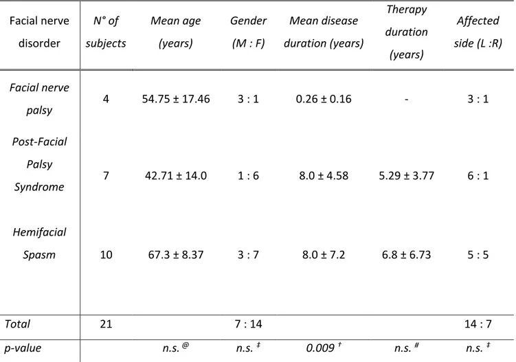
Demographic and clinical data ...40
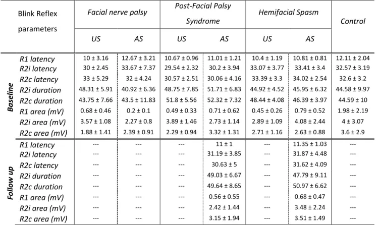
Blink Reflex ...42
Blink Reflex Recovery Cycle ...44
Masseter inhibitory reflex ...48
Discussion ...49
Conclusions ...54
Maria Paola Cabboi. Brainstem excitability in Hemifacial Spasm and Post-Facial Palsy
Synkinesias and effects of Botulinum Toxin. Tesi di dottorato in Scienze Biomediche,
Università degli Studi di Sassari.
Summary
Hemifacial Spasm (HFS) and Post-facial Palsy Syndrome (PFPS) are disorders of the facial nerve. HFS is characterized by involuntary intermittent contractions and synkinesis of muscles supplied by the facial nerve; PFPS is a clinical condition that follows a peripheral facial palsy (FP), often of unknown origin, and presents with synkinesis as well. The synkinesis has its electrophysiological hallmark in the lateral spread of the blink reflex.
HFS and PFPS seem to be, in one manner, two different expressions of a common neurophysiological substrate, sharing some clinical aspects but being different in others. Nevertheless, currently available data still lack direct evidence to precisely locate the site of the lateral spread that causes a widespread facial contraction. Objectives of our study were to evaluate excitability changes in the facial and trigeminal pathways of the affected and unaffected side in HFS and PFPS in order to obtain information on the respective underlying pathophysiological mechanisms and to evaluate possible modifications in excitability of brainstem circuits after the treatment with botulinum toxin.
To these aims ee assessed the Blink Reflex, Blink Reflex Recovery Cycle and Masseter Inhibitory Reflex in patients affected by HFS, PFPS, FP and in a group of healthy controls.
Data obtained revealed that, in our patients, the trigemino-facial pathway is anatomically intact on both the affected and unaffected sides. To a more detailed analysis, data suggest a central hyperexcitability pattern that occurs either in patients with HS and PFPS. However, differently from PFPS, in our HFS group, we did not
Maria Paola Cabboi. Brainstem excitability in Hemifacial Spasm and Post-Facial Palsy
Synkinesias and effects of Botulinum Toxin. Tesi di dottorato in Scienze Biomediche,
Università degli Studi di Sassari.
evidence an excessive excitability on the unaffected side.
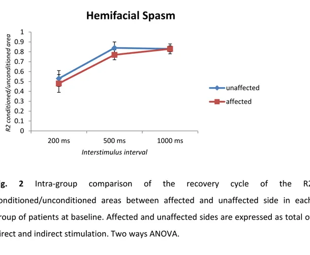
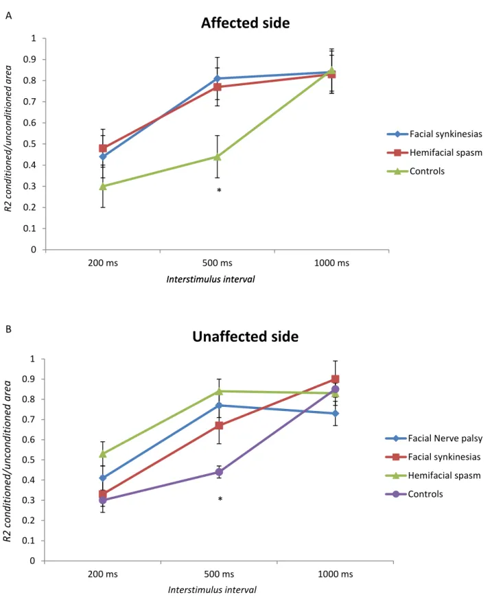
In both HFS and PFPS groups, the blink reflex recovery cycles was enhanced in the affected side, following both direct and indirect stimulation and, also on the unaffected side, both from direct and indirect evocation on the HFS, PFPS and FP groups.
Based on these data, we may conclude that the enhanced sensitivity of the blink reflex circuitry in the context of the synkinetic disorders, is not mediated specifically by a dysfunctional trigeminal afference. The absence of any significant difference between the blink reflex recovery cycles obtained from stimulation of affected and unaffected side and the normality of the masseteric inhibitory reflex arch on both the affected and unaffected sides, supports this idea.
After botulin toxin treatment, a decrease in the amplitude and duration of the R2 response, along with a longer dalay, revealed the expected effects of chemodenervation on the affected side in PFPS group, but not in the HFS group. Surprisingly, the latter showed a contralateral hyperexcitability, which suggests the occurrence of central adaptative phenomena. The absence of modifications on the brainstem interneuronal pool implicated in the recovery cycle of the blink reflex after treatment with botulinum toxin, suggests that the documented effect of botulinum beyond the peripheral action is likely to occur in the central nervous system at higher level than the brainstem. Indeed, the mechanisms responsible for the inhibition of the conditioned response are considered to be segmental and thus, although mostly referred to the interneuronal net of the lateral reticular formation, are potentially influenced to a higher conditioning.
In conclusion, it is conceivable that, clinical and electrophysiological similarity patterns between PFPS and HFS, could be both interpreted as evidence for a change in brainstem hyperexcitability, which in turn can be attributed to a consequent high-ranking influence. Therefore, we can conclude that the study of trigemino-facial
Maria Paola Cabboi. Brainstem excitability in Hemifacial Spasm and Post-Facial Palsy
Synkinesias and effects of Botulinum Toxin. Tesi di dottorato in Scienze Biomediche,
Università degli Studi di Sassari.
reflex and, specifically, of its recovery cycle, appears to provide an objective demonstration of function abnormalities of the inhibitory brainstem interneurons in the context of the synkinetic phenomenon. This can be viewed like a central compensatory attempt in response to a facial damage, probably more serious, but temporary, in the case of PFPS and subtler, but persistent, in the case of HFS. Therefore, our data move to not exclude any of the two possibilities but admits the presence, in either case, of an excessive activation of brainstem networks, in turn inducted and/or simply amplified by an additional effect of peripheral origin.
Maria Paola Cabboi. Brainstem excitability in Hemifacial Spasm and Post-Facial Palsy
Synkinesias and effects of Botulinum Toxin. Tesi di dottorato in Scienze Biomediche,
Università degli Studi di Sassari.
Introduction
Facial nerve lesions are some of the most distressing benign lesions of the peripheral nervous system, since paralysis of hemifacial muscles leads to defective spontaneous blinking, with consequent eye dryness and corneal damage, defective lip and mouth movements with consequent lack of control of food and liquids ingestion, and finally deficit in the expression of emotions which heavily impacts on nonverbal communication leading to social withdrawal. In case of non complete recovery this condition can become chronic and produce in the patient physical and social maladaptive reactions.
Idiopathic facial palsy (FP), post-facial palsy syndrome (PFPS) and essential hemifacial spasm (HFS) are the most common disorders of the facial nerve.
In the case of PFPS, patients exhibit synkinesis, myokimias and involuntary simultaneous contractions of all hemifacial muscles that may resemble those of essential HFS, a chronic condition characterized by involuntary and synchronous contractions of hemifacial muscles.
The facial nerve is one of the most complex nerves in the human body.
The facial nucleus is divided into four cell groups: dorsomedial, ventromedial, intermediate, and lateral. Cells in the intermediate group innervate the frontalis, orbicularis oculi, corrugator supercilli, and zygomaticus; cells in a small medial group innervate the stapedius muscle (Carpenter MB and Sutin J. Human Neuroanatomy. Baltimore: Williams & Wilkins; 1983: 385-386).
At its exit from the brainstem it appears to be formed by two roots: -the facial nerve itself, which carries out fibers to all facial muscles
Maria Paola Cabboi. Brainstem excitability in Hemifacial Spasm and Post-Facial Palsy
Synkinesias and effects of Botulinum Toxin. Tesi di dottorato in Scienze Biomediche,
Università degli Studi di Sassari.
-the intermediate nerve of Wrisberg, which carries parasympathetic fibers to salivary glands (submandibular and pterygopalatine ganglion).
Near the cerebellopontine angle, the facial nerve enters the petrosal bone at the internal auditory meatus. The most relevant structure in the petrosal course of the facial nerve is the geniculate ganglion, located at the genu of the facial nerve, between the labyrinthine and the tympanic segments. Inside the petrosal course the great, the small and the external petrosal nerves emerge. Then, in the mastoid segment, the nerve branches out into the stapedius nerve and the chorda tympani branch.
After emerging from the skull through the stylomastoid foramen, the facial nerve releases collaterals to the posterior auricular muscle, the posterior belly of the digastric muscle and to the stylohyoid muscle. Finally, the nervous trunk enters the parotid gland where it further divides into a frontozygomatic (upper) and cervicofacial (lower) division at the pes anserinus goose’s foot .
After that division the nerve divides into its five major terminal branches: the frontal, zygomatic, buccal, marginal mandibular and cervical. These nerve branches go on to innervate the muscles of facial expression.
Hemifacial spasm
Clinical features
HFS is a movement disorder of the seventh cranial nerve characterised by involuntary clonic and/or tonic contractions of the muscles of facial expression, usually unilaterally, beginning in the periorbital musculature (90%), but later
Maria Paola Cabboi. Brainstem excitability in Hemifacial Spasm and Post-Facial Palsy
Synkinesias and effects of Botulinum Toxin. Tesi di dottorato in Scienze Biomediche,
Università degli Studi di Sassari.
progressing to involve the perioral, platysma and other facial muscles (Chaudhry et al., 2015). This condition, although first described by Gowers in 1888, received its current terminology by Babinski in 1905 who, at the same time, also described a other hara teristi feature of this disease, thereafter k o as the other Babinski sig that is, he the or i ular o uli o tra ts a d the e e loses, the internal part of the frontalis contracts at the same time, and the eyebrow rises during eye occlusion (Babinski, 1905). This typical feature distinguishes HFS from blepharospasm in which this sign is absent (Chaudhry et al., 2015).
Although traditionally perceived as a benign illness, HFS can lead, in severe cases, to functional blindness and disturbed sleep because, unlike other movement disorders, it persists during sleep. Furthermore symptoms increase during times of stress, daily activities like reading, speaking and sometimes eating whereas they abate with relaxation techniques and occasionally touching some parts of the face (sensory tricks) (Brissaud, 1895). Other rare features may include paroxysmal click sounds caused by the contraction of the stapedius muscle (Schwarze et al., 1995), hearing loss, neuropathic pain with the characteristic of trigeminal neuralgia (a combination k o as ti douloureu a d su tle fa ial pals Wa g a d Ja ko i ; Yalto & Jankivic, 2011). The twitching may be induced by 2-3 minutes of hyperventilation, which presumably causes respiratory alkalosis and decreases the interstitial calcium levels, triggering ectopic excitation (Nielsen 1984). In some instances there may also be long-lasting, till almost continuous muscle contractions. In rare cases (<1%) it is bilateral but not symmetrical (Jannetta & Kassam, 1999) indeed, in these cases, the side affected secondarily becomes involved after a long interval – from several months to years - and the movements on each side are usually asynchronous, typically having less severe manifestations on the side being involved later (Jannetta & Kassam, 1999).
Natural history of HFS is chronic with gradual worsening. The first symptom, classically, is a unilateral spasm in the upper face and, more precisely, an intermittent
Maria Paola Cabboi. Brainstem excitability in Hemifacial Spasm and Post-Facial Palsy
Synkinesias and effects of Botulinum Toxin. Tesi di dottorato in Scienze Biomediche,
Università degli Studi di Sassari.
twitching of the eyelid muscle that can lead to involuntary forced closure of the eye Wa g, . This is t pi all asso iated ith the ele atio of the e e ro s other Ba i ski sig . The spas a the graduall spread to other fa ial us les o the same side. Spontaneous resolutions occur rarely: it is estimated that less than 10% of cases heal completely (Ehni & Woltman, 1945).
The diagnosis of this disorder is clinical, but it requires investigations to rule out an underlying secondary cause, such as electromyography for denervation of facial nerve and nuclear magnetic resonance of the brain for demyelination or space occupying lesions near the brainstem. Patients candidate to surgical intervention need a magnetic resonance angiography with special high resolution sequences (Nielsen, 1985).
Differential diagnosis includes facial tic, postparalytic HFS, facial myokymias, partial continuous epilepsy, essential blepharospasm, late dyskinesia, oromandibular dystonia, masticatory spasm and psychogenic disorders.
The most popular choice of treatment of HFS is botulinum toxin: a large number of trials have validated the successful outcomes of this therapy with improvements in as many as 75-100% of individuals with HFE (Defazio et al., 2002). Other options include oral pharmacotherapy (anticonvulsants such as carbamazepine, clonazepam, gabapentin, phenytoin and other drugs like baclofen, anticholinergics, haloperidol) and, in selected cases, microsurgical decompression.
Epidemiology
Epidemiological studies, usually focused on the primary form, have estimated the main prevalence of the disorder to be from 9.8 to 11 per 100,000 people with a clear predominance in women and prevalence typically for an involvement of the left side
Maria Paola Cabboi. Brainstem excitability in Hemifacial Spasm and Post-Facial Palsy
Synkinesias and effects of Botulinum Toxin. Tesi di dottorato in Scienze Biomediche,
Università degli Studi di Sassari.
of the face (Auger & Whisnant, 1990; Nilsen et al., 2004).
Age of onset has a range between the fifth to sixth decades of life, with only 1-6% of presentations before the third decade (Tan & Chan, 2006); however, a presentation before the age of 40 should prompt search for an underlying secondary cause (Tan & Chan, 2006). Rare familiar cases are reported, suggesting that some patients may be genetically predisposed to developing HFS (Carter et al., 1990; Micheli et al., 1994).
Pathophysiology and electrophysiology
The pathophysiological mechanism of HFS is still unclear and the results of various studies are contradictory.
The most common identified cause of HFS reported in the literature is that of an ectatic or aberrant blood vessel (mostly the anterior inferior cerebellar artery) (Soriano-Baron et al., 2015), which compresses the facial nerve at the root entry/exit zone. This area is the nerve segment between the central and peripheral nervous system where there is a transition of the cells responsible for myelination and the nerve lacks an epineurium, being protected by an arachnoid membrane only (Girard et al., 1997). Consistent with this hypothesis, patients improve after surgical intervention at the posterior fossa (Jannetta et al., 1977; Soriano-Baron et al., 2015). Nevertheless, it is interesting that clinical improvements are observed after surgical intervention in patients without any evidence of the compressing vessel. Moreover neurovascular conflicts can be identified in up to 25% of healthy controls, suggesting that compression by a vessel alone is insufficient to produce HFS (Tan et al., 1999). Several electrophysiological studies have been put forward to explain how the compression of the facial nerve leads to hemispasm: although there is a general belief that the hyperexcitability responsible for HFS involves the facial nerve or the
Maria Paola Cabboi. Brainstem excitability in Hemifacial Spasm and Post-Facial Palsy
Synkinesias and effects of Botulinum Toxin. Tesi di dottorato in Scienze Biomediche,
Università degli Studi di Sassari.
nucleus (Møller & Jannetta, 1985; Møller & Jannetta, 1984), currently available data still lack direct evidence to precisely locate the site of lateral spread that causes a widespread facial contractions (Kameyama et al., 2016).
No conduction block is usually observed in the facial nerve and no denervation signs are found in the muscles of the hemiface affected; the electrophysiological hallmark of HFS is the spread of the blink reflex to muscles other than orbicularis oculi (Kimura et al., 1975). The proposed explanation for the phenomenon is the lateral spread: an abnormal response of the orbicularis oris muscle to sopraorbital nerve stimuli; unwanted synkinetic muscle activity is the most common expression of this lateral spread of excitation.
It is likely that this response is generated through ephaptic activation of neighbouring demyelinated or poorly myelinated fibers of the facial nerve innervating the perioral muscles, by those that naturally carry the reflex volley to the orbicularis oculi (Nielsen, 1984). Ephaptic transmission occurs when transmission in one axon generates action potentials in a parallel axon. This has been attributed to the passage of inputs through a demyelination zone: the slowing of conduction velocity in demyelinated fibers gives time enough for a neighbouring hyperexcitable axon to reach the firing threshold. Similarly abnormal responses have been observed in patients with peripheral facial palsy.
Alternatively, some authors have raised the hypothesis that some synkinetic hemifacial responses triggered by trigeminal nerve stimulation, may be sort of ephaptic facio-facial responses (and not a trigemino-facial reflex) generated after antidromic activation of small motor axon terminals of the facial nerve near the trigeminal afferents and innervating the orbicularis oculi and/or other facial muscles (Montero et al., 1996). To support this hypothesis, the same work group applied step by step stimuli, as in the inching technique, from the supraorbital zone towards the malar zone, following the zygomatic branch of the facial nerve. The striking observation was that the "R1-like" response recorded in the orbicularis oris
Maria Paola Cabboi. Brainstem excitability in Hemifacial Spasm and Post-Facial Palsy
Synkinesias and effects of Botulinum Toxin. Tesi di dottorato in Scienze Biomediche,
Università degli Studi di Sassari.
decreased when the stimulating electrode was moved away from the site of emergence of the supraorbital branch of the trigeminal nerve to a site closer to the facial nerve. With this maneuver the length of the facio-facial axon reflex arc was shortened whereas the length of the trigemino-facial reflex arc was increased. These findings are consistent with the hypothesis of ephaptic activation of facial nerve axon terminals (Montero et al., 2007). A study conducted more recently supports the view that the generation of an abnormal muscle response requires the participation of an antidromic motor impulse (Kameyama et al., 2016).
A second possibility proposed to explain the origin of lateral spread is co-activation of many abnormally hyperexcitable motoneurons following antidromic invasion or reflexive activation of the facial nucleus (Møller & Jannetta, 1985). Specifically, Misawa and coworkers, obtained an abnormal muscle response after subthreshold stimulation of the facial nerve: this result suggested the role of aberrant cutaneous afferent volleys via the trigeminal nerve, which enhanced the reflexive excitability of the facial motoneurons, rather than antidromic activation of the facial nerve (Misawa et al., 2006).
There is some evidence that facial motoneurons are hyperexcitable in HFS. A probably extrinsic irritation of the facial nerve at the posterior fossa generates an antidromic input to facial motoneurons that causes excitability changes and spo ta eous or refle firi g of oto euro s after a ki dli g effe t (Møller, 1987; Martinelli et al., 1992).
To support the hypothesis of a facial motor neuron hyperexcitability, Valls-Solè and Tolosa, in 1989, studied blink reflex responses in patients with HFS to single and paired (conditioning and test) stimuli at both sides of the face. In response to single stimulus, the late responses (R2) were of larger size on the side of the spasm compared with the uninvolved side and with controls, to either ipsilateral or controlateral stimulation. With paired stimuli, the recovery curve of the blink reflex resulted enhanced on the side of the spasm and also on the contralateral side: this
Maria Paola Cabboi. Brainstem excitability in Hemifacial Spasm and Post-Facial Palsy
Synkinesias and effects of Botulinum Toxin. Tesi di dottorato in Scienze Biomediche,
Università degli Studi di Sassari.
result suggests an enhanced excitability of brainstem interneurons involved on the pathway of the blink reflex and/or an enhanced excitability of facial motoneurons (Valls-Sole & Tolosa, 1989).
More recently, Eekhof et al. (1996), investigated R1 and R2 blink reflex responses to single and paired stimuli in a group of patients with HFS. To single stimulus, amplitude and area of R1 and R2 responses showed no significant differences between patients and controls, providing evidence that the blink reflex circuits are functionally intact in these patients. To paired stimulation, R2 recovery curve was significantly enhanced on the affected side at intervals from 0.21 to 0.5 seconds and R2 index (calculated as the mean recovery of peak amplitude values at interstimulus intervals of 0.5, 0.3 and 0.21 s) was enhanced in 50% of patients on the affected side and in 20% of them on the unaffected side (Eekhof et al., 1996).
These results provided additional evidence that, in patients with HFS, there is a bilateral involvement of the brainstem (facial motor neurons and/or brainstem interneurons) in at least some patients as the cause of the involuntary contractions (Eekhof et al., 1996).
In 2005, Oge and coworkers, studied the blink reflex recovery curves bilaterally (that is, from stimulus on the symptomatic and asymptomatic sides) in patients affected by HFS and PFPS to directly compare the electrophysiological excitability characteristics of the facial nucleus and related structures (Oge et al., 2005).
Perfectly in line with abo e e tio ed e tral h pothesis , the authors re ealed that both HFS and PFPS groups showed an early blink reflex recovery, more prominent in PFPS patients, when stimulations and recordings were made on the symptomatic side. Interestingly, the same tendency to early recovery was also present in both groups when supraorbital nerve on the symptomatic side was stimulated and the responses from the asymptomatic side were recorded.
Maria Paola Cabboi. Brainstem excitability in Hemifacial Spasm and Post-Facial Palsy
Synkinesias and effects of Botulinum Toxin. Tesi di dottorato in Scienze Biomediche,
Università degli Studi di Sassari.
Peripheral facial palsy and post-facial palsy syndrome
Introduction and clinical features
Unilateral peripheral facial palsy is one of the most common neurologic disorders affecting the cranial nerves and it is the most common cause of facial paralysis worldwide: it is thought it accounts for approximately 60-75% of cases of acute unilateral facial paralysis (Katusic et al., 1986). Although it is a benign and self-limiting inflammatory condition, it has a considerable impact on the patient because it is a disfiguring disorder (Newadkar et al., 2016) .
Peripheral facial palsy, in the majority of cases, is of unknown origin and is described as idiopathic facial paralysis or Bell's palsy (BP). Clinically it is characterized by a rapid onset, a unilateral lower motor neuron type of facial deficit, partial or complete, of muscles of facial expression for both, voluntary and emotional responses. Differently, in supranuclear lesions, although voluntary facial movements are impaired, there is some sparing of the frontalis and orbicularis oculi muscles because of the bilateral cortical representation. Depending on the site of the lesion, other defects may be associated, such as hyperacusis or loss of taste. This is due to the fact that facial nerve also contains the nerve to the stapedius muscle and the chorda tympani nerve supplying taste sensitivity to the anterior two-thirds of the tongue, and also parasympathetic secretomotor fibers to lacrimal and salivary glands. In some patients pain may be present and, sometimes, this symptom tends to precede palsy. However, if pain is significant and accompanies BP, the patient is believed to have the diagnosis of Ramsey-Hunt syndrome, probably caused by varicella zoster infection.
With the loss of facial function, patients have an incomplete eye closure, with a consequent cornea damage, and epiphora. Patients fail to control their lip and
Maria Paola Cabboi. Brainstem excitability in Hemifacial Spasm and Post-Facial Palsy
Synkinesias and effects of Botulinum Toxin. Tesi di dottorato in Scienze Biomediche,
Università degli Studi di Sassari.
mouth, thereby affecting their ability to speak, eat and drink. The loss of facial muscular to e a ha e a sig ifi a t egati e i pa t o a perso ’s self-image and the way he/she interacts with others in society.
BP is a li i al diag osis a d is largel a diag osis of e lusio therefore the li i ia ’s history and physical examination is directed toward ruling out diseases within this differential diagnosis. Many conditions can produce isolated facial nerve palsy, more frequently structural lesions in the ear or parotid gland, Guillain-Barrè syndrome, Lyme disease, otitis media, Ramsay-Hunt syndrome, sarcoidosis, central nervous system lesions and some influenza vaccines. But, although these conditions can present as isolated facial nerve palsy, they usually have additional features that distinguish them from BP.
After the clinical diagnosis, patients with BP do not require imaging studies unless they fail to recover as expected (that is if there is no recovery after 3 months from the onset) or even worse, or show atypical new dysfunctions. For those patients who have complete facial paralysis, electrophysiological testing may be considered.
Treatment of BP should be conservative and guided by severity and probable prognosis in each particular case. Studies have shown the benefit of high dose corticosteroids in the early phase but not of antiviral treatment, although it has been used in recent years (Lockhart et al., 2009) and of physical therapy to improve recovery of facial deficits. The late phase of treatment is directed toward treating and preventing synkinesis, contractures, spasms or autonomic dysfunctions such as crocodile tears. Currently, based on a lack of strong evidence, surgical decompression of the nerve is not recommended as a standard approach.
Fortunately, prognosis of BP is of a near-to-complete recovery in approximately 80% of cases; 15% experiences some kind of permanent nerve damage and only 5% remains with severe sequelae (Tate & Tollefson, 2006). Usually some recovery occurs in the first 3 weeks.
Maria Paola Cabboi. Brainstem excitability in Hemifacial Spasm and Post-Facial Palsy
Synkinesias and effects of Botulinum Toxin. Tesi di dottorato in Scienze Biomediche,
Università degli Studi di Sassari.
PFPS is a condition that follows a facial nerve lesion intense enough to cause a severe paralysis, that is if a significant percentage of axons undergo wallerian degeneration. PFPS is characterized by synkinesis between facial muscles, myokimia and unintended mass contractions of hemifacial muscles (Valls‐Solé & Montero, 2003). The severity of PFPS may range from a subclinical stage, only evident in electrophysiological examinations, to a complex disturbance of voluntary and automatic activation of facial muscles.
Synkinesis is a typically documented recording, with surface electrodes. It consists of bursts of electromyography activity in the orbicularis oris during spontaneous blinking. The same is seen with voluntary contraction of an individual muscle of the face, that leads to an involuntary and simultaneous contraction of other hemifacial muscles, leading to unwanted facial movements. Needle electrode recording may also show interesting phenomena indicating the complexity of synkinesia: in a few patients also movements that were not intended to activate the facial muscles at all (i.e. neck movements or accommodation of some body parts) trigger some motor unit action potentials in the orbicularis oris, which can also be seen to fire in bursts time-locked to the inhalation phase of breathing (Valls-Solé, 2002; Pavesi et al., 1994).
In some cases, very uncomfortable mass contractions are triggered by facial movements, leading to what patients describe as a spasm. In these cases, patients can experience severe episodes of muscular pain, tension and spasms induced by common maneuvers such as eating, smiling or speaking (Valls-Sole et al., 1991). This condition can mimic those observed in essential HFS, but a careful clinical and neurophysiological examination allows separation of both entities. First of all, on the basis of the clinical history, more than 65% of patients with primary HFS initially have contractions of periocular muscles alone, whereas more than 72% of patients with the post-paralytic syndrome report involvement of the upper and lower facial muscles, including platysma, simultaneously (Colosimo et al., 2006). Furthermore, in
Maria Paola Cabboi. Brainstem excitability in Hemifacial Spasm and Post-Facial Palsy
Synkinesias and effects of Botulinum Toxin. Tesi di dottorato in Scienze Biomediche,
Università degli Studi di Sassari.
essential HFS, the involuntary twitches are not necessarily triggered by voluntary or automatic muscle contractions, whereas in the post-paralytic syndrome mass contractions are always brought about by facial muscles activation. The two entities, finally, differ at the electrophysiological evaluation. Some differences can be observed already with the simple recording of the compound muscle action potential, which may be smaller in patients with history of facial palsy. At the EMG analysis the two clinical entities are completely different: the muscular discharges are of lower frequency and more irregular in post-facial palsy syndrome than in essential HFS (Valls‐Solé & Montero, 2003). Finally, the R2 component of the blink reflex obtained in the orbicularis oculi on stimulation of the supraorbital nerve is larger on the affected side in case of HFS, instead is larger in the non affected side in case of peripheral facial palsy (Manca et al., 2001).
Epidemiology
BP is a relatively common condition with an incidence rate of approximately 25 per 100.000, or about 1/60 people in a lifetime (Katusic et al., 1986). It represents the most common disorder affecting the facial nerve (Wolfson, 2009) and is responsible for about 80% of all facial mononeuropathies (Goroll & Mulley, 2009). BP affects the right and left sides of the face fairly equally and bilateral paralysis is rare (Vakharia & Vakharia, 2016). BP may occur at any age, although it is more frequent in adults than children, with similar incidence rates reported among males and females (Bosco et al., 2011). Particularly susceptible groups include diabetics, pregnant women, elderly and patients with hypothyroidsm. Furthermore, 4% to 8% of cases have an associated family history (Wolfson, 2009).
Most patients with BP experience spontaneous resolution: a clinically important improvement occurs within 3 weeks in 85% of people and within 3 to 5 months in the
Maria Paola Cabboi. Brainstem excitability in Hemifacial Spasm and Post-Facial Palsy
Synkinesias and effects of Botulinum Toxin. Tesi di dottorato in Scienze Biomediche,
Università degli Studi di Sassari.
remaining 15%. Overall, 71% of people will fully recover facial muscle function (61% of people with complete palsy, 94% of people with partial palsy). The remaining 29% are left with mild to severe residual facial muscle weakness, 17% with contracture, and 16% with hemifacial spasm or synkinesis (Peitersen, 1982; Holland & Bernstein, 2011). Incomplete recovery of facial expression may have a long-term impact on quality of life. The prognosis for children with Bell's palsy is generally good, with a high rate (>90%) of spontaneous recovery, in part because of the high frequency of partial paralysis. However, children with complete palsies may suffer poor outcomes as frequently as adults.
Pathophysiology and electrophysiology
Early phase of damage
Controversy surrounds the etiology of BP. The cause of paresis still remains unknown even if the etiology has been discussed by several authors and herpes viruses (particularly herpes simplex virus 1, HSV-1, and herpes zoster virus, HZV) seemed to be the most plausible infective agent (Holland & Bernstein, 2011). These reports are in agreement with finding of HSV-1 DNA in the endoneural fluid of the facial nerve by polymerase chain reaction during the acute phase of BP (Murakami et al., 1996). Herpes zoster-associated facial palsy more frequently presents as zoster sine herpete (without vesicles), although 6% of people will subsequently develop vesicles, depicting the Ramsay-Hunt syndrome (Furuta et al., 2001). Thus, treatment plans for the management of Bell's palsy should recognize the high incidence of HZV, which is associated with worse outcomes. More recently the inactivated intranasal influenza vaccine has also been linked with BP in a case-control study: the authors reported a strong temporal and specific association between vaccine and risk of developing BP
Maria Paola Cabboi. Brainstem excitability in Hemifacial Spasm and Post-Facial Palsy
Synkinesias and effects of Botulinum Toxin. Tesi di dottorato in Scienze Biomediche,
Università degli Studi di Sassari.
(Mutsch et al., 2004). There was no association of the palsy with parenteral flu vaccines. Other known documented infectious cause BP but, although the viral etiology is accepted, given the circumstantial evidence, the exact etiology still is considered to be unclear.
Histopathological correlates of a BP are inflammatory infiltrates with swollen macrophages containing demyelination debris, suggesting an inflammatory process with axonal damage (Liston & Kleid, 1989). Inflammation of the facial nerve initially results in reversible neuroapraxia, but Wallerian degeneration may occur.
Although BP is clearly a mononeuropathy of the facial nerve, some patients report pain or numbness on the paretic side of their face or the perioral area, suggesting a trigeminal nerve involvement, but little is known about this condition. There are only a few reports in literature suggesting an electrophysiological trigeminal dysfunction (Hanner et al., 1986). However, depending on the neurophysiologic test employed to study specifically the trigeminal function (like masseter inhibitory reflex, MIR), no trigeminal nerve involvement – at least of the second division - can be detected in BP (Uluduz et al., 2010).
Neurophysiological examination is helpful at all stages in patients with BP, not only because it provides cues for the prognosis and suggests the nature of the underlying pathological process, but also in order to provide the basis for a pathophysiological explanation of related abnormalities of movement control.
At the early phase (few days) the typical neurophysiological pattern is the absence of responses in the affected orbicularis oculi after electrical stimulation of the supraorbital nerve of either side (Kimura et al., 1976): this indicates that, at this stage, the lesion is certainly limited to the facial nerve and that it does not involve the trigeminal nerve, as suggested by a normal response in the orbicularis oculi of the contralateral side. At the same time, the latency and size of the compound muscle action potential of the facial nerve are completely normal, unless the lesion lies in the
Maria Paola Cabboi. Brainstem excitability in Hemifacial Spasm and Post-Facial Palsy
Synkinesias and effects of Botulinum Toxin. Tesi di dottorato in Scienze Biomediche,
Università degli Studi di Sassari.
extracranial segment (such as in the case of parotid tumor) or in the terminal branch (Turkof et al., 2003; Guntinas-Lichius et al., 2004). An intrapetrosal facial nerve conduction block can be confirmed with magnetic stimulation over the posterior fossa at the parieto-occipital region and at the stylomastoid foramen, but this methodic is not easy to apply because it induces large artifacts (Rösler et al., 1991; Glocker et al., 1994).
At about 10 days, the functional loss of axons in the affected side is revealed as a reduction of the compound muscle action potential from nasalis muscle: this is the most informative data for determination of the prognosis (Glocker et al., 1994). A needle electromyographic (EMG) recording performed 20 days after onset allows to approximate a measure of denervation, confirmed by finding of fibrillation potentials and positive sharp waves as a consequence of the degree of axonal damage.
Subsequent evaluations provide evidences for abnormal regeneration and changes in motoneuronal excitability, that configure the PFPS (Montserrat & Benito, 1988; Valls-Sole et al., 1992).
At all these stages of the disease, it is also convenient to perform the neurophysiological examination in both paralyzed and non-paralyzed sides, not only for comparison between the damaged and the non-damaged nerve, but also to obtain information on adaptive changes occurring in the contralateral side of paralysis. Indeed, although lack of movement in the paralysed side of the face is the prominent sign of facial palsy, a few patients complain of excessive spontaneous blinking in the contralateral eye (Pastor et al., 1998). Starting from this observation, Pastor and coworkers measured the spontaneous blinking rate in 20 patients with peripheral facial palsy, between 24 and 72 hours after onset, finding a significant enhancement. This enhanced blinking rate may be an attempt of compensatory mechanism to protect the cornea of the paralysed side from dryness and it is
Maria Paola Cabboi. Brainstem excitability in Hemifacial Spasm and Post-Facial Palsy
Synkinesias and effects of Botulinum Toxin. Tesi di dottorato in Scienze Biomediche,
Università degli Studi di Sassari.
obviously evident on the nonparalyzed side only. Also, the EMG activity and eyelid movements, recorded simply with surface electrode during spontaneous blinking, can give interesting information. Normally this recording consists of a slow negative wave accompanied by a small burst of EMG activity on the rising phase: in patients with peripheral facial palsy, the recording of the paralysed side showed a small slow wave as a result of weakness of levator palpebrae; in the non-paralysed side the slow wave was characterized by an EMG burst of larger amplitude than normal (Valls‐Solé & Montero, 2003).
Excessive blinking may also be accompanied by hemifacial hyperactivity. This observation has led to a few reports on the occurrence of contralateral blepharospasm induced by BP (Chuke et al., 1996; Baker et al., 1997). In particular, Chuke and coworkers in 1996 described a case of blepharospasm-like symptoms appearing contralaterally to an eyelid weakened by facial nerve palsy, that improved with implantation of a golden weight to assist closure of the paretic eyelid. The authors proposed that hyperexcitable blink reflex could be a maladaptive consequence of adaptive system, i.e. a physiological adaptive control mechanisms of the efficacy of the blink reflex, as suggested by the fact that aiding closure of the weak eyelid led to a reduction of the maladaptive excitability of the blink circuit. Changes in central excitability was attributed to excessive afferent input from the cornea of the paretic side and, since blinking is a conjugate behavior, the bilateral nature of the blink system-adaptive control was not unexpected (Chuke et al., 1996). The following year, Baker described 4 patients with BP and blepharospasm and, on the basis of eyelid movements kinematic study, proposed a correlation model for BP– induced blepharospasm in in which a blink adaptive system may produce the maladaptive consequence of eyelid spasms (Baker et al., 1997).
To identify electrophysiological correlates for adaptive changes suggested by previous kinematic studies, Syed and coworkers, evaluated the excitability of the brainstem interneuronal circuitry in people with persistent or resolved unilateral
Maria Paola Cabboi. Brainstem excitability in Hemifacial Spasm and Post-Facial Palsy
Synkinesias and effects of Botulinum Toxin. Tesi di dottorato in Scienze Biomediche,
Università degli Studi di Sassari.
facial weakness (Syed et al., 1999). The finding of an enhanced recovery curve of the blink reflex in patients with residual weakness (but not in patients who recovered) suggested a role for abnormal afferent input in maintaining sensitization (Syed et al., 1999).
Further evidence in the same direction was reported simply analysing the size differences between the R2 component of the nonparalyzed side from direct (R2) and indirect (R2c) stimulation: contrary to what usually observed in healthy subjects, the mean R2c/R2 ratio was significantly larger in patients with recent peripheral facial palsy (Manca et al., 2001).
The causal relationship between sensitization of reflex pathways and abnormal motor control is supported by experimental animals studies in which rats who sustained a 6-hydroxydopamine lesion of the striatum, developed blepharospasm after peripheral facial nerve lesion. The authors postulated that, in a predisposing condition such as a lesion of the basal ganglia, an excessive trigeminal input from corneal irritation subsequent to facial weakness, would lay the grounds for the abnormal motor behavior (Schicatano et al., 1997). These data identified a crucial role of subcortical structures in the integration of sensory input, like as basal ganglia and cerebellum (Schicatano et al., 2002).
Facial nerve regeneration phase and synkinesis
After a complete muscle denervation subsequent to facial nerve paralysis, EMG signs of regeneration can be seen at about 3 months after onset of symptoms as small amplitude polyphasic potentials (Cossu et al., 1999). These action potentials are visible during automatic or voluntary contraction, but they are elicited with difficulty by electrical stimulation of the damaged in-growing nerve, notoriously markedly hypoexcitable because poorly myelinated (Moldovan et al., 2006).
Maria Paola Cabboi. Brainstem excitability in Hemifacial Spasm and Post-Facial Palsy
Synkinesias and effects of Botulinum Toxin. Tesi di dottorato in Scienze Biomediche,
Università degli Studi di Sassari.
proximal stu ps, elo gati g ithi the Bű g er a ds a luster of S h a ells enclosed around a Schwann cell basement membrane). As the growing axonal cone advances, proximal segments are progressively myelinated.
However, during this regeneration processes of a motor nerve, there is a risk of two main types of error: the axon regenerates in a funiculus going to another muscle or the axon regenerates, simultaneously, into one or more funiculi going to different muscles that may have antagonistic functions or to parasympathetic axons of the intermediary nerve of Wrisberg. Lacrimation and hemifacial sweating - the so-called Fray syndrome - subsequent to muscle activation is one of the possible consequences of such reinnervation errors (Guntinas-Lichius et al., 2006).
In reality, abnormal axonal growth cannot be the sole cause of reinnervation abnormalities: a series of morphological and functional changes in the cell body, known as chromatolysis, was observed for the first time since in 1892 by Nissl and successively confirmed by other studies (Tetzlaff et al., 1996; Graeber et al., 1993). Indeed, after axonal lesion, facial motor neurons change their phenotype from a transmitter to a regenerative mode, undergoing ultrastructural changes that make them hyperexcitable (Tetzlaff et al., 1996). Anatomic evidence of the disruption of central control mechanisms was confirmed when it was discovered that early and brisk proliferation of microglia occurs when the facial nerve is cut and that these microgliacytes remove most of the intact synapses from the neuronal surface membranes within 4 days (Blinzinger & Kreutzberg, 1968). It was later found that this synapse stripping might have greater effects on inhibitory synapses (Lux & Schubert, 1975). More recently, synaptic stripping in the human facial nucleus has been observed 3 months after a severe facial nerve injury (Graeber et al., 1993). It has also been shown that rapid genetic changes in messenger RNA with unique patterns in the facial motor neurons occur within 5 hours from axotomy (Moran & Neely, 1996). In this context, the trigeminal stimulation would be easily induced by action potentials at level of particularly excitable facial motor neurons (Valls-Solé, 2013).
Maria Paola Cabboi. Brainstem excitability in Hemifacial Spasm and Post-Facial Palsy
Synkinesias and effects of Botulinum Toxin. Tesi di dottorato in Scienze Biomediche,
Università degli Studi di Sassari.
To better understand alterations at the level of motoneuronal cells of a damaged facial nerve, Cossu and coworkers, recorded simultaneously, with needle electrode, the spontaneous, voluntary and reflex activity of orbicularis oculi and orbicularis oris muscles in patients with recent history of facial palsy (between 75 and 90 days after damage), that is, in the period before the ensuing of peripheral compensatory mechanisms, such as lateral spread. All patients showed: involuntary firing of motor unit, synkinetic activation of orbicularis oris muscle induced by spontaneous, automatic or voluntary movements of facial muscles, reflex responses in orbicularis oris and, simultaneously, in orbicularis oculi, induced by electrical stimulation of ipsilateral and contralateral trigeminal or facial nerve and also peripheral nerve (Cossu et al., 1999) .
These results revealed that, at least some of the functional abnormalities of regenerating axons, are due to a change in the excitability state of facial motorneurons and that enhanced motoneuronal activity is present precociously (from the very first connection between growing axons and denervated muscles) to offer a pathophysiological substrate for the development of PFPS (Cossu et al., 1999). A study of blink reflex and lateral spreading in patients with synkinesia after BP has supported this possibility as an increased R1 amplitude ratio of orbicularis oris/orbicularis oculi was recorded (Eekhof et al., 2000). In physiological conditions, the amplitude of R1 response recorded at orbicularis oris muscle is smaller than R1 at orbicularis oculi. This was explained by the findings that direct cortical projections upon the lower face muscles are less so upon the upper facial muscles. In contrast to this, patients with history of BP showed an increased amplitude of R1 response in the orbicularis oris. These results were explained calling into question a loss of inhibitory cortical control (Eekhof et al., 2000) and were in line with previous results of enhanced F-waves and larger R2 responses in PFPS cases, on the symptomatic side (Valls-Sole et al., 1992).
Maria Paola Cabboi. Brainstem excitability in Hemifacial Spasm and Post-Facial Palsy
Synkinesias and effects of Botulinum Toxin. Tesi di dottorato in Scienze Biomediche,
Università degli Studi di Sassari.
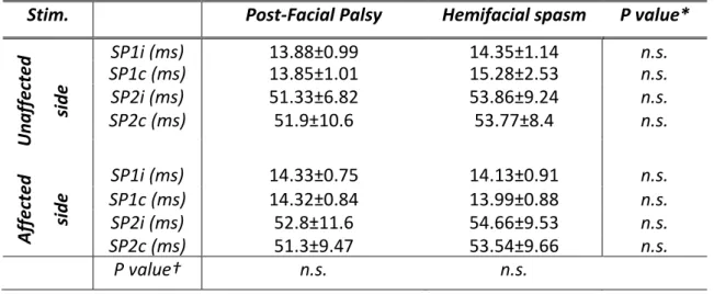
related brainstem reflexes in PFPS and HFS to evaluate excitability changes in the facial pathways. To this purpose they investigated F-waves, blink reflex recovery curve and magnetically elicited silent period. F-waves of facial nerve were enhanced in PFPS and HFS on the affected side. At the study of BRRC the authors found, in both group of patients, early recruitment of R2, more prominent in PFPS patients and if recorded and/or stimulated at the symptomatic side. An early recruitment was also found, although to a lesser extent, on the asymptomatic side. Finally, duration of silent period was nearly equal on both sides. In conclusion these results revealed an exceeding hyperexcitability of blink reflex bilaterally, more prominent to stimuli and/or to recording on the affected side and it was ascribable to an increased excitability of brainstem structures, probably at facial nuclei level. Absence of differences of magnetically elicited silent periods from affected/non-affected sides implies, according to the interpretation of the authors, that cortical excitability changes do not take place in these disorders, confirming that they are confined to the brainstem level (Oge et al., 2005).
Regenerating activity, described in the abovementioned studies, may take place not only in the affected facial nerve but also in the contralateral non-affected side: recordings of EMG activity made with concentric needle electrode at the orbicularis oris muscle of the nonparalyzed side, showed a significant proportion of enhanced jitter and blocking (Valls‐Solé & Montero, 2003). This data demonstrate that a regenerative process in the non injured facial nerve exists, inducted, most likely, by contralateral chemical changes and motoneuronal reorganization that has been shown to occur in distant muscles after local denervation induced by botulinum toxin
Maria Paola Cabboi. Brainstem excitability in Hemifacial Spasm and Post-Facial Palsy
Synkinesias and effects of Botulinum Toxin. Tesi di dottorato in Scienze Biomediche,
Università degli Studi di Sassari.
Botulinum toxin
Botulinum toxin is produced by anaerobic fermentation of the bacterium Clostridium Botulinum, during growth and reproduction. More precisely, Clostridium produces eight immunologically distinct serotypes (A-H) but the most widely used for therapeutic application is the neurotoxin type A preparation, although studies indicate that botulinum neurotoxin serotypes B and F may be useful for the treatment of selected cases (Chen et al., 1998; Brin et al., 1999).
In nature, BoNT-A is synthesized as macromolecular protein complexes that are referred to as progenitor toxins and consist of nontoxic accessory proteins (NAPs) bound to the 150-kD active neurotoxin. Currently, on the market, there are three botulinum neurotoxin type A products: onabotulinum toxin A, abobotulinum toxin A and incobotulinum toxin A.
The different products of BoNT-A vary in molecular weight (from 300 to 900 kD) depending on the composition of NAPs and the manufacturing process, whereas the toxin moiety is the same in all pharmaceutical preparations.
To exert its pharmacological effect, the 150-kD neurotoxin must dissociate from NAPs, with the exception of incobotulinum which contains only the 150-kD neurotoxin.
Therapeutical power, expressed in biological units, is related to the quantity of toxin required to achieve a median lethal dose (LD50) unit and is conditioned by many factors (mouse strain, sex, age, volume and route of injection etc.). Moreover, the LD50 units of BoNT products are not strandardized across manufacturers (Frevert, 2015).
Maria Paola Cabboi. Brainstem excitability in Hemifacial Spasm and Post-Facial Palsy
Synkinesias and effects of Botulinum Toxin. Tesi di dottorato in Scienze Biomediche,
Università degli Studi di Sassari.
Mechanisms of action and clinical uses
The efficacy of botulinum neurotoxin therapy is primarily due to the injection method delivery and the high affinity of cholinergic nerves for neurotoxin uptake.
Botulinum toxin acts at the neuromuscular junction to inhibit the presynaptic release of the acetylcholine neurotransmitter by the following steps:
- fast, specific and irreversible binding to acetylcholine reptors on presynaptic nerve surface
- uptake of toxin into cell vesicle
- translocation of toxin into cytosol across vesicle membrane
- toxin-activated proteolysis which, in turn, blocks the release of acetylcholine (Orsini et al., 2015).
After local injection, the toxin spreads into contiguous areas, increasing the risk of adverse effects. Although, distant spread is uncommon.
In clinical use, BoNT-A is precisely injected into specific muscles to cause a temporary chemodenervation of the skeletal muscle in those entities characterized by excessive muscle contraction.
More precisely, an injection of BoNT-A into a muscle, will reduce the alpha motoneuron activity on the extrafusal muscle fibers and thus muscle contraction. Simultaneously, muscle spindles, are also inhibited by BoNT-A through the inhibition of the gamma motor neuron control of the spindle intrafusal fibers and subsequent reduction of the Ia fiber afferent signal. This attenuated Ia signal, then, reduces the feedback to the alpha motor neurons and, subsequently, also muscle activity of non-injected muscles (the injection causes a profound reduction of spasticity in areas larger than expected and not related to the zone of diffusion) (Filippi et al., 1993).
Maria Paola Cabboi. Brainstem excitability in Hemifacial Spasm and Post-Facial Palsy
Synkinesias and effects of Botulinum Toxin. Tesi di dottorato in Scienze Biomediche,
Università degli Studi di Sassari.
The overall impact of a long-term reduction of alpha, gamma and Ia neurons, may lead to secondary effects on the central nervous system. The work group of Delgado-Garcia, studying effects of BoNT in extrinsic ocular muscles of the cat, found an elimination of inhibitory postsynaptic potentials and a reduction of gephyrin-immunoreactive clusters (glycine-receptor-clustering protein): these findings indicate that the long-term paralysis of a muscle may induce the reorganization of central programs and the appearance of compensatory movements (Moreno-López et al., 1997).
Finally, BoNT therapy has been reported to alleviate pain in various conditions, with or without concomitant excess muscle contractions.
Clinical observations of antalgic effect of botulinum were made, early, in patients infiltrated for cervical dystonia in which the pain relief exceeded the motor benefit (Tsui et al., 1986; Brin et al., 1987). Successively, the antinociceptive effect was observed on headache (Binder et al., 2000), neck and back pain (Foster et al., 2001) and myofascial pain (Porta, 2000).
This antalgic effect of BoNT was explained by the in vitro and in vivo demonstration of reduction of neuropeptide release (Mustafa et al., 2013).
Local injections of BoNT have been widely used as a symptomatic treatment for movement disorders since the eighties, when it was used to correct strabismus (Scott, 1981) and dystonic blepharospasm (Fahn et al., 1985). Successively BoNT owes its effectiveness to the treatment of many motor disturbances, including upper limb dystonia and cervical dystonia (Cohen et al., 1989; Jankovic & Schwartz, 1990; Costa et al., 2005).
Since 1980, animal studies have provided evidence that the toxin inhibits, in cortical slices, the release of acetylcholine in cerebral cortex synaptosomes whereas, morphological studies, have shown that BoNT, after intramuscular injection, accumulates into spinal motoneurons (Habermann, 1974; Wiegand et al., 1976;
Maria Paola Cabboi. Brainstem excitability in Hemifacial Spasm and Post-Facial Palsy
Synkinesias and effects of Botulinum Toxin. Tesi di dottorato in Scienze Biomediche,
Università degli Studi di Sassari.
Simpson, 1981). In 1993, results from animal studies, induce to supposed that the toxin also blocks the intrafusal motor end plate and provided evidence that the toxin reduces the spindle afferent discharge without altering muscular tension (Filippi et al., 1993). Patie ts ith riter’s ra p, seg e tal upper li d sto ia a d, surprisingly, also patients with cervical dystonia not involving the limbs, have an abnormally decreased forearm reciprocal inhibition (Nakashima et al., 1989; Panizza et al., 1989; Deuschl et al., 1992). Based on these data, in 1995, Priori and Colleagues, studied reciprocal inhibition between forearm flexors and extensor muscles, before and after BoNT-A injection, in patients with upper limb dystonia, to see whether botulinum toxin treatments alters segmental motor system function (Priori et al., 1995). After treatment, the second abnormal phase of reciprocal inhibition increased and the authors postulated an indirect effect of BoNT on spinal cord circuitry, probably through changes affecting the tonic sensory inflow coming from the injected muscles (Priori et al., 1995). Similar results were found in patients with essential tremor treated with infiltration of the neurotoxin (Modugno et al., 1998). More recently, Pauri and coworkers, investigated changes in motor evoked potentials (MEPs) in patients with leg spasticity treated whit BoNT-A and found an increased MEP latency and a prolonged central conduction time. The authors attributed these findings to central changes in spinal motoneuron responsiveness to cortical descending inputs (Pauri et al., 2000).
In the same line are results of a study in which, using transcranial magnetic stimulation (TMS), the topography of the primary motor cortex projections to the upper li us les i patie ts ith riter’s ra p, as apped (Byrnes et al., 1998). After treatment with BoNT the work revealed a reorganization of the cortical map, that was distorted in shape and in extension before treatment. These changes were reversible when the clinical benefit wore off. This study suggested that changes in primary motor cortex could be secondary to abnormal afferent inputs that the injected BoNT may have transiently modulated (Byrnes et al., 1998).
Maria Paola Cabboi. Brainstem excitability in Hemifacial Spasm and Post-Facial Palsy
Synkinesias and effects of Botulinum Toxin. Tesi di dottorato in Scienze Biomediche,
Università degli Studi di Sassari.
The paired-pulse TMS technique, that employs a conditioning-test paradigm, demonstrated, in patients with upper limb dystonia, a normalization of intracortical inhibition, that was increased before treatment. Similarly to the abovementioned stud a out orti al appi g of us les’ orti al represe tatio , three o ths after injection, values of cortical excitability dropped again to pre-treatment levels (Gilio et al., 2000).
Also, positron emission tomography (PET) was used to investigate possible changes in cortical activation induced by BoNT-A. PET activation studies showed, in patients with dystonia before treatment, an overactivity of the striatum and of non-primary motor areas and an hypoactivity of the primary motor cortex in patients with dystonia. After BONT-A injection, clinical benefit was accompanied by an increased activation of parietal cortex and supplementary motor areas, suggesting a cortical reorganization secondary to the temporary deafferentation (Ceballos-Baumann et al., 1997).
In 2011, the group of work of Kojovic, used TMS to study the effects of botulinum toxin on associative plasticity in patients with primary dystonia (Kojovic, 2011). Indeed, abnormal sensorimotor cortical plasticity is one of the mechanisms that contribute to the pathophysiology of dystonia (Quartarone et al., 2003; Edwards et al., 2006). The authors, hypothesizing that BoNT-A injection might normalize enhanced plasticity, studied the response to the paired associative stimulation (PAS) protocol in patients with cervical dystonia, before and after treatment. Before BoNT injection, PAS significantly facilitated MEP amplitude and, after one month, this facilitation was suppressed, whereas after 3 months it partially recovered (Kojovic, 2011). To explain these results, the authors postulated that the effect of botulinum might be secondary to changes in motor maps and, in turn, consequent to changes in afferent inputs from treated muscles. Such findings should not be surprising, since a considerable body of evidence demonstrates the importance of afferent input in modulating cortical organization and its excitability (Ridding & Rothwell, 1995; Ziemann, Corwell, et al., 1998; Ziemann, Hallett, et al., 1998).
Maria Paola Cabboi. Brainstem excitability in Hemifacial Spasm and Post-Facial Palsy
Synkinesias and effects of Botulinum Toxin. Tesi di dottorato in Scienze Biomediche,
Università degli Studi di Sassari.
The altered afferent input, therefore, may be indirectly responsible for functional changes in central motor mechanisms at both spinal and supraspinal levels. Again, animal studies evidenced that intramuscular injection of BoNT-A comes from a retrograde-tracing approximately 48 hours after injecting radiolabelled BoNT into the cat gastrocnemius muscle with a distal-prossimal gradient of radioactivity up to sciatic nerve, ipsilateral ventral roots and, finally, spinal cord. In a smaller amount radioactivity may spread to contralateral spinal roots and spinal cord (Wiegand et al., 1977).
Studies conducted to evaluate changes in brainstem excitability in patients with ra ial d sto ia, like lepharospas , after BoNT i je tio , did ’t sho a redu tio in the recovery cycle of the blink reflex: in a study by Valls-Solè and colleagues, it appears equally abnormal before and after local injection of botulinum toxin suggesting that there is little influence of muscular afferents upon the excitability of brainstem interneurons (Valls-Sole et al., 1991).
However, after botulinum treatment, patients with blepharospasm, besides a reduction of the spasm intensity, also notice a reduction of the spasm frequency and this effect is difficult to explain on the basis of muscle paresis alone.
The dissimilar effects of botulinum injection on the blink reflex, on reciprocal inhibition and cortical reorganization, might depend on the different segmental motor organization of the brainstem (for the absence of muscle spindles in facial muscles) and spinal cord.
In complete contrast with these data are the results of a study which investigated the effects of BoNT on long-term potentiation-like plasticity of the trigeminal blink reflex circuit in patients with blepharospasm. Indeed, repetitive bursts of high-frequency stimulation to the supraorbital nerve, applied to normal subjects, can induce a long-lasting facilitation of the blink reflex (Mao & Evinger, 2001): such facilitation is more pronounced if the electrical stimulation is applied on patients with dystonic disorder,
Maria Paola Cabboi. Brainstem excitability in Hemifacial Spasm and Post-Facial Palsy
Synkinesias and effects of Botulinum Toxin. Tesi di dottorato in Scienze Biomediche,
Università degli Studi di Sassari.
hereas it is or alized otuli u to i treat e t that redu es li ki g a d hence restores plasticity of the trigeminal complex toward normal values (Quartarone et al., 2006).
Maria Paola Cabboi. Brainstem excitability in Hemifacial Spasm and Post-Facial Palsy
Synkinesias and effects of Botulinum Toxin. Tesi di dottorato in Scienze Biomediche,
Università degli Studi di Sassari.
Objectives of the study
HFS and PFPS are the most common disorders of the facial nerve. HFS is characterized by involuntary intermittent contractions and synkinesis of muscles supplied by the facial nerve. PFPS is a clinical condition that follows a peripheral facial palsy, often of unknown origin, and presents with synkinesis as well. Therefore, from a certain point of view, HFS and PFPS, although being two different nosological e tities, a e oth lassified as a s ki eti disorders . The s ki eti phenomenon has its electrophysiological hallmark in the lateral spread of the blink reflex, that is found in both conditions (Eekhof et al., 2000; Oge et al., 2005). The presence of synkinesis and lateral spread is one of the causes of confusion in distinguishing HFS and PFPS from each other.
The pathogenesis of HFS and PFPS, despite their clinical similarity, are thought to be different.
In literature, the most common cause identified of HFS is a neuro-vascular conflict between a cerebellar artery and facial nerve at the root entry/exit zone (Soriano-Baron et al., 2015). However, it can be identified only in a part of patients and also in healthy controls, therefore, compression by vessel alone is insufficient to produce HFS (Tan et al., 1999). The typical electrophysiological observation giving support to the suspicion of chronic nerve lesion, likely a demyelination, in the proximal part of the facial nerve, is ephapsis (transmission in one axon generates action potential in a parallel axon) (Nielsen, 1984a). However, the presence of ephaptic responses, does not completely settle the question on whether the responses are generated at the peripheral side of compression or at central level. A discovery of an increased size as well as of a faster recovery curve of the R2 component of the blink reflex on the