Università degli Studi di Sassari
Ph.D SCHOOL IN VETERINARY SCIENCES
QUALITY AND SAFETY OF FOOD
CYCLE XXX
2016/2017
Pharmacokinetic studies of “off-label” drugs in food producing animals.
Quantification of drug residues in different organic matrices.
Ph.D Coordinator: Prof. Naitana Salvatore Coordinator: Prof. De Santis Enrico
Tutor: Prof. Giorgi Mario Ph.D student: Dr. De Vito Virginia
Index
Abstract
1. Introduction 1
1.1 The risk analysis framework 2
1.1.1 Risk assessment 2
1.1.2 Risk management 4
1.1.3 Risk communication 4
1.2 Major and minor animal species in veterinary medicine 5
1.2.1 Off-label drugs and dosage regimens 7
1.2.2 Species variabilities in poultry 9
1.2.2.1 Drug residues in eggs. 9
1.2.3 Species variabilities in caprine 11
1.3 Pharmacokinetics and drug residues 13
1.4. The analytical methods 16
2. Chemical and pharmacological properties of drugs of interest 18
2.1 Antibiotic drugs 18
2.1.1 Quinolones and fluoroquinolones 22
2.1.1.1 Chemical properties 23
2.1.1.2 Structure-activity relationship 24
2.1.1.3 Mechanism of action 25
2.1.1.5 Antimicrobial resistance 27
2.1.1.6 Levofloxacin 29
2.1.2 Macrolides 30
2.1.2.1 Tulathromycin 32
2.2 Pain relief drugs 34
2.2.1 Opioid receptor agonists 35
2.2.1.1 Tapentadol 36
2.2.2 Non Steroidal Anti Inflammatory Drugs 38
2.2.2.1 NSAID Mechanism of Action 38
2.2.2.2 Meloxicam 39
3. Ex vivo antibacterial activity of levofloxacin against E. coli and its pharmacokinetic profile following intravenous and oral administrations
in broilers 41
3.1. Aim of the study 42
3.2 Material and methods 42
3.2.1. Chemicals, reagents and solutions 42
3.2.2. Animal experiment 42
3.2.3 Analytical method 44
3.2.3.1 Instrumentation and chromatographic conditions 44
3.2.3.2 Sample preparation 45
3.2.4 Pharmacokinetic analysis 46
3.2.5 Serum protein binding 47
3.2.6 Pharmacodynamic analysis 47
3.2.6.1 Isolation of Escherichia coli 47
3.2.6.2 Determination of MIC and MBC values in broth and in serum 48
3.2.6.3 In vitro bacterial killing curves 48
3.2.6.4 Ex vivo bacterial killing curves 49
3.2.7 PK/PD integration and optimal dose determination 49
3.2.8 Statistical analysis 51
3.3. Results 51
3.3.1 Pharmacokinetics analysis 51
3.3.2 Disposition of levofloxacin in tissues 54
3.3.3 Serum protein bound 54
3.3.4 Pharmacodynamic analysis 55
3.3.4.1 MIC and MBC determination 55
3.3.4.2 In vitro bacterial killing curves 55
3.3.4.3 Ex vivo bacterial killing curves 56
3.3.5 PK/PD integration 57
3.3.5.1 PK/PD modelling and optimal dose determination 58
4. Determination and validation of two analytical methods for quantification of tulathromycin in plasma goats. Pharmacokinetic study
of tulathromycin in lactating goats. 65
4.1 Aim of the study 68
4.2 Material and methods 68
4.2.1 Development of a new analytical method for the determination and
quantification of TU by the HPLC-FL. 68
4.2.1.1 Chemicals, reagents and solutions 68
4.2.1.2 Chromatographic conditions 69
4.2.1.3 Sample preparation 70
4.2.2 Development of a new analytical method for the determination and
quantification of TU by the HPLC/MS-MS. 71
4.2.2.1 Chemicals, reagents and solutions 71
4.2.2.2 Chromatographic conditions 71
4.2.2.3 Animal experiment for the analytical validation 75
4.2.2.4 Sample preparation 75
4.2.2.5 Statistical analysis and pharmacokinetic evaluation 76
4.2.3 PK study in lactating goats 76
4.2.3.1 Animal experiment 76
4.2.3.2 Pharmacokinetic analysis 77
4.2.3.3 Statistical analysis and pharmacokinetic evaluation 77
4.3.1 Development of a new analytical method for the determination and
quantification of TU by the HPLC-FL 77
4.3.1.1 Optimization of the analytical method 77
4.3.2 Development of a new analytical method for the determination and
quantification of TU by the HPLC-MS/MS 79
4.3.2.1 Optimization of the analytical method 79 4.3.2.2 Animal experiment for the analytical validation 81
4.3.3 PK study in lactating goats 82
4.4 Discussion and conclusion 87
5. Pharmacokinetics of tapentadol in laying hens and its residues in
eggs after multiple oral dose administration. 91
5.1 Aim of the study 92
5.2 Materials and methods 93
5.2.1 Chemicals, reagents and solution. 93
5.2.2 Animal experiment 93
5.2.3 Analytical method 95
5.2.3.1 Instrumentation and chromatographic conditions 95
5.2.3.2 Sample preparation 96
5.2.3.3 Sample quantification 97
5.2.5 Statistical analysis 98
5.3 Results 98
5.3.1 Pharmacokinetics of tapentadol 98
5.3.2 Disposition of TAP in eggs (albumen and yolk) 101
5.4 Discussion and conclusion 102
6. Pharmacokinetics of meloxicam in lactating goats (Capra hircus) and its quantification in milk after a single intravenous and intramuscular
injection. 106
6.1 Introduction 106
6.2 Aim of the study 107
6.3 Materials and methods 107
6.3.1 Chemicals and reagents 107
6.3.2 Animal experiment 108
6.3.3 Analytical method 109
6.3.3.1 Instrumentation and chromatographic conditions 109
6.3.3.2 Sample quantification 110
6.3.3.3 Plasma sample preparation 110
6.3.3.4 Milk sample preparation 111
6.3.4 Pharmacokinetic analysis 111
6.4 Results 112
6.4.1 Pharmacokinetics of meloxicam 113
6.4.2 Disposition of MEL in milk 117
6.5 Discussion and conclusion 118
Abstract
Four studies were carried out in the present thesis. In the first study 65 poultry chickens were randomly divided into 4 groups: group A (n = 20) and B (n = 20) received the drug orally (PO), in group C (n = 20) the drug was injected by intravenous (IV) route, whereas group D (n = 5) was the control group. The analysis of plasma samples and residues in organs were performed through the HPLC-FL instrument. The second study was divided into 2 phases where at the same 6 lactating goats were given tulathromycin drug before gestation and during lactation postpartum period. In the first phase of the study, tulathromycin was administered at 2.5 mg/kg b.w. by IV and subcutaneous (SC) routes. In the second phase of the study, the same animals were administered with a single IV dose. The analysis of plasma samples and organs were performed through the HPLC-MS/MS instrument. In the third study, 20 laying hens were divided into 3 groups: group A (n=6), B (n=6) and C (n=8). During the first phase group A and B was administered the drug tapentadol by IV and PO routes at the dose of 1 and 5 mg/kg b.w. respectively. In the second phase of study, group C received the drug by PO at a dose of 5 mg/kg b.w. for 5 consecutive days. The eggs were collected for 30 days from the beginning of the experiment. Plasma, egg yolk and album samples were analysed using the HPLC-FL instrument. In the fourth study, 6 lactating goats were divided into 2 groups: group A (n = 3) and B (n = 3) where meloxicam was administered by IV and intramuscular (IM) routes at a dose of 0.5 mg/kg b.w. Milk samples were taken up to 168 h for quantification of drug residues in the organic matrix. The plasma and milk samples were analysed using the HPLC-DAD instrument.
1
1. Introduction
Over the last decades, animal production science supported the modernization of food animal husbandry in order to provide adequate food supply to meet the demands of a growing population. Food producing animals bred in optimized conditions supply a large amount of food derivatives (milk, eggs, meat, honey and fish) and produce foodstuffs that are safe for the human consumption. Furthermore, in the recent years, society has become progressively more concerned about food producing animals suffering and aware of the need of disease prevention and treatments. The use of veterinary drugs are necessary to meet the challenges of providing adequate amount of food but also to prevent and treat food producing animals diseases. However, this is not obtained without the risk for human health associated with drugs residues that remain in the tissues of treated animals at the time of slaughter or residues in animal derived products (De Vito, 2015; Giorgi et
al., 2016; Beyene, 2016).
In the United States of America (USA), the Food and Drug Administration (FDA) is the regulatory body that sets maximum permitted concentrations for veterinary drug residues, known as tolerances. In the European Union (EU), the equivalent regulatory body is the European Medicines Agency (EMA), which publishes maximum residue limits (MRLs) of the selected marker residue that have been set by the Committee for Medicinal Products for Veterinary Use (CVMP) (Baynes
et al., 2016). The marker residue is usually the parent drug but can be a drug metabolite or the total
concentration of several metabolites (Lees & Toutain, 2012). In CVMP position paper is reported that “substances capable of pharmacological action are substances which are
pharmacodynamically active at the dose at which they are administered to the target animal by means of the veterinary medicinal product in which they are included”. While, residues are “pharmacologically active substances which remain in foodstuffs obtained from animals to which the veterinary drug has been administered” (EMA/CVMP/SWP/355689/2006).
1.1 The risk analysis framework
During the past 30 years, a risk analysis framework for food safety has been developed. It comprises three major elements: risk assessment, management and risk communication.
1.1.1 Risk Assessment
The assessment of dietary risk is conceptualized in the present equation (Eq. 1):
Eq. 1 Risk = Hazard x Exposure
Where, “risk” is the probability of harm occurring to the consumer; “hazard” refers to the chemical residue representing a source of potential harm attributable to its intrinsic properties; “exposure” refers to the dietary exposure to the chemical residue.
It is important to underline that the above relationship highlights a fundamental difference between “hazard” and “risk”:
“Hazard is the inherent property of an agent or situation having the potential to cause adverse effects when an organism, system, or (sub) population is exposed.”
“Risk is the probability of an adverse effect in an organism, system, or (sub) population caused under specified circumstances by exposure to an agent.”
Under the normal physiological conditions, following drug administration, most of the drugs are metabolized in order to facilitate the elimination. The parental drug and its metabolites are excreted in urine and a lesser extent via faeces. However, these substances may also be found in milk, eggs and meat (Beyene, 2016). Indeed, established MRLs information are needed relating to how each drug product is to be used. It includes the recommended dose and frequency of administration and application, pharmacokinetic and metabolic studies in experimental and food producing animals, residue depletion studies with radiolabelled drugs in target animals, a description of analytical methods for determination and quantification of residues, including the marker residue, and studies
3 designed to assess the impact of residues of antimicrobial agents on food processing (Reeves, 2010). When residue levels exceed, the MRLs values consumer could develop adverse health effects. Potential adverse health effects can include allergic reactions, immune-depressive effects, blood dyscrasias, mutagenic effects, carcinogenicity, teratogenic effects and cardiovascular toxicity (Nebbia, 2009). Differently, residue levels that are below the MRLs are considered safe when food at that level is consumed daily for a lifetime.
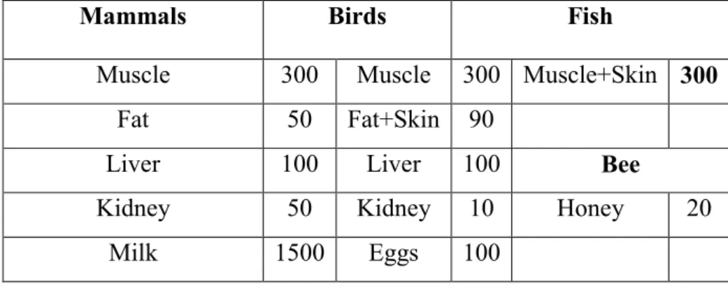
Derivation of the MRLs requires algorithms and several toxicological, pharmacological, and microbiological data packages. The risk assessment process is a standard battery of safety studies in animals and humans as well as in vitro studies that are used to determine the acceptable daily intake (ADI). The resulting toxicological ADI is often determined from the lowest no-observable-adverse-effect level (NOAEL) and lowest-observable-no-observable-adverse-effect level (LOAEL) obtained from the animal and human studies. These NOAELs and LOAELs are often adjusted by uncertainty factors to account for species differences (10 for animal to human extrapolations respectively) and intra-species differences (10 for variability within a population). The ADI is then adjusted with food consumption values for various organic matrices (300 g for muscle, 100 g for liver, 50 g for kidney, 50 g for fat, and if a dairy approval,1500 g for milk) to obtain MRLs for each matrix (Table 1). This requires kinetic data for each matrix, to be sure that the total food basket of residues at each matrix results in less than the ADI. (EMA/CVMP/SWP/355689/2006; Baynes et al., 2016).
Table 1. Values of ADI (gr or mL) for the MRL evaluations (EMA/CVMP/SWP/355689/2006)
Mammals Birds Fish
Muscle 300 Muscle 300 Muscle+Skin 300
Fat 50 Fat+Skin 90
Liver 100 Liver 100 Bee
Kidney 50 Kidney 10 Honey 20
Milk 1500 Eggs 100
1.1.2 Risk management
Good practice in the use of veterinary drugs is defined as the official recommended or authorised usage, including withdrawal periods, approved by national authorities (Codex Alimentary Commission Procedural Manual, 18th edition, 2008). The withdrawal period (also referred to as withdrawal time or withholding period) is the interval between the time of the last administration of a veterinary drug and the time when the animal can be safely slaughtered for food, or collected milk and eggs safely for the human consumption. Compliance with the withdrawal period provides an high degree of assurance to both producers and consumers that the concentration of residues in foods derived from treated animals will not exceed the MRLs. Withdrawal periods are typically assigned on the basis of results of a residue depletion study using non-radiolabelled drug in which the veterinary drug product proposed for marketing is administered at the highest label rate, the shortest dosing interval and for the longest duration. These conditions represent a worst-case scenario for residue depletion (Reeves, 2010).
1.1.3 Risk communication
Evaluation reports are necessary to document the risks and the proposed approaches to manage the identified risks. Based on these reports and dialogue with the risk assessors, risk managers gain a clear understanding of the involved hazards and risks, the basis of decisions taken in the risk
5 assessment, and the implications of the proposed strategy for managing the risks. The regulator must ensure that producer organizations and consumers understand the identified risks and the proposed risk mitigation strategy. Consumer engagement, including the presentation of programmes that provide an understanding of the regulatory processes, serves to assure consumers that drug residues in animal derived foods do not pose a health risk (Reeves, 2010).
1.2 Major and minor animal species in veterinary medicine
One of the most important challenge of veterinary medicine is the necessity to treat many types of animals including livestock, companion animals, working animals, sport animals, laboratory animals etc. This leads to the necessity of a rational dosing regimen of each species influenced by anatomy, biochemistry, physiology and behaviour of a given species. Moreover, some species show considerable differences within and between breeds in pharmacokinetic (PK) and pharmacodynamic (PD) profiles (Toutain et al., 2010).
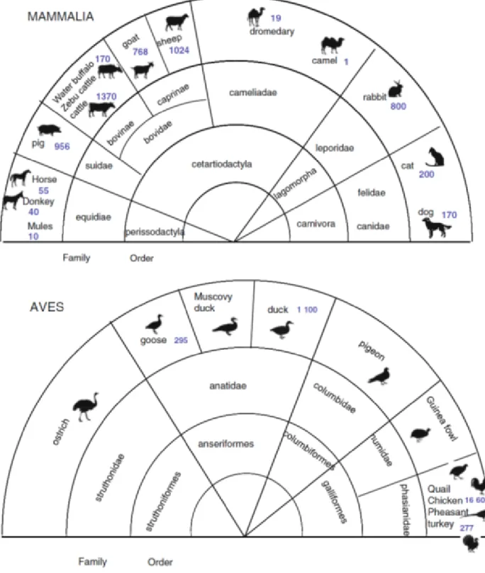
Concerning livestock species, nowadays it has been identified more than 40 domestic species. The World Watch List for Domestic Animal Diversity (WWL-DAD:3) issued by FAO (2000) provides inventories of the species and breeds of the domestic animals used for food production. Thirteen species contribute to most of the world’s food and agricultural production and are considered of veterinary interest (Figure 1).
7 For these reasons, some species are classified as major and others as minor species by the regulatory agencies in Europe and the US (Table 2).
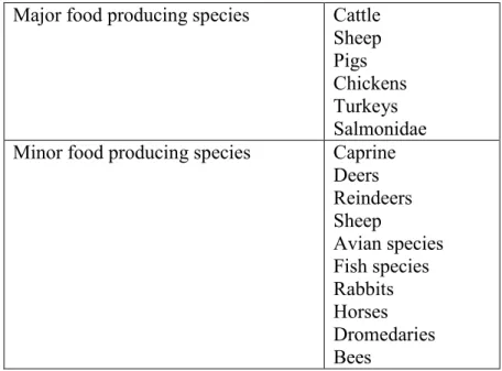
Table 2. Major and minor food producing species (EMEA/CVMP 2003) Major food producing species Cattle
Sheep Pigs Chickens Turkeys Salmonidae Minor food producing species Caprine
Deers Reindeers Sheep Avian species Fish species Rabbits Horses Dromedaries Bees
1.2.1 Off-label drugs and dosage regimens
Veterinary pharmacology still presents a reduced drug armamentarium for both major and minor animal species. Off-label drugs represent an opportunity to treat more and more new categories of disease. They refer to the use of an approved drug in a manner that is not in accordance with the approved label directions; they occur when a drug, only approved in human medicine, is used in animals; when a drug, approved for one species of animal, is used in another one; when a drug is used to treat a condition for which it was not approved, or the use of drugs at levels in excess of recommended dosages. Off-label drugs in food-producing animals present particular conditions. The veterinarian may not use an approved human drug if an animal drug approved for use in food-producing animals can be used. Finally, if scientific information on the human food safety aspect of the use of the drug in food-producing animals is not available, the veterinarian must take
appropriate measures to assure that the animal and its food products will not enter the human food supply (Beyene, 2016).
It is clear that the lack of new innovative therapies in veterinary medicine and the multiplicity of species of veterinary interest with their large interspecies differences in PK and PD profile requires some modelling tools to extrapolate PK and PD parameters between species. Allometric scaling is one of these approaches based on the influence of physiological properties of body size, metabolism and PK data ; on the contrary, PD data are considered negligible. It is a useful method to provide a first estimation of a dosage regimen, but it is predictive only if the differences between animal species are qualitative rather than quantitative. Especially for some minor species, excluded from the major drug companies that do not wish to grant a local marketing authorization, it is possible to extrapolate the dose from major species thanks to PK data. It consists to scale the approved dose in the major species against ratio of clearance of the two species in question (Eq.2) (Toutain & Bousquet-Melou, 2009):
Eq. 2 Dose new specie = Dose reference specie x Clearance new specie Clearance reference specie
Another approach to determine the optimal dosage regimens are PK/PD studies which calculate the effective dose (Eq 3.):
Eq. 3 ED = Cl x EC
9 Where “ED” is an efficacious dose, “Cl” is the plasma (total) clearance, bioavailability is referred to a particular route of administration and “EC” is the efficacious plasma concentration. This approach can predict the optimal rational dose, but also predicts the rate of depletion of residues from foods derivate (Toutain et al., 2010).
1.2.2 Species variabilities in poultry
Although, some poultry species such as chicken and turkey are considered as a major food-producing animals, PK and PD variability among poultry species (major and minor species) is large enough to consider that each species must be treated in its own right.
Taking in consideration the poultry medications, most of the drugs approved or given in off-label manner to these animal species are administered orally (90% of all treatments). To achieve an effective collective treatment in poultry, special attention must be given to poultry feeding and drinking behaviours and to the species characteristics of digestive tract physiology. Drinking water is the preferred mode of drug administration, especially for antibiotics, because diseased birds tend to stop eating but usually continue to drink. The alternative to the drinking administration is the added of drug in food by a pre-mix formulation. On the other hand, additional biological information such as body weight, age, and gender, but also environmental (lighting period and temperature) and managerial factor such as the composition of the diet, are necessary to determine an appropriate dosage regimen and to predict the rate of depletion of residues from meat and eggs (Toutain et al., 2010).
1.2.2.1 Drug residues in eggs.
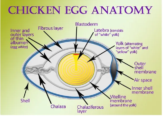
Figure 2 show the anatomy of the avian eggs. Eggs are constituted by three mainly components: yolk, albumen, and shell. Yolk has the longest development time. Precursors to yolk lipoproteins are produced in the liver and transported through circulation to the yolk follicles in the ovary. In an actively laying hen, several follicles at varying developmental stages reside simultaneously in
the ovary. Before an egg is laid, the yolk undergoes a stage of rapid growth, in which it increases in size exponentially over 10 days. Drugs that deposit in the yolk are rapidly accumulate during this time and can be present in successive eggs for 10 or more days following treatment. Following yolk maturation, the albumen is laid down over a period of 2-3 h and can also serve as a residue accumulation site. The egg shell is added, after that albumen proteins are deposited and diluted with water. The egg development process is similar across species of poultry and game birds, although the rates of development vary (Montesissa & Nebbia, 2009; Goetting et al., 2011).
Figure 2. Illustration of egg anatomy
Concerning drug residues, many drugs deposit preferentially in the yolk or albumen, depending on the drug’s physicochemical properties. Some characteristics that affect the distribution of residues are the drug’s affinity to bind to plasma proteins, hydrophobicity or hydrophilicity, and
11 the ability to move through different tissue types. However, a drug’s kinetic properties cannot always be predicted from its chemical properties (Montesissa & Nebbia, 2009; Goetting et al., 2011).
1.2.3 Species variabilities in caprine
As previously observed in Table 2 caprine are considered as minor species, though domestic goat (Capra aegagrus hircus) has many characteristics that make it a valuable livestock species. First of all, goats can survive on range land and plant species that other ruminants cannot. According to the Food and Agriculture Organization of the United Nations, over 90% of the world's goats are in developing countries with world’s goat population over 861,000,000 head (Clothier, 2010). Actually, this animal species is considered as a minor species by the regulatory agencies in Europe and USA, but goats present a worldwide population large enough to confer a status of major species (Toutain et al., 2010). Indeed, while goats represent a growing segment of agriculture, their numbers are far lower than those of other livestock species and many of their health issues cannot be addressed with EMA or FDA-approved medications (Clothier, 2010). Consequently, many drugs are administered to goats in an extra-label manner with no scientific information on drug behaviour, with a potential toxicity and with an inadequate withdrawal periods for drug removal from products marketed for human consumption.
Goats have many unique differences in anatomy, physiology and product biochemistry, from sheep and cattle which support the contention of many unique qualities of dairy goat products for human nutrition. Goat milk and its products of yogurt, cheese and milk-powder have three-fold significance in human nutrition:
• Treating people afflicted with cow milk allergies and gastro-intestinal disorders, which is a significant segment in many populations of developed countries.
• Filling the gastronomic needs of consumers, which is a growing market share in many developed countries.
The physiological and biochemical facts of the unique qualities of goat milk are just barely known. The high levels in goat milk of short and medium chain fatty acids have recognized medical values for many disorders and diseases of people. Moreover, the enrichment of short and medium chain fatty acids in goat butter, and their greater concentration compared to cow butter, could have become a valued consumer item supporting the idea of promoting goat butter (Haenlein, 2004). Species differences in drug action and effect reflect differences in target functions (anatomy, physiology, pathology) and/or target receptors (Toutain et al., 2010). For example, goats are frequently used as models for studying respiratory disease in cattle due to the similarity of these two species in susceptibility to inciting causes, bacterial pathogens, and mechanisms of pathology but differences in serotype, capsular type, and species of recovered bacteria (Clothier, 2010). Moreover, goats have generally a more active metabolism than sheep or cattle. This is linked to their respective feeding behaviour where goats are natural browsers that can stand on their hind legs or even climb trees. They preferably eat leaves, shrubs, flowers and fruits, thus choosing the most nutritious available food but also the portions of plants containing many toxic alkaloids that need to be metabolised by a hepatic first pass effect. In contrast, cattle are a non-selective bulk feeder that grazes non-selective grass, generally low in term of alkaloid content.
13
1.3 Pharmacokinetics and drug residues
Pharmacokinetics is the science that describes quantitative changes in drug concentration in the body over time as a function of administered dose. It is usually based on subjecting serum/plasma concentration-time data to mathematical models in order to determine quantitative terms, which describe absorption, distribution, metabolism, and excretion of the drug and its metabolites. Residue depletion profiles are linked to administered doses of drugs through their pharmacokinetic profiles (Lees & Toutain, 2012).
Several fundamental parameters are critical to establishing valid models from which other parameters can be determined. Clearance (Cl), is the amount of drug removed per unit of time; it is the most important parameter to determine since it dictates the dose and frequency of drug administration needed to reach a steady-state concentration. The volume of distribution (Vd) dictates how broadly the drug mobilizes throughout the body and is proportional to the amount of drug in the body; it is generally small if most of the drug is retained in the blood stream and large if the drug disseminates to tissues. Bioavailability (F%) is the percentage of drug that will reach the intended destination, which is generally the blood stream. Physiochemical traits of a drug such as size, lipid solubility, degree of ionization, and protein binding properties determine how it will traverse cell membranes and arrive at a target location, how it will persist at that location, and how it will be removed from that location. F% is considered complete for drugs administered intravenously, but must be determined for those administered by other routes (intramuscular, subcutaneous and oral routes). In this case, F% will influence Vd and Cl; consequently, they will become apparent clearance (CL/F) and apparent volume of distribution (Vd/F). Moreover, the area under the time-concentration curve (AUC) gives an estimation of total drug exposure. Half-life (t½) is the time needed to remove half of the drug from the compartment under study (usually plasma but can also be applied to a specific tissue), and is dependent on both CL and Vd. The
elimination rate of a drug is estimated by the determining the terminal slope (λz) of the time-concentration curve. Finally, the maximum drug time-concentration (Cmax) and time to reach maximum drug concentration (Tmax) are derived directly from the time-drug concentration plot (Bauer, 2004; Gabrielsson & Weiner, 2000; Goodman & Gilman, 2006; Rescigno, 2003).
Pharmacokinetic analysis is used to estimate parameters that characterize the behaviour of a drug and can be done experimentally or through the use of modelling. Pharmacokinetic modelling attempts to match drug behaviour with mathematical equations that can be used to predict drug activity in other populations. Non-compartmental PK models of experimental results consider the entire body as a single “vessel” into which the drug is administered and from which the drug is removed. These models make the fewest assumptions about how the drug is distributed without the need to describe movement from one location to another. On the contrary, compartmental analysis uses kinetic models to predict the time-concentration plot and considers drug entry and elimination from various segments of the body, such as blood, fat, or central nervous system (Bauer, 2004; Gabrielsson & Weiner, 2000; Goodman & Gilman, 2006; Rescigno, 2003).
Anti-microbial and anti-inflammatory drugs are used extensively in farm animal species for prophylaxis (administration to prevent disease), metaphylaxis (treatment of animals not showing clinical signs of disease but in contact with those which are), and therapy (treatment of animals displaying clinical signs of disease). For regulatory purposes, pharmacokinetic profiles are established in healthy animals; small numbers of animals, usually of a single breed, of similar age, and possibly of the same gender are used with an intensive sampling schedule (Lees & Toutain, 2012). However, it is clear that the PK behaviour is influenced by chemistry properties of the drug, dose, route of administration, but also age, gender and pathological conditions and the animals of interest. The absorption and distribution processes, the bioavailability of the drug and its metabolites in different organs and tissues and finally the elimination and depletion of the drug are used to evaluate the persistence of the residue in the inoculum site, tissues, organs and other
15 organic matrices used for the production of foodstuffs (Montesissa, 2009). Moreover, concentrations of veterinary drug residues depend on a range of properties such as lipid solubility and acidic/basic characteristics, which influence the passive diffusion of drugs across cell membranes. High lipid solubility drugs (for example fluoroquinolones and macrolides) cross cell membranes readily by passive diffusion to penetrate into intra- as well as extracellular compartments of tissues. Moderate to high lipid solubility drugs, similarly, generally enter readily into all water compartments of tissues. On the other hand, drugs of low lipophilicity generally do not readily enter cells, so that these drugs are located mainly or solely in the extracellular compartment of tissues. Concerning the acid/base characteristics of the molecule, take in consideration that intracellular fluid pH is lower than extracellular fluid pH (7.0 versus 7.4), weak bases penetrate readily into cells, by the classical Henderson-Hasselbalch mechanism of ion/diffusion trapping, whereas weak acids do not.
The CVMP (CE n. 2377/90) has issued specific guidelines for pharmacokinetics studies and residues depletion to define the behaviour of the different drugs in animal species and to establish rules of experimental design. All these information are needed to establish a maximum residue tolerable to ensure safety to the consumers in a day-to-day exposure condition and to harmonized the MRLs of the different countries of EU to a maximum required performance limit of the analytical methods (Montesissa, 2009). The concept “zero tolerance” is equivalent to the idea of total absence of residual amounts. Obviously, because of the improvement of analytical techniques, the value of zero becomes smaller and smaller that depicts the limits corresponding to the sensitivities of part per million (ppm), part per billion (ppb) and part per trillion (ppt). As a result, by using the high efficacy analytical methods such us the high performance liquid chromatography, it can be concluded that there are nearly always detectable residues but an extremely low concentrations that are not inevitably toxic for consumers (Beyene, 2016).
1.4 Analytical methods
The analytical methods for the determination of residues in different matrices are conventionally subdivided in 3 levels:
- The third level provides qualitative information. These level represent the screening method used to determine the presence/absence of drug to a concentration of interest. These methods are characterized by a high productivity, low cost, ease of use and not high technologies. An example of analytical method that belongs to this level is the test ELISA.
- The second level provides quantitative information. The analytical methods that belong to this level are confirmatory tests. These methods are characterized by a high technology, validation of methods and preparation of samples depending to the chemistry properties. Example of the present method is the high performance liquid chromatography (HPLC) with different detector (ultraviolet, spectrofluorometric and diode array detectors)
- The first level provides information with elevate significance. The analytical methods that belong to this level are confirmatory test. Example of analytical methods are gas chromatography and liquid chromatography with mass spectrometry detector (GC-MS/MS and LC-MS/MS) (Nachtmann, 2009).
The analytical methods utilized in the present thesis are:
- High performance liquid chromatography with spectrofluorometric detector (HPLC-FL) and diode array (HPLC-DAD). The spectrofluorometric detector, is used to determine fluorescent substances by a chromatographic curve obtained recording a signal at a specific emission and excitation wavelengths. The diode array detector allows to scan simultaneously at discrete intervals and a predefined electromagnetic range.
17 -Liquid chromatography mass spectrometry (LC-MS/MS). The mass spectrometry detector is able to provide information on the structure of the molecule under examination on the basis of the fragmentation products obtained under controlled and reproducible conditions.
2. Chemical and pharmacological properties of drugs of interest
2.1 Antibiotic drugs
As mentioned in chapter 1, it’s necessary to have greater knowledge of the pharmacokinetics of drugs in different animal species, but also are needed more information about the biological and toxic effects induced by these molecules in both the animal and human (Odore & Badino, 2009). Pharmacodynamics is the study of the relationship between drug exposure and the biological effects of the drug on the host or, in the case of an antibiotic, on the bacterial target.
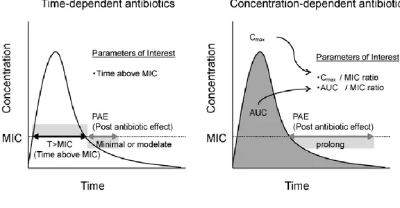
Parameters used to evaluate PD effects of antibiotics include the minimum inhibitory concentration (MIC) against specific bacterium in vitro, the post-antibiotic effects (PAE) and kinetics of bacterial killing (Figure 3) (Gabrielsson & Weiner, 2000; Rescigno, 2010; Godinho et
al., 2005; Wise, 2001).
The MIC is defined as the lowest concentration of antimicrobial that suppresses bacterial growth in a defined incubation period (Catry et al., 2007). The MIC determination involves an incubation of a bacterial suspension with a range of doubling antibiotic concentrations for a specified time and how their growth is inhibited. The MIC is considered the “gold standard” of susceptibility test. Depending on the quantitative antimicrobial susceptibility as indicated by the MIC value, bacteria, in relationship with the drug, are categorized into “clinically susceptible (S),” “intermediate (I),” or “resistant (R)” (Mouton et al., 2012). Moreover, the mean bactericidal concentration (MBC) can be identified by sub-culturing the bacterial antibiotic suspension used in MIC testing onto antibiotic-free media to determine what concentration of antibiotic is required for a complete bacterial killing. Bactericidal antibiotics have MBC that are very close to MIC values, since their mode of action results in irreversible damage to the bacterium; bacteriostatic drugs have MBC that
19 are much greater than MIC. Kill-kinetic assays are an established method to determine the degree of bacterial killing. The most common chosen levels of killing are MBC90 (90% killed), MBC95 (95% killed), and MBC99 (99% killed) (Andrews, 2001; Firsov et al., 1997; Stratton et al., 1987).
Figure 3. PK and PD parameters
Antibiotics are traditionally classified into each one of the following three categories: time-dependent, concentration-time-dependent, and time-dependent with a post-antibiotic effect, based on PK/PD activity (Figure 3 and 4). The activity of time-dependent antimicrobials relies on the length of time that drug levels are at or above the MIC (T>MIC) of the specific pathogen being treated. More the drug levels persist above the MIC, more effective is the antibiotic treatment. Common time-dependent antibiotics include penicillins, cephalosporins, and older macrolides. Concentration-dependent antimicrobials depend on the maximum level of antimicrobial above the MIC (Cmax/MIC) of the pathogen being treated. An effective treatment is based upon maximizing plasma or tissue concentrations of the drug and benefits from the suppression of bacterial growth that continues for a period even after drug levels fall below the MIC. Aminoglycosides,
tetracyclines, and fluoroquinolones are members of this category. The concentration-effect relationship is empirically described by the sigmoidal Emax model (Hill equation Eq. 4):
Eq. 4 E(t) = E0 + Emax∗C(t)
EC50 H+C (t)
Where “E(t)” is the effect observed for a given concentration (C) at time (t). “Emax” is the maximal effect attributable to the drug. “EC50” is the plasma concentration producing 50% of Emax; “H” is the Hill coefficient which describes the steepness of the sigmoidal relationship between the concentration and the effect. “E0” is the rate of background response in the absence of drug (such us that achieved by the host immune response) (McKellar et al., 2004; Martinez et al., 2013). The third category of antibiotics, which includes streptogramins, ketolides, and newer macrolides, has characteristics of both, with time-dependent killing mechanisms and PAE. PAE is based on the concentration of drug, the time that bacteria is in contact with the drug, and its mechanism of action. Total drug exposure is evaluated by the area under the time-concentration curve (AUC) and represents a mathematical summation of drug concentrations in the compartment being assessed. The AUC/MIC ratio is frequently used to determine the drug efficacy (Toutain et al., 2002; Martinez et al., 2013; McKellar et al., 2004; Van Bambeke & Tulkens, 2001; Gabrielsson & Weiner, 2000; Li & Zhu, 2002).
The AUC/MIC ratio may be used to compute a dose using the following equation (Eq. 5):
Eq. 5 𝐷ose( pre day) =Cl x (
AUC
MIC)breakpoint x MIC 90
21 where “Dose (per day)” is the daily maintenance dose at steady state; “Cl” is the plasma clearance expressed as L/h. “(AUC/MIC)breakpoint” the targeted AUC/MIC value (e.g. 125 h) provides a dose achieving an average plasma concentration over 24 h in the steady state equal to approximately five times MIC (125h/24h). “MIC90” the 90th percentile of the MIC distribution. “fu” (from 0 to 1) the free (unbound) fraction (only the free antibiotic plasma concentration is a relevant plasma surrogate of the free antibiotic concentration at the biophase level) and F the absolute bioavailability (from 0 to 1) (Toutain et al., 2007; Lees et al., 2015)
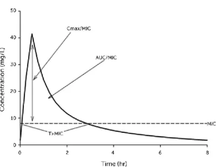
Figure 4. PK/PD analysis
When PK/PD indexes are not in these values, mutated bacterial strains exhibit resistance, as the mutated species typically has MIC values 4 to 8 times higher than the MIC of the Wild type species (Papich & Rivere, 2009).
2.1.1 Quinolones and fluoroquinolones
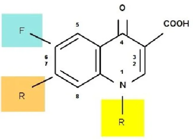
Figure 5. Fluoroquinolones chemical structure
Quinolones and fluoroquinolones, comprise a large and expanding group of synthetic antimicrobial agents. The first drug of this class, nalidixic acid, was discovered in 1962 and was a modification of a compound isolated during the production of the anti-malaria drug, chloroquine. Its antibacterial spectrum of activity was restricted to the Enterobacteriaceae and, because of limitations in absorption and distribution, the drug was effective solely for the treatment of urinary tract infections. In the 1980s, the addition both of a fluorine molecule at the 6-position of the basic quinolone structure and a piperazine substitution at the 7-position was found to enhance quinolone antibacterial activity, gaining effectiveness against Pseudomonas aeruginosa and Gram-positive, cocci, and to increase the extent of oral drug absorption and tissue distribution. Products possessing this fluorine molecule are known as the fluoroquinolones (Figure 5).
Fluoroquinolones belongs to the third generation class of quinolones. This class of drugs exhibited an increased antibacterial activity against the Enterobacteriaceae and other Gram-negative bacteria (such as P. aeruginosa) compared to the first and second generation of quinolones. It had some activity against certain Gram-positive cocci. Structural changes associated with the third generation increased their oral bioavailability and systemic distribution. Quinolones fitting into this category include norfloxacin, ciprofloxacin, enrofloxacin, danofloxacin, difloxacin and
23 marbofloxacin. Likewise, the fourth generation drugs belonging to the quinolones, maintained the favourable characteristics of the third generation drugs while exhibit an increase activity against Gram-positive bacteria, anaerobes and mycobacteria. These compounds also exhibited excellent oral bioavailability, a prolonged terminal elimination half-life, lower central nervous system toxicities and exhibit fewer interactions with the cytochrome P450 (CYP 450) system (Ball, 2000). Fluoroquinolones are a class of bactericidal drugs and have of a concentration-dependent nature, showing a rapid and profound killing over a wide range of concentrations (Martinez et al., 2013). Therefore, the action is evaluated by Cmax/MIC ratios greater than 10 and AUC/MIC whose value must be between 100 and 125, in order to have a good antibacterial activity and to limit the development of peripheral resistances. (Toutain et al., 2002; Wright et al., 2000).
2.1.1.1 Chemical properties
Fluoroquinolones are amphoteric molecules weakly soluble in water for the presents of a zwitterion form with the carboxylic acid and the basic amine. The pKa values range from 5.5 to 6.3 for carboxylic acid and 7.6 to 9.3 for amine group. In acid pH condition both groups are protonated, while at a basic pH condition the carboxylic acid is in the form of anion. For this reason, the fluoroquinolones tend to be more water-soluble with acid or basic pH, while at physiological pH they exhibit minimum water solubility. Finally, fluoroquinolones have a high melting point, generally greater than 200 ° C, so the crystallized form is very stable (Papich & Riviere, 2009; Brighty & Gootz, 2000).
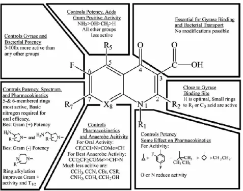
2.1.1.2 Structure–activity relationship
Figure 6. Structure–activity relationship of fluoroquinolones
Figure 6 shows the general structure-activity relationship of the fluoroquinolones. The carboxyl group in position 3 and the ketone at position 4 are essential for antibacterial activity. Fluoride in position 6 gives a wider spectrum action, a greater power, a greater input capacity in the bacterial cell. The N substitution in position 1 with an ethyl, cyclopropyl, fluorophenyl, leads to a wider spectrum of action. Substitution at position 7, with a piperazine, increases antibacterial activity against Pseudomonas. At position 8, it is preferable to have carbon instead of nitrogen to limit side effects at the central nervous system level and increase activity against staphylococci, while substitutions with a methoxy or cyan group, in this position, can increase the activity against anaerobic bacteria (Papich & Riviere, 2009).
25 2.1.1.3 Mechanism of action
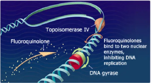
Figure 7. Fluoroquinolones mechanism of action
Figure 7 shows the fluoroquinolones mechanism of action. Fluoroquinolones damage bacterial DNA and lead to defects in negative supercoiling. This effect was linked to inhibition of DNA gyrase activity, an enzyme found in all bacteria. In concert with other proteins, gyrase catalyzes changes in the degree of double-stranded DNA supercoiling. In this capacity, it plays a vital role in DNA packing, replication and transcription.
The effect of fluoroquinolones on bacterial proliferation suggests three mechanisms of cell killing: 1. Mechanism A: common to all quinolones. This requires RNA and protein synthesis and is only effective against dividing bacteria. Mechanism A appears to involve the blocking of replication by the gyrase–quinolone complex on DNA.
2. Mechanism B: does not require RNA and protein synthesis and can act on bacteria that are unable to multiply. Mechanism B (chloramphenicol insensitive) can be correlated with dislocation of the gyrase subunits that constrain the ternary complex.
3. Mechanism C: requires RNA and protein synthesis but does not require cell division. Mechanism C may correlate with trapping of topoisomerase IV complexes on DNA.
Moreover, survival curves show that when the fluoroquinolone concentration is near the minimal inhibitory concentrations (MIC) of the bacterium, the drug has a static effect on bacterial growth (bacteriostatic). As the drug concentration increases relative to the MIC of the bacterium, bacterial killing increases up to a certain drug concentration (termed the optimum bactericidal concentration). As concentrations exceed the optimum bactericidal concentration, further increases in drug concentration can lead to a decrease in bacterial killing (Martinez et al., 2006). 2.1.1.4 Pharmacokinetics of fluoroquinolones
Fluoroquinolones show to be well absorbed and distributed in tissues and body fluids after oral subcutaneous and intramuscular administrations in different animal species. However, food intake and tissue binding can decrease its absorption. High and major concentrations of fluoroquinolone are found in the kidney and liver. This can be due to the fluoroquinolones high lipophilicity, passing well the cell membranes and are not easily expelled. In addition, fluoroquinolones show the ability to be concentrate more on leukocytes that allows them to be transported to the site of infection, acting more on infected tissues than healthy ones (Papich & Riviere, 2009).
The main metabolites of fluoroquinolones are the 3-carboxylated derivatives that are produced by kidney and the hydroxylated derivatives. The letter compounds are more water-soluble, and therefore more easily eliminated than the parental drug. This type of metabolism produces both active and inactive products.
The main route of elimination of the fluoroquinolones is kidney. This drug having a partially acidic feature, are eliminated for active tubular secretion, reaching higher urinary concentrations than serum. They are re-absorbed in a higher percentage in the animal species that have acid urine, while in herbivores the urine alkalinity conditions give a faster elimination. Finally,
27 fluoroquinolones and their metabolites can also be eliminated by the bile duct (Odore & Badino, 2009).
2.1.1.5 Antimicrobial resistance
Antimicrobial resistance is a complex problem that is likely to require attention at many levels. The most frequently developed resistance to fluoroquinolones is related to:
i) modification of the target structure, that is, the mutation of the gyrA gene, which encodes for the subunit A of DNA-gyrase, resulting in the transcription of a different subunit that is not recognized by fluoroquinolone; ii) mutation of the paro-gene (responsible for breaking DNA) of Topoisomerase IV. This type of alteration is common in Gram positive bacteria; iii) alteration of the outer membrane permeability, which involves the bacterial cell, a cross-resistance with other antibiotics that use porinic channels to enter the periplasmic space. This mechanism is typical of some Gram negative strains (Ferrero et al., 1995).
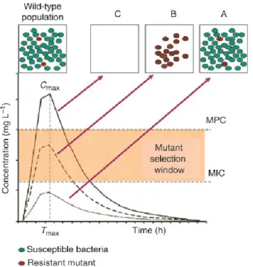
Issues concerning the optimal dose to administered are addressed by the mutant selection window hypothesis. This hypothesis maintains that drug-resistant mutant subpopulations present prior to initiation of antimicrobial treatment are enriched and amplified during therapy when antimicrobial concentrations fall within a specific range (the mutant selection window). The upper boundary of the mutant selection window is the MIC of the least drug susceptible mutant subpopulation, a value called the mutant prevention concentration (MPC). The lower boundary of the mutant selection window is the lowest concentration that exerts selective pressure, often approximated by the minimal concentration that inhibits colony formation by 99% (MIC99) (Drlica & Zhao, 2007). Figure 8 shows the mutant selection window (MSW) characterized by three discrete concentrations. A: The MIC of the wild-type bacteria
B: Concentration above the MIC of the wild-type bacteria, where there is a plateau in killing due to the survival of the least susceptible microbial subpopulation of the first-step resistant variant. C. Concentrations at which even the least susceptible organism are killed (MPC). Above MPC all bacteria are inhibited or eliminated. The concentration of the administered drug should therefore remain as low as possible in this range (MSW) and pass over the MPC threshold to obtain good therapy with excellent results (Martinez et al., 2013; Canton et al., 2006)
29 2.1.1.6 Levofloxacin
Figure 9. Levofloxacin
Levofloxacin belongs to third generation fluoroquinolones (Figure 9). It is an optical isomer of ofloxacin having two-fold higher antimicrobial activity than the parent compound. Compared with other fluoroquinolones it has more pronounced bactericidal activity against Pseudomonas,
Enterobacteriaceae and Klebsiella,spp, several species of staphylococci, streptococci including S. pneumoniae, bacteroides, clostridium, haemophilus, moraxella, legionella, mycoplasma and chlamydia (North et al., 1998). The bactericidal effect of levofloxacin is obtained through a
reversible linkage with DNA excretion and subsequent inhibition of bacterial DNA replication (Figure 10) (Fu et al., 1992).
Levofloxacin distributes well in all body tissues and fluids of the respiratory tract, skin, urine and prostrate, and its uptake by cells makes it suitable for use against intracellular pathogens. However, it penetrates poorly into the central nervous system. Like all fluoroquinolones, levofloxacin shows a concentration-dependent killing mechanism, whereby the optimal effect is attained by the administration of high doses over a short period of time. This concentration-dependent killing profile is associated with a relatively prolonged postantibiotic effect. The drug undergoes a limited metabolism in rats and humans and is primarily excreted by kidney mainly as active drug. Inactive metabolites (N-oxide and demethyl metabolites) represent <5% of the total dose (Goudah & Abo-El-Sooud, 2008) The pharmacokinetics of levofloxacin has been fully investigated in humans (Chulavatnatol et al., 1999), rabbits (Destache et al., 2001), cats (Albarellos et al., 2005) and calves (Dumka & Srivastava, 2006, 2007). Currently, Levofloxacin is successfully used in human medicine for the treatment of infections of upper and lower respiratory tract, genitourinary system, skin and soft tissue (Patel et al., 2009).
2.1.2 Macrolides
The macrolide class of antibiotics contain a 14-, 15-, or 16-membered macrocyclic lactone ring to which amino sugars are attached (Odore & Badino, 2009). The unique structure of macrolides facilitates rapid dissemination from the central compartment to lung tissues which make them useful in the treatment of bacterial pneumonia (Clothier, 2010). Macrolides are classified in natural macrolides, originally isolated from Streptomyces such as erythromycin and semi-synthetic macrolides like clarithromycin and azithromycin.
Macrolides present a bacteriostatic action for the binding to the 50S ribosomal subunit, preventing translocation of the growing peptide and blocking protein synthesis (Figure 11).
31 Figure 11. Mechanism of action of macrolides
Due to its mechanism of action the spectrum of action includes Gram positive bacteria, mycoplasma, rickettsia, chlamydia, some protozoa such as amoeba, toxoplasmosis and
cryptosporidium. Due to its basic nature and liposolubility, macrolides are generally well absorbed
orally. The percentage of plasma protein binding is not high (about 20-40%) in different animal species. Macrolides distribute well in all organs and tissues except the SNC, spreading intracellularly to reach concentrations higher than plasma concentrations. The basic nature of this class of drugs led to higher concentrations in saliva and liquids with acidic pH such as milk where they reach levels up to 3 times higher compared to plasma concentrations. In the peripheral compartment, macrolides concentrations are elevated, particularly in the lung, liver, kidney and spleen. Macrolides are bio-transformed in different percentages through N-demethylation cytochrome P450 dependent reactions. The metabolites are eliminated predominantly by bile
(about 80%) in conjugated compound forms and may undergo the entero-hepatic uptake (Odore & Badino, 2009). This class of drugs is used in farm animals for respiratory disease, streptococcal and staphylococcal mastitis in ruminants, arthritis in bovine, enteritis haemorrhagic in the pig and oral cavity infections (Odore & Badino, 2009).
2.1.2.1 Tulathromycin
Figure 12: Tulathromycin
Tulathromycin (Figure 12) is an antimicrobial drug belongs to the class of semi-synthetic macrolide consisting of a regioisomeric, equilibrated mixture of a 13-membered ring azalide (10%) and a 15-membered ring azalide (90%) but in aqueous media the drug exists as an equilibrated mixture. The drug molecule has three nitrogen/amine functional groups representing the first member of a novel sub–class of macrolides known as triamilides. Tulathromycin is a basic molecule with a pKa in a range from 8.6 to 9.6 and it is approximately 50 times more soluble in hydrophilic than hydrophobic media. This physicochemical feature allows the unionized fraction of the drug to easily penetrate into tissues from plasma and to accumulate in compartments with acidic conditions (a phenomenon known as ion trapping). This may explain, at least in part, the
33 large volume of distribution of the drug and the accumulation of the drug in alveolar epithelial lining fluid and bronchoalveolar lavage cells. This activity can provide unique therapeutic advantage in treating bacterial respiratory infections in livestock species (Villarino et al., 2013a). Finally, tulathromycin has been shown to accumulate in leukocytes (primarily macrophages and neutrophils) in greater amounts than older macrolides (Evans, 2005).
As a macrolide, tulathromycin exerts its mechanism of action through binding to the 50S subunit of bacterial ribosomes and blocking peptidyl transferase which results in dissociation of transfer RNA (tRNA), cessation of peptide translocation, and blockage of protein synthesis (Benchaoui et
al., 2004; Evans, 2005). Although this drug, like other macrolides, is classified as bacteriostatic
agent when tested against Staphylococcus aureus and E. coli (Pfizer, 2004), it can also exhibit bactericidal activity at higher concentrations (Benchaoui et al., 2004; Evans, 2005; Nowakowski
et al., 2004). This effect is dictated by the microorganism involved in the infection and the
concentration-time profile of the drug at the site of infection. In particular, tulathromycin present a bactericidal effect at 4x and 8x the MIC against M. haemolytica, A. pleuropneumoniae and P.
multocida (Villarino et al., 2013b).
The pharmacokinetics of tulathromycin, have been studied in different animal species such us mice, cattle, pigs, goats and foals. At label dose (2.5 mg/kg) in cattle and swine species, tulathromycin is characterized by a rapid rate of absorption and large systemic availability (90%) after IM and SC administration. In plasma, tulathromycin has a long terminal half-life, ranging across domestic species from 60 to 140 h and a remarkable large volume of distribution (>10 L/kg). In pigs, most of the dose is eliminated as unchanged by biliary and renal excretion. Protein binding studies indicate that the unbound fraction of tulathromycin ranges from 53% to 68%. Lung pharmacokinetic studies in cattle, pigs and horses reveal an extraordinary capacity of tulathromycin to accumulate in lung tissue, where the lung homogenate concentrations showed to be 50-fold higher than in plasma concentration. Finally, the antimicrobial efficacy of
tulathromycin has been addressed in several published studies including data on cattle, swine, horses, and goats.
Tulathromycin is safe when used according to label directions (Pfizer, 2005) but studies of toxicity in bovine, swine and caprine species have revealed minor clinical and injection site histopathological changes that have been resolved over time (Pfizer, 2005; Clothier et al., 2010). Moreover, hypersalivation, head shaking, and pawing at the ground have been reported at therapeutic doses (Pfizer, 2005).
2.2 Pain relief drugs
Pain is a sensory process that results from tissue damage and is intended to prevent further tissue damage that follows the injury. Pain activates numerous physiological reactions that often induce negative effects on well-being and behaviour, as well as on the growth and reproduction of the animals. However, the main obstacle to the pain management, in farm animals, is to recognise, quantify and evaluate the pain status of each individual and treat without disturbing the whole group of animals (Guatteo et al., 2012). In veterinary medicine, recently, pain has been shown to affect animal welfare and production, and the interest in the field of analgesia has been drastically increasing (Lee et al., 2014). Therefore, the desirable medication associated with the pain status of each individual will be important to treat pain in farm animals. A similar specific approach designed to minimize pain in laboratory animals is also required in farm animals. Nowadays, the concepts of “Replacement, Reduction and Refinement”, called the ‘3Rs’ (Russell & Burch, 1959), used in the design of animals experiments to minimise unnecessary pain, is the basis of a new approach the ‘3S’ “Suppress, Substitute and Soothe pain”. This approach is used to review existing practical solutions and find new solutions to eliminate or alleviate pain in farm animals. It is based on the possibility to “suppress” the procedures or environments that are a source of pain, to
35 “substitute” such procedures by others causing less pain and to “soothe” pain when it cannot be avoided (Guatteo et al., 2012).
Animal welfare issues in food production are now being driven by animal activists, food companies and consumers. Consequently, animal welfare assurance programs have been developed and are encoded in non-mandatory codes or guidelines, government regulations, inter-governmental agreements and corporate programs (Fraser, 2006). One of the measures aimed at improving animals welfare is pain management by using anti-inflammatory drugs. These substances can be effective in suppressing or preventing inflammation, treating allergy, lowering fever and reducing pain. The classes of anti-inflammatory drugs differ because of their actions towards the biochemical mediators that are released during inflammation and that propagate the inflammatory response (De Vito, 2015).
2.2.1 Opioid receptor agonists
One of the main classes of anti-inflammatory drugs most used in veterinary medicine for food-producing animals are opioid receptor agonists. Unfortunately, the drugs in these classes range present typical side effects. Compounds that activate opioid receptors, in particular the Mu Opioid Receptor (MOR) subtype has been used for decades in the treatment from moderate to severe pain (Meldrum, 2003). Extensive clinical experience with the prototypical MOR agonist morphine indicates that, although this compound is very effective against acute pain, it may be less effective for conditions precipitating chronic pain, especially neuropathic pain or pain of inflammatory origin. This reduced effectiveness for chronic pain is due to the MOR down regulation with long-term therapies; a substantial increase in dosing is required in order to maintain a clinically satisfactory analgesic effect (Dickenson & Suzuki, 2005). Furthermore, morphine has a limited therapeutic window, its analgesic effect generally coincides with several side effects (nausea, emesis, constipation and respiratory depression) which limit its usefulness especially in cases of
chronic pain. Another therapeutic approach has been to combine the MOR activation with an additional compound that provides analgesia via a different mechanism of action, resulting in an opiate-sparing effect. Activation of the noradrenergic descending pain inhibitory pathway could be considered a candidate for such an additional mechanism (Wang et al., 2008). Active ingredients that inhibit the reuptake of noradrenalin (NA) are effective analgesics, particularly in chronic pain conditions. The first molecule showing this dual mechanism of action was tramadol. It produces MOR activation as well as inhibition of serotonin (5HT) and NA reuptake. Nowadays, tramadol is widely used in veterinary clinical practice but its efficacy is still controversial for its faster metabolism to inactive metabolites N-desmethyl tramadol (M2) and O,N-didesmethyl tramadol (M5), from different animal species such as goats, dogs, horses, lamas, alpacas, peafowl, hawks than in cats (Giorgi, 2012).
2.2.1.1 Tapentadol
Figure 13. Tapentadol
Tapentadol is a novel opioid drug launched at the end of 2011on the European market for human use (Figure 13). This drug, like tramadol, presents a dual mechanism of action: the mu-opioid receptor agonism and the inhibition of noradrenaline-reuptake. Based on its unique mechanism of action, it has been proposed as the first representative of a new pharmacological class of centrally acting analgesics: the MOR agonist, NA Reuptake Inhibitor (MOR-NRI) (Kress, 2010). Interestingly, even though its MOR affinity is 50-fold lower than that of morphine, this reduces to
37 a 2-3-fold difference after systemic administration. This finding, consistent across different pain relief evaluation models, may be due to a better brain penetration of tapentadol, but also suggests that the NA reuptake-inhibitory property, contributes to a more potent analgesia that would be expected solely from its MOR agonism (Tzschentke et al., 2006). Given the moderate affinity of tapentadol at the MOR and the opioid sparing effect of tapentadol NRI component, it seems logical that this drug would produce fewer opioid-related side effects than classical MOR agonists, such as morphine. Indeed, compared to morphine, tapentadol produces much less nausea and vomiting in ferrets, the duration of these side effects was also shorter (Tzschentke et al., 2009). Furthermore, the threshold dose for these effects was 100 times higher for tapentadol than for morphine. In vivo and in vitro preclinical pharmacological studies demonstrated that tapentadol displays weak anticholinercic activity and a negligible 5HT reuptake inhibition but a pronounced NA reuptake inhibition (Tzschentke et al., 2006). Following its chronic administration, tolerance development took much longer compared with morphine (Ahlbeck, 2011). The PK features of tapentadol have been tested in rodents and humans. In summary, the drug is almost completely absorbed after oral administration but undergoes high levels of phase II metabolism glucuronidation, limiting oral bioavailability at 8 and 32% in rats and humans, respectively. Phase I biotransformation is negligible and does not produce active metabolites. Finally, tapentadol has shown no potential for CYP450 induction or inhibition (Terlinden et al., 2007). However, tapentadol still has some disadvantages. It has weak anti-muscarinic activity, which produces a well known adverse effects. Tapentadol is a weak blocker of 5-HT3 receptor (e.g., as mirtazapine, metoclopramide and ondasteron), as yet it has not been determined whether this property is helpful or harmful. It has very low oral bioavailability although animal species deficient of glucuronic acid, might be much higher (Giorgi, 2012).
2.2.2 Non steroidal anti Inflammatory drugs
Another major classes of anti-inflammatory drugs most used in veterinary medicine for food-producing animals are the non-steroidal anti-inflammatory drugs (NSAIDs). The NSAIDs are widely used because of their ability to reduce inflammatory process. NSAIDs can be classified into several groups according to their chemical structure. The propionic acid derivatives (Ketoprofen, Carprofen, Vedaprofen , Naproxen); the anthranilic acid derivatives (Tolfenamic acid, Mefenamic acid); the nicotinic acid derivatives (Flunixin); the pyrazolones
(Phenylbutazone]) and the acetic acid derivatives (Diclofenac); the class of oxicams
(Meloxicam) (Dubreil-Chéneau et al., 2011). In veterinary practice, NSAIDs are used in the treatment of musculoskeletal disorders, coliform mastitis, pulmonary diseases and enteritis in several animal species. NSAIDs, however, because of their toxicity, can affect the gastro-intestinal, hematopoietic and renal systems. Gastro-intestinal ulcerations are the most common and serious side effect of NSAIDs, especially in cases of overdose or chronic abuse (Kim et al., 2014; 2015).
2.2.2.1 NSAID mechanism of action
The major mechanism of action by which the NSAIDs elicit their therapeutic effects (antipyretic, analgesic, and anti-inflammatory activities) is inhibition of prostaglandin (PG) synthesis. Specifically NSAIDs competitively (for the most part) inhibit cyclooxygenases (COXs), the enzymes that catalyze the synthesis of cyclic endoperoxides from arachidonic acid to form. Two COX isoenzymes have been identified: COX-1 and COX-2. COX-1, expressed constitutively, is synthesized continuously and is present in all tissues and cell types, most notably in platelets, endothelial cells, the GI tract, renal microvasculature, glomerulus, and collecting ducts. Thus COX-1 is important for the production of prostaglandins of homeostatic maintenance, such as platelet aggregation, the regulation of blood flow in the kidney and stomach, and the regulation of gastric acid secretion. Inhibition of COX-1 activity is considered a major contributor to NSAID