Università degli Studi di Ferrara
DOTTORATO DI RICERCA IN
"Farmacologia e Oncologia Molecolare"
CICLO XXII
COORDINATORE Prof. Pier Andrea Borea
THE A
3ADENOSINE RECEPTOR: A LINK BETWEEN
INFLAMMATION AND CANCER
Settore Scientifico Disciplinare BIO/14
Dottorando Tutore
Dott.ssa Sacchetto Valeria Dott.ssa Gessi Stefania
Contents
Pag.
General introduction 5
References 49
Aim of the thesis 0 69
CHAPTER 1 71
A3 adenosine receptor regulation of cells of the immune
System and modulation of inflammation.
Abstract 72
A3 adenosine receptor effects on neutrophil function 72
A3 adenosine receptor effects on eosinophil function 80
A3 adenosine receptor effects on lymphocyte function 84 A3 adenosine receptor effects on monocyte-macrophage function 90 A3 adenosine receptor effects on dendritic cell function 94
Conclusion 98
References 99
CHAPTER 2 109
Modulation of MMP-9 in glioblastoma cells by A3 adenosine receptors.
Introduction 0110
Materials and Methods 111
Results 117 Discussion 136 References 141 111 List of publications 148 Acknowledgements 149
ADENOSINE
Adenosine is a nucleoside composed of a molecule of adenine attached to a ribose sugar molecule (ribofuranose) via a β-N9-glycosidic bond (Figure 1).
Figure 1 – Chemical structure of Adenosine
Adenosine is an endogenous nucleoside-signalling molecule, which, by acting on specific membrane receptors produces a number of physiological and pathophysiological effects in both the central nervous system and peripheral organs. Under normal conditions, adenosine is continuously formed intracellularly as well as extracellulalry. The intracellular production is mediated either by an intracellular 5’-nucleotidase, which dephosphorylates AMP or by hydrolysis of S-adenosyl-homocysteine (Fredholm et al., 2001). Adenosine generated intracellular is transported into the extracellular space mainly via specific bi-directional transportes through facilitated diffusion that efficiently evens out the intra-and extracellular levels of
N N N N NH2 O OH OH H H H H HO
adenosine. The dephosphorylation of extracellular AMP to adenosine, mediated by ecto-5’-nucleotidase, is the last step in the enzymatic chain that catalyzes the breakdown of extracellular adenine nucleotides, such as ATP, to adenosine. Ectonucleotidases include ectonucleoside triphosphate diphosphohydrolase which can hydrolyze ATP or ADP, ectonucleotide pyrophosphatase/phosphodiesterases, alkaline phosphatases and 5’-nucleotidases (Zimmermann, 2000). When adenosine levels in the extracellular space are high, adenosine is transported into cells by means of transportes. It is then phosphorylated to AMP by adenosine kinase or degraded to inosine by adenosine deaminase. Adenosine deaminase, but not adenosine kinase, is also present in the extracellular space (Fredholm et al., 2001). Another potential source of extracellular adenosine is cAMP, which can be released from neurons and converted by extracellular phosphodiesterases into AMP and thereafter by an ecto-5’-nucleotidase to adenosine. The transport of adenosine by facilitated diffusion is equilibrative and bidirectional, meaning that the net transport of adenosine either into or out of the cell depends upon the adenosine concentration gradient in both sides of the membrane. Inhibition of adenosine transport can, therefore, inhibit either adenosine release or adenosine uptake, depending upon the intra- and extracellular levels of adenosine (Baraldi et al., 2008). However, since the extracellular formation of adenosine from released adenine nucleotides constitutes a second source of adenosine, which is not affected by transport inhibition, the transport inhibitors usually cause an icrease in the extracellular adenosine levels. Under hypoxic and ischemic conditions there is a marked increase in cytoplasmatic adenosine leading to an intense release of adenosine, which is inhibited by adenosine uptake inhibitors (Parkinson et al., 2002).
Excitatory amino acid-mediated release of adenosine is certainly involved; however, of greater importance is probably the fact that whenever intracellular levels of adenine nucleotides fall as a result of excessive energy use, the intracellular levels of adenosine
will rise dramatically (Fredholm et al., 2001). For example, following hypoxia there is a decrease of intracellular ATP, accompanied by an accumulation of 5’-AMP and subsequently adenosine: The nucleoside is thereafter transported into the extracellular space via the transporters. Furthermore, when the intracellular level of adenosine is very high, adenosine simply diffuses out of cells. Direct release of intracellular adenine nucleotides, such as ATP, that is thereafter converted extracellularly by ecto-ATPase and ATPdiphosphohydrolase (apyrase) to AMP and dphosphorylated by ecto-5’-nucleotidase to adenosine, should also be considered (Zimmermann et al., 2000). Adenosine is neither stored not released as a classical neurotransmitter since it does not accumulate in synaptic vescicles, being released from the cytoplasm into the extracellular space through a nucleoside transporter. The adenosine transportes also mediate adenosine reuptake , the direction of the transport being dependent upon the concentration gradient at both sides of the membrane (Fredlhom et al., 2001). Since it is not exocytotically released, adenosine behaves as an extracellular signal molecule influencing synaptic transmission withiìout itself being a neurotransmitter, i.e. modulates the activity of the nervous system at celluar level presynaptically by inhibiting or facilitating transmitter release, postsynaptically by hyperpolarizing or depolarizing neurons and/or exerting nn-synaptic effects. Adenosine, therefore, belongs to the group of neiromodulators.
Adenosine receptors
Four adenosine receptor (AR) subtypes (A1, A2A, A2B, and A3) have been cloned and pharmacologically characterized, all of which are G protein-coupled receptors (GPCRs). (Figure.2)
Adenosine receptors can be distinguished according to their preferred mechanism of signal transduction: A1 and A3 receptors interact with pertussis toxin-sensitive G proteins of the Gi and Go family; the canonical signaling mechanism of the A2A and of the A2B receptors is stimulation of adenylyl cyclase via Gs proteins. In addition to the coupling to adenylyl cyclase, all four subtypes may positively couple to phospholipase C via different G protein subunits (Fredholm et al, 2001; Ciruela et al, 2010). (Table.1) Furthermore it has been demonstrated that adenosine, through interaction with adenosine receptors, mediated phosphorylation of MAPK kinase family.
Considering the overall protein structure, ARs display the topology typical of GPCRs. Sequence comparison between the different GPCRs revealed the existence of different receptor families sharing no sequence similarity even if specific fingerprints exist in all GPCR classes. However, all these receptors have in common a central core domain consisting of seven transmembrane helices (TM1-7), with each TM composed of 20–27 amino acids, connected by three intracellular (IL1, IL2, and IL3) and three extracellular (EL1, EL2, and EL3) loops. Two cysteine residues (one in TM3 and one in EL2), which are conserved in most GPCRs, form a disulfide link which is possibly crucial for the packing and for the stabilization of a restricted number of conformations of these seven TMs. Aside from sequence variations, GPCRs differ in the length and function of their N-terminal extracellular domain, their C-terminal intracellular domain, and their intracellular loops. Each of these domains provides very specific properties to these receptor proteins. Particularly, consensus sites for N-linked glycosylation exist on the
extracellular regions of ARs, although the precise location of the sites for this post-translational modification varies amongst the AR subtypes. The carboxyl-terminal tails of the A1AR, A2BAR, and A3AR, but not A2AAR, possess a conserved cysteine residue that may putatively serve as a site for receptor palmitoylation and permit the formation of a fourth intracellular loop (Moro et al., 2005).
The A1AR, A2BAR, and A3AR are very similar in regard to the number of amino acids composing their primary structure, and in general, these AR subtypes are among the smaller members of the GPCR family. For example, the human homologs of the A1AR, A2BAR, and A3AR consist of 326, 328, and 318 amino acid residues, respectively. Conversely, the human A2AAR is composed of 409 amino acids. It should be noted that the size of ARs deduced from their primary amino acid structure frequently is not consistent with the mass estimated by polyacrylamide gel electrophoresis of the expressed proteins. The post-translational glycosylation of ARs, which may vary in a cell type-dependent fashion, likely accounts for these discrepancies. The human A1AR and human A3AR display 49% overall sequence identity at the amino acid level, while the human A2AAR and human A2BAR are 45% identical (Fredholm et al, 2001).
Figure.2 G protein-coupled receptors (GPCRs). OUT
G
αβγ
αβγ
αβγ
αβγ
s
G
αβγ
αβγ
αβγ
αβγ
s
AC AC INA
2A,A
2Bβγ
βγ
βγ
βγ
G
αααα
i
βγ
βγ
βγ
βγ
G
αααα
i
AC AC IN OUTA
1,A
3 cAMP cAMPTable.1
A
1adenosine receptors
The A1 receptor is widely expressed throughout the body, having its highest expression in the brain, spinal cord, atria and adipose tissue (Ciruela et al., 2010). Via adenosine A1ARs, adenosine reduces heart rate, glomerular filtration rate, and renin release in the kidney; it induces bronchoconstriction and inhibits lipolysis (Elzein and Zablocki, 2008). Adenosine A1Rs can be coupled to different pertussis toxin-sensitive G proteins, which mediate inhibition of adenylate cyclase and regulate calcium and potassium channels, as well as inositol phosphate metabolism (Fredholm et al., 2001). A1ARs and A2AARs are primarily responsible for the central effects of adenosine (Dunwiddie and Masino, 2001). In addition to their postsynaptic locations in different brain regions, A1ARs can be found presynaptically and modulate neurotransmitter release. Presynaptic A1ARs are the prototype of GPCRs, the stimulation of which decreases the probability of neurotransmitter release. The main mechanism of A1AR-mediated inhibition of exocytosis is a direct inhibitory effect on voltage-dependent Ca2+ channels (Moore et al., 2003). A1AR displays two different affinities for agonist, which have classically been attributed to a different coupling to heterotrimeric G proteins. According to this two independent site model, coupled receptor–G protein complexes display high affinity for agonists and uncoupled receptors display low affinity. The reported cluster-arranged cooperative model predicts that the high- and low-affinity sites are a consequence of the negative cooperativity of agonist binding and do not seem to be related to the content of G protein-coupled or –uncoupled receptors (Franco et al., 1996). Like other GPCR members, A1AR expression is regulated in response to agonist or antagonist stimulation. Desensitization of A1ARs has been described in intact animals and in cell cultures. Prolonged administration of A1AR agonists to animals leads to functional desensitization of A1ARs in guinea pig heart, rat adipocytes, rat atrial muscle, and rat brain (Moro et al., 2006). The reduced functional response is attributable to a net loss of
A1ARs or down-regulation, a decrease in the proportion of A1ARs displaying the high-affinity state for agonists, and a decrease in the content of Gi proteins. The loss of binding sites on the cell membrane owing to internalization of A1ARs is a slower event. Ser/Thr phosphorylation seems to be related to short-term clustering and desensitization, as well as long-term internalization of A1ARs (Ciruela et al., 1997).
A
2Aadenosine receptors
The A2AAR exists in a wide variety of organs including major peripheral tissues (e.g., liver, heart, lung, and the immune system) and the central nervous system (CNS) (Lee et al., 2003). In the developing rat brain, expression of the A2AAR is transiently regulated in various areas (e.g., the striatum, cortex, and hippocampus), perhaps implying a role of adenosine in neuronal development. Soon after neurogenesis, the A2AAR is highly expressed by striatal neurons and co-localizes with the D2 dopamine receptor in GABAergic striatopallidal neurons (Ferrè et al., 2008). In addition to the intense expression in the striatum, low levels of A2AAR are found in many brain regions (e.g., the cortex and hippocampus) and it has been suggested that adenosine acting at the A2AAR regulates important neuronal functions including neuronal protection and synaptic transmission (Ferrè et al., 2008). Regulation of A2AAR gene expression is therefore likely to play an important role in neuronal development, basal ganglia activity, and many other peripheral functions. In the CNS, l-DOPA enhanced the gene expression of the striatal A2AAR in 6-OHDA-lesioned rats (Tomiyama et al., 2004). Treatment with an antagonist of the NMDA receptor (memantine) was also reported to elevate the transcript level of striatal A2AARs (Marvanova and Wong, 2004). The adenosine A2AAR couples primarily to members of the Gs family. Like other GPCRs it can also interact with other G proteins if the receptor is very over-expressed, but the evidence for such coupling in vivo is not compelling. In striatum the A2AAR interacts
with Golf proteins (Corvol et al., 2001). It is not known if there are significant differences in receptor affinity or in signaling dependent on which of the two partners (or which variant of Gs) the receptor interacts with. There are instances where other G protein pathways have been implicated, and it will be important to determine if this alternate coupling is a regulated process, for example via phosphorylation. There is no compelling reason to assume that this GPCR coupling to members of the Gs family would signal in anything but a canonical way. Thus, most effects are probably due to activation of adenylyl cyclase and generation of cAMP. The A2AAR can recruit β-arrestin via a GRK-2 dependent mechanism (Khoa et al., 2006). This is influenced by activation of cytokine receptors, which cause reduced desensitization of the A2AAR (Khoa et al., 2006).
One key target of PKA is the cAMP responsive element-binding protein (CREB) which is critical for many forms of neuronal plasticity as well as other neuronal functions (Josselyn and Nguyen, 2005). Phosphorylation of CREB at Ser133 by PKA activates CREB and turns on genes with cAMP responsive elements (CRE sites) in their promoters. One important feature of CREB is that it is a point of convergence for the cAMP/PKA and MAPK pathways. Stimulation of the A2AARs counteracts the inhibition of neurite outgrowth due to MAPK blockade (Cheng et al., 2002). Stimulation of the A2AAR alone also activates the Ras/Raf-1/MEK/ERK signaling through PKA-dependent and PKA-independent pathways via Src- and Sos- mediated mechanisms, respectively (Schulte and Fredholm, 2003). Interestingly, phosphorylation/activation of CREB has been shown to compete with nuclear factor-κB (NFκB) p65 for an important co-factor, CBP. Phosphorylated CREB was therefore proposed to mediate the anti-inflammatory effect of the A2AAR receptor and inhibition of NFκB by A2AAR activation during acute inflammation in vivo was demonstrated (Fredholm et al., 2007).
An interesting observation is that activation of A2AAR receptor facilitates activities of adenosine transporters via a PKC-dependent pathway in the hippocampus, and thus reduces the level of extracellular adenosine available for A1AR activation (Pinto-Duarte et al., 2005). In addition, PKC was shown to play a key role in mediating the enhancement of noradrenaline release by the A2AAR in rat tail artery (Fresco et al., 2004). Activation of multiple signaling pathways by the A2AAR appears to contribute to its diverse and complex functions in various tissues.
A
2Badenosine receptors
A2BAR mRNA was originally detected in a limited number of rat tissues by Northern blot analysis, with the highest levels found in cecum, bowel, and bladder, followed by brain, spinal cord, lung, epididymis, vas deferens, and pituitary. The use of more sensitive reverse transcriptase-polymerase chain reaction techniques revealed a ubiquitous distribution of A2BAR (Spicuzza et al., 2006). mRNA encoding A2BAR was detected at various levels in all rat tissues studied, with the highest levels in the proximal colon and lowest in the liver. In situ hybridization of A2BARs showed widespread and uniform distribution of A2BAR mRNA throughout the brain (Dixon et al., 1996).
Pharmacological identification of A2BARs, based on their low affinity and characteristic order of potency for agonists, also indicates a widespread distribution of A2BARs. In brain, functional A2BARs are found in neurons and glial cells. Although there is no evidence that A2BAR are present in microglia, there is ample data that show that they are expressed in astrocytes and in different glioma cell lines (Fiebich et al., 1996). The expression of A2BARs in glial cells, which represent a majority of the brain cell
population, can explain the original observation that slices from all brain areas examined showed an adenosine-stimulated cAMP response.
Functional A2BARs have been found in fibroblasts and various vascular beds, hematopoietic cells, mast cells, myocardial cells, intestinal epithelial and muscle cells, retinal pigment epithelium, endothelium, and neurosecretory cells (Gessi et al., 2005). Although activation of adenyl cyclase is arguably an important signaling mechanism for A2AARs, this is not necessarily the case for A2BARs, as other intracellular signaling pathways have been found to be functionally coupled to these receptors in addition to adenyl cyclase. In fact activation of adenosine A2BARs can increase phospholipase C in human mast cells and in mouse bone marrow-derived mast cells. A2BAR activation also elevates inositol triphosphate (IP3) levels, indicating this receptor can couple also to Gq-proteins. A2BARs have been implicated in the regulation of mast cell secretion and, gene expression, intestinal function, neurosecretion, vascular tone and in particular asthma (Varani et al., 2005).
A
3adenosine receptors
The A3 adenosine receptor (A3AR) is the only adenosine subtype which was cloned before its pharmacological identification. It was originally isolated as an orphan receptor from rat testis, having 40% sequence homology with canine A1 and A2A subtypes (Meyerhof et al., 1991) and was identical with the A3AR later cloned from rat striatum (Zhou et al., 1992). Homologs of the rat striatal A3AR have been cloned from sheep and human, revealing large interspecies differences in A3AR structure. For example, the rat A3AR presents only 74% sequence homology with sheep and human A3AR, while there is 85% homology between sheep and human A3AR. This is reflected in the very different pharmacological profiles of the species homologs, especially in
terms of antagonist binding that has made characterization of this adenosine subtype difficult. Recently equine A3AR has been cloned and pharmacologically characterized. Sequencing of the cDNA indicated that it has a high degree of sequence similarity with that of other mammalian A3AR transcripts, including human and sheep (Brandon et al., 2006).
The A3AR has been mapped on human chromosome 1p21-p13 (Atkinson et al., 1997) and consists of 318 aminoacid residues. Murrison et al. (1996) determined that the A3AR gene contains 2 exons separated by a single intron of about 2.2 kb. The upstream sequence does not contain a TATA-like motif, but it has a CCAAT sequence and consensus binding sites for SP1, NF-IL6, GATA1 and GATA3 transcription factors. Involvement of the latter in transcriptional control of this gene would be consistent with a role of the receptor in immune function. The A3AR is a G-protein-coupled receptor (GPCR) characterized by its C-terminal portion facing the intracellular compartment and 7 transmembrane spanning domains. In contrast to other adenosine receptors, the C-terminal region presents multiple serine and threonine residues, which may serve as potential sites of phosphorylation that are important for rapid receptor desensitization upon agonist application (Palmer & Stiles, 2000). Phosphorylation leads to a decrease of the number of receptors in the high-affinity state and a decrease of agonist potency to inhibit adenylyl cyclase activity. At the same time, the receptor is reversibly internalized in an agonist-dependent fashion (Trincavelli et al., 2002a).
The A3AR has widely distributed its mRNA being expressed in testis, lung, kidneys, placenta, heart, brain, spleen, liver, uterus, bladder, jejunum, proximal colon and eye of rat, sheep and humans. However, marked differences exist in expression levels within and among species. In particular rat testis and mast cells express high concentrations of A3AR mRNA, while low levels have been detected in most other rat tissues (Gessi et
al., 2008). Lung and liver have been found as the organs expressing high levels of A3AR mRNA in human, while low levels have been found in aorta and brain. Lung, spleen, pars tuberalis and pineal gland expressed the highest levels of A3AR mRNA in sheep.
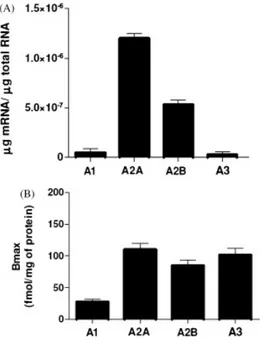
The presence of A3AR protein has been evaluated through radioligand binding, immunoassay or functional assay in a variety of primary cells, tissues and cell lines (Gessi et al., 2008). In the mouse brain a widespread, relatively low level of A3AR binding sites was found (Jacobson et al., 1993). Similar data were obtained in the rat and in gerbil and rabbit brain. Electrophysiological and biochemical evidence suggested the presence of A3ARs in the rat hippocampus and cortex, and functional studies also indicated its presence in the brain. In cardiomyocytes, there was no direct evidence of the presence of A3ARs but several studies reported that it was responsible for cardioprotection in a variety of species and models, including isolated cardiomyocytes and isolated myocardial muscle preparations (Peart and Headrick, 2007). In lung parenchyma and in human lung type 2 alveolar-like cells (A549), the A3AR was detected through radioligand binding and immunohistochemical assays (Varani et al., 2006).
The classical pathways associated with A3AR activation are the inhibition of adenylyl cyclase activity, through the coupling with Gi proteins, and the stimulation of phospholipase C (PLC), inositol triphosphate (IP3) and intracellular calcium (Ca2+), via Gq proteins (Fredholm et al., 2001). However, more recently additional intracellular pathways have been described as relevant for A3AR signaling. For example, in the heart, A3AR mediates cardioprotective effects through ATP-sensitive potassium (KATP) channel activation. Moreover, it is coupled to activation of RhoA and a subsequent stimulation of phospholipase D (PLD), which in turn mediates protection of
cardiac myocytes from ischemia (Mozzicato et al., 2004). In addition, in different recombinant and native cell lines, A3AR is involved, like the other adenosine subtypes, in the modulation of mitogen-activated protein kinase (MAPK) activity (Schulte and Fredholm, 2003). A3AR signaling in Chinese Hamster Ovary cells transfected with human A3AR (CHO-hA3) leads to stimulation of extracellular signal-regulated kinases (ERK1/2). In particular, A3AR signaling to ERK1/2 depends on βγ release from pertussis toxin (PTX)-sensitive G proteins, phosphoinositide 3-kinase (PI3K), Ras and mitogen-activated protein kinase kinase (Schulte and Fredholm, 2003). It has been reported that A3AR activation is able to decrease the levels of PKA, a downstream effector of cAMP, and of the phosphorylated form of PKB/Akt in melanoma cells. This implies the deregulation of the Wnt signaling pathway, generally active during embryogenesis and tumorigenesis to increase cell cycle progression and cell proliferation (Fishman et al., 2002). Involvement of the PI3K/PKB pathway has been linked with preconditioning effects induced by A3AR activation in cardiomyocytes from newborn rats (Germack and Dickenson, 2005). An elegant study has recently documented a role of A3AR in cell survival signaling in resveratrol preconditioning of the heart. This study provides evidence that resveratrol preconditions the heart through the activation of adenosine A1 and A3AR, transmitting a survival signal through both the PI3K-Akt-Bcl2 and, only in the case of A3AR, cAMP response element-binding protein (CREB)-Bcl2 pathways (Das et al., 2005). Subsequently it has been demonstrated that CREB phosphorylation occurs through both Aktdependent and -independent signaling. Activation of PI3K-Akt-pBAD by A3AR has been observed recently in glioblastoma cells leading to cell survival in hypoxic conditions (Merighi et al., 2007). Further studies indicate that A3AR activation by interfering with PKB/Akt pathways can decrease interleukin-12 (IL-12) production in human monocytes (la Sala et al., 2005). Collectively, these findings demonstrate that several intracellular
mechanisms are involved following A3AR stimulation, the understanding of which may be essential and crucial for explaining the different aspect of its activation.
Therapeutic potential of A
3adenosine receptors
Neuroprotection versus neurodegeneration
Considerable interest has been shown in understanding the involvement of A3AR in normal and pathological conditions of the CNS despite its low expression in the brain (Rivkees et al., 2000). Even though the function of A3AR in the CNS has been controversial in terms of protective versus toxic actions, actually several data point towards a neuroprotective effect. Firstly, a dual role of A3AR was described in a model of global ischemia in gerbils where acute preischemic administration of the agonist N6 -(3-iodobenzyl)-adenosine-5′-N-methylcarboxamide (IB-MECA) caused a severe depression of cerebral blood perfusion, worsening of neuronal damage and postischemic mortality, while its chronic administration induced a significant improvement of postischemic cerebral flow and neuron protection (Von Lubitz et al., 1994). In line with the results obtained after acute treatment, in rat cerebellar granule neurons high concentrations of Cl-IB-MECA were able to induce lactate dehydrogenase release, neuronal cell death and augmented glutamate-induced neurotoxicity through a pathway involving inhibition of cyclic AMP production (Sei et al., 1997). It was then observed that the effect of IB-MECA depended on the timing of treatment as administration of IB-MECA 20 min prior to transient middle cerebral ischemia increased the infarct size, whereas its addition 20 min after ischemia resulted in a significant decrease of damage, leading the authors to define the cerebroprotective effect of A3AR a “right thing at a wrong time” (Von Lubitz et al., 2001). It has been speculated that the deleterious effects
caused by acute preischemic treatment with IB-MECA were the consequence of a series of adverse events triggered immediately prior to the occlusion such as release of inflammatory mediators, breakdown of the blood–brain barrier integrity and Ca2+ influx. In contrast, the neuroprotective effects obtained when the A3 agonist treatment was performed following a focal insult were related to astrocyte activation or to a direct neuroprotective action (Von Lubitz et al., 2001). One of the main factors contributing to the overall neuroprotective profile of chronic treatment with A3AR agonists was found to be the reduction in post-ischemic expression of nitric oxide (NO) synthase, the enzyme involved in NO generation (Von Lubitz et al., 1999). Other beneficial effects associated with chronic A3AR stimulation were the increase of glial fibrillary acidic protein [(GFAP), astrocyte proliferation and preservation of the ischemia-sensitive microtubule-associated protein 2 (MAP-2) (Von Lubitz et al., 1999)]. Destructive and protective actions of A3AR stimulation have also been demonstrated in experiments in astroglial cells where Cl-IB-MECA at nanomolar doses was responsible for “trophic effects” related to reorganization of actin cytoskeleton, while in the micromolar range was a mediator of apoptosis ([Abbracchio et al., 1997], [Abbracchio et al., 2001], [Appel et al., 2001] and [Di Iorio et al., 2002]). Such apparently opposing effects have been reconciled by hypothesizing that adenosine-induced cell death that occurs during severe metabolic stress by A3AR activation might isolate the most damaged areas to favor those parts of the brain that still retained a chance for functional recovery, supporting the role of adenosine as a “retaliatory metabolite” (Von Lubitz, 1999). Later it was speculated that desensitization/down-regulation of the A3AR may be the basis of cytoprotection, suggesting a role for this receptor in induction of cell death (Trincavelli et al., 2002a). Recently a study performed in primary cortical cultures demonstrated that Cl-IB-MECA antagonized the hypoxia-mediated decrease in cell viability. Moreover, when given in vivo before focal cerebral ischemia, it reduced cerebral infarction while it
was inactive in A3 knock-out (A3KO) mice. Furthermore A3KO mice after ischemia presented an increase in cerebral infarction in comparison to wild-type animals suggesting that A3AR mediate a tonic protective condition during ischemia (Chen et al., 2006b). In contrast, A3AR activation did not affect neuronal death triggered by kainate and cyclothiazide in primary cultures of cortical neurons (Rebola et al., 2005). Contrasting results have been reported also about how A3AR activation might influence neuronal activity in rat brain. Dunwiddie and coworkers (1997) demonstrated that in the CA1 region of the rat hippocampus A3AR has no direct effect on synaptically evoked excitatory responses, while it induced heterologous desensitization of A1AR, thus limiting adenosine-mediated cerebroprotection. Others suggested that A3AR activation in cortical neurons mediated a depression of synaptic transmission by inhibiting glutamate release additionally to and independently from the A1 receptors, thus providing neuroprotection ([Brand et al., 2001], [Lewerenz et al., 2003] and [Lopes et al., 2003b]). It was also found that activation of A3AR by endogenous adenosine inhibited synaptic transmission during hypoxia in rat cortical neurons (Hentschel et al., 2003), and the inhibitory function of A3AR activation was in agreement with an in vivo report showing that A3AR has depressant effects on locomotor activity in behavioral tests (Jacobson et al., 1993). However, on the other hand it has been observed that Cl-IB-MECA facilitates epileptiform discharges in the CA3 area of immature rat hippocampal slices, suggesting that activation of A3AR following a rise in endogenous adenosine facilitates excitation, thus limiting the known inhibitory and neuroprotective effects of adenosine in immature brain (Laudadio & Psarropoulou, 2004). Genetic suppression of A3AR enhanced some aspects of motor function, suppressed pain processing at supraspinal levels and showed an increase in neurodegeneration in response to repeated episodes of hypoxia, suggesting the possible use of A3 agonists in the treatment of ischemic and degenerative conditions of the CNS (Fedorova et al.,
2003). Different evidences suggest that a part of neuroprotection induced by A3AR derives from its modulation of the brain immune system (Haskó et al., 2005). It has been reported that functional A3AR are expressed in mouse microglia cells, where their activation is responsible for a biphasic effect on ERK1/2 phosphorylation (Hammarberg et al., 2003) and in murine astrocytes where A3AR stimulation induces the release of the neuroprotective chemokine CCL2 (Wittendorp et al., 2004). Moreover, in lipopolysaccharide (LPS)-treated BV2 microglial cells A3AR activation suppresses tumor necrosis α (TNF-α) production by inhibiting PI3K/Akt and nuclear
factor-κB (NF-factor-κB) activation, suggesting that selective ligands of this receptor may be of
therapeutic potential for the modulation and possible treatment of brain inflammation (Lee et al., 2006a). Even though for some aspects the role of A3AR in the CNS seems less confusing now than in the past, there are many aspects yet that need clarification before a role of A3 agonists in therapy can be envisioned.
Cardioprotection versus cardiotoxicity
To date several pieces of evidence support the conclusion that activation of A3AR is crucial for cardioprotection during and following ischemia–reperfusion and it has been suggested that a consistent part of the cardioprotective effects exerted by adenosine, once largely attributed to the A1 receptor, may now be in part ascribed to A3AR activation (Headrick & Peart, 2005). Even though there is a low expression of A3AR in myocardial tissue, a number of studies have demonstrated that acute treatment with agonists induced protective “anti-ischemic” effects (Auchampach et al., 1997a; Tracey et al., 1997; Thourani et al., 1999a; Ge et al., 2006 and Xu et al., 2006). The molecular mechanism of A3AR cardioprotection has been attributed to regulation of mitochondrial (mito) KATP channels (Thourani et al., 1999b; Shneyvays et al., 2004 and Peart and Headrick, 2007).
In addition Shneyvays et al. (2005) demonstrated that in cultured rat myocytes Cl-IB-MECA delayed the dissipation of the mitochondrial membrane potential (∆ψ) and decreased the elevated intracellular calcium concentrations induced by hypoxia. These effects prevented irreversible cardiomyocyte damage and confirmed previous results showing that A3AR activation protected cardiomyocytes treated with doxorubicin via inhibition of calcium overload and prevented cardiomyocyte death during incubation in high extracellular calcium concentrations (Shneyvays et al., 2001 and Shneyvays et al., 2004). As for the timing of cardioprotection, some studies have indicated that protection occurred post-ischemia, through inhibition of neutrophil-induced reperfusion injury or inhibition of myocyte apoptotic cell death (Jordan et al., 1999 and Maddock et al., 2002), while others found that preischemic A3AR activation was effective and necessary for cardioprotection (Thourani et al., 1999a). Auchampach et al. demonstrated that A3 agonism was able to trigger an anti-infarct response with either pre- or postischemic treatment (Auchampach et al., 2003). Moreover, it has been reported that A3AR activation is able to mimic or induce myocardial preconditioning, meaning that transient stimulation of the A3AR before induction of ischemia leads to both an early and a delayed protection (Peart & Headrick, 2007). The first condition has been shown to require mito KATP channel activation through PKC, 1,2-diacylglycerol (DAG), PLD and RhoA, but also reduction of caspase 3 and increase of cell survival through MEK/ERK1/2 and PI3K pathways (Parsons et al., 2000; Sato et al., 2000; Lee et al., 2001; Nakai et al., 2001; Germack and Dickenson, 2004 and Germack and Dickenson, 2005). In addition it has been reported that resveratrol preconditions the heart through A3AR signaling that triggers a survival effect mediated by the Akt-Bcl2-Bad signaling pathway and also by another survival signal mediated via Akt-dependent and independent CREB phosphorylation (Das et al., 2005a and Das et al., 2005b). In terms of delayed preconditioning some authors reported a role for mito KATP channels but not for
nitric oxide synthase (iNOS), while other acquired evidence of NF-κB and iNOS involvement (Takano et al., 2001 and Zhao and Kukreja, 2002). Pharmacological preconditioning (early and late) obtained after A3AR activation is clinically important, but cardioprotection is even more relevant when it occurs at reperfusion (Auchampach et al., 2003 and Xu et al., 2006). This situation, called post-conditioning, has been demonstrated for IB-MECA through inhibition of the mitochondrial permeability transition pore (mPTP) opening via PI3K/Akt inactivation of glycogen synthase kinase (GSK-3β; Kin et al., 2005 and Park et al., 2006). The cardioprotective effects of A3 receptors were also detected in A3AR-overexpressing mice where after in vivo regional ischemia and reperfusion, infarct size was lower than in wild-type mice (Black et al., 2002). In these animals A3AR overexpression decreased basal heart rate and contractility, preserved ischemic ATP and decreased postischemic dysfunction (Cross et al., 2002). On the other hand, the results obtained with mice carrying higher transgene copy numbers suggested that basal signaling was increased when the A3AR was expressed at higher levels, leading to the development of a dilated cardiomyopathy. Paradoxically, in contrast with pharmacological evidence of A3-induced cardioprotection the first studies carried out in mice in which the A3AR was genetically disrupted demonstrated an improvement of cardiac function as revealed from a smaller myocardial infarct size (Guo et al., 2001). Cerniway and colleagues (2001) reported similar beneficial effects in A3KO mice, which they initially ascribed to the absence of a proinflammatory action of A3AR mediated through mast cell degranulation. Subsequently, it was suggested that this effect might be due to compensatory changes that developed in the KO mice due to the chronic absence of A3AR (Harrison et al., 2002). In this respect, recent evidence obtained by using pharmacological agents and genetic methods suggests that Cl-IB-MECA protects against myocardial ischemia/reperfusion injury in mice via A3AR activation. These conclusions were
supported by experiments with a selective A3 antagonist and through evaluation of the A3 agonist effects on A3KO mice. Interestingly, in this paper by using congenic (C57BL/6) A3KO mice, deletion of the A3 gene itself had no effect on ischemic tolerance, suggesting that previous opposite results from the same group (Guo et al., 2001) were probably explained by differences in the genetic background of the mice rather than specific deletion of the A3 gene. Additional studies using wild-type mice treated with compound 48/80, a condensation product of p-methoxyphenetyl methylamine with formaldehyde, to deplete mast cell contents excluded the possibility that Cl-IB-MECA was cardioprotective by releasing mediators from mast cells (Ge et al., 2006) and supported the idea that therapeutic strategies focusing on A3AR subtype may represent a novel and useful approach for protection of the ischemic myocardium. The A3-mediated cardioprotection remains a mystery if one thinks of its cellular location. Literature data reported that myocardial A3AR expression in the mouse is very low and below the detection limits of radioligand binding or northern blot techniques (Black et al., 2002). It is also surprising that mice overexpressing the A3AR reveal only 12 fmol/mg of protein and that animals with 66 fmol/mg of protein present negative effects like dilated cardiomyopathy, meaning that the level of A3AR is very critical for heart function. Therefore, on the one hand it is possible to hypothesize that given the strong cardioprotective effects and the low cardiac expression, this receptor must be very efficiently coupled to protective intracellular signaling pathways. On the another hand, it is also conceivable that cardioprotection might derive at least in part from activation of A3AR expressed in other cells (Jacobson & Gao, 2006; Fig. 2).
Anti-inflammatory versus proinflammatory effects
The interest in the elucidation of A3AR involvement in inflammation is attested by the large amount of experimental work carried out in cells of the immune system and in a variety of inflammatory conditions. However, as in the SNC or in the cardiovascular system the A3AR subtype appears to have a complex or “enigmatic” role, as both proinflammatory and antiinflammatory effects have been demonstrated. One of the first evidence for a role of A3AR in increasing inflammation derived by studies in mast cells where it was found that its activation was responsible for release of allergic mediators (Ramkumar et al., 1993 and Fozard et al., 1996). In addition, it has been reported that A3AR mRNA was higher in lung tissue of patients with airway inflammation and that A3AR activation mediates rapid inflammatory cell influx into the lungs of sensitized guinea pigs (Walker et al., 1997 and Spruntulis and Broadley, 2001). It has been reported that A3AR activation in RBL-2H3 mast cells inhibits apoptosis and may have a profound effect on survival of inflammatory cells expressing A3AR in inflamed tissues, thus contributing to inflammatory cell expansion (Gao et al., 2001). Moreover, antigen-dependent degranulation of bone marrow-derived mast cells was found to be mediated by A3AR (Reeves et al., 1997), and the ability of Cl-IB-MECA to potentiate antigen-dependent mast cells degranulation was lost by using mice lacking A3AR, suggesting a role for antagonists as antiasthmatic agents (Salvatore et al., 2000). The involvement of A3AR in mast cells degranulation has been further confirmed in murine lung mast cells where it was dependent from Ca++ elevations through Gi and PI3K coupling (Zhong et al., 2003). However, in contrast with these findings it has been demonstrated that in the rat parenchymal strip, where contraction in response to adenosine is mast cell mediated, the receptor involved shows similarities to the A3AR but Cl-IB-MECA is a high affinity
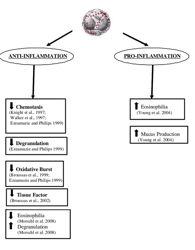
antagonist and MRS 1523 and MRS 1191 are inactive at concentrations that substantially exceed their affinities for the rat A3AR by adding further twist to A3AR pharmacology (Wolber & Fozard, 2005). Moreover, it has been shown that human and canine mast cells degranulation was mediated by A2BAR instead of A3AR (Feoktistov and Biaggioni, 1995; Auchampach et al., 1997b and Ryzhov et al., 2004). This discrepancy reflects the low human and rat overall coidentity at the amino acid level of A3AR and questions the role of the A3AR as a target for asthma therapy. Nevertheless the high expression of A3AR in other cells involved in allergic diseases and asthma still gives reason to suggest a role for antagonists in the treatment of these pathologies. In particular transcript levels for the A3 subtype are elevated in the lungs of asthma and COPD patients, where expression is localized to eosinophilic infiltrates. Interestingly, similar findings were made in the lungs of adenosine deaminase deficient (ADA−/−) mice that exhibited adenosine-mediated lung disease. Treatment of ADA−/− mice with MRS 1523, a selective A3 antagonist, prevented airway eosinophilia and mucus production. Similar results were obtained in the lungs of ADA/A3 double KO mice, suggesting that A3 signaling plays an important role in regulating chronic lung disease and that A3 antagonism may provide a mechanism for reducing eosinophilia (Young et al., 2004). These results are in contrast with experiments performed in human eosinophils ex vivo, where chemotaxis such as degranulation and superoxide anion production were reduced by A3AR activation (Ezeamuzie & Philips, 1999). This discrepancy may be due to differences in mouse and human eosinophils or to differences attributed to the ex vivo nature of the chemotaxis experiments performed. Additional studies of A3-mediated effects on mouse eosinophils ex vivo confirmed the results observed in human cells, suggesting that diminished airway eosinophilia seen in the lungs of ADA−/− mice following disruption of A3AR is not a cell autonomous effect of eosinophils. Rather A3 disruption in ADA−/− mice is likely to affect the expression
and activity of key regulatory molecules from other cells that present A3AR and that affect eosinophil migration (Young et al., 2004). For example A3AR are expressed on murine mast cells, airway macrophages and epithelial cells, all of which might affect eosinophil migration. However, levels of key regulatory cytokines such as 5 and IL-13, or chemokines including eotaxin I, thymus- and activation-regulated chemokine (TARC) and monocyte chemotactic protein-3 (MCP3) were not affected by A3 removal in ADA−/− mice, pointing perhaps to the involvement of A3 subtype in the regulation of other key modulators of eosinophil migration, such as cell adhesion molecules, extracellular matrix elements and proteases (Young et al., 2004). The molecular mechanisms by which A3 signaling may affect eosinophil chemotaxis are not known, but may involve the regulation of intracellular calcium (Khono et al., 1996a). In addition to influencing chemotaxis, A3 engagement might also affect eosinophils survival. It has been reported that A3 subtype can protect rat mast cells from apoptosis by a pathway involving PI3K and phosphorylation of PKB. In the same way, activation of A3AR on eosinophils may promote their survival at sites of inflammation. However, the functional role of the A3 subtype in the pathogenesis of asthma remains controversial and differences in the pharmacology of A3 subtype from different species render it difficult to understand whether an A3AR agonist or antagonist could be needed to improve the treatment of asthma. At this regard, a recent paper by Rimmer and coworkers reports the effect of a novel A2A agonist/A3AR antagonist in the treatment of allergic rhinitis through a randomized, double-blind, placebo-controlled study (Rimmer et al., 2007). Unfortunately, this ligand appears to have limited clinical benefit in both the early- and late-phase response to intranasal allergen challenge, even though it reduced the release of some mediators after allergene challenge. However, as correctly pointed out by the authors, the study presented a number of shortcomings. As an example the dose of the drug was limited by the narrow therapeutic index, due to side
effects like tachycardia, raising the possibility that higher doses of new compounds with fewer side effects might be more efficacious. Alternatively, it is possible that future studies targeting a different receptor, perhaps the A2B, or using dual antagonists versus A3/A2BAR will be more successful (Press et al., 2005).
Discrepancy between anti- and proinflammatory effects induced by A3AR have been observed also in other cell types. For example, A3AR are expressed in human neutrophils where they are involved together with A2A in the reduction of superoxide anion generation (Bouma et al., 1997 and Gessi et al., 2002). However, recently an elegant study by Chen et al. reported that neutrophils rapidly hydrolyze released ATP to adenosine that then acts via A3-subtype adenosine receptors, which are recruited to the leading edge, to promote cell migration (Chen et al., 2006a and Linden, 2006).
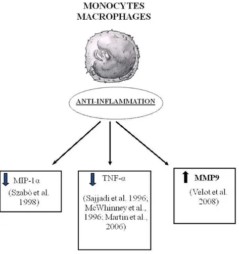
In addition to a role of A3AR in increasing inflammation, evidence that A3AR decrease inflammation have also been reported in literature. As an example, it has been shown that A3AR suppress TNF-α release induced by endotoxin CD14 receptor signal transduction pathway from human monocytes and murine J774.1 macrophages (Le Vraux et al., 1993 and McWhinney et al., 1996). Moreover, in a macrophage model the A3AR was the prominent subtype implicated in the inhibition of LPS-induced TNF-α production (Sajjadi et al., 1996). This effect was associated with changes in stimulation of the activator protein-1 (AP-1) transcription factor, whereas it was independent of MAPK and NF-κB, PKA, PKC and PLC. This was not confirmed in BV2 microglial cells where A3-mediated inhibition of LPS-induced TNF-α expression was associated with the inhibition of LPS-induced activation of PI3K/Akt and NF-κB pathway (Lee et al., 2006a). The inhibitory effect induced by A3AR on TNF-α production was also assessed in A3KO mice where the A3 agonist was unable to reduce TNF-α levels in contrast with its effect in wild-type animals (Salvatore et al., 2000). Recently, it has
been reported that in mouse RAW 264.7 cells the A3 subtype inhibits LPS-stimulated TNF-α release by reducing calcium-dependent activation of NF-κB and ERK 1/2 (Martin et al., 2006) while in peritoneal macrophages, isolated from A3KO mice, the ability of IB-MECA to inhibit TNF-α release was not altered in comparison to wild-type mice (Kreckler et al., 2006). In this study, the inhibitory effect was exerted through the activation of A2A and A2B agonists as recently demonstrated also in human monocytes (Zhang et al., 2005 and Haskó et al., 2007). The discrepancy observed among these papers might not depend on species differences, being in both cases mouse cells, but by other factors including the source of the cells, and/or the inflammatory stimulus used. However in spite of these contrasting results, one of the best potential therapeutic applications of the regulatory role of A3 activation on TNF-α release has been found in the treatment of arthritis. A3AR agonists exert significant antirheumatic effects in different autoimmune arthritis models by suppression of TNF-α production (Baharav et al., 2005). The molecular mechanism involved in the inhibitory effect of IB-MECA on adjuvant-induced arthritis included receptor down-regulation and deregulation of the PI3K–NF-κB signaling pathway (Fishman et al., 2006 and Madi et al., 2007). Previous studies also demonstrated that A3AR activation inhibited macrophage inflammatory protein (MIP)-1α, that is a C-C chemokine with potent inflammatory effects, in a model of collagen-induced arthritis, providing the first proof of concept of the adenosine agonists utility in the treatment of arthritis (Szabò et al., 1998).
In agreement with an antiinflammatory role for the A3AR, it has been recently demonstrated that A3AR activation decreases mortality and renal and hepatic injury in murine septic peritonitis (Lee et al., 2006b). Higher levels of endogenous TNF-α were observed in A3KO mice after sepsis induction, in comparison to wild-type animals and IB-MECA significantly reduced mortality in mice lacking the A1 or A2A but not the
A3AR, demonstrating specificity of the A3 agonist in activating A3 subtype and mediating protection against sepsis-induced mortality (Lee et al., 2006b). A similar mortality reduction associated with a decrease of IL-12 and interferon-γ (IFN-γ) production induced by A3AR activation was previously observed by Haskó et al. (1998) in endotoxemic mice. In addition other investigators reported reduced inflammation and increased survival following A3 activation in 2 murine models of colitis (Mabley et al., 2003). Furthermore, a protective role for A3AR in lung injury following in vivo reperfusion has been recently reported (Matot et al., 2006). In contrast, it has been demonstrated that A3AR activation exacerbates renal dysfunction and mice lacking A3AR have been found to have better renal function following renal ischemia reperfusion injury (Lee and Emala, 2000 and Lee et al., 2003). A3 receptors have been found to be up-regulated in ocular ischemic diseases and in conditions associated with oxidative stress. Their activation lead to the regulation of chloride channels in nonpigmented ciliary epithelial cells, suggesting that A3 agonists would increase aqueous humor secretion and thereby intraocular pressure in vivo, while antagonists may represent a specific approach for treating ocular hypertension (Mitchell et al., 1999; Okamura et al., 2004 and Schlotzer-Schrehardt et al., 2005).
Antitumor versus tumorigen effects
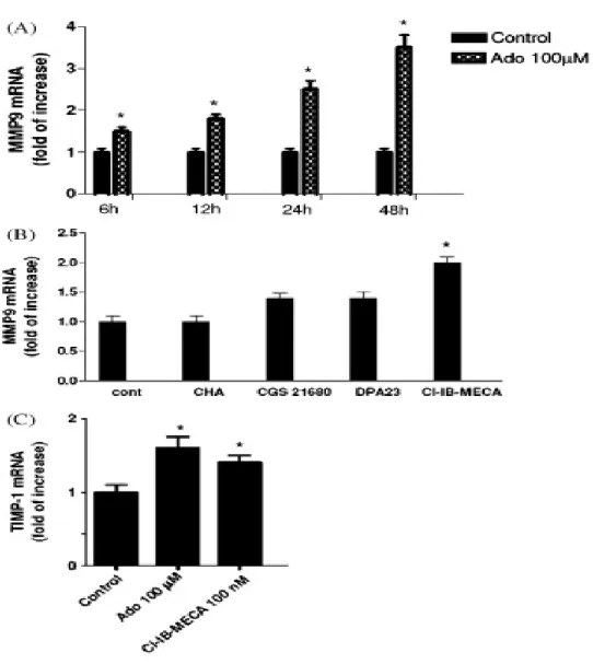
A very interesting area of potential application of A3 ligands concerns cancer therapies. The possibility that A3 adenosine receptor plays a role in the development of cancer has aroused considerable interest in recent years (Fishman et al., 2002 and Merighi et al., 2003 and Gessi et al., 2008). The A3 subtype has been described in the regulation of the cell cycle and both pro- and antiapoptotic effects have been reported depending on the
level of receptor activation (Jacobson, 1998; Yao et al., 1997; Gao et al., 2001; Merighi et al., 2005a; Nakamura et al., 2006 and Gessi et al., 2007). Starting from the observation that muscle tissues are resistant to tumor metastases, it was reported that one of the active components of muscle cell conditioned medium was adenosine, that exerted a differential effect on tumor and normal cell growth (Fishman et al., 1998); this inhibition was removed when the A3AR was blocked, while it was mimicked following A3AR agonist stimulation. The mechanism was found to involve inhibition of telomerase activity and arrest in the G0/G1 phase of the cell cycle, leading to a cytostatic effect (Fishman et al., 2000). In addition, it was demonstrated that A3AR inhibited tumor growth by regulation of the WNT pathway (Fishman et al., 2004). The WNT pathway, active during embryogenesis and tumorigenesis, mediates cell cycle progression and cell proliferation. A key modulator of this pathway is represented by GSK-3β that is crucial for β-catenin phosphorylation. β-Catenin induces the transcription of genes fundamental for cell cycle progression such as c-myc and cyclin D1. Upon exposure of tumor cells to the A3 agonist, a decrease in the protein expression level of A3AR and the downstream effectors PKA and PKB was observed. Consequently, the GSK-3β protein level increased, resulting in the destabilization of β-catenin and the subsequent suppression of cyclin D1 and c-myc expression (Fig. 4). IB-MECA treatment also induced down-modulation of the expression of NF-κB, known to regulate the transcription of cyclin D1 and c-Myc (Fishman et al., 2003 and Fishman et al., 2004). Moreover A3AR agonist treatment induced inhibition of tumor growth both in vitro and in vivo, gave a synergistic effect in combination with chemotherapy and exhibited a myelostimulatory effect by inducing G-CSF production by mononuclear cells, thus leading to the development of A3 agonists in clinical trials for colon carcinoma (Jacobson & Gao, 2006). Other authors found inhibition of cell proliferation or induction of apoptosis by treating cells with the A3 agonist, but the effects generally
were observed only at micromolar doses and the involvement of the A3 subtype was questioned (Wen and Knowles, 2003; Panjehpour and Karami-Tehrani, 2004; Merighi et al., 2005a and Nakamura et al., 2006). In this respect several observations may be underlined: (i) it has been demonstrated that Cl-IB-MECA at micromolar doses inhibits cell proliferation and this effect is reduced by blocking the receptor, supporting a role for the A3 subtype (Merighi et al., 2005a); (ii) it has been previously reported that IB-MECA, at micromolar doses in breast cancer cells, inhibits cell proliferation through interaction with receptors different from the adenosine subtypes such as estrogen receptor α (Lu et al., 2003); (iii) at micromolar doses Cl-IB-MECA loses its selectivity for A3 receptors and the complicating presence of interaction with other adenosine subtypes might be involved in the final response; (iv) the difference between the effects induced by low and high doses of Cl-IB-MECA could be attributed to the receptor desensitization of A3 receptors that has been demonstrated by other authors in various cell systems (Trincavelli et al., 2002a). Conversely, it has been demonstrated that A3AR in retinal ganglion cells was obligatory for life (Zhang et al., 2006), and it has been recently observed in colon cancer cells that after treatment with ADA, Cl-IB-MECA increased cell proliferation through the activation of A3 subtype and involvement of ERK1/2 (Gessi et al., 2007). It is important to underline that all these experiments have been performed in normoxic conditions. From another point of view, hypoxia that is typical of solid tumors (Vaupel et al., 1989), creates conditions that, on one hand, are conducive to the accumulation of extracellular adenosine and, on the other hand, stabilize hypoxia-inducible factors, such as HIF-1α ([Blay et al., 1997], [Semenza, 2000], [Hockel and Vaupel, 2001], [Linden, 2001], [Minchenko et al., 2002], [Fredholm, 2003] and [Sitkovsky et al., 2004]). HIF-1, the most important factor involved in the cellular response to hypoxia, is up-regulated across a broad range of cancer types and is involved in key aspects of tumor biology, such as angiogenesis,
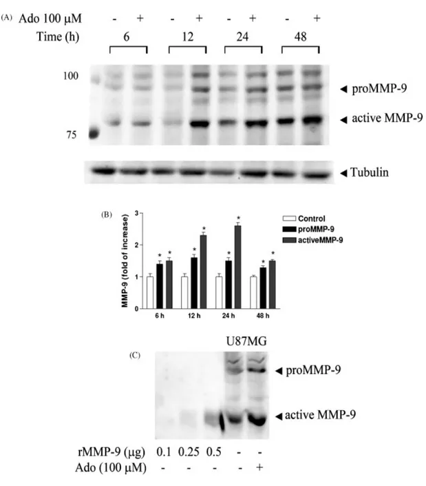
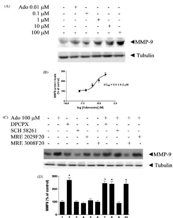
invasion and altered energy metabolism (Semenza, 2003). HIF-1 is a heterodimer composed of an inducibly expressed HIF-1α subunit and a constitutively expressed HIF-1β subunit (Epstein et al., 2001). HIF-1α and HIF-1β mRNAs are constantly expressed under normoxic and hypoxic conditions (Wiener et al., 1996). The unique feature of HIF-1 is the regulation of HIF-1α expression: it increases as the cellular O2 concentration is decreased (Semenza, 2000 and Minchenko et al., 2002). During normoxia, HIF-1α is rapidly degraded by the ubiquitin proteasome system, whereas exposure to hypoxic conditions prevents its degradation (Minchenko et al., 2002). A growing body of evidence indicates that HIF-1 contributes to tumor progression and metastasis (Welsh and Powis, 2003 and Hopfl et al., 2004). Immunohistochemical analyses have shown that HIF-1α is present in higher levels in human tumors than in normal tissues (Zhong et al., 1999). Interestingly, it has been demonstrated that A3AR are also overexpressed in cancer tissues in comparison to normal mucosa (Gessi et al., 2004a). Furthermore, attention has been paid to responses to chronic hypoxia that involve adenosine-induced changes in the transcription regulator HIF-1 expression. In particular, the correlation between adenosine receptor stimulation and HIF-1α expression modulation in hypoxia has been investigated. Adenosine increases HIF-1α protein expression in response to hypoxia in human melanoma, glioblastoma and colon cancer cells (Merighi et al., 2005b; Merighi et al., 2006 and Merighi et al., 2007b). These results indicate that the cell surface A3AR transduces extracellular hypoxic signals into the cell interior. Increased HIF-1α protein synthesis through the activation of the Akt or MAPKinase pathways is a common theme accounting for the up-regulation. To evaluate how A3AR accumulates HIF-1α in hypoxia, the signaling pathway generated by A3AR stimulation has been investigated and it was found that MAPKinase activity is required for the HIF-1α expression increase induced by A3AR activation (Fig.3). Furthermore, as HIF-1α plays a key role in inducing angiogenesis, we
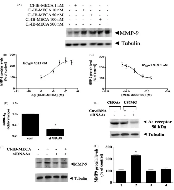
have also studied the role of adenosine in mediating the production of VEGF in tumor cells. Activation of the A3AR in glioblastoma and colon cancer cells stimulates VEGF expression (Merighi et al., 2006 and Merighi et al., 2007b), whereas this receptor subtype promotes VEGF downregulation in PC12 pheochromocytoma cells (Olah & Roudabush, 2000). It has been proposed that the effect of VEGF on new capillary formations is facilitated by the concomitant stimulation of A2B and A3 receptors that induce the expression of angiopoietin-2 (Feoktistov et al., 2003). Indeed, the activation of A3 receptors results in increased expression of angiopoietin-2 in mast and melanoma cells (Feoktistov et al., 2003 and Merighi et al., 2005b). Although adenosine may contribute rather little to the increase in VEGF induced by hypoxia, it may contribute as much as 50% to angiogenesis (Adair, 2005). This could mean that adenosine acts also independently of VEGF, something that is not unlikely given the involvement of multiple cell types and multiple angiogenetic factors. Recent studies indicate that pharmacological inhibition of HIF-1α and particularly of HIF-regulated genes, that are important for cancer cell survival, may be more advantageous than therapeutic approaches based on HIF-gene inactivation. In this regard, A3AR antagonists are able to block HIF-1α, angiopoietin-2 and VEGF protein expression accumulation in hypoxia, indicating a new approach for the treatment of cancer, based on the cooperation between hypoxic and adenosine signals.
Immunosuppressive versus immunostimulating effects
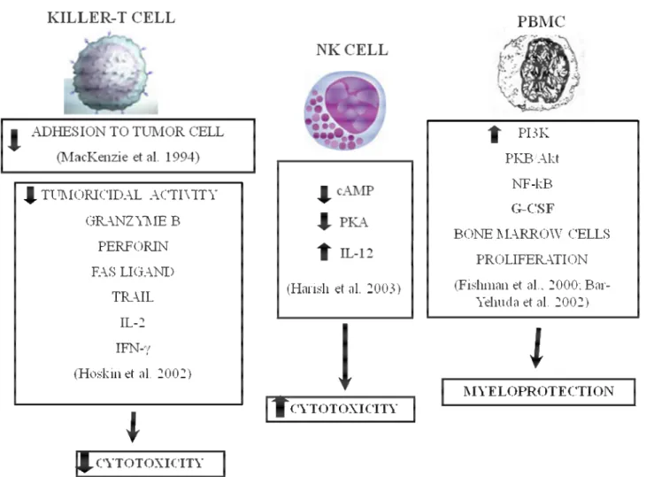
The ability of immune cells to fight tumor cells is fundamental for successful host defense against cancer. Adenosine, whose concentration increases within hypoxic regions of solid tumors, may interfere with the recognition of tumor cells by cytolytic effector cells of the immune system (Blay et al., 1997 and Merighi et al., 2003). Adoptive immunotherapy with lymphokine-activated killer (LAK) cells has shown some promise in the treatment of certain cancers that are unresponsive to conventional treatment approaches. However, colon adenocarcinomas tend to respond poorly to LAK therapy, possibly as a result of tumor-induced immunosuppression. It has been demonstrated that colon adenocarcinoma cells inhibited anti-CD3-activated killer cell induction through the production of a tumor-associated soluble factor that was distinct from transforming growth factor beta or prostaglandins (Hoskin et al., 1994a). Therefore, adenosine was indicated as a possible inhibitor of killer T-cell activation in the microenvironment of solid tumours (Hoskin et al., 1994b and Hoskin et al., 1994c). Indeed, evaluating the adhesion of murine spleen-derived anti-CD3-activated killer (AK) lymphocytes to syngeneic MCA-38 colon adenocarcinoma cells it was found that adenosine reduced adhesion by up to 60% (MacKenzie et al., 1994). The inhibitory effect of adenosine was exerted on AK cells and not on the MCA-38 targets and the agonist potency profile indicated that the A3 receptor subtype might be responsible for the inhibition of adhesion. The authors suggested that this mechanism of immunosuppression, secondary to tissue hypoxia, may be important in the resistance of colorectal and other solid cancers to immunotherapy. In addition the same authors demonstrated that adenosine plays a strong inhibitory effect on the induction of mouse cytotoxic T cells (Hoskin et al., 2002). Diminished tumoricidal activity correlated with
reduced expression of mRNAs coding for granzyme B, perforin, Fas ligand and TNF-related apoptosis-inducing ligand (TRAIL). IL-2 and IFN-γ synthesis by AK-T cells was also inhibited by adenosine. The inhibitory effect of adenosine on AK-T cell proliferation was also blocked by an A3 receptor antagonist suggesting that adenosine acts through A3 receptors to prevent AK-T cell induction. Tumor-associated adenosine may act through the same mechanism to impair the development of tumor-reactive T cells in cancer patients. Therefore, the suppression of T-killer cell function suggests that adenosine may act as a local immunosuppressant within the microenvironment of solid tumors. Subsequently, it was reported that adenosine partially inhibits the interaction of T lymphocytes with tumor cells by blocking the function of integrin α4β7 that is the major cell adhesion molecule involved in the adhesion of T cells to syngeneic MCA-38 adenocarcinoma cells (MacKenzie et al., 2002). The effect of adenosine has been investigated on the expression of costimulatory molecules by T cells in resting and activated conditions. One of the most important costimulatory molecules present on the T cells surface are CD2 and CD28 acting in concert to achieve optimal costimulation of T lymphocytes during interaction with antigen presenting cells. It has been demonstrated that adenosine interfered with activation-induced expression of the costimulatory molecules CD2 and CD28 in a way IL-2 dependent but not involving the accumulation of intracellular cAMP, possibly by activating the A3 subtype (Butler et al., 2003). However, recent data obtained from studies using adenosine receptor KO mice examined the capability of adenosine and its analogues to inhibit the ability of LAK to defeat tumor cells. This work demonstrated that adenosine and adenosine A2A ligands suppress the cytotoxicity of LAK cells in parallel with their ability to increase cAMP levels. These effects were produced by interfering with both perforin-mediated and Fas ligand-mediated killing pathways. Studies with LAK cells generated from A1 and A3AR KO mice indicated the lack of any involvement of these adenosine subtypes in the
inhibitory effect exerted by adenosine, whereas LAK cells obtained from A2AR KO mice were resistant to the inhibitory effect of the nucleoside. Only very high concentrations of the non selective agonists 5-N-ethylcarboxamide adenosine (NECA) or 2-chloroadenosine (CADO) produced mild inhibition of LAK cytotoxicity that were possibly induced through A2B activation, suggesting the predominant role of the A2A subtype in inhibition of LAK cell toxicity (Raskovalova et al., 2005). Therefore, the authors indicate the use of A2A antagonists to increase the efficacy of immunotherapy (Fredholm, 2007). In contrast to the immunosuppressive role of adenosine in the environment of solid tumors, it has been reported that A3AR activation stimulates the proliferation of bone marrow cells in vitro. This effect was induced through the adenosine-mediated G-CSF production by peripheral blood mononuclear cells (PBMC). In vitro studies were also confirmed by in vivo experiments, which revealed an increase in leukocyte and neutrophil numbers, when adenosine was administered before chemotherapy (Bar-Yehuda et al., 2002). The molecular mechanism at the basis of G-CSF production included the upregulation of PI3K, PKB/Akt and NF-κB. In addition, it has been observed that Cl-IB-MECA potentiates the activity of NK cells in naïve and tumor bearing mice through the induction of IL-12 production; this effect was dependent on inhibition of cAMP levels and PKA expression. IL-12 is a potent stimulant of NK cells and is a cytotoxic factor that exerts a potent anti-tumor effect in vivo. It induces IFN-γ production by activated T and NK cells and augments cytotoxic activity of these cells via perforin, Fas and Trail-dependent mechanisms. Therefore, A3AR activation enhances NK cell activity and probably NK cell-mediated destruction of tumor cells (Harish et al., 2003). This antitumor effect played in immune cells is in line with other findings of the same group demonstrating a direct inhibitory action of A3 receptor activation on tumor cell growth (Fishman et al., 2003)