PhD Course in Biochemistry
XXX Cycle (Academic Years 2014-2017)
Hydrogen sulfide metabolism in cancer and
homocystinuria: towards the development of new
pharmacological strategies
PhD student
Francesca Malagrinò
Tutor
PhD coordinator
Dr. Alessandro Giuffrè
Prof. Francesco Malatesta
ACKNOWLEDGMENTS
I would like to thank all people who supported and ispirated me during during these PhD years.
Firstly, I want to turn my thanks to my supervisor, Dr. Alessandro Giuffrè, for his rigourous scientific advices and for his precious academic guidance.
I would like to thank Prof. Paolo Sarti, who has always encouraged and motivated me with great enthusiasm.
My best gratitude goes, moreover, to Dr. Pierpaolo Ceci, Dr. Elisabetta Falvo and Dr. João B. Vicente, for the scientific support and
affectionate help.
I want to thank Prof. Elena Forte, Dr. Marzia Arese and Dr. Daniela Mastronicola for their insightful comments and precious encouragement.
For the stimulating scientific discussions, the loving closeness and their sincere friendship, a special thank goes to Dr. Maria Chiara Magnifico and Dr. Karim Zuhra.
I would like to thank, furthermore, the coordinator of the PhD in Biochemistry, Prof. Francesco Malatesta, for his meticulous guidance.
Thanks to all of my colleagues and friends of these PhD years, in particular Giulia, Filippo, Alessia, Vincenzo, Serena, Ilaria, Matilde, Daniela, Ludovica and Giulia for their precious closeness.
Last but not least, thanks to my dear mother, my grandparents, my aunt Linda and my uncle Giovanni for the love and continous support.
This work was partly supported by Ministero dell’Istruzione, dell’Università e della Ricerca (MIUR) of Italy (PNR-CNR Aging Program 2012–2014 and PRIN 20158EB2CM_003 to Alessandro Giuffrè).
My PhD was funded by a Sapienza University of Rome fellowship for three years
SCIENTIFIC COMMUNICATIONS
Publications
F. Malagrinò, K. Zuhra, L. Mascolo, D. Mastronicola, J.B. Vicente, E. Forte, A. Giuffrè. “Effect of hypoxia on mitochondrial hydrogen sulfide catabolism”, to be submitted
E. Falvo*, F. Malagrinò*, A. Arcovito, F. Fazi, G. Colotti, E. Tremante, P. Di Micco, G. Fracasso, A. Giuffrè and P. Ceci. “A novel ferritin-based nanocarrier for efficient mitoxantrone encapsulation and selective delivery to cancer cells”, to be submitted
J.B. Vicente, H. G. Colaço, F. Malagrinò, P. E. Santo, A. Gutierres, T. Bandeiras, P. Leandro, J. A. Brito and A. Giuffrè. “A clinically relevant variant of the human hydrogen sulfide-synthesizing enzyme cystathionine ß-synthase: increased CO reactivity as a novel molecular mechanism of pathogenicity?”, Oxidative Medicine and Cellular Longevity (2017), 2017, 8940321.
J.B Vicente, F. Malagrinò, M. Arese, E. Forte, P. Sarti and A. Giuffrè. “Bioenergetic relevance of hydrogen sulfide and the interplay between gasotransmitters at human cystathionine beta-synthase”, Biochim. Biophys. Acta – Bioenergetics (2016), 1857, 1127-38.
* Equally contributing authors
Selected communications by the candidate at scientific conferences
F. Malagrinò. “H2S metabolism in colon cancer cells: effect of hypoxia”, BeMM Symposium (2017) – Rome, Italy. (poster presentation)
F. Malagrinò, K. Zhura, D. Mastronicola, L. Mascolo, J.B. Vicente, E. Forte, A. Giuffrè. “Effect of hypoxia on H2S metabolism in colon cancer cells”, 59th
Congress of the Italian Society for Biochemistry and Molecular Biology (2017) – Caserta, Italy. (poster
communication)
F. Malagrinò, K. Zhura, D. Mastronicola, L. Mascolo, J.B. Vicente, E. Forte, A. Giuffrè. “Hypoxia and H2S metabolism in colon cancer cells”, Mitochondrial Physiology, from organelle to organism (2017) – Copenhagen, Denmark. (poster presentation)
F. Malagrinò, H. G. Colaço, K Zuhra, P. E. Santo, E. Forte, A. Gutierres, M. Arese, T. M. Bandeiras, P. Sarti, P. Leandro, J. A. Brito, J. B. Vicente and A. Giuffrè. “Novel pathogenic mechanisms from investigation of human hydrogen sulfide metabolism”, First
international meeting of the Italian Group of Bioenergetics and Biomembranes (2017) – Catania, Italy. (oral presentation)
J.B. Vicente, H.G. Colaço, F. Malagrinò, M. Arese, E. Forte, P. Sarti, P. Leandro, A. Giuffrè. “S-adenosyl-L-methionine and the cross-talk between the bioenergetically relevant gasotransmitters H2S, NO and CO at human cystathionine β-synthase”, 19th
European
Bioenergetics Conference (2016) - Riva del Garda, Italy. (poster
OBJECTIVES 1
BACKGROUND 7
1. Physico-chemical properties of H2S 9
2. H2S, the new gasotransmitter 10
2.1. Synthesis 11
2.1.1. Endogenous H2S production - Enzymes and pathways 11
2.1.1.1. Focus on cystathionine β-synthase 21
2.1.2. H2S production by the gut microbiota 31
2.2 Catabolism 31
2.2.1. Mitochondrial H2S oxidation - Enzymes and pathways 32
2.2.2. Mitochondrial H2S oxidation in colonocytes 37
3. Physiological and pathological role of H2S 38
3.1. H2S, a Janus-faced molecule 39
3.2. Bioenergetics 40
3.2.1. H2S as mitochondrial substrate or inhibitor 40
3.3. Cancer 46
3.3.1. H2S and colorectal cancer 46
3.4. Hypoxia 48
3.4.1. H2S as an oxygen sensor 48
3.5. Metabolic diseases 50
3.5.1. H2S in hyperhomocysteinemia/homocystinuria 50
4.1 H2S metabolism as a drug target 53
4.2 Ferritin-based nanoparticles as a new drug delivery system 55
RESULTS AND DISCUSSION 65
Paper1: “Bioenergetic relevance of hydrogen sulfide and the interplay between gasotransmitters at human cystathionine ß-synthase” 67
Paper 2: “A clinically relevant variant of the human hydrogen
sulfide-synthesizing enzyme cystathionine ß-synthase: increased CO reactivity as a novel molecular mechanism of pathogenicity?” 117
Paper 3: “Effect of hypoxia on mitochondrial hydrogen sulfide
catabolism” 161
Paper 4: “A novel ferritin-based nanocarrier for efficient mitoxantrone
encapsulation and selective delivery to cancer cells” 192
CONCLUSIONS 215
MATERIALS AND METHODS 221
1. Materials 223
1.1. Chemical reagents 223
1.2. Human cell lines 224
1.3. Expression vectors 226
2. Methods 228
2.1.1. Human cell cultures 228
2.1.2. Hypoxic treatment 228
2.1.3. Trypan blue dye exclusion test 229
2.1.4. Mycoplasma detection using RT-PCR 229
2.1.5. Protein extraction 231
2.2. Cell proliferation assays 232
2.3. Bacterial cell cultures 233
2.3.1. Generation of competent cells 233
2.3.2. Transformation of competent cells 234
2.4. Recombinant proteins 234
2.4.1. Expression and purification of full-length CBS wild-type
and p.P49L mutant 235
2.4.2. Expression and purification of truncated CBSΔ409-551
mutant p.P49L 236
2.4.3. Expression and purification of the PASE-derivative of human
ferritin (HFt-PASE) 238
2.4.3.1. Encapsulation of mitoxantrone in HFt-based nanocarriers 240
2.5. Preparation of gasotransmitters stock solutions 241
2.5.1. Sulfide solution 241
2.5.2. Carbon monoxide solution 242
2.5.3. Nitric oxide solution 242
2.6. Protein quantification and immunodetection 243
2.6.1. Bicinchoninic acid (BCA) and Bradford assays 243
2.6.2. SDS-electrophoresis 244
2.6.3. Western blot analysis 244
2.7. Citrate synthase activity determination 245
2.8. H2S consumption measurements 246
2.9. H2S synthesis measurements 249
2.9.1. Amperometric method 249
2.9.2. Lead acetate assay 251
2.10. Equilibrium titrations 251
2.11. Stopped-flow absorption spectroscopy measurements 253
2.12. Data Analysis 254
ABBREVIATIONS 257
REFERENCES 261
OBJECTIVES
Hydrogen sulfide (H2S), similarly to the other two gasotransmitters carbon monoxide (CO) and nitric oxide (NO), plays a fundamental role in human (patho)physiology, acting as a signalling molecule at low concentrations or exerting toxic effects at higher levels (Wang, 2012). Interestingly, H2S plays such a dual role also on mitochondrial function, inhibiting cytochrome c oxidase (complex IV) at higher concentrations or stimulating mitochondrial respiration at lower concentrations. Sulfide oxidation by mitochondria is indeed coupled to electron injection into the mitochondrial respirator chain and thus stimulates cellular respiration, while preventing toxic accumulation of sulfide (Szabo, 2014). Because H2S is crucially involved in the regulation of cellular homeostasis, the bioavailability of this gaseous signalling molecule must be finely controlled through a correct balance between its synthesis and breakdown (Kabil & Banerjee, 2014). H2S biologic effects are interestingly interconnected with O2 and the action of the other two gasotransmitters CO and NO. Perhaps not surprisingly, abnormally increased or decreased H2S levels are widely reported to be associated with various pathologies (Hellmich & Szabo, 2015; Wallace & Wang, 2015). Hence the relevance of investigating the molecular mechanisms that control the cell bioavailability of H2S and its cross-talk with O2, CO and NO, as well as the specific role of H2S in human diseases.
One of the goal of the present thesis work has been to gain insight into the molecular bases of classic homocystinuria. This is a rare metabolic disease caused by mutations in the gene of cystathionine β-synthase (CBS), one of the major H2S-synthesizing enzymes in humans (Mendes, 2014). CBS activity regulates homocysteine levels in humans and was previously shown to be fine-tuned by both the allosteric stimulator S-adenosyl-L-methionine and the inhibitory action of CO and NO (Vicente, 2014; Vicente, 2016). Here, we aimed to investigate the structural and functional properties of a CBS variant (p.P49L) causing a mild form of classic homocystinuria. The goal was to test if the disease is associated to altered regulatory mechanisms, in particular regarding the cross-talk between H2S and CO.
Endogenously produced H2S has been recently shown to stimulate the energy metabolism and proliferation in cancer cells. In light of this information, we aimed to evaluate the effect of hypoxia, a common feature in the tumour microenvironment,on the ability of cells to dispose H2S. Because hypoxic conditions are associated to enhanced H2S synthesis and higher H2S levels have been reported to have key protective effects against hypoxia and ischemia/reperfusion damage, it seems relevant to assess the effect of hypoxia on the H2S breakdown, mostly occurring at a mitochondrial level. To this end, we performed high-resolution respirometric measurements on a cell model of colon cancer (SW480).
Finally, in order to identify new pharmacological interventions against colorectal cancer (CRC), we have focused our attention on the study of human ferritin (HFt)-based drug delivery systems. HFt can interact with and be internalized by the transferrin receptor 1 (TfR1) overexpressed in many cancer cells, including colon cancer cells. As HFt-based constructs can efficiently entrap chemotherapeutic molecules, these nanodevices represent innovative drug delivery systems (Falvo, 2016). In this thesis work we aimed to improve HFt as a drug-delivery system and test the efficacy of novel HFt-based nanocarriers on a suitable cell model of colon cancer progression (SW480 and SW620 cells). The long-term of this research is to exploit these nanocarriers as delivery systems for drugs with solubility issues or high toxicity.
BACKGROUND
1. Physico-chemical properties of H2S
Hydrogen sulfide is a compound with chemical formula H2S and a
molecular weight of 34.08. H2S is a colourless, flammable, corrosive and water-soluble (~100 mM at 1 atm and 25 °C) gas characterized by a typical smell of rotten eggs (Nagy, 2014). The sulfur is a chalcogen element, positioned in group 16 of the periodic table. The electron configuration 1s2 2s2 2p6 3s2 3p4 corresponds to 6 valence electrons and a vacant 3d orbital. Therefore, sulfur can have oxidation states from −2 to +6. The oxidation state of sulfide sulfur is −2 which makes H2S a reducing species, with a strong nucleophilic character (Nagy, 2014). In biological systems, hydrogen sulfide reacts with oxidising species, like reactive oxygen species (ROS) and reactive nitrogen species (RNS) (Hancock & Whiteman, 2016), and can generate persulfides, recently discovered to have signalling biological effects. Further, H2S directly or indirectly (via persulfide) promotes protein sulfhydration, which consists in the addition of a persulfide moiety on protein cysteine residues by reduction of protein-disulfides or oxidation of thiol groups by the HS● radical species (Kimura, 2017; Nagy, 2013; Nagy, 2014). Persulfides and polysulfides are common contaminants of H2S aqueous solutions. Aqueous solutions of H2S are prepared by dissolving sulfide salts. The sodium salts NaHS and Na2S are very soluble in water (s ≅ 19g/100g H2O at 20°C) and, for this reason, are often used as H2S donors in biological
studies. In aqueous solution, H2S dissociates into hydrosulfide anion (HS‒), sulfide (S2-) and 2 protons (H+), according to the following equilibria:
H2S ⇋ HS− + H+ pKa = 7.05
HS− ⇋ S2− + H+ pKa > 14
Consistently, at physiological pH 7.4 the [H2S]/[HS‒] ratio is 0.45 and, therefore, one third of H2S is present in the undissociated form (H2S) and two thirds as hydrosulfide anion HS‒, whereas the amount of the sulfide anion (S2-) is negligible.
The chemical undissociated specie H2S is highly liposoluble and highly volatile, in contrast to HS‒ and S2- that can form strong bonds with the solvent (Nagy, 2014; Ono, 2014; Wang, 2012).
2. H2S, the new gasotransmitter
Along with carbon monoxide (CO) and nitric oxide (NO), hydrogen sulfide (H2S) belongs to a small group of gaseous signalling molecules, termed gasotransmitters (Wang, 2002). H2S is able to diffuse across biological membranes and dose-dependently regulates multiple physiological processes. While displaying toxic effects at high (μM) concentrations, it acts
as a direct biological mediator (Wang, 2012) and a bioenergetic ‘fuel’ at low (nM) concentrations. Additionally, H2S plays a critical role in several pathological processes (Kimura, 2014; Wang, 2012). Therefore, the bioavailability of this gaseous molecule must be finely regulated through a correct balance between its synthesis and breakdown (Kabil & Banerjee, 2014). Notably, sulfide biosynthesis proved to be regulated by CO and NO (Taoka, 1999, Vicente, 2014; Vicente, 2016) suggesting a cross-talk between the three gasotransmitters which appears to be fundamental for the maintenance of cellular homeostasis (Wang, 2012).
2.1. Synthesis
2.1.1 Endogenous H2S production - Enzymes and pathways
In mammals, H2S is enzymatically produced by the pyridoxal 5’-phosphate (PLP)-dependent enzymes cystathionine β-synthase (CBS) and cystathionine γ-lyase (CSE or CGL), and the mitochondrial enzyme 3-mercaptopyruvate sulfurtransferase (MST) or by the peroxisome-localized D-amino acid oxidase (DAO) (Kabil & Banerjee, 2014; Shibuya & Kimura, 2013; Singh & Banerjee, 2012; Shibuya, 2013). CBS and CSE are involved
in the trans-sulfuration pathway of methionine, a cytosolic metabolic
pathway leading to the synthesis of cysteine from homocysteine with 11
formation of cystathionine as intermediate. H2S is a secondary product of these reactions.
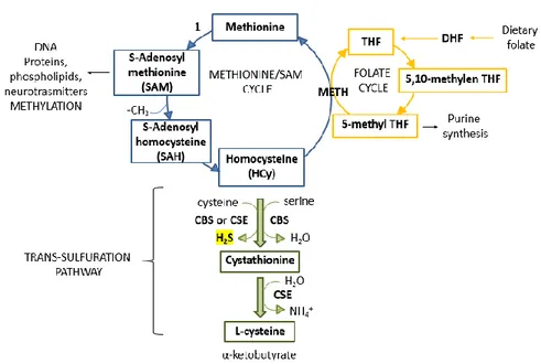
Figure 1. Methionine/SAM cycle (blu), folate cycle (yellow) and trans-sulfuration pathway
(green).
CBS- and CSE- catalysed reactions
The first metabolite in the transulfuration pathway is homocysteine (HCy), which is also an intermediate of the SAM/methionine cycle. Dietary methionine provides cells with methyl groups through the generation of S-adenosylmethionine (SAM or AdoMet) which transfers a methyl group to different acceptors generating S-adenosyl homocysteine (SAH or AdoHcy).
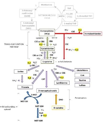
AdoHcy is reversibly hydrolysed to adenosine and homocysteine. HCy has then two fates; it can be remethylated by methionine synthase (MS or METH) regenerating methionine and thus completing the cycle, or can enter the trans-sulfuration pathway (fig.1). In mammals, the trans-sulfuration pathway is irreversible; therefore, sulfur exits from methionine cycle and can be used to generate cysteine, H2S and other sulfur-containing metabolites. In particular, cysteine becomes the limiting substrate for the synthesis of glutathione (GSH), coenzyme A (CoA), taurine and sulfate (SO42-). GSH together with its oxidized form (GSSG) is an important antioxidant compound implicated in the control of cellular redox homeostasis. Coenzyme A can transfer acetyl groups and it is involved in energy metabolism (e.g., in the Krebs cycle and fatty acids oxidation), while taurine and sulfate are the final metabolites derived from the oxidative degradation of the cysteine in excess via cysteine dioxygenase (CDO) (Kabil, 2014). The reactions involved in the trans-sulfuration pathway and H2S generation are reported below (fig. 2).
Figure 2. Reactions involved in the production of H2S and other sulfur metabolites.
Trans-sulfuration pathway (green arrows); CSE-catalysed synthesis of H2S from homocysteine (red
arrows); Generation of H2S, 3-mercaptopyruvate and other sulfur metabolites from
L-cysteine (violet arrows); MST-catalysed reaction of H2S synthesis (blue arrows);
Contribution of DAO to H2S synthesis (yellow arrows).
The first reaction of transulfuration pathway is catalysed by CBS and leads to formation of cystathionine. The enzyme uses serine and homocysteine as substrates and produces one molecule of water as
product (reaction 1). CBS is a member of the -family of PLP-containing enzymes which are typically able to catalyse -replacement or -elimination reactions. OH NH3 O O NH3 O O SH NH3 O O S NH3 O O H2O
L-serine L-homocysteine L-cystathionine
Reaction 1. Canonical-elimination reaction catalysed by CBS.
Du Vigneaud and co-workers were the first ones to note the production of H2S in the presence of homocysteine during his studies on trans-sulfuration pathway (Binkley & Du Vigneaud, 1944). CBS can indeed catalyse the condensation of homocysteine and cysteine (derived from diet, via degradation of endogenous proteins or via trans-sulfuration pathway) producing cystathionine and one molecule of H2S (Chen, 2004) (reaction 2).
SH NH3 O O NH3 O O S NH3 O O H2S L-cysteine L-cystathionine NH3 O O SH L-homocysteine
Reaction 2. Alternative reaction catalysed by CBS, yielding H2S from cysteine and
homocysteine.
Homocysteine is a substrate of many “accessory” reactions involved in H2S generation. In particular, CSE catalyses the synthesis of -ketobutyrate, ammonia and H2S from homocysteine and H2O (reaction 3)
NH3 O O SH L-homocysteine O O O NH4 H2S -ketobutyrate H2O
Reaction 3. - elimination reaction catalysed by CSE yielding H2S and -ketobutyrate
from homocysteine.
or the condensation of two molecules of homocysteine generating homolanthionine and H2S (reaction 4) (Singh, 2009).
NH3 O O S H2S L-homolanthionine NH3 O O SH L-homocysteine NH3 O O SH L-homocysteine NH3 O O
Reaction 4. -replacement reaction catalysed by CSE, yielding H2S and homolanthionine
from homocysteine. Homolanthionine is a biomarker of
hyperhomocisteinemia/homocystinuria (Chiku, 2009).
The second and last step of the trans-sulfuration pathway is the generation of cysteine from cystathionine and H2O catalysed by CSE (react. 5).
NH3 O O S NH3 O O L-cystathionine O O O 2NH4 -ketobutyrate H2O SH O O O L-cysteine
Reaction 5. Canonical elimination reaction catalysed by CSE.
Cysteine in turn is a substrate of alternative reactions catalysed by CSE yielding H2S and ultimately pyruvate (reaction 6) or L-lanthionine (reaction
7) (Singh, 2009). SH O O NH3 H2O O OH O NH3 H2S O O O NH4
L-cysteine L-serine Pyruvate
Reaction 6. elimination reaction catalysed by CSE.
SH O O NH3 S O O NH3 H2S L-cysteine L-lanthionine SH O O NH3 L-cysteine NH3 O O
Reaction 7. - replacement reaction catalysed by CSE. L-lanthionine is a biomarker of hyperhomocisteinemia/homocystinuria. (Chiku, 2009).
Furthermore, CBS proved to catalyse H2S production through -replacement of cysteine by several compounds (-mercaptoethanol, dithiothreitol, cysteamine and methanethiol) with formation of the corresponding thioethers (Singh, 2009).
MST- and DAO- catalysed reactions
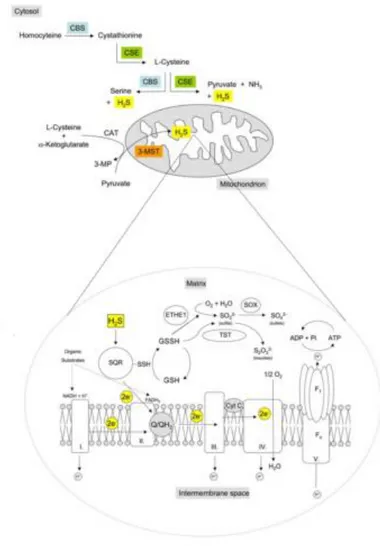
Cysteine can also be converted to 3-mercaptopyruvate via a transamination reaction catalysed by cysteine (or aspartate) aminotransferase (CAT). In the cytosol and in the mitochondria matrix, 3-mercaptopyruvate is desulfurated by 3-mercaptopyruvate sulfurtransferase (MST), which has a redox-active cysteine residue in the active site (MST-SH), to generate pyruvate and persulfidated enzyme (MST-SSH). Sulfur is then transferred by the NADPH-dependent enzyme thioredoxin reductase (TrxR) to the preferred reducing acceptor, thioredoxin (Trx) or glutathione (GSH), generating H2S (Kabil & Banerjee, 2014). Alternatively 3-mercaptopyruvate can be generated through the oxidative deamination of cysteine catalysed by D-amino acid oxidase (DAO) in peroxisomes (Shibuya & Kimura, 2013), leading to H2S generation (Shibuya, 2013).
Biological relevance
The catalytic promiscuity of the enzymes responsible for H2S production may be viewed as a manner to ensure H2S generation from different substrates and under several physiological conditions (Singh, 2009). The regulation of H2S generation is expected to be a physiological mechanism for augmenting or diminishing sulfur metabolites levels on demand. Indeed, the physiological relevance of this pathway is both set on the production of important cysteine-derived metabolites, such as glutathione and H2S, and on the prevention of toxic accumulation of homocysteine (Kabil, 2014).
Compound Human plasma levels (Kabil, 2014)
Methionine 20-100 µM
AdoMet Low micromolar or trace quantities
Homocysteine Low micromolar or trace quantities
Cystathionine Low micromolar or trace quantities
Cysteine ~100-200 µM
Taurine ~50-90 µM
GSH and GSSG ~100-200 µM
H2S
Low nanomolar
(*except aorta, 20- to 100-fold higher)
Table 1. Plasma levels of sulfur compounds derived from the CBS- and CSE-catalysed
reactions.
Tissue and cellular localization of H2S synthesis enzymes
The relative contribution of CBS, CSE and MST to H2S synthesis varies among different tissues, depending on the expression levels of the enzymes and the concentration of their respective substrates. In terms of tissue localization, CSE is expressed in the cardiovascular system but also in the liver, kidney, uterus, placenta, pancreatic islet, lung and gastrointestinal tract. CBS is preferentially expressed in the brain and other tissues, such as liver, kidney, pancreas, lung and gastrointestinal tract; more recently, it was also detected in the cardiac tissue. MST is expressed in the central system, mostly in glial cells, but it is also detected in the vascular smooth muscle, cardiomyocytes, kidney and liver (reviewed in Wallace & Wang 2015). Finally, DAO is localized in brain and kidney (Shibuya & Kimura 2013; Shibuya, 2013). In terms of cellular localization, CBS and CSE are mostly cytosolic enzymes, whereas MST is present in mitochondria and has a role in H2S-based energy production, as confirmed by Szabo and collaborators (Modis, 2013) (see Bioenergetics). However, under specific conditions both
CBS and CSE can be found in mitochondria or nuclei, and MST in the cytosol (Fu, 2012; Teng, 2013) (see Hypoxia).
2.1.1.1 Focus on cystathionine β synthase (CBS)
Structure
Human cystathionine β-synthase (hCBS) is a homotetrameric enzyme characterized by a complex domain structure and peculiar regulatory mechanism. Each 63 kDa subunit contains 551 amino acid residues organized in three functional domains (Meier, 2001). The N-terminal domain harbours a hexacoordinate heme with cysteine 52 (C52) and histidine 65 (H65) as endogenous Fe ligands; the central catalytic domain binds the pyridoxal-5’-phosphate (PLP) cofactor, whereas the C-terminal domain consists of a motif, named “Bateman module” (Bateman, 1997), that binds the positive regulator S-adenosyl-L-methionine (AdoMet) and is responsible for CBS tetramerization (Ereño-Orbea, 2013). Each CBS subunit binds three different substrates (homocysteine, serine and cysteine) and is further regulated allosterically by AdoMet, leading to an increase in catalytic activity by approximately two- to five-fold (Ereño-Orbea, 2014).
Figure 3. Protein domains and crystallographic structure of monomeric hCBS. Image from
(Ereño-Orbea, 2013).
Limited proteolysis of the full-length enzyme yields the `catalytic core' of CBS (amino acid residues 40-413). The reduction in size is accompanied by a significant increase in the specific activity of the enzyme and a change in its oligomerization state. In particular, removal of the regulatory C-terminal regulatory domain leads to a dimer with increased activity (four-fold higher kcat) unable to bind AdoMet. The truncated form of
CBS (45 kDa) lacking the C-terminal domain is a dimer which does not exhibit the aggregating properties which make physical studies on the wild-type full-length protein (63 kDa) difficult (Meier, 2001).
Regulation of enzymatic activity and cross-talk between gasotransmitters
The regulation of the enzymatic activity of hCBS is physiologically relevant (see Metabolic disease and Cancer) (Banerjee, 2003). The effectors and mechanisms involved in the activity modulation are reported below.
Pyridoxal-5’-phosphate (PLP)
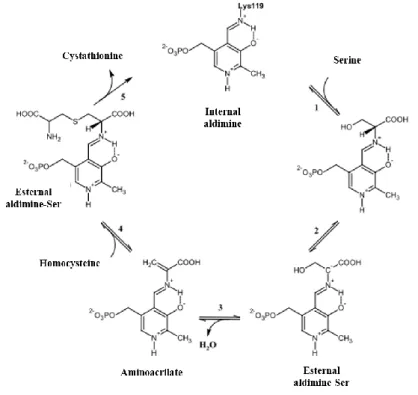
The pyridoxal-5’-phosphate (PLP) is present under two tautomeric forms; enolimine and ketoimine (Garret, 1998). The pyridoxal-5’-phosphate (PLP)-dependent enzymes catalyse several metabolically critical transformations including trans-aminations and crucial reactions in the metabolism of amino acids and amino sugars. Members of this protein family share a common first reaction step: the formation of an aldimine intermediate (via Schiff base) between the PLP cofactor and the substrate (e.g., the α-amino group of α-amino acids) (Toney, 2012). Following this initial step, each enzyme catalyses specific reactions, such as α- and β-decarboxylation, racemization, transamination, aldolic reaction, β- and γ-elimination, etc.
(Toney, 2012). hCBS belongs to the -family of the PLP-containing enzymes which are typically able to catalyze -replacement or -elimination reactions (Meier et al., 2001). In CBS, the PLP is deeply buried between the N- and C-terminal domains, and the active site is accessible only via a narrow channel. The cofactor is linked to the protein through the -amino group of Lys119 forming a Schiff base named `internal aldimine' (fig. 4). The nitrogen of the pyridine ring generates a hydrogen bond with the O of serine 349 (Ser349). Another hydrogen bond is formed between the 3’-hydroxyl group of PLP and the N of asparagine 149 (Asn149). The phosphate binding loop is located between -strand 8 and -helix 8 and forms an extended hydrogen bonding network with several amino acid residues, including threonine 257 (Thr257) and threonine 260 (Thr260) (Meier, 2001).
Figure 4. PLP in the CBS catalytic site.
In the presence of the substrate (serine or cysteine), the formation of a new aldimine intermediate (via Schiff base) between the PLP and the aminoacidic substrate (L-homocysteine) takes place. The protonation of the Schiff base, stabilized by a hydrogen bond with the oxygen of the ring, leads to increased acidity of the proton linked to Cα. The ketoenamine is key to PLP reactivity because, when its imine bond is protonated, it facilitates the nucleophilic attack by serine or cysteine. (1). The carbanion forming by the loss of hydrogen bond to Cα is stabilized by electronic delocalization on the pyridinic ring, where the positive charge on the nitrogen atom trapped the electrons (2). The -elimination of the -OH group of serine (or -SH group of cysteine) leads to formation of the amino acrylate intermediate and elimination of one molecule of H2O (or H2S) (3). The carbon atom in position of the amino acrylate intermediate undergoes nucleophilic attack by homocysteine generating cystathionine (4) that is eventually released (5) (fig.
5) (Weeks, 2009)
Figure 5. Reaction mechanism of CBS. Image adapted from (Weeks, 2009).
S-adenosyl-L-methionine (SAM or AdoMet)
Full-length CBS contains a C-terminal regulatory domain of ~140
residues, including the so-called `CBS domain' or ‘Bateman domain’ of 53 residues (Bateman, 1977). The C-terminal domain of CBS contains an autoinhibitory region harbouring the active site upon binding of the allosteric activator AdoMet (Ereño-Orbea et al., 2014). The C-terminal regulatory domain occludes the entrance to the catalytic site of the complementary monomer (fig. 6). Therefore, removal of the regulatory region in the truncated dimeric CBS leads to increased activity (Ereño-Orbea, 2014).
Figure 6. Mechanism of CBS allosteric regulation by AdoMet. Image from (Ereño-Orbea,
2014).
The presence of pathogenic missense mutations in this region often does not impair enzyme activity, but typically interferes with the binding of AdoMet and/or the enzyme activation by AdoMet (see Metabolic diseases) (Mendes, 2014).
Heme
In hCBS, the heme has been proposed to act not only as a redox sensor able to bind exogenous ligands (CO and NO) and modulate CBS activity (reviewed in Singh, 2010), but also to structurally stabilize CBS and improve its folding (Oliveriusova, 2002). The B-type heme of hCBS is in a
hydrophobic pocket located in the N-terminal domain between the α-helices 6 and 8. The heme iron, both in oxidized (FeIII) and reduced (FeII) state, coordinates the four pyrrolic rings of the protoporphyrin IX cofactor, whereas axially it binds the imidazolic nitrogen atom of histidine 65 (His65) and the thiolate moiety of cysteine 52 (Cys52), generating a low-spin hexacoordinate complex with octahedral geometry (Taoka, 2001). The enzyme is fully active when the heme is in the ferric state (FeIII), whereas heme reduction has been reported to promote a slow inactivation of the enzyme, that has been attributed to a ligand switch process (Cherney, 2007). Notably, the CBS heme in the reduced state is able to bind the gasotransmitters NO and CO resulting in a reversible inhibition of the enzyme (Vicente, 2014; Taoka, 2001). The communication between the heme and PLP active site has been attributed to molecular interactions with the α-helix 8 (Singh, 2010). As previously reported, the heme axially binds Hys65 and Cys52. The cysteine 52 establishes electrostatic interactions with arginine 266 (Arg266) at the end of this helix. Upon reduction, the heme increases its affinity for exogenous ligands, like CO and NO (Weeks, 2009). Moreover, AdoMet was found to further increase the CO affinity of the ferrous heme in human CBS (Vicente, 2016). Despite the low redox potential (-350mV) of the full-length CBS heme (Singh, 2010), in the presence of NADPH the human enzyme methionine synthase reductase (MSR) is able to catalyze the reduction of the CBS heme
in vitro. This finding shows that reduction of the CBS heme may occur under
physiological conditions (Kabil, 2001), making the enzyme susceptible to inhibition by the other gasotransmitters in vivo.
CO inhibition
CO binding to ferrous CBS is characterized by two dissociation constants, Kd1 =0.7–1.5 μM and Kd2= 45–68 μM (Taoka, 1999), attributed to differences in the heme microenvironment (Taoka, 1999) and/or an anti-cooperative effect between adjacent monomers within a functional CBS dimer (Puranik et al., 2009). The reaction follows a biphasic time course with a major slow phase, the rate constant of which is hyperbolically dependent on CO concentration yielding a limiting value klim= 0.012 – 0.017 s−1. This rate constant has been attributed to the slow rate-limiting dissociation of the endogenous Cys52 thiolate ligand from the ferrous heme iron (Puranik, 2009).
NO inhibition
NO binds to ferrous CBS at rates orders of magnitude higher than CO, and it dissociates from the ferrous heme iron much more slowly than CO (Vicente, 2014). Altogether, these data show that CBS has a higher affinity for NO than for CO. The kinetics of NO binding is not limited by the off-rate
of the Cys52 ligand and NO was proposed to initially attack the Fe from the His65 side, rather than from the Cys52 side (Vicente et al., 2014).
O2 binding
The ferrous heme [Fe(II)] of CBS can be rapidly oxidized by O2. Under normoxic conditions, the protein with ferric heme [Fe(III)] is a target of Lon protease, one of the most expressed protease at the mitochondrial level involved in the regulation of the mitochondrial protein turnover. Under hypoxic condition, the Lon protease does not bind ferrous CBS [Fe(II)], determining an accumulation of the CBS inside mitochondria (see Hypoxia and Synthesis) (Teng, 2013).
Heme spectral properties
Upon reduction, the Soret band of the CBS heme shifts from 428 nm to 449 nm. On CO binding to the ferrous CBS heme, the band further shifts to 422 nm, resulting in a hexacoordinate ferrous-CO adduct with the endogenous Cys52 ligand displaced. NO binding to ferrous CBS results instead in a notable broadening of the Soret band, accompanied by an intensity decrease and a shift to ~395 nm, which have been attributed to formation of a high-spin pentacoordinate ferrous- NO adduct with both endogenous ligands displaced (Vicente, 2014; Taoka, 2001).
2.1.2 H2S production by the gut microbiota
The gut microbiota is a major source of H2S. In particular, anaerobic sulfate-reducing bacteria produce sulfide from the metabolism of dietary proteins in the lumen of the human large intestine (reviewed in Blachier, 2010; Carbonero, 2012). Approximately, one millimolar has been reported for the total sulfide content, but a much lower value (60 μM) has been reported for free H2S. Due to the potentially toxic sulfide levels, colonocytes have been adapted in order to neutralize the sulfide content and, therefore, show an unusually high sulfide oxidizing activity (Goubern, 2007; Lagoutte, 2010; Leschelle, 2005) (see Mitochondrial H2S oxidation in colonocytes). Alterations of H2S metabolism in colonocytes are associated to pathological condition discussed in H2S and colorectal cancer (Carbonero, 2012).
2.2. Catabolism
As H2S is an important signalling molecule exerting dose- and time-dependent biological effects, the balance between hydrogen sulfide synthesis and consumption rates plays a critical role in the maintenance of cellular homeostasis (Módis, 2014; Szabo, 2014). H2S, is the only gasotransmitter
that is enzymatically catabolised (Hildebrandt & Grieshaber, 2008) by two different pathways: it can be oxidized inside the mitochondria (Jackson, 2012) or it can enter the cytosolic methylation pathway (Weisiger 1980).
2.2.1. Mitochondrial H2S oxidation - Enzymes and pathways
Oxidative phosphorylation: a brief overview
The oxidative phosphorylation takes place at the inner mitochondrial membrane where the four complexes (I-IV) of the electron transport chain (ETC), as well as the electron transporters coenzyme Q (CoQ) and cytochrome c (cyt c), and the ATP synthase, are located. The oxidative phosphorylation couples the electron flux derived from the oxidation of NADH and succinate, with the ADP phosphorylation, thus generating ATP. In a first step, electrons transported by NADH and succinate are transferred to Complex I (NADH:ubiquinone dehydrogenase) or Complex II (succinate dehydrogenate or SDH) and subsequently, through Complex III (cytochrome
bc1 or ubiquinol:cytochrome c reductase), cytochrome c and Complex IV (cytochrome c oxidase or CcOX), are then transferred to the final acceptor, molecular O2. The process is overall accompanied by the vectorial translocation of 10 protons (for one molecule of NADH) or 6 protons (for
one molecule of succinate or FADH2) from the mitochondrial matrix side to the intermembrane space. The electron transport chain contributes to the formation of an electro-chemical gradient across the inner membrane which generates the proton motive force (PMF) used by ATP synthase (ATPase or complex V) to generate ATP from ADP and Pi (2.5 molecules of ATP per NADH or 1.5 molecules of ATP per FADH2) (Nicholls & Ferguson, 2013). More recent studies have shown that the oxidation of H2S is associated with the concomitant injection of electrons into the mitochondrial electron chain, thus sustaining the electron flow and promoting the ATP synthesis at lower concentrations of sulfide (Goubern, 2007).
Hydrogen sulfide catabolic pathway
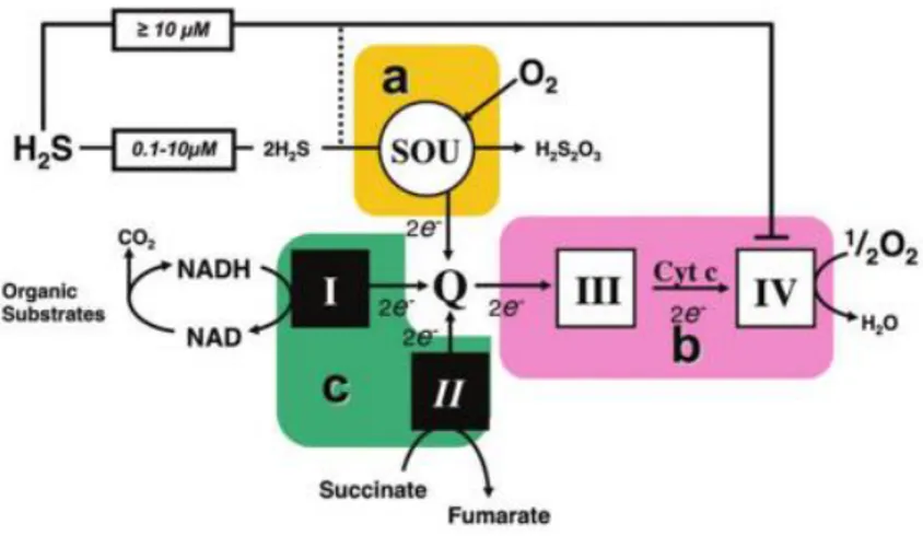
The mitochondrial sulfide oxidation pathway is involved in the breakdown of sulfide, with the concomitant stimulation of mitochondrial respiration (Goubern, 2007) (see Bioenergetics). The pathway has been initially identified in the lugworm Arenicola marina (Hildebrandt and Grieshaber, 2008). This enzymatic system responsible for H2S metabolism at the mitochondrial level, also named sulfide-oxidizing unit (SOU), couples the oxidation of 2 sulfide molecules with the consumption of 1.5 molecule of oxygen (Goubern, 2007; Lagoutte, 2010). The system comprises the
mitochondrial inner membrane-associated enzyme sulfide:quinone oxidoreductase (SQR) and the mitochondrial matrix-localized enzymes persulfide dioxygenase or ethylmalonic encephalopathy 1 (ETHE1), thiosulfate sulfurtransferase or Rhodanese (TST or Rhod) and sulfite oxidase (SOx). Overall the pathway oxidizes H2S leading to formation of a variety of species, including sulfite (SO32-), thiosulfate (S2O32-), sulfate (SO42-) and glutathione persulfide (GSSH) (Kabil & Banerjee, 2014; Libiad, 2014; Szabo, 2014).
The first step of sulfide metabolism is catalysed by sulfide:quinone oxidoreductase (SQR). In the catalytic site of SQR, a reactive cysteine disulphide transfers a sulfur atom derived from H2S to glutathione (GSH) generating glutathione persulfide (GSSH). Concomitantly, the sulfide-derived electrons are transferred via a flavin adenin dinucleotide (FAD) moiety to coenzyme Q, thus leading to injection of two electrons into mitochondrial chain and reduction of 0.5 molecule of O2 to H2O by CcOX. The sulfur-acceptor has been a topic of discussion in the literature (Jackson, 2012; Libiad, 2014): it was proposed to be reduced glutathione (GSH), sulphite (SO32-) or an unknown acceptor. Recently, GSH has been proposed as the preferred sulfur acceptor for SQR-mediated H2S oxidation under physiological conditions (Libiad, 2014). Indeed, increased GSH synthesis by administration of the cysteine precursor N-acetylcysteine improves H2S
clearance in patients with ethylmalonic encephalopathy caused by mutation in the ETHE1 gene, implicating GSH as the persulfide carrier in the sulfide oxidation pathway (Tiranti, 2009).
Subsequently, the persulfide dioxygenase (ETHE1), a mononuclear non-heme iron enzyme structurally belonging to the family of metallo-β-lactamases, uses O2 as co-substrate to convert the GSSH into sulfite and GSH. GSSH and sulfite are further converted into thiosulfate and GSH by thiosulfate sulfur transferase (Rhod). Rhodanese has a single redox-active cysteine in the active site and catalyses several reactions involving numerous substrates, but the generation of thiosulfate appears to be the most catalytically favourable reaction at physiologically relevant substrate concentrations (Kabil & Banerjee, 2014; Libiad, 2014).
The last step in the sulfide oxidation pathway is catalysed by sulfite oxidase (SOx, Fig. 1B) (Johnson-Winters, 2010).
Overall, the mitochondrial SOU couples the oxidation of two H2S with the consumption of 1.5 molecules of O2: the stoichiometry is thus 1.5/2 = 0.75, as reported by Laguette and co-workers (Lagoutte, 2010).
Figure 7. Hydrogen sulfide synthesizing-enzymes, Sulfide-oxidizing unit (SOU) and
electron transport chain (ETC). Image from (Szabo, 2014).
A large variety of mammalian cell types (including colonocytes, macrophages, hepatocytes, neurons, etc.) have been assayed for their ability to consume H2S at the mitochondrial level (Goubern, 2007; Lagoutte, 2010; Mimoun, 2012), revealing a large variability among cell types in terms of SOU activity. The activity is high in cells physiologically exposed to relatively high H2S levels, such as colonocytes, but absent or hardly detectable in other cell types, such as neuroblastoma or other nervous system- derived cell lines (Lagoutte, 2010).
2.2.2. Mitochondrial H2S oxidation in colonocytes
The gut microbiota produces sulfide in a concentrations ranging from high micromolar to low millimolar as reviewed in (Blachier, 2010) (see H2S production by the gut microbiota). The effect of sulfide on the metabolism of
colonocytes was studied in the HT29 cell line derived from human colon cancer (Leschelle, 2005). After 24 hours exposure to high levels of sulfide (1 mM), HT29 cells have shown a general suppression of mitochondrial bioenergetics (decreased CcOX activity and increased proton leak), enhanced anaerobic glycolysis (decreased cell proliferation and increased lactate release) and unchanged Ki of cytochrome c oxidase (0.3 μM). Therefore,
colonocytes appear well adapted to the sulfide-rich colon environment in that they are able to neutralize the exogenous sulfide (Leschelle, 2005; Szabo, 2014). When sulfide exposure is maximal, indeed, complex I, complex II and SQR compete for coenzyme Q. However, according to the redox conditions, Complex I can work in a reverse mode in the presence of sulfide (Leschelle, 2005). This process appears to be specific to colonocytes (Lagoutte, 2010). In a previous study, reversal of Complex II activity was also suggested (Goubern, 2007). When Complexes I or II operate in reverse mode, oxidation of sulfide is favoured as these repiratory complexes can accept electrons from coenzyme Q, even in the presence of severe CcOX inhibition by high sulfide concentrations. The impact of these reverse reactions on ATP production is null or negative; indeed, reversal of Complex I activity would cause uncoupling (Lagoutte, 2010). However, when complete inhibition of Complex IV takes place, the cell must use anaerobic glycolysis to produce energy (Szabo, 2014).
3. Physiological and pathological role of H2S
Hydrogen sulfide (H2S) has emerged as an important signalling molecule. The lipid-soluble nature of H2S allows it to target a large variety of proteins in different cellular compartments. Due to its chemical versatility,
H2S can interact with protein sulfhydryl groups and metals (reviewed in (Wallace & Wang, 2015). Hydrogen sulfide, therefore, has unique properties that make it a selective and powerful modulator of a large range of biological targets, with stimulatory or inhibitory effects.
3.1. H2S, a Janus-faced molecule
At low (nM) concentration H2S ensures inflammatory, anti-oxidant and cytoprotective responses, whereas at higher (μM) concentrations it can lead to toxicity and eventually cell death. Indeed, high levels of H2S promote pro-inflammatory pathways and inhibit CcOX, thus blocking the electron transfer through the respiratory chain with the induction of pro-oxidant and DNA-damaging effects eventually culminating in cell death (Kimura, 2015; Wallace & Wang, 2015). Furthermore, H2S has a very interesting dual role on vascular remodelling. It was indeed reported to induce smooth muscle relaxation in the portal vein and the thoracic aorta, associated with opening of ATP-sensitive K+ channels. However, H2S pre-treatment was also found to induce vasoconstriction by scavenging endothelial NO. The response of blood vessels to H2S depends on the vessel type, exposure time and concentration. Another variable that influences the effects of H2S on the smooth muscle of blood vessels is O2, with low and
high concentrations inducing vasodilation and vasoconstriction, respectively (see Hypoxia) (Kabil, 2014; Wallace & Wang, 2015). However, the major and perhaps most interesting dual role induced by sulfide is the one exerted on cell bioenergetics.
3.2. Bioenergetics
3.2.1. H2S as mitochondrial substrate or inhibitor
In mitochondria, H2S exerts a dual effect on cell bioenergetics. At high (high μM) concentration, it blocks mitochondrial respiration by inducing a potent, reversible and non-competitive inhibition of complex IV (CcOX) (Cooper & Brown, 2008); at the opposite, at lower concentrations (from high nM to low μM), H2S is a substrate for most mitochondria (see Mitochondrial
H2S oxidation) (Goubern, 2007; Lagoutte, 2010), thus stimulating cell
respiration. H2S has therefore been recognized as the first inorganic substrate used by mitochondria to generate ATP (Yong and Searcy, 2001). This makes H2S a very interesting molecule from a bioenergetic viewpoint, able to induce opposite effects on mitochondrial respiration depending on its bioavailability.
H2S as a mitochondrial substrate
The first evidence for mitochondrial sulfide oxidation associated with oxygen consumption and ATP synthesis was obtained by investigating the invertebrate Solemya reidi (Powell and Somero, 1986). Following this observation, Bouillaud and co-workers have reported the first evidence of sulfide stimulation of energy metabolism in a human cell model (human colon adenocarcinoma cell lines) (Goubern, 2007). As described above (see
Mitochondrial H2S oxidation), H2S oxidation by the sulfide-oxidizing unit (SOU) occurs with the injection of electrons in the ETC. The major contribution of this pathway to cellular bioenergetics arises from the SQR-catalysed reduction of coenzyme Q.
Figure 8. Scheme of sulfide oxidation. Complexes of the mitochondrial respiratory chain are
numbered with roman numerals. H2S can either induce a stimulation of electron flow via
SQR in the SOU or inhibit mitochondrial oxygen binding at Complex IV, thereby stopping
electron flow. Image from (Szabo et al., 2014).
That sulfide-derived electrons enter the mitochondrial respiratory chain at the level of coenzyme Q is demonstrated by the finding that, whereas inhibition of Complex I with rotenone has not effect on sulfide catabolism, inhibition of Complex III or Complex IV by antimycin A or cyanide, respectively, blocks mitochondrial sulfide oxidation (Goubern, 2007). Complexes I and II therefore compete with SQR for the reduction of coenzyme Q, while complexes III and IV are required for sulfide oxidation. Interestingly, the maximal rate of sulfide oxidation was found to increase with cell differentiation (Mimoun, 2012) and a maximal rate of sulfide oxidation approaching the theoretical limit was observed, under which condition all the electrons travelling in the respiratory Complexes III–IV are expected to come from H2S via SQR.
Biological relevance
Compared to other substrates of the mitochondrial respiratory chain, H2S oxidation has a relative low energetic yield, due to the relatively high cost in terms of oxygen [injection of electrons into coenzyme Q is associated
with the consumption of additional O2 by ETHE1 (Hildebrandt and Grieshaber, 2008)] and the low contribution in terms of electrons injected into respiratory chain. On the other hand, H2S diffuses freely across cell membranes, it does not need any ‘biochemical preparation’ (which has an energy cost), the affinity of SQR for sulfide is high and, in addition, in the gut an abundant sulfide production occurs with no energy cost. All that guarantees that sulfide oxidation takes place with high efficiency at low concentrations of sulfide, thereby preventing its toxic accumulation (Szabo, 2014). Additionally, sulfide was suggested to serve as an ‘emergency’ substrate. Indeed, although in terms of quantitative bioenergetics it is clearly inferior to carbon metabolism, under physiological or stress conditions, H2S production by endogenous enzymes can contribute to cellular bioenergetics (Fu, 2012; Goubern, 2007). A bioenergetic role has been clearly demonstrated for 3-MST, the enzyme that is responsible for the majority of mitochondrial H2S production, which shows preferential mitochondrial localization as described above (see Synthesis). The role of 3-MST in the regulation of cellular bioenergetics is supported by a large body of evidences (Módis, 2013). SQR silencing suppresses both basal and 3-MP mediated stimulation of bioenergetic function (Módis, 2013). Interestingly, oxidative stress impairs the bioenergetic role of 3-MST (Módis, 2013). Also CSE and CBS contribute to the physiological regulation of bioenergetic function, since
the H2S produced in cytosol by these enzymes can freely diffuse across the cell. Additionally, since the distribution of both CSE and CBS is cell type- and tissue-dependent, effects of CBS/CSE are also cell type- and organ-dependent. Under conditions of cell dysfunction (such as hypoxia), CSE sustains cellular bioenergetics by translocating to the mitochondria (Fu, 2012), while in colon cancer cells (HCT116 cell model) it is the endogenous H2S production by CBS that supports cellular bioenergetics and cell proliferation (Szabo, 2013). Intriguingly, CBS was also found to accumulate in mitochondria under hypoxic/ischemic conditions (see Cancer and
Hypoxia), although the physiological relevance of this process needs to be
established.
H2S as a mitochondrial inhibitor
Sulfide inhibition of isolated mitochondrial CcOX is reversible, potent (Ki = 0.2 μM at pH 7.4) and non-competitive in that H2S affects the enzyme Vmax, but not the KM for O2 (Petersen, 1977). Interestingly, sulfide inhibition of CcOX is pH dependent, the Ki dropping from 2.6 μM to 0.07 μM as the pH decreases from 8.05 to 6.28 (Nicholls, 1982). Consistently, CcOX inhibition by sulfide is more effective under acidosis conditions (Szabo, 2014), probably because under these conditions H2S prevails over the
HS− form. The active site of CcOX, being located in an apolar environment, is expected to preferentially bind electroneutral species, such as H2S, or proton-neutralized anionic species (HS− + H+) (Rich, 1996), both favoured at lower pH. (Nicholls, 2013). H2S inhibits CcOX with a low Ki value. This notwithstanding, inhibition of respiration in isolated mitochondria or intact cells requires much higher H2S concentrations (µM, see (Leschelle, 2005). The sulfide-mediated inhibition of CcOX leads to decreased electron flow through the ETC and thus to a decline in ATP production (Hill, 1984). This effect has been well documented both in vivo and in cell models (Leschelle, 2005). The inhibitory effect has been observed at 10-100 µM H2S depending on the experimental approach. Indeed, quantitative information on sulfide oxidation and related bioenergetic effects was often obtained in these studies by supplying sulfide at selected injection rates rather than as a single bolus, an approach reviewed in (Abou-Hamdan, 2015). After tissue homogenates exposure to a single bolus of NaHS, the complex I activity has been shown to return to its original level within 10-30 min. Due to the opposite effects of sulfide on mitochondrial respiration, stimulation of oxygen consumption and mitochondrial energization is best appreciated at low sulfide concentrations, non-inhibitory towards CcOX (Abou-Hamdan, 2015).
3.3. Cancer
Recent studies revealed increased expression of various hydrogen H2S-producing enzymes in colon, ovarian, prostate and breast cancer cells (reviewd in Szabo, 2013), and a new role of H2S in cancer has emerged (Chao 2016; Hellmich & Szabo, 2015; Szabo, 2015). H2S seems to have a dual role also in the cancer, promoting angiogenesis and proliferation and sustaining cellular bioenergetics or, at the opposite, displaying a cytotoxic effect and becoming deleterious for the tumour cell at higher concentration (Szabo, 2014a) (see H2S metabolism as drug target).
3.3.1. H2S in colorectal cancer (CRC)
Colonocytes are exposed to high levels of H2S produced by the gut microbiota (Blachier, 2010). These cells therefore show efficient systems to metabolize H2S (Lagoutte, 2010). On the other hand, high levels of H2S in the gut have been associated to ulcers, chronic inflammation, DNA damages and colon cancer (CRC- colorectal cancer) (Carbonero, 2012). In particular, tissues isolated by patients with CRC display a reduced expression of Rhod, one of the enzymes involved in sulfide catabolism, underlining the importance of the role of H2S metabolism in CRC (Ramasamy, 2006). More recent studies have shown in cancer cells an over-expression of the H2S-generating enzyme CBS and a functional role of CBS-derived H2S, which
promotes proliferation through the stimulation of cellular bioenergetic at both mitochondrial and cytosolic level (Hellmich & Szabo, 2015; Szabo, 2013; Szabo & Hellmich, 2013). In the mitochondrion, H2S acts as a metabolic ‘fuel’ injecting electrons into mitochondrial chain (Szabo, 2013; Szabo & Hellmich, 2013), whereas in the cytosol, hydrogen sulfide stimulates the glycolytic activity (Mustafa, 2010).
Additionally, inhibition (with aminooxiacetic acid, AOAA) or silencing of CBS in colon cancer cells HCT116 decreases bioenergetic functions in vitro. Implantation into the mice of CBS-silencing colon cancer cells, reduces tumour growth and inhibits peritumour angiogenesis (Szabo, 2013) in vivo. Furthermore, interestingly, inhibition (with AOAA) or silencing of CBS also sensitizes the cancer cells to chemotherapy (Chao & Zatarain, 2016; Chao, 2014). In colon cancer cell lines SW480, instead, high expression levels of CSE were observed. Inhibition (with propargylglycine, PAG) or silencing of CSE in SW480 cell lines reduces the proliferation in vitro and tumour growth
in vivo (Fan, 2014).
Therefore, H2S produced by CBS or CSE promotes bioenergetics, proliferation, migration, invasiveness, and neoangiogenesis, thus creating the ideal conditions for tumour cell survival (Cai, 2010; Hellmich & Szabo, 2015; Szabo & Hellmich, 2013). Following these studies, scientific interest in
H2S metabolism has increased in the perspective of developing new pharmacological interventions in the therapy of cancer (Hellmich, 2015).
3.4. Hypoxia
Hypoxia is a common factor of the microenvironment of solid tumours that is associated to drug-resistance and malignancy (reviewed in Muz, 2015). Under hypoxic conditions a central role is played by the hypoxia inducible factor (HIF) which appears to be directly involved in the up- or down-regulation of key enzymes in pathways implicated in cancer metabolism and thus in cell survival (Semenza, 2013). The relationship between hypoxia and metabolic reprogramming in cancer has been widely reported and is reviewed in (Masson & Ratcliffe, 2014).
3.4.1. H2S as an oxygen sensor
The interplay between the two gaseous molecules H2S and O2 has been thoroughly investigated (reviewed in Olson, 2015; Wu, 2015). H2S mimics hypoxia-induced responses like vasodilation (Zhao, 2001), neo-angiogenesis (Papapetropoulos, 2009) and the expression of HIF-1a (Beaumont, 2016). Furthermore, the H2S levels proved to be regulated by the O2 concentration in tissues. Olson et al. proposed a model in which both cytosolic synthesis and mitochondrial oxidation of sulfide are regulated in an
O2-dependent manner (Olson, 2015). The higher H2S levels in hypoxic/ischemic conditions is primarily due to the over-expression and/or activity stimulation of the H2S-synthesizing enzymes (Takano, 2014; Kolluru, 2015). Under hypoxic conditions both CBS and CSE were shown to display increased activity due to release of CO inhibition, following activity down-regulation of the isoform 2 of heme-oxygenase (enzyme involved in the heme degradation generating CO) (Morikawa, 2012; Yuan 2015). Hypoxia also leads to the translocation of CSE from the cytosol to mitochondria, where CSE uses approximately three-fold higher concentrations of L-cysteine to produce H2S (Fu, 2012). In addition, whereas under normoxic conditions CBS is degraded inside mitochondria by Lon protease, upon hypoxic stress Lon protease cannot recognize the deoxygenated/ferric haem group in CBS, and therefore CBS is not degraded. This leads to accumulation of CBS in the mitochondrion and increased mitochondrial H2S levels (Teng, 2013). The enhanced production of H2S occurring under hypoxia may be interpreted as a mechanism to ensure H2 S-mediated protection against ischemia injuries and against the cytotoxic and inflammatory damages induced by hypoxic conditions (Takano, 2014; Kolluru, 2015; Morikawa, 2012; Hine, 2016). Enhanced H2S levels proved to be a compensatory response to hypoxia promoting vasodilation (Yuan, 2015; Morikawa, 2012) in many O2 sensitive tissues. On the other hand, hypoxia
makes complex IV more sensitive to H2S inhibition (Matallo, 2014; Abou-Hamdan, 2016) and induces notable metabolic changes: it enhances anaerobic glycolysis and reduces mitochondrial mass and activity (as a result of enhanced mitophagy and reduced mitochondrial biogenesis) (Solaini, 2010; Zhang 2008, Wu 2015),thus shiftingthe action of H2S from protective to detrimental.
3.5. Metabolic diseases
Alterations of H2S metabolism are also involved in pathological metabolic dysfunctions (Wallace & Wang, 2015), like lipid metabolism disorders, liver diseases, diabetes and homocystinuria. The role of hydrogen sulfide metabolism in homocystinuria is reported below.
3.5.1. H2S in hyperhomocysteinaemia and homocystinuria
Hyperhomocysteinemia is a clinical metabolic condition characterized by elevated homocysteine plasma levels which have been linked to a variety of human diseases, including heart attack, stroke and osteoporosis. The major causes of hyperhomocysteinaemia are deficiencies in vitamins B4, B9 and B12 and mutations in the genes encoding 5-methyltetrahydrofolase or CBS (Maron and Loscalzo, 2009). CBS is involved in the trans-sulfuration pathway of methionine and H2S metabolism and regulates both homocysteine
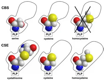
and H2S levels in the human body (see Synthesis). Banerjee and collaborators, have demonstrated that the H2S generation catalysed by cystathionine γ-lyase (CSE) increases progressively with the grade of hyperomocysteinemia. Indeed, at severely elevated homocysteine (200 mM), both -elimination (reaction 5) and the replacement (reaction 4) reactions of homocysteine catalysed by CSE lead to the generation of H2S with concomitant production of homolanthionine and lanthionine, respectively. The elevated H2S production by CSE may contribute to the hyperhomocysteinemia-associated cardiovascular disturbances. Based on these data, the inhibition of CSE has been suggested as a pharmacological strategy to treat hyperhomocysteinemia, and homolanthionine and lanthionine were proposed as novel biomarkers of the disease (Chiku, 2009)
Figure 9. Proposal mechanism for the CSE selective affinity for homocysteine. Image from
(Chiku, 2009).
Classical homocystinuria (OMIM#236200) is an inborn error of metabolism associated with mutations in the in CBS gene. It has a variable incidence of 1:1.800 to 1:900.000 and is biochemically detected by markedly high homocysteine and methionine levels in plasma and urine. The clinical presentation includes complications in the vascular, neurological and skeletal systems (Mudd, 1975) and an oxidative stress condition due to the high plasmatic levels of homocysteine. The major therapeutic approach for classical homocystinuria consists in dietary methionine restriction and administration of pyridoxine (vitamin B6), a precursor of the PLP cofactor essential for CBS activity (Mudd, 1975). However, a significant part of patients (approximately half) does not respond to this treatment (Schiff & Blom, 2012). The CBS mutations identified in patients with classical homocystinuria are often missense mutations resulting in single amino acid substitutions which often affect the enzyme folding and/or activity (Hnízda, 2012). Additionally, some of these mutations have been shown to affect the positive allosteric regulation of CBS by AdoMet, pointing to such dysregulation as a new pathogenic mechanism in classical homocystinuria (Mendes, 2014). A novel therapeutic approach is currently under development based on enzyme replacement therapy using PEGylated recombinant CBS (Bublil, 2016).
4. New pharmacological strategies in cancer
4.1. H2S metabolism as drug target
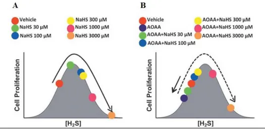
CBS has recently emerged as a drug target in cancer (Druzhyna, 2016). CBS-derived H2S was indeed shown to play a key role in the survival and proliferation of tumour cells. Different strategies have been proposed for the treatment of cancer, based on the dual effect of sulfide on cell bioenergetics. The dose-dependent effect of H2S on cancer has been demonstrated in colon adenocarcinoma cell lines using increasing concentration of H2S in the presence or absence of aminooxiacetic acid (AOAA), an inhibitor of CBS (fig. 10). These studies showed enhanced cell proliferation induced by H2S, the effect being related to the balance between endogenous and exogenous H2S.
Figure 10. Biphasic (bell-shaped) dose-response of H2S on tumour cell proliferation. Image
from (Hellmich, 2015).
Based on these studies two possible pharmacological approaches, both aiming to reduce the cell proliferation, seem applicable: i) increasing the H2S production using either H2S-donors or inhibitors of the H2S-consuming enzymes, or ii) reducing the H2S generation using inhibitors of H2S-synthesis enzymes (Chao, 2016, Chao, 2014; Asimakopoulou, 2013; Druzhyna, 2016; Hellmich, 2015).