Pagina 2
Riassunto
Pag. 4
Riassunto
L’identificazione precoce di diatomee fitoplanctoniche del genere
Pseudo-nitszchia è di grande importanza per il fatto che questo genere
annovera numerose specie in grado di produrre acido domoico, una neurotossina responsabile dell’ ASP (Amnesic Shellfish Poisoning), sindrome che causa gravi disturbi neurologici nell’uomo e mortalità massiva della fauna marina.
Come per le altre diatomee, l’identificazione delle diverse specie di
Pseudo-nitzschia avviene, ove possibile, mediante l’osservazione di
caratteristiche morfologiche cellulari distinguibili al microscopio ottico, sebbene caratteri sistematici importanti si rilevano soprattutto dalle osservazioni ultrastrutturali del frustulo (parete cellulare silicea delle diatomee) attraverso il microscopio elettronico.
Negli ultimi anni, le analisi genetiche e molecolari, hanno rilevato la presenza di un’ elevata diversità genetica nell’ambito del genere
Pseudo-nitzschia, con la presenza di numerose specie geneticamente affini (cladi)
definite specie pseudo-criptiche e criptiche, che presentano, rispettivamente, lievi o nessuna differenza ultrastrutturale, ma con apprezzabile diversità genetica. Queste differenze si riflettono anche nella capacità di queste specie di produrre o meno acido domoico e quindi di essere o meno neurotossiche.
Attualmente, l’identificazione di alcune alghe tossiche come anche l’individuazione di specie criptiche, è resa possibile attraverso l’impiego di markers molecolari usati su acidi nucleici estratti (analisi di regioni variabili del rDNA, sia nelle regioni codificanti che negli ITS, analisi degli
Riassunto
Pag. 5
rRNA, tecniche di DNA microarray); di recente sono state sperimentate anche tecniche di identificazione molecolare su cellula intera (Whole Cell- WC) con uso di sonde fluorescenti (Fluorescent In Situ Hybridization-FISH). In particolare la WC-FISH, metodologia adottata in questo lavoro di tesi, prevede l’uso di brevi sequenze nucleotidiche (oligoprobes) marcate con fluorocromi, specifiche per regioni target dell’rRNA presenti nella subunità maggiore (LSU) o minore (SSU) del ribosoma. Queste regioni sono infatti molto variabili e riescono a identificare i diversi ceppi e/o cladi.
La metodologia WC-FISH è stata messa a punto partendo da protocolli già presenti in letteratura che sono stati da noi integrati e modificati. Nella prima fase di calibrazione e test delle diverse sonde, sono state impiegate monocolture di specie criptiche e/o pseudocripitiche del genere
Pseudo-nitzschia, isolate e coltivate in laboratorio. Per ogni coppia
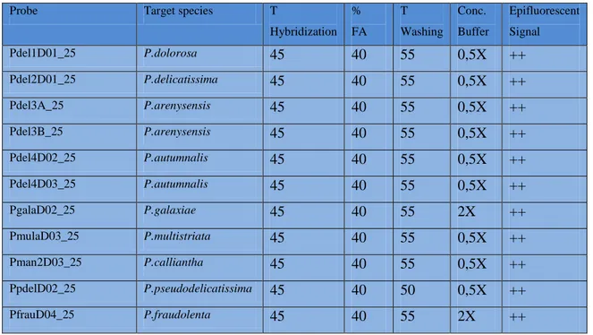
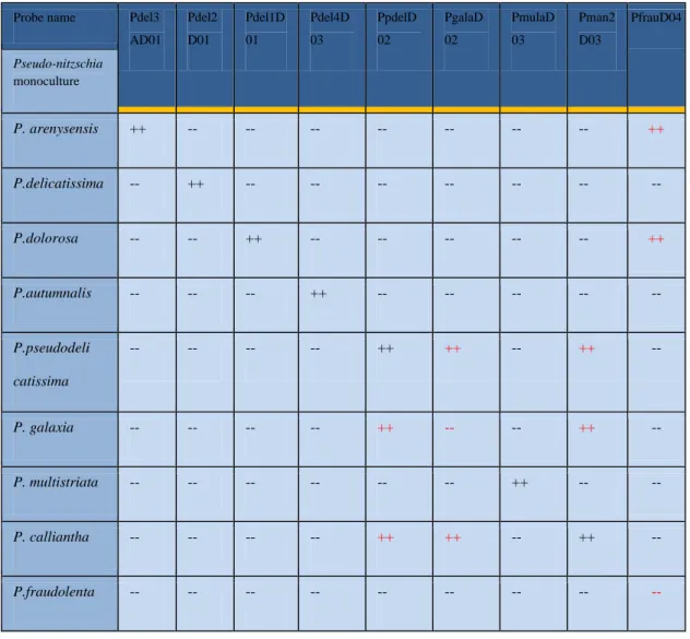
oligoprobe/specie target è stata effettuata un’accurata fase di calibrazione allo scopo di ottenere un segnale di marcatura ottimale ed eliminare i fenomeni di cross-ibridazione delle diverse sonde con i cladi affini. Tali operazioni hanno consentito di validare, di tutte le sonde disponibili, 4 sonde in grado di individuare in maniera specifica, le specie criptiche P.
arenysensis, P. autumnalis sp. nov., P. delicatissima e P. dolorosa,
appartenenti originariamente al raggruppamento P. delicatissima (P.
delicatissima complex).
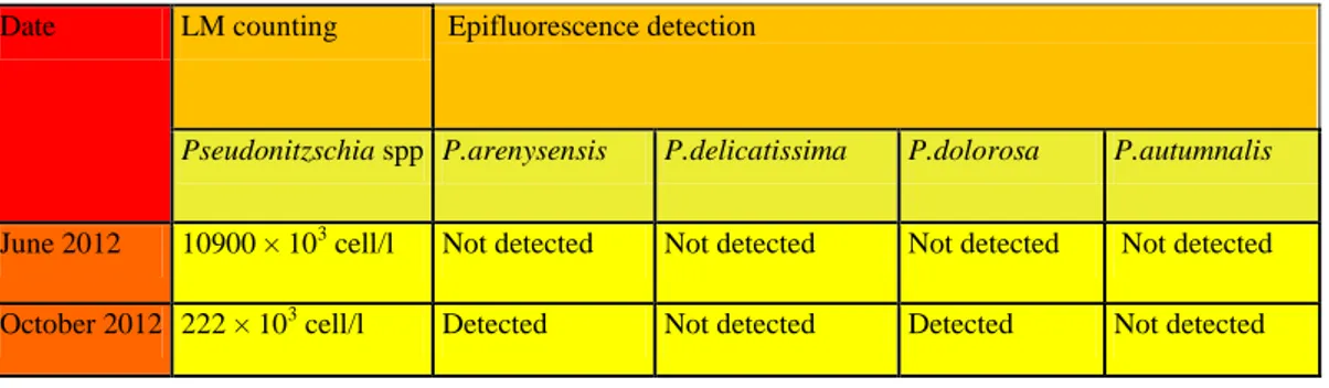
La seconda parte del lavoro ha riguardato il trasferimento della tecnica su campioni naturali (Field-test) al fine di verificare la sua eventuale applicazione. Le sonde sono state impiegate anche su campioni ambientali in corrispondenza di fioriture di Pseudo-nitzschia.
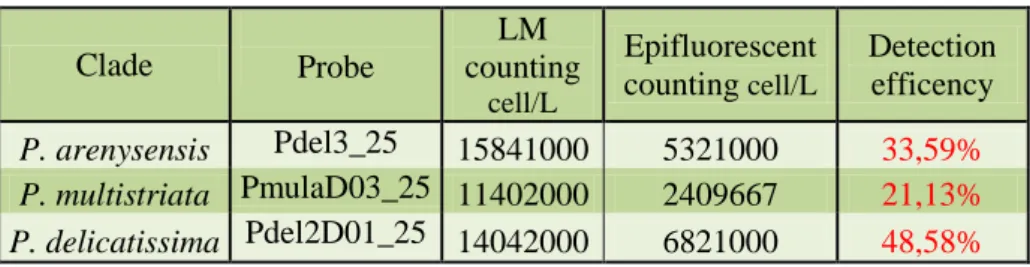
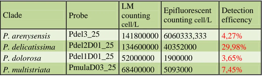
Sui diversi campioni naturali, l’effettiva sensibilità e specificità della WC-FISH è stata valutata comparando i risultati ottenuti con quelli di
Riassunto
Pag. 6
identificazione tradizionale in microscopia ottica (metodo Utermöhl). Complessivamente i field-test hanno evidenziato un buon accordo tra i due metodi dal punto di vista soprattutto qualitativo. Dal punto di vista quantitativo (stima delle abbondanze dopo WC-FISH vs metodo Utermöhl) i nostri dati mostrano una correlazione tra l’efficienza di detection della WC-FISH e numero di cladi presenti nel campione. In particolare, l’efficienza del metodo WC-FISH diminuisce con l’aumentare del numero di specie criptiche, come verificato da successivi esperimenti in laboratorio. Questa criticità del metodo rispetto alle stime quantitative, trova la sua spiegazione nel fatto che, nonostante l’accurato processo di calibrazione effettuato a monte, le diverse sonde, avendo sequenze molto simili, competono per gli stessi siti target delle specie affini del P. delicatissima complex.
Altri fattori possono inoltre influire sulla stima quantitativa con metodo WC-FISH: raccolta delle cellule per filtrazione che determina una perdita di densità cellulare; presenza di materiale organico nei campioni naturali che ostacola l’accesso delle sonde ai siti target; stato fisiologico delle cellule che influenza la quantità di rRNA disponibile e quindi l’intensità del segnale fluorescente.
Nonostante questi limiti, possiamo comunque concludere che la metodologia WC-FISH sia uno strumento economico e rapido per l’identificazione di specie criptiche e pseudo-criptiche, anche in campioni ambientali, e che le sonde da noi utilizzate siano un valido strumento per l’identificazione molecolare di specie potenzialmente tossiche del genere
Riassunto
Index
Pag. 8
Index
Riassunto……….4
1. Introduction and aim of work………...10
2. Algae………14
2.1 Heterokontophyta………16
2.2 Class Bacillariophyceae………..17
2.3 Genus Pseudo-nitzschia………..20
2.4 Toxic Pseudo-nitzschia and domoic acid………...25
3. Methodologies for the study of phytoplankton………29
3.1 Traditional methods………29
3.2 Molecular tools………...32
3.3 Molecular markers………..37
3.4 Fluorescent In Situ Hybridization (FISH) and Whole Cell-FISH (WC-FISH)……….39
3.5 Molecular probe ribosomal RNA………44
4. Materials and methods ………48
4.1 Algal cultures and culture conditions……….48
4.2 Whole Cell Fluorescent In Situ Hybridization (WC-FISH): application of probes to algal cultures……….50
4.3 Cross-reactivity test of probes……….53
4.4 WC-FISH on simulated field sea-water sample………...54
4.5 WC-FISH applied to field sample………54
Index
Pag. 9
5. Results………56
5.1 Establishment and optimization of WC-FISH protocol……..56
5.2 Specificity assessment of the probes on the target species: calibration of hybridization conditions………58
5.3 Cross-reactivity test of probes……….61
5.4 WC-FISH test on simulated natural samples………64
5.5 WC-FISH test on natural samples………65
5.6 Application of the probes on Calabrian field samples………71
6. Discussion………..73
References ………...80
Photographic plates ….………93
Introduction and aim of work
Pag. 10
Introduction and aim of work
The fast and secure identification of phytoplankton, especially of toxic species, is important from an ecological and economical point of view. Identification usually necessitates other time and cost-intensive techniques, such as electron microscopy, pigment analysis with high-performance liquid chromatography (HPLC), or sequencing of conserved genes sequences before a definitive identification can be made of particularly difficult taxa.
Diatoms are unicellular heterokont algae known most notably for their elaborately ornamented cell walls of opaline silica. The scanning electron microscope (SEM) revolutionized diatoms systematics, revealing taxonomically important ultrastructural features that were otherwise hidden from the light microscope. Most recently, the application of molecular biological techniques to systematic studies of diatoms has revealed still more variation, and these methods now play an important role in the discovery and delimitation of diatom species. This is evident in the increasingly common discovery of ‘‘cryptic species”, morphologically similar but genetically distinct species that, at some point, shared the same specific epithet. In some cases, DNA sequence data have indeed provided compelling evidence for the existence of cryptic species, in which the rates of morphological and molecular evolution have apparently been decoupled. In other cases, DNA sequence data corroborate the subtle morphological differences and allow to differentiate multiple species from what previously had been considered one. These species are sometimes referred to as ‘‘pseudo-cryptic’’.
Pseudo-nitzschia (Heterokonta, Bacillariophyceae) is a cosmopolitan
genus of chain-forming, pennate, planktonic diatoms. The genus is highly
Introduction and aim of work
Pag. 11
diverse, and is composed of several groups (clades) of genetically closely related species that are often indistinguishable in light microscopy (cryptic species). A number of species within this genus are associated with production of domoic acid, a neuroexcitatory amino acid responsible for a human illness known as Amnesic Shellfish Poisoning (ASP).
Pseudo-nitzschia are strongly elongated cells, which form distinctive
stepped chains by overlapping cell ends-features that make the genus readily recognizable under the light microscope. However, individual species within the genus are much harder to discriminate, requiring detailed morphologic observations of valve fine structure. Characters such as cell length, valve outline and width in valve view may be observed in fresh or preserved material under the light microscope, but the finest features, which in many cases are diagnostic for making species determinations, are only revealed under scanning or transmission electron microscopy (SEM or TEM). Thus, identification and enumeration of particular Pseudo-nitzschia, especially in natural populations harboring potentially toxic and nontoxic species, is difficult and time consuming. In turn, correlating the abundance and distribution of particular Pseudo-nitzschia with manifestation of domoic acid in the plankton or food web is problematic.
In recent years, several new species have been circumscribed that are not easily distinguishable using light microscopy (LM) and often also using electron microscopy (SEM or TEM ). All these species are referred to as pseudo-cryptic because of the subtle morphological dissimilarities between them, mainly consisting of slight differences in the ultrastructure of the poroids in the valve striae, a character that is only visible using TEM. Identification of pseudo-cryptic species is obviously critical when some of them are toxic, as in the case of Pseudo-nitzschia.
The ability to produce toxin that may be incurred in genetic diversity, but also in environmental changes such as nutrient status and growth. For this
Introduction and aim of work
Pag. 12
reason, monitoring programs are needed able to identify and distinguish species pseudo-cryptic with different potential for toxicity.
While the Pseudo-nitzschia genus can be easily distinguished by the chains of cells joined by overlapping poles, even in live phytoplankton samples, species identification is much more difficult and often requires SEM and/or TEM in order to see the distinguishing morphological features. Electron microscopes are not readily available or appropriate for routine monitoring, and the identification of Pseudo-nitzschia species requires a high level of expertise. These impediments have led to the development of identification tools that require less specialized equipment and expertise. Therefore, all focus on development of new methods instead of traditional methods to facilitate the identification and quantification of harmful algae.
Presently, the most widely used detection methods include Fluorescence In Situ Hybridization (FISH) (Chen et al., 2008; Huang et al., 2008), real-time PCR (Shi et al., 2010; Park and Park, 2010), Sandwich Hybridization Assay (SHA) (Mikulski et al., 2008; Diercks et al., 2008), loop-mediated isothermal amplification (Zhang et al., 2009), Sandwich Hybridization integrated with Nuclease Protection Assay (Zhen et al., 2009) (NPA-SH) and Nucleic Acid Sequence-Based Amplification (NASBA) (Ulrich et al., 2010).
Phytoplankton species identification by Whole-Cell Fluorescent In Situ Hybridization (WC-FISH) with specific fluorochrome-labeled probes followed by fluorescence microscopy, offers a faster alternative for species identification. Based on conserved and variable region of the RNA of the ribosomal small and large subunit (SSU, LSU rRNA), signature sequences of varying specificity can be found, which has been used to develop probes for the identification of phytoplankton at various taxonomic levels from classes down to species or strains.
Introduction and aim of work
Pag. 13
In this study we applied the Whole-Cell Fluorescent In Situ Hybridization (WC-FISH) on cryptic species and / or pseudo-cryptic of the genus Pseudo-nitzschia. We used fluorescent labeled probes directed against the ribosomal RNA (rRNA) of the 28S large subunit (LSU). The work was carried out with the aim to evaluate the possibility of applying the WC-FISH of natural samples and thus obtain the visual discrimination of all species cryptic/pseudo-cryptic of Pseudo-nitzschia in order to obtain a semi-quantitative estimation of the presence of potential toxic species.
Algae: general featutes
Pag. 14
2 Algae: general features
Something like two-thirds of the earth's surface are covered by ocean and sea and in them live the photosynthetic organisms calls “Algae”. Algae occur on shores and cost, attached to the bottom (benthic species), or live suspended in the water itself (planktonic species), down to a depth of around 150 m, depending on the transparency of the water. Altogether, the algae probably account for more than half the total primary production worldwide and virtually, all aquatic organism are dependent on this production.
Algae are extremely important not only ecologically, but also phylogenetically. It is thought that all the major groups (Phyla and Divisions ) of animals and plant originated in the sea, and even today this is where one can find representatives of many ancient evolutionary lineages. Thus, if we are to be able to understand the diversity and the phylogeny of the plant world, it is of fundamental importance, indeed essential, to investigate the algae.
There had been an enormous increase of knowledge about the morphology and cytology of algae. These have led to a complete re-evaluation of the algal systematic and its phylogenetic interpretation. In particular, there has been an enormous increase in our knowledge of the cytoskeleton, especially in relation to the flagellar apparatus and the processes of mitosis and cytokinesis.
Also, ultrastructural and molecular genetic data, particularly the latter, have completely revisioned the systematic of Algae. For example, nucleotide sequence of ribosomal RNAs or their nuclear genes and the structure and sequences of the chloroplast genome are important new
Algae: general featutes
Pag. 15
sources of data, from which phylogenetic trees can be constructed and compared with trees derived from morphological data. It has been possible, for instance, to test the hypothesis of the endosymbiotic origin of chloroplast and mitochondria.
The names of the division and classes of Algae often contain a reference to the color of the organism included in them: Cyanophyta, blue-green algae; Rhodophyta, red algae; Chrysophyceae, golden algae; Phaeophyceae, brown algae; Chlorophyceae, green algae. The kinds and combinations of photosynthetic pigment present accordingly have an important role in algae classification.
The chemical nature of the storage products and cell walls also plays an important part in the definition of the various algal groups. The biochemical characters we have just mentioned are correlated with many other characters, drawn especially from the cytology and morphology of the organisms. Important criteria include, for instance, the presence or absence of flagellate cells, the structure of the flagella and flagellar roots, the pattern and course of mitosis (nuclear division) and cytokinesis (cell division), the presence or absence of an envelope of endoplasmic reticulum around the chloroplasts, and the possible existence of a connection between this envelope and the nuclear membrane. A summary of the mentioned characters and algal group is reported in the table 1.
Algae: general featutes
Pag. 16 2.1 Division Heterokontophyta
The principal feature of the Heterokontophyta (Stramenopiles) is the flagellate cells heterokont, bearing a long pleuronematic flagellum, which is directed forwards during swimming, and a shorter smooth flagellum, which lacks the stiff hairs of the pleuronematic flagellum and points backwards along the cell. The pleuronematic flagellum is called mastigonemes. The mastigonemes are composed of glycoprotein (Bouck 1971) and are synthesized in cisternae of the endoplasmic reticulum (Dodge 1975; Moestrup 1982). The chloroplast is enclosed not only by its own double membrane, but also by a fold of endoplasmic reticulum. The chloroplast contain chlorophyll a, c1 and c2. The principal accessory pigment is fucoxanthin or vaucheriaxanthin. The main reserve polysaccharide is chrysolaminaran.
The Heterokontophyta constitute a natural group and includes diverse organisms with very different morphology: from the multicellular brown algae of Laminariales, to unicellular silica- walled diatoms. The division Heterokontophyta contains nine algal classes: Chrysophyceae, Parmaphyceae, Sarcinochrysidophyceae, Xanthophyceae, Eustigmatophyceae, Bacillariophyceae, Raphidophyceae,
Algae: general featutes
Pag. 17
Dictyochophyceae, Phaeophyceae. As Pseudo-nitszchia belong to the Bacillariphyceae, here we report some more informations about this Class.
2.2 Class Bacillariophyceae
Different characters distinguish the Bacillariophyceae (fig.1) from other classes within the Heterokontophyta. All species are unicellular or colonial coccoid algae. Each cell is encased by a unique type of cell wall, which is siliceous and takes the form of a box with an overlapping lid; this is termed the frustule. The only flagellate cells formed in the Bacillariophyceae are the male gametes of the centric diatoms. Here there is a single pleuronematic flagellum (Manton et al.1966). The chloroplasts are usually golden-brown, because the chlorophyll is masked by the accessory pigment fucoxanthin. Species that reproduce sexually, have a diplontic life cycle, with gametic meiosis.
Fig.1 Different cells of Diatoms.
The Bacillariophyceae are widespread in both marine and freshwater habitats. There are well over 250 genera of living diatoms, with around 100000 species (Round, Crawford 1990). Diatoms occur in the sea, in
Algae: general featutes
Pag. 18
freshwater, on damp rocks, or on soil. The phytoplankton of the oceans consist to a large extend of diatoms, and in temperate or cold parts of the oceans, particularly in nutrient- rich water, diatoms are largely responsible for the very high primary productivity that occurs. The constant ‘rain’ of dead diatom frustules to the bottom of these highly productive parts of the ocean results in the accumulation of diatoms oozes (Seibold and Berger 1982). Large fossil deposits from past geological periods are now mined as ‘diatomite’ or ‘diatomaceus earth’, which is used for filters, deodorants and decolouring agents, and as an abrasive, for instance, in toothpaste.
The very extend of the world’s oceans makes it easy to see what an enormous role diatoms must play in the production of organic material through photosynthesis. This is underlined still further when one considers that almost all other marine life is directly or indirectly depended on this primary production for its food.
The class Bacillariophyceae contains two major groups, the centric diatoms (Centrales) (fig.3) and the pennate diatoms (Pennales) (fig.2), which distinguished each other on the basic differences in cell wall symmetry and structure. The silica shell (frustule) of a pennate diatom is elongate and usually bilaterally symmetrical in face (valve) view, with a lanceolate or elliptical outline. Te frustule consist of two halves: the hypotheca and epitheca (the overlapping lid). The epitheca consist in turn of two parts called epivalve and hoop-like side wall, epicingulum. Similarly, the hypotheca, consist of a hypovalve and hypocingulum. The epicinculum and epivalve are separated by a suture as are the hypocinculum and hypovalve. The valves are beautifully structured and ornamented, and these characters are species- specific and have a great systematic importance. Some pennate diatoms also exhibit a fissure along their longitudinal axis. This is known as a raphe, and is involved in gliding
Algae: general featutes
Pag. 19
movements made by diatom cells; motile diatoms always possess a raphe. In many other pennate diatoms the pattern appears at first sight to consist of lines (striae), which are regularly spaced and arranged roughly parallel to the transapical plane. The lines can sometimes be seen to be composed of many separate dots and the electron microscope reveals that the dots are pores (areolae) through the silica.
Fig.2 Pennales diatom. SEM photograph.
The cell wall of centric diatoms have the structure of the valve, and frequently radially symmetrical, the frustules often resembling a petri dish. As in pennate diatoms, the cells are ornamented with species-specific patterns and structures. In many centric diatoms the valves contain radial rows of small, more-or-less hexagonal chambers, called ‘loculate areolae’.
Algae: general featutes
Pag. 20 2.3 Genus Pseudo-nitzschia
The genus Pseudo-nitzschia (fig.4) was originally defined by Peragallo and Peragallo (1900) from the genus Nitzschia and has been subjected to many taxonomic changes over the last century based on frustule morphology.
Fig.4 Pseudo-nitzschia. Light Microscopy photograph.
Fifty years after being defined, Pseudo-nitzschia was reduced to a section of the genus Nitzschia on the basis of its raphe and motility. Eventually, Pseudo-nitzschia was again separated from Nitzschia as a distinct genus by Hasle (1994) based on morphological characters and later supported by analysis of the 18S ribosomal RNA (rRNA) (Douglas et al. 1994).
Pseudo-nitzschia, like many pennate diatoms, can reproduce sexually.
Clonal cultures of Pseudo-nitzschia will gradually decrease in cell size over time and eventually die if they do not undergo sexual reproduction. This is due to vegetative cell division and splitting of the frustule between two daughter cells. The halves of the frustule fit together like a glass Petri dish, with one side slightly smaller than the other. The daughter cell that receives the smaller of the two frustules will grow a new second frustule inside the first. This cell will be smaller than the initial parent cell. In this way, the average dimensions of the cell gradually decrease until they become so
Algae: general featutes
Pag. 21
small the culture can no longer survive. However, if cells undergo sexual reproduction, cell size is restored.
Pseudo-nitzschia is a cosmopolitan genus, however, some tropical and
polar species exist as well as coastal and oceanic species (Skov et al. 1999, Hasle 2002). The genus has also a global importance due to its production of the neurotoxin domoic acid that can cause Amnesic Shellfish Poisoning and Domoic Acid Poisoning. It has been recorded from nearly every major marine and estuarine environment and domoic acid has been found in the tissue or feces of organisms in multiple trophic levels in the oceans.
Studies show that blooms of Pseudo-nitzschia spp. are increasing in frequency and duration due to changes in coastal nutrients (Anderson et al. 2006, Parsons et al. 1999). It is often found in areas of upwelling or nutrient enrichment. Approximately 12 Pseudo-nitzschia species are documented domoic acid producers. The reason for domoic acid production is not fully understood. Laboratory analyses show cultures of
Pseudo-nitzschia produce domoic acid under silicate or phosphate limitation, but
not nitrogen or light limitation. Field studies in the Pacific Ocean and laboratory studies have found increased domoic acid production under conditions of iron limitation.
Many species of Pseudo-nitzschia are found over a wide range of salinity and temperature (P. pungens) while other species are restricted to a narrow environmental regime (P. prolongatoides and P. turgiduloides). Blooms of
Pseudo-nitzschia happen relatively frequently, in some regions seasonally,
and in a wide variety of locations (fig.5).
Pseudo-nitzschia blooms can be stimulated by nutrients from two
sources: upwelling or mixing events and riverine inputs. Both sources stimulate Pseudo-nitzschia blooms at concentrations of 8-22 μM NO3‾,
Algae: general featutes
Pag. 22
2.4-35 μM Si, 0.2-2 μM PO4‾ (Dortch et al. 1997, Scholin et al. 2000, Trainer et al. 2000, Loureiro et al. 2005), but in different temperature and salinity regimes.
Fig.5 Toxigenic species of Pseudo-nitzschia. Symbols of those species that have been demonstrated to produce domoic acid in culture are circled, and are shown at the locations from which they were isolated. Areas along coastlines marked with red are locations where closures of shellfish harvesting due to elevated levels of domoic acid (>20 mg DA g_1 wet weight of shellfish tissue), or animal mortalities, have occurred. Modified and updated from Thessen (2007) and Trainer et al. (2008). Symbols outlined in blue indicate a species description that was made before major taxonomic revisions were implemented for P. delicatissima and P. pseudodelicatissima (From Trainer, 2012).
Different species in natural populations can demonstrate distinct correlations with environmental characteristics, which suggests seasonal succession of species or regional specificity (Fryxell et al. 1997). Many species may coexist, but different growth and loss rates can lead to complex bloom dynamics and seasonal succession. Several studies looking at molecular data have found potential cryptic species or ecotypes within morphological species (Amato et al. 2007, Amato et al. 2008).
Algae: general featutes
Pag. 23 Pseudo-nitzschia is an important primary producer at the base of the
food web. It is consumed directly by a wide variety of organisms from heterotrophic dinoflagellates to planktivorous fish. It can form dense blooms and be an important source of food for these primary consumers, thereby introducing domoic acid into higher trophic levels. As a hydrophilic molecule, domoic acid (DA) does not bioaccumulate. Instead, DA is concentrated in the digestive system with little transfer to surrounding tissues and can be quickly eliminated from the body. The toxin is moved through the food chain during blooms when primary consumers with guts full of Pseudo-nitzschia are eaten by secondary consumers.
During the last decade, an increasing number of studies combining morphological and molecular characters for studying taxonomy and evolution of Pseudo-nitzschia have been published and species have been newly described or emended ( Lundholm et al., 2003, 2006; Amato et al., 2007; Amato and Montresor, 2008; Churro et al., 2009). This has revealed that cryptic and pseudo-cryptic species are a more commonly encountered phenomenon than previously considered (Orsini et al., 2004; Lundholm et al., 2003, 2006; Amato et al., 2007; Quijano- Scheggia et al., 2009b). Cryptic species are morphologically identical, but genetically different, whereas pseudo-cryptic species, apart from the genetic diversity, show minor morphological differences that are only detected by detailed examination (Mann and Evans, 2007). However, correct species assignment provides a possibility for understanding complex patterns of physiological parameters, e.g. toxin production, biogeographical patterns and species succession in field studies.
Today, species identification is easier with the developments of PCR tools, e.g. microarray or simpler methods like ARISA (Automated
Algae: general featutes
Pag. 24
Ribosomal Intergenic Spacer Analysis) analyses (McDonald et al., 2007; Hubbard et al., 2008; Medlin and Kooistra, 2010; Kudela et al., 2010).
Different regions of the genome have been targeted to assess the phylogeny of Pseudo-nitzschia: LSU, ITS1, 5.8S and ITS2 of the ribosomal DNA and rbcL, the large subunit of RuBisCo (Lundholm et al., 2002a,b, 2006; Orsini et al., 2004; Amato et al., 2007). The nuclear-encoded internal transcribed spacers (ITS), comprising ITS1, 5.8S and ITS2, have been the most widely used, as they give highly supported separation of species, and ITS2 is useful as a barcode sequence (Evans et al., 2007; Moniz and Kaczmarska, 2009). So, in the last decade, a number of species complexes have been identified in Pseudo-nitzschia (Trainer et al., 2012). For example, Pseudo-nitzschia delicatissima has been split into well separated pseudo-cryptic species: P. delicatissima, P. decipiens and P. dolorosa. However, it was recently recognized that P. delicatissima, as previously defined, actually consists of two separate clades, representing two cryptic species: P. delicatissima and P. arenysensis (Orsini et al., 2004; Lundholm et al., 2006; Amato et al., 2007; Quijano-Scheggia et al., 2009b). The two cryptic species are morphologically indistinct, but comparisons of the secondary structure of ITS2 support that the species are separate entities. Furthermore, the species differ with respect to physiological parameters, e.g. temperature required to sexualize the genotypes and differences in growth rates (Kaczmarska et al., 2008; Quijano- Scheggia et al., 2009b).
Algae: general featutes
Pag. 25 2.4 Toxic Pseudo-nitzschia and domoic acid
The ecological position of Pseudo-nitzschia is in part related to its ability to produce domoic acid (DA). While most studies on DA production are focused on the harmful effects and gross levels of DA produced, the possible role of DA in the intracellular regulation of Pseudo-nitzschia physiology has also been considered.
Pseudo-nitzschia was not recognized as a toxic diatom until the first
documented incident of Amnesic Shellfish Poisoning (ASP) occurred in Prince Edward Island, Canada in 1987 when residents ate domoic acid contaminated mussels (Mytilus edulis) from Cardigan Bay estuaries (Bates et al. 1989). Out of 250 reported illnesses, 107 met the case definition for ASP (Perl et al. 1990). Common symptoms were vomiting, abdominal cramps, diarrhea, incapacitating headache and loss of short-term memory (Perl et al. 1990).
Due to its variable toxicity and cosmopolitan distribution,
Pseudo-nitzschia poses a unique management challenge worldwide. An effective
monitoring program must include Pseudo-nitzschia identification and enumeration, domoic acid quantification and testing of potential seafood vectors. Since the presence of Pseudo-nitzschia does not guarantee the presence of domoic acid, abundance data alone are rarely a sufficient basis for management decisions. Instructions on how to monitor for
Pseudo-nitzschia spp. and domoic acid can be found in the Manual on Harmful
Marine Microalgae, edited by G. M. Hallegraeff, D. M. Anderson and A. D. Cembella. Traditional plankton sampling techniques are typically useful for Pseudo-nitzschia monitoring; however, there are situations when this sampling method may be inadequate. Molecular probes have come into use to increase speed of identification and resolution between genetically
Algae: general featutes
Pag. 26
distinct strains. There are several types of rRNA targeted probes: whole-cell hybridization (labeling of intact whole-cells), fluorescent in-situ hybridization (FISH), sandwich hybridization (measuring DNA in cell homogenate) and PCR methods (PCR replication of targeted genome).
Domoic acid (MW 311) (fig.6) is a water-soluble, heat stable analogue of the amino acid glutamate (Hatfield et al. 1995, Leira et al. 1998). It was first isolated from Chondria armata and named after the Japanese word for seaweed – domoi (Mos 2001). DA is toxic to vertebrates because it binds to neurons twenty times more powerfully than ordinary neurotransmitters, resulting in a massive depolarization of the neuron.
Fig. 6 Molecular structure of Domoic acid.
DA is quantified by HPLC and UV detection. If shellfish contain more than 20 mg kg-1 DA, harvesting is stopped until three consecutive samples spaced out over at least 2 weeks have < 20 mg DA kg-1 (Todd 2003). Countries with limits for DA in shellfish are Canada, USA, New Zealand, Chile, Peru and EU member states. The limit is 20 mg kg-1 edible meat. A limit of 30 mg DA kg-1 in cooked viscera of Dungeness crab has been set by the FDA in the United States.
In order to minimize economic impact of DA contamination, the EU allows harvest of scallops with whole body burdens of DA between 20 and 250 mg kg-1 if they are sold after total removal of the hepatopancreas.
Algae: general featutes
Pag. 27
There are many methods of detection of DA. However, the limit of detection of the mouse bioassay for ASP toxin is 40 mg DA kg-1 shellfish, above the regulatory limit of 20 mg DA kg-1 shellfish (Fernandez et al. 2009).
A heavily studied aspect of Pseudo-nitzschia physiology is domoic acid (DA) production. It is commonly known that growth phase, nutrients, temperature, irradiance and bacteria play a role in DA production. Many studies show Pseudo-nitzschia cultures produce little DA until cell division has stopped. In batch cultures, DA production often starts at the onset of stationary phase and DA content of the cells peaks about one week later. Some cultures produce DA during late exponential phase, possibly because this is a period of transition when some cells have stopped growing and are producing DA while other cells are still dividing. In continuous culture, toxin content increases when growth is slowed by decreasing the dilution rate (Pan et al. 1996). This growth effect means that many factors that slow growth would also indirectly increase toxin production. Studies showing that an increased pH will increase toxin production also show that growth rate decreases under these circumstances, making it difficult to know the effect of pH alone (Lundholm et al. 2004). Cultures grown on urea have higher DA production, but also have a slower growth rate. The increase in toxin production when growth slows must be taken into account when investigating factors that affect DA production (Trainer 2012).
Nutrient limitation is widely used to induce DA production in culture, with Si and P limitation commonly used. Metabolism typically decreases with lower temperatures, but many enzymes involved in the production of DA have differing temperature optima. For example, RUBISCO, the enzyme that fixes atmospheric carbon, operates optimally at a higher temperature than nitrate reductase, which transforms NO3‾ into NO2‾
Algae: general featutes
Pag. 28
(Trainer 2012). Both fixed carbon and reduced nitrogen are required for DA synthesis.
Temperature has an obvious effect on DA production by regulating the speed of multiple enzyme reactions within the cell. No studies have examined the effects of rapid changes in temperature on DA production, although rapid temperature changes can uncouple the light and dark reactions of photosynthesis.
Irradiance is, of course, a very important control on DA production since it provides the energy necessary for biosynthesis. Irradiances below 100 μmol photons m-2sec-1 can lead to decreased DA production, a trait that has consequences for mass culture of toxic Pseudo-nitzschia (Whyte et al. 1995).
In the over two decades since the first deaths associated with the DA outbreak in Prince Edward Island, Canada, considerable progress has been achieved in all areas of the science of Pseudo-nitzschia. The rapid initial characterization of the toxin, and the clear establishment of the causative genus, have opened the door to extensive, hypothesis-driven research on factors that lead to toxic bloom development and ways to avoid future incidence of ASP.
Methodologies for the study of phytoplankton
Pag. 29
3 Methodologies for the study of phytoplankton
3.1 Traditional methods of analysis of the phytoplankton
The research on phytoplankton ecology are generally based on counting and identifications of phytoplankton cells on natural samples. In this way, a lot of natural samples are collected, fixed and conserved for successively microscopic analysis. The type of sampling is chosen in relation to the program objective: for qualitative or semi-quantitative analysis, the net sampling are used (fig.8); for quantitative estimation in discrete depth along the profile vertical, samples are collected with Niskin or Vandhorn type bottles (fig.7).
Fig.7 Niskin bottles. Fig.8 Plankton net sampling.
The sampling by net allows to filter a large volumes of water, but it is also selective and, depending from the size of the mesh, a determinate fraction of phytoplankton can be sampled. Samples collected with nets have the advantage of containing a large amount of material on which to conduct the preliminary taxonomic investigations. A sampling carried out in this way makes it possible to filter a large amount of water by concentrating the phytoplankton organisms in a reduced volume. However,
Methodologies for the study of phytoplankton
Pag. 30
quantitative studies conducted on samples collected with this procedure have shown poor reliability because many species of small size they escape through the meshes, even in the case in which the meshes are particularly fine.
The quantitative sampling by Niskin and/or Vandhorn bottles, allows to estimate parameters such as cell density and biomass. The fixed samples remain virtually unchanged, if kept in appropriate conditions, making it a major database system that can be employed at a later date. For longer period of preservation, some problems occur for the siliceous frustules of diatoms and for the coccoliths (Zingone et al., 1990). One of the most widely used fixatives is formalin at final pH neutral or slightly basic. Lugol's solution, less toxic than formalin, is suitable for very long periods of conservation. Lugol's solution is appropriate for the preservation of small flagellates, and for slightly silicified diatoms, while it is less suitable for Coccolithophores.
The successive cells counting is performed on a sub-sample and the more traditional method involves the observation and counting of phytoplankton cells by an inverted microscope (Fig.9).
Methodologies for the study of phytoplankton
Pag. 31
This method, as first described by Utermohl (1958), is utilized to identify and enumerate the phytoplankton community from many different types of aquatic habitats. In this method the algae are allowed to settle onto the base of the settling chamber.
A recent evaluation of the quality control of the Utermohl method was performed by Rott et al. (2007). Similar estimates on the precision of the method (determination of the number of cells to be classified, often around 200 cells per sample) as well as the criteria for the validation of the analysis have been discussed in the guides European standards (EN 15204, 2006).
During the microscopic analysis, the taxonomic identification of algal cells is realized through dichotomous keys that allow the identification of algal organisms in the sample.
The Utermohl method is difficult to apply for some taxa. In fact, the ultrastructural morphological characteristics on which is based the taxonomy of some group (e.s. diatoms, dinophyta, ecc.) needs the scanning electron microscope (SEM) and / or transmission (TEM) (Fig.10).
Methodologies for the study of phytoplankton
Pag. 32
These morphological characteristics concern mainly the structure of the wall or cell envelope which presents a considerable variety in different taxonomic groups (Hasle et al., 1997).
The choice of the use of a SEM and/or a TEM is linked to the type of structure that it will be analyze. The SEM shows a lower power of resolution than the TEM, but allows to obtain three-dimensional images and is therefore particularly useful for the observation of the external surface of whole cells such as the theca of dinoflagellates, the frustule of diatoms or coccoliths of Coccolithophores. Alternatively, the TEM allows the observation of samples of reduced thickness (ultrathin sections) and is therefore suitable for observing cellular content and organules features. In this case the high power of resolution displays morphological details important for algal taxonomical studies. The disadvantages of the use of electron microscopy are that these techniques require time-consuming preparation sample and are often laborious and expensive.
3.2 Molecular tools
Small photosynthetic organisms are responsible for the bulk of primary production both in oceanic as in neritic waters. These organisms play also a great roles in many biogeochemical processes that regulate our global climate.
Approximately less than 10% of the known biodiversity in the marine protistan community is known and new groups being discovered regularly (Kim et al. 2011). Many cosmopolitan species are very difficult to identified, with little or no morphological markers to separate them (Medlin, 2013). Their small size and paucity of morphological markers, the inability to bring many into culture, and the difficulty of obtaining samples for long term seasonal studies in open ocean environments has hampered our knowledge of phytoplankton diversity and population structure (Medlin
Methodologies for the study of phytoplankton
Pag. 33
and Kooistra, 2010). The advent of molecular biological techniques has greatly enhanced our ability to analyze phytoplankton populations.
Molecular tools, in general, offer the possibility to estimate biodiversity at all levels, e.g., kingdom/class/family/species level, in a comparatively small environmental sample. In some cases, even a few milliliters of seawater may be enough. Moreover, some of the techniques are very sensitive, e.g., they offer the possibility to detect single cells in a sample.
Depending on the question(s) being asked, the molecular tools to answer them differ greatly (tab.2).
In general, molecular techniques have some significant advantages over traditional methods:
1. High sensitivity, enabling the researcher to detect even single specific cells among thousands of others.
2. Dead or non-culturable cells can be analyzed.
3. Species-specific data (such as sequences) can be obtained without the need to culture or even isolate a species (Medlin and Kooistra, 2010)
As with all methods, molecular ones also contain certain disadvantage. The harvesting of cells through filtration or centrifugation may be harmful for fragile organisms, which thus may escape the analysis. In PCR-based approaches, biases are evident concerning the choice of (universal) primers, PCR conditions (e.g., the amount of DNA or primers used, annealing temperature, cycle number, etc.), machines or enzymes used., Hybridization experiments are susceptible to hybridization conditions (temperature, salt concentration, time) or base composition and subsequent detection of fluorescence may be hampered by autofluorescence (Medlin and Kooistra, 2010).
Methodologies for the study of phytoplankton
Pag. 34
Tab.2 Molecular tools for study phytoplankton. From: ISPRA “Manuale del fitoplancton”2009.
General assessment of comparative biodiversity in a larger number of samples can be achieved with DNA fingerprinting methods based on denaturing or temperature gradient gel electrophoresis (DGGE, TGGE) (Muyzer and De Wall 1993; Wright and Bentzen 1994) or single strand conformation polymorphisms (SSCP) (Schwieger and Tebbe 2008). These methods can discern strains, that differ by only a few DNA base pairs, by performing first a PCR, followed by a polyacrylamide gel electrophoresis
Methodologies for the study of phytoplankton
Pag. 35
with a denaturant or temperature gradient (Coyne et al. 2001; Etscheid and Riesner 1998; Godhe 2002; Muyzer et al. 1993). The method is based on the melting behavior of DNA sequences in the gel with increasing concentrations of denaturing substances or increasing temperature (Etscheid and Riesner 1998; Van Hannen et al. 1998). The benefits are obviously the rapidness, the high sensitivity, specificity, and good resolution to study a population structure, whereas the drawbacks are the non-quantitative detection due to the PCR and the utilization of neurotoxin acrylamide (Biegala et al. 2003; Godhe 2002; Van Hannen et al. 1998).
Real-Time PCR or Quantitative PCR (QPCR) is a modification of conventional PCR protocols and allows the product formation in the PCR reaction to be monitored in-situ by fluorescence (Marie et al. 2006). This PCR approach can be used to determine the abundance of specific groups. The simple approach is the utilization of nucleic acid dyes (e.g., SYBR Green or Ethidiumbromide), which bind to the newly synthesized double-stranded DNA as soon as it is formed during PCR. The incorporation is measured in a special thermocycler device and compared to a standard. The major drawback is that the dye will unspecifically bind to all double-stranded DNA including any unexpected PCR products, which may lead to errors in the calculation. The main advantage are the detection accuracies over a large dynamic range, the fast analysis and the large sample throughput (Countway and Caron 2006; Johnson et al. 2006; Zhu et al. 2005). However, it destroys cells, and the equipment and components are expensive (Godhe 2002). It requires sophisticated controls and calibrations (Johnson et al. 2006).
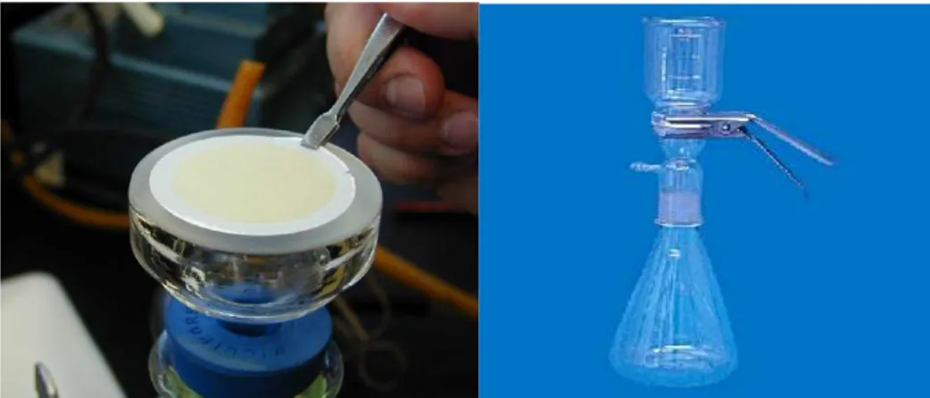
Presence or absence of a known species can be monitored with species-specific probes using chemiluminescent detection and DNA dot blot techniques or, more sophisticated, with fluorescent in-situ hybridization (FISH). DNA microarrays (fig.11) or so-called DNA chips are one of the
Methodologies for the study of phytoplankton
Pag. 36
most powerful innovations in microbiology. Microarrays were introduced in the mid 1990s primarily for the detection and monitoring of gene expression (Schena et al. 1995; Schena et al. 1996). The application of sequences onto the surface of a glass slide with special surface properties in an ordered array is based on a minimized, but high throughput form of a dot-blot (Gentry et al. 2006; Ye et al. 2001). The DNA microarray experiment is performed by chip production, sample isolation and preparation, hybridization and data analysis. Prior to the hybridization, the target nucleic acid is labeled with a fluorescent dye, which can be incorporated directly to the nucleic acid or via indirect labeling of other substances (Cheung et al. 1999; Metfies et al. 2006; Southern et al. 1999).
Methodologies for the study of phytoplankton
Pag. 37
The hybridization pattern is captured via fluorescent excitation in a special device, the microarray scanner (Ye et al. 2001). One of the major drawbacks of this method is a possible cross-reaction from unspecific binding. The probes are designed to be specific to known sequences but there are a high number of unknown environmental sequences. As a consequence, species without a probe on the chip can also be overlooked (Gentry et al. 2006). Furthermore, the development of a functional chip is time-consuming, expensive and all probes on one chip need to work specifically under the same hybridization conditions (Boireau et al. 2005; Feriotto et al. 2002; Metfies et al. 2006).
3.3 Molecular marker
A molecular marker is a genetic trait of DNA or in the case of proteins (isozymes) used to distinguish between individuals or groups by the marker's different alleles ( Medlin and Koistra 2010).
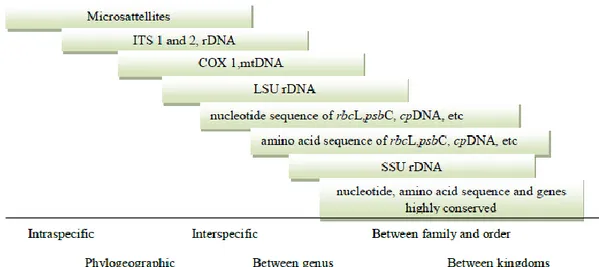
Photosynthetic organisms usually contain three different genomes: the nuclear, the plastid and the mitochondrial genomes (fig.12). Each has its own unique set of genes, each of which evolves at different rates.
Methodologies for the study of phytoplankton
Pag. 38
Microsatellites (MS) or simple sequence repeats (SSR), are the most powerful molecular markers available (Burke et al., 1991; Wright et al., 1994). Microsatellites are short sequences of one to six nucleotides, e.g., (CT)n or (CAG)n, that are repeated five to dozens and sometimes hundreds of times and are found in great abundance dispersed all over the genomes of all organisms investigated so far. This abundance together with the large number of alleles, resulting from high mutation rates because of their special, regular structure, makes them highly useful molecular markers at the population level (Alpermann 2009).
Distinction of individuals at the species level can be obtained using highly variable molecular markers, such as ITS sequences (internal-transcribed spacer) or microsatellites.
At higher taxonomic levels, slower evolving coding regions, such as the ribosomal RNA genes (small subunit or SSU, large subunit or LSU) and the large subunit of RUBISCO (ribulose 1,5-bisphosphate carboxylase,
rbcL), are commonly used, but other genes, such as the plastid psaA, psaB, psbC and tufA genes, are increasing (Medlin and Koistra 2010).
The SSU rRNA gene is often the gene of choice for cloning and is the gene most commonly used as a phylogenetic yardstick. The method allows the exhaustive description of biodiversity in a sample down to the species level. Also the resulting sequence information may serve as a basis for developing specific oligonucleotide probes necessary for subsequent methods like FISH (Woese 1987).
This molecule, besides the availability of a huge dataset (the current ARB database contains over 300,000 prokaryotic and eukaryotic sequences aligned by secondary structure), offers some advantages:
1. Universally present with the same function in all organisms;
2. Variable regions of conservation, which enables design of primers or probes to be designed;
Methodologies for the study of phytoplankton
Pag. 39
3. Many copies, which makes PCR easy or genome application for later PCR also easy;
4. No evidence for lateral gene transfer, so vertical descent is analyzed by phylogenetic methods (Woese 1987).
Also the resulting sequence information may serve as a basis for developing specific oligonucleotide probes necessary for subsequent methods like FISH. Probes are short oligonucleotides of normally 16-24 bp length that are hundred percent homologous only to a complementary sequence in a gene of the species of interest and differ by at least one position to all other organisms. In hybridization experiments, these probes can therefore be used to identify species of interest by binding to the target's sequence and later detection by a probe-attached label, e.g., Digoxigenin (DIG) or a fluorochrome like Fluorescein (Toebe 2013). The next section reports extensively about this technique.
3.4 Fluorescent In Situ Hybridization (FISH) and Whole Cell-FISH (WC-FISH)
In situ hybridization is a powerful technique that enables the
visualization of nucleic acid probes on target tissue, cells, nuclei and chromosomes, so that the location of the nucleic acid (DNA, RNA) can be determined as in vivo. The ability to detect nucleic acid in situ enables: construction of physical maps of chromosomes; analysis of chromosome structure and aberrations; determination of the spatial and temporal expression of genes (Toebe 2013)
Understanding in situ hybridization requires knowledge of molecular biology, genetics, immunochemistry ad histochemistry.
Methodologies for the study of phytoplankton
Pag. 40
The main steps are outlined in figure 13 and initially involve the preparation of biological material and the labeling of a nucleic acid sequence to from the probe. Labeling a nucleic acid involves the incorporation of either a radioactive or non-radioactive marker which can be detected. Both probe and material are then denatured to make all nucleic acid single- stranded. Finally the sites of hybridization are detected and visualized; the detection methods depend on the type of label attached to the probe.
Fig.13 Basic steps of fluorescence in situ hybridization. The sample is first fixed to stabilize the cells and permeabilize the cell membranes. The labelled oligonucleotide probe is then added and allowed to hybridize to its intracellular targets before the excess probe is washed away. The sample is then ready for single-cell identification and quantification by either epifluorescence microscopy or flow cytometry ( Aman and Fuchs 2008).
Two types of non- radioactive in situ hybridization procedures can be distinguished: the direct and the indirect methods. In the direct method the
Methodologies for the study of phytoplankton
Pag. 41
label that has been incorporated into the nucleic acid can be visualized directly once in situ hybridization has been completed. The recently introduced fluorochrome- labelled nucleotides (fluorescein isothiocyanate FITC, rhodamine TRITC, Cyanine Cy3), which can be enzymatically incorporated into the nucleid acid, are becoming extremely important because of the simplicity of detection.
Successful in situ hybridization experiment require that the material is adequately preserved and that the target sequences and tissue morphology are maintained. In addition, the tissue has to be permeable to probe and detection reagents. Once harvested the material should be fixed quickly to minimize endogenous nuclease activity and other degradation processes. Rapid fixation is particularly important when RNA is to be detected because RNA is very sensitive to the degrading activity of RNase. The fixation step serves to preserve tissue morphology and minimize loss of nucleic acids. Two categories of fixative are predominantly used: cross-linking fixative (glutaraldehyde, formaldehyde) or protein- precipitating fixatives (ethanol or methanol mixed with acetic acid). The fixative chosen depends on the material and probe being used, the method of imaging probe hybridization sites and the level of sensitivity required.
The in situ hybridization reaction exploit the kinetics of nucleic acid duplex formation. Nucleic acid duplexes form by hydrogen bonding between two complementary nucleic acid strands. The stability of the duplex under defined conditions can be determined by calculating its melting temperature (Tm). The Tm is the temperature at which one-half of the duplex molecules become dissociated or “melted” into single strands.
The stability of the hybrid nucleic acid is dependent on a number of factors:
Methodologies for the study of phytoplankton
Pag. 42 The proportion of guanine and cytosine (%GC). GC pairs are more stable than AT pairs because they are bonded together by three as opposed to two hydrogen bonds. For this reason, DNA with a high GC content is more stable than DNA which is rich in AT, and more energy is required to separate the two strands.
Length of hybrid nucleic acid. In general, a long duplex is more stable than a shorter one because it is held together by more hydrogen bonds. Oligonucleotide probes have fewer hydrogen bonds binding the duplex, and this reduces the duplex stability.
Typically, labeled nucleic acid probes are made up in a hybridization mix containing formamide , salts, dextran sulphate, with optional incorporation of blocking DNA or tRNA, sodium dodecyl sulphate and bovine serum albumin. Formamide is used in denaturation and hybridization solution to enable a reaction temperature that is not damaging to tissue morphology. Formamide also regulates stringency. Salts in solution are used to regulate the ionic strength of hybridization and denaturation solution and to help stabilize the nucleic acid duplex. Dextran sulphate can increase the hybridization reaction rate by factor of 3. It functions by forming a matrix in the hybridization mixture which concentrates the probe without affecting the stringency. Unlabelled blocking DNA or RNA is included to block probe hybridization to non- specific sites. Sodium dodecyl sulphate (SDS) helps in probe penetration by acting as a wetting agent. Bovine serum albumin (BSA) can reduce some non-specific probe hybridization.
The precise amount of target nucleic acid available for in situ hybridization is difficult to assess because of unknown effects of the nucleic acid conformation and its interaction with associated molecules, particularly protein. The rate hybridization depends on the probe length,
Methodologies for the study of phytoplankton
Pag. 43
complexity of sequence and concentration. In general, long probes may result in a slower rate because of limited diffusion into the material.
Post-hybridization washes are usually carried out in a slightly more stringent solution that the hybridization mixture to denature and remove weakly bound probe, leaving only perfectly or nearly perfectly matched nucleotides in the duplex.
FISH with rRNA-targeted oligonucleotide probes was first used for identification, localization, and quantification of defined microbial populations in complex samples (Amann et al.,1995). The thousands of ribosomes provide enough targets for probe binding and therefore, strong enough signals to be detected. In picoplankton and also in bacterial cells, which often show weaker signals because of their small size and therefore lower ribosome content, techniques like catalyzed reporter deposition– fluorescence in situ hybridization (CARD-FISH) can be used to boost the signal strength up to a detectable level (Schönhuber et al., 1997; 1999).
Higher group level probes, species, or even strain specific oligonucleotide probes for FISH are available for many taxa and enable the differentiation of morphological similar co-occurring species, especially for harmful algal bloom (HAB) species ( Mikulski et al. 2008; Anderson et al. 2005; Miller and Scholin 1998; Chen 2011).
In these paper the FISH is applied to the whole algal cells and so we can refer as Whole Cell FISH. For the successful detection it is necessary to fix the cells on a filter or in a culture for preservation and to permeabilize the membrane before the application of probes. The advantages are definitely the rapid quantitative detection and visualization of algal species in a mixed field sample and the discrimination of closely related species or strains with even similar appearance (Godhe 2002;
Methodologies for the study of phytoplankton
Pag. 44
Groben et al. 2004; John et al. 2003a; John et al. 2005; Metfies et al. 2006). Furthermore the morphology of the cells is conserved and thus also different external life stages can be recognized (Godhe 2002).
One drawback of this method is that cellular rRNA content may vary under different environmental conditions and could have an impact on the fluorescence signal. Further challenges are the autofluorecence of photosynthetic cells, non-specific binding of probes, difficulties with penetration of thick cell walls of resting cysts and non-stability of rRNA molecules (DeLong 1998; Garcés et al. 1998; Godhe 2002; Medlin and Simon 1998; Rice et al. 1997).
In the last years , several works have performed WC-FISH for detection of HAB specie. Among these species, notably members belonging to the dinoflagellate genera Alexandrium (Toebe et al. 2013a; Tang et al. 2012; Touzet et al. 2011; Hosoi-Tanabe and Sako 2006; Anderson et al. 2005),
Prorocentrum (Hou et al. 2009), Cochlodinium (Mikulski et al. 2008) and Karenia (Mikulski et al. 2005), or species of the diatom genus Pseudo-nitzschia (Parsons et al. 1999; Rhodes et al. 1998; Miller and Scholin
1998), as well as members of the haptophyte genus Prymnesium (Toebe et al. 2006) were monitored by FISH analysis in their marine environment.
3.5 Molecular probes ribosomal RNA
The continually growing number of available algal 18S and 28S rDNA sequences, e.g. from GenBank (Benson et al. 2011), makes it possible to design rRNA oligonucleotide probes which specifically target the SSU (or 18S) or LSU (or 28S) ribosomal DNA (rDNA) from higher group down to species level (Groben et al. 2004; Lange et al. 1996).
Methodologies for the study of phytoplankton
Pag. 45
The rRNA provides some characteristics, which predestines this molecule as a phylogenetic marker. First of all, the rRNA is an integrated part of the ribosome, the protein factory of each cell, and has the same function in every organism. The rRNA molecules must have developed in early stages of life and thus are evolutionarily conserved even in its two- and three-dimensional structures. Although the primary structure is highly conserved throughout all organisms, some regions in the rRNA are more conserved than others (Thiele et al., 2011).
The conserved sequence stretches on the rRNA provide a variety of technical advantages, it is highly abundant in cells, making it suitable as a target for in situ hybridization studies. Also the resulting sequence information may serve as a basis for developing specific oligonucleotide probes necessary for subsequent methods like FISH. Probes are short oligonucleotides of normally 16-24 bp length that are hundred percent homologous only to a complementary sequence in a gene of the species of interest and differ by at least one position to all other organisms. In hybridisation experiments, these probes can therefore be used to identify species of interest by binding to the target's sequence and later detection by a probe-attached label, e.g., Digoxigenin (DIG) or a fluorochrome like Fluorescein.
The full-cycle rRNA approach was first developed as a phylogeny-based toolbox for cultivation-independent studies of microbial diversity and ecology. After DNA extraction, the 16S rRNA genes are amplified by polymerase chain reaction (PCR) using conserved primers. The amplified rRNA genes are singularized by cloning in a plasmid vector and transformation in competent Escherichia coli cells. They are then sequenced and submitted to sequence databases. Comparative sequence analysis is the basis for the design of oligonucleotide probes. Finally, these
Methodologies for the study of phytoplankton
Pag. 46
probes can be applied to environmental samples using FISH techniques (Amann et al., 1995).
For FISH, a probe length of 15–25 nucleotides, most often 18 nucleotides is common. The GC content of a newly designed probe should be between 50% and 70%, since a higher GC content could result in unspecific binding. The GC content of probe sequences influences their melting behavior (Thiele et al., 2011). A problem that should be considered during the design of FISH probes is target site accessibility (Fuchs et al., 2001). Although the secondary structure of the ribosome is highly conserved and a consensus map could be developed from the 16S rRNA accessibility studies, each probe should be checked on their respective target group of organisms to ensure high probe signals (Behrens et al., 2003). In the last step of synthesis, a fluorochrome is added to the 5I end of the oligonucleotide. A range of fluorochromes is available for the labeling of nucleic acids. Standard labels for in situ hybridization are the green fluorescein and the red tetramethylrhodamine derivatives (Tab.3).
The optimal hybridization conditions need to be established to guarantee high specificity and good sensitivity of the probe. For this, a series of hybridizations is performed at increasing stringency either by increasing the temperature of hybridization or by increasing the concentrations of a denaturing agent such as formamide in the hybridization buffer.
Methodologies for the study of phytoplankton
Pag. 47
Table 3 Dye labels frequently used for oligonucleotide probes and their characteristics. Excitation (±10 nm) Emission (±10 nm) Molecular weight (Da) ɛ (mol-1 cm-1) Carboxy-fluorescein (FAM) 492 518 376 79 000 Fluorescein 490 520 389 77 000 Alexa 488 494 517 643 71 000 Atto 488 501 523 90 000 CY3 512/552 565/615 766 150 000 Alexa 546 554 570 1079 112 000 Carboxytetramethyl-rhodamine (TAMRA) 540 565 466 91 000 Alexa 594 590 617 820 92 000 Atto 590 (rhodamine-derivative) 594 624 12 000 CY5 625–650 670 792 250 000 Alexa 647 651 672 1250 270 000
Materials and methods
Pag. 48
4 Materials and methods
4.1 Algal cultures and culture conditions
Pure cultures of P. dolorosa, P. arenysensis, P. autumnalis, P.
delicatissima, P.galaxia, P. fraudolenta, P. manni, P. pseudodelicatissima,
and P. multisriata employed in this study were kindly provided from Stazione Zoologica “Anton Dohrn” (Naples). All cultures were grown in 50 ml flasks containing 35 ml Guillard’s f/2 medium (Guillard, 1975) (Table 4). The cultures were kept at 22°C on a 16:8 h light:dark cycle with light provided by cool white fluorescent tubes at a photon flux density of 100μmol m-2 s-1.
Tab.4 Composition of Guillard’s f/2 medium.