Research Article
Antioxidant Treatment Reduces Formation of Structural
Cores and Improves Muscle Function in RYR1
Y522S/WT
Mice
Antonio Michelucci,
1,2Alessandro De Marco,
1,2Flavia A. Guarnier,
3Feliciano Protasi,
1,4and
Simona Boncompagni
1,21Center for Research on Aging and Translational Medicine (CeSI-MeT), University G. d'Annunzio of Chieti, 66100 Chieti, Italy
2Department of Neuroscience, Imaging, and Clinical Sciences (DNICS), University G. d'Annunzio of Chieti, 66100 Chieti, Italy
3Department of General Pathology, University Estadual de Londrina, 86057-970 Londrina, PR, Brazil
4Department of Medicine and Aging Science (DMSI), University G. d'Annunzio of Chieti, 66100 Chieti, Italy
Correspondence should be addressed to Simona Boncompagni; [email protected] Received 6 April 2017; Accepted 13 June 2017; Published 10 September 2017
Academic Editor: Marko D. Prokić
Copyright © 2017 Antonio Michelucci et al. This is an open access article distributed under the Creative Commons Attribution License, which permits unrestricted use, distribution, and reproduction in any medium, provided the original work is properly cited.
Central core disease (CCD) is a congenital myopathy linked to mutations in the ryanodine receptor type 1 (RYR1), the sarcoplasmic
reticulum Ca2+release channel of skeletal muscle. CCD is characterized by formation of amorphous cores within musclefibers,
lacking mitochondrial activity. In skeletal muscle of RYR1Y522S/WTknock-in mice, carrying a human mutation in RYR1 linked
to malignant hyperthermia (MH) with cores, oxidative stress is elevated and fibers present severe mitochondrial damage and
cores. We treated RYR1Y522S/WTmice with N-acetylcysteine (NAC), an antioxidant provided ad libitum in drinking water for
either 2 or 6 months. Our results show that 2 months of NAC treatment starting at 2 months of age, when mitochondrial and fiber damage was still minimal, (i) reduce formation of unstructured and contracture cores, (ii) improve muscle function, and
(iii) decrease mitochondrial damage. The beneficial effect of NAC treatment is also evident following 6 months of treatment
starting at 4 months of age, when structural damage was at an advanced stage. NAC exerts its protective effect likely by lowering
oxidative stress, as supported by the reduction of 3-NT and SOD2 levels. This work suggests that NAC administration is
beneficial to prevent mitochondrial damage and formation of cores and improve muscle function in RYR1Y522S/WTmice.
1. Introduction
Central core disease (CCD), one of the most common human congenital myopathies, is an inherited neuromuscular dis-order characterized by hypotonia and proximal muscle weakness, which cause motor developmental delay in chil-dren [1, 2]. Diagnosis of CCD is confirmed by histological examination of muscle biopsies showing amorphous cen-tral areas (i.e., cores) lacking glycolytic/oxidative enzymes and mitochondria [3]. Disorganization of contractile and sarcotubular systems is also typical in cores [4]. To date, management of patients/children is essentially supportive and based on physiotherapic approaches and no curative treatments are available. Hence, a deeper understanding of the molecular mechanisms underlying mitochondrial
damage and formation of cores in CCD is needed to
develop effective therapeutic interventions.
In humans, about 90% of CCD cases are linked to muta-tions in the ryanodine receptor type 1 (RYR1) gene [5],
encoding for the sarcoplasmic reticulum (SR) Ca2+ release
channel of skeletal muscle. RYR1 is a large protein of about 2200 kDa specifically localized in calcium release units (CRUs), the intracellular junctions formed by the close appo-sition of transverse-tubules (TT) to the SR. RYR1 in CRUs is part of macromolecular complex deputed to excitation-contraction (EC) coupling, the mechanism that allows
trans-duction of the action potential into Ca2+release from the SR
[6, 7]. Mutations in RYR1 gene, which causes abnormalities
in the opening probability of the Ca2+channel, are also often
associated to malignant hyperthermia (MH) susceptibility,
Volume 2017, Article ID 6792694, 15 pages https://doi.org/10.1155/2017/6792694
an inherited pharmacogenetic subclinical myopathy, char-acterized by a life-threatening hypermetabolic response to commonly used halogenated/volatile anesthetics (i.e.,
halo-thane, isofluorane) [8, 9]. An association between CCD
and MH exists as individuals with MH may have muscle biopsies with cores [10, 11], while CCD patients may be at risk for hyperthermic episodes during anesthetic
proce-dures, as also confirmed by in vitro caffeine-halothane
contracture testing (either IVCT, in vitro contracture test, or the CHCT, caffeine-halothane contracture test), per-formed on muscle biopsies [2, 12–14].
In the 2006, an animal model (RYR1-Y522S knock-in mice) carrying a human gain-of-function mutation associated to MH, skeletal muscle weakness, and formation of cores was generated and characterized [10]. Heterozygous Y522S mice
(RYR1Y522S/WT) suffer lethal MH crises when acutely exposed
to both anesthetics and heat [15, 16] and develop structural
cores [17]. Muscles of RYR1Y522S/WTmice also exhibit a marked
temperature-dependent increase in resting myoplasmic Ca2+,
excessive oxidative stress [16], and enhanced mitochondrial
superoxideflashes activity [18]. The current view of the
molec-ular mechanisms underlying the phenotype of RYR1Y522S/WT
mice is that the Y522S mutation promotes a significant increase of the opening probability of the RYR1 channel,
which causes SR Ca2+leak and overproduction of reactive
oxy-gen and nitrooxy-gen species (ROS and RNS), as a consequence of
the increased Ca2+-dependent mitochondrial activity. In turn,
excessive ROS and RNS would determine nitrosylation/glu-tathionylation of RYR1, oxidative modifications responsible of further increase of RYR1 opening probability [16]. The feed-forward mechanism triggered by ROS/RNS would in principle play a pivotal role: (i) acutely, in anesthetic- and heat-induced lethal MH episodes and (ii) chronically, in mito-chondrial damage and development of cores, two types of structural alterations that resemble CCD in humans [17].
Here, we treated RYR1Y522S/WT mice with
N-acetylcysteine (NAC), a potent antioxidant provided ad libitum in drinking water (1% w/v) for either 2 months (2–4 months of age, starting when mitochondrial and fiber damage was still minimal) or 6 months (4–10 months of age; starting when structural damage was already at an advanced stage) [17], and evaluated the effect of this pharmacological treatment on formation of cores, muscle function, mitochon-drial damage, and levels of oxidative stress in extensor digi-torum longus (EDL) muscles. The results collected in this
study indicate that NAC administration is beneficial to
prevent/reduce mitochondrial damage and formation of
cores and improve muscle function in RYR1Y522S/WT mice.
2. Materials and Methods
2.1. RYR1Y522S/WTMice. Heterozygous Y522S mice were
gen-erated as previously described [15]. Mice were housed in
microisolator cages at 20°C in a 12 hr light/dark cycle and
provided free access to water and food. Animals used in this study were all males, and analyses were carried out in EDL muscles. All experiments were conducted according to the Directive of the European Union 2010/63/UE, and animal protocols were approved by the Committee on the Ethics of
Animal Experiments of the University of Chieti (15/2011/ CEISA/COM). All surgeries were made to minimize animal suffering; animals were anesthetized and then sacrificed by cervical dislocation.
2.2. NAC Treatment. RYR1Y522S/WT mice were randomly
assigned to the experimental groups: untreated or
NAC-treated RYR1Y522S/WT mice. NAC (Sigma-Aldrich, Milan,
Italy) was provided in vivo in drinking water at thefinal
con-centration of 1% weight/volume (1% w/v) as previously described [19] for either 2 months (from 2–4 months of age) or 6 months (from 4–10 months of age).
2.3. Preparation and Analysis of Samples in Light and
Electron Microscopy (EM). EDL muscles were dissected,fixed
at room temperature with 3.5% glutaraldehyde in 0.1 M
NaCaCo buffer (pH 7.2), and kept at 4°C infixative until
fur-ther use. Small bundles offixed muscles were then postfixed,
embedded, stained en-block, and sectioned as previously
described [17, 20]. For EM, ultrathin sections (~50 nm) were
examined after staining in 4% uranyl acetate and lead citrate, with a Morgagni Series 268D electron microscope (FEI Company, Brno, Czech Republic), equipped with Megaview III digital camera (Olympus Soft Imaging Solutions GmbH, Munster, Germany) at 60 kV. For histology, 700 nm thick sections were stained in a solution containing 1% Tolui-dine blue O and 1% sodium borate tetra in distilled water
for 3 min on a hot plate at 55–60°C. After washing and
dry-ing, sections were finally mounted with mounting medium
(DPX Mountant for histology, Sigma-Aldrich, Milan, Italy). 2.4. Quantitative Analysis in Histology and EM Preparations. Consider the following:
(1) Histological sections were examined under direct illumination and/or phase contrast optics with a
Leica DMLBfluorescence microscope (Leica
Micro-system, Vienna, Austria), and individual EDLfibers
were visually scored for the presence of either unstructured or contracture cores as in [17]. (2) Number/area, size, and volume of mitochondria (and
number/area of CRUs and mitochondria CRU pairs)
were determined in EDL fibers as follows: (a)
Mitochondrial volume was determined using the well-established stereology point-counting technique [21, 22] in micrographs taken from transversal sec-tions at magnification 7100x. Briefly, after superim-posing an orthogonal array of dots at a spacing of
0.20μm to the electron micrographs, ratio between
numbers of dots falling within mitochondrial profiles
and total number of dots covering the whole image
was used to calculate the relativefiber volume
occu-pied by mitochondria. (b) Number of severely dam-aged mitochondria was counted in longitudinal sections, and their frequency was reported as an
aver-age number in 100μm2 and as % of mitochondria
evaluated. Mitochondria were classified as severely damaged as previously described [17]. (c) Average
mitochondria was measured in longitudinal sections using the analysis software of the EM digital camera (Olympus Soft Imaging Solutions GmbH, Munster, Germany). Severely damaged mitochondria, included in (b), were excluded from this analysis. (d) Number/ area of CRUs, mitochondria, and mitochondria CRU pairs was also evaluated in longitudinal sections and
reported as the average number in 100μm2.
2.5. Immunolabeling and Confocal Microscopy (CM). EDL
muscles were dissected from sacrificed mice and fixed with
paraformaldehyde 2% for 1-2 hrs at room temperature. Small bundles of muscles were processed for double immunostain-ing as previously described [23]. Primary antibodies used (a) mouse monoclonal anti-RyR1/RyR3 34C (1 : 20) (Develop-mental Studies Hybridoma Bank, University of Iowa, Iowa, USA) and (b) rabbit polyclonal antimitochondrial preprotein translocases of the outer membrane, TOM20 (1 : 50) (Santa Cruz Biotechnology, Inc., Dallas, TX, USA). Secondary anti-bodies used (a) Cy5-labeled goat anti-mouse IgG and (b) Cy3-labeled goat anti-rabbit IgG (Jackson ImmunoResearch Laboratories, West Grove, PA, USA). Images were acquired using a Zeiss LSM510 META laser-scanning confocal micro-scope system (Zeiss, Jena, Germany) equipped with Zeiss Axiovert 200 inverted microscope and a Plan Neofluar oil-immersion objective (63X/1.3 NA). Negative controls for each immunostaining assay were performed by immu-nolabeling of samples with only secondary antibodies. 2.6. Quantitative Plasma and Serum Analyses. Blood levels of creatine kinase (CK) and lactate dehydrogenase (LDH),
markers offiber damage, were spectrophotometrically
mea-sured in serum (CK) and plasma (LDH) samples obtained from mice as previously described [19], by using a Screen Touch Master spectrophotometer (Hospitex Diagnostic, Sesto Fiorentino, Italy).
2.7. Calpain Activity. The activity of calpain was measured in total hind limb muscle homogenates, by a chemilumines-cence assay using a Calpain Protease Assay kit (Calpain-Glo Protease Assay®, Promega; Madison, WI, USA). The assay provides a proluminescent calpain substrate, in a buffer system optimized for calpain and luciferase activities. During the assay, calpain cleavage of the substrate generates a glow-type luminescent signal produced by the luciferase reaction. In this homogeneous, coupled-enzyme format, the signal is proportional to the amount of calpain activity present in the sample [24]. In this study, total hind limb muscle homog-enates were prepared at a concentration of 6.25 mg/mL in
10 mM KH2PO4 buffer, pH 7.4 in 0.9% NaCl and finally
processed according to the manufacturer’s instructions. Results are expressed as calpain activity/mg of muscle tissue. 2.8. Carbonyl Protein Content. Protein carbonyl group for-mations are classic and immediate biomarkers of oxidative
modification to proteins, which may promote
disorganiza-tion of contractile structures [25, 26]. 2,4-Dinitrophenylhy-drazine (DNPH) tagging of protein carbonyls has been one of the most common measures of oxidative stress and
consequent protein damage. Carbonyl protein content was measured as previously described [25], with modifications. Briefly, a mix of total hind limb muscles (50 mg/mL) was homogenized in 50 mM phosphate buffer, 1 mM ethylenedia-mine tetraacetic acid, pH 7.4, and tissue samples were
centri-fuged at 600g/10 min/4°C. A volume of 200μL of DNPH was
added to 200μL of supernatant and incubated at room
tem-perature. After a 30 min incubation, 100% trichloroacetic acid (TCA) was added and samples were placed on ice for 5 min and then spinned at maximal speed for 2 min. Super-natants were discarded without disturbing pellets, which
were washed in cold acetone and placed at−20°C for 5 min.
Then, acetone was carefully removed, and pellets were dis-solved in 0.5 mL 6 M guanidine hydrochloride to be read at 375 nm. To calculate the protein carbonyl content, the
fol-lowing formula was used: C = [(OD 375 nm)/6.364× (100)]
nmol/well, where 6.364 is the extinction coefficient using the enclosed 96-well plates in mM (=22 mM-1
cm-1× 0.2893 cm path length in well). Results were expressed
as nmol carbonyl/mg of total protein, which were quantified in each sample at 280 nm.
2.9. Western Blot Analyses. For assessment of 3-nitrotyrosyne (3-NT) content, mixed muscles from hind limb were homog-enized on ice in a buffer containing 50 mM Tris-HCl, pH 7.4; 1% NP-40; 0.25% sodium deoxycholate; 150 mM NaCl; 1 mM EDTA; 1 mM PMSF; protease inhibitors. After
centri-fugation at 10000g for 15 min at 4°C, supernatants were
collected and total protein concentration was determined using Bio-Rad Protein assay (Bio-Rad laboratories, CA).
40μg of total proteins was separated by SDS-PAGE in a
10% polyacrylamide gel, followed by western blotting using anti 3-NT mouse monoclonal antibody (1 : 500, Merk Millipore, Milan, Italy) and a horseradish peroxidase-(HRP-) conjugated anti-mouse secondary antibody (Merck Millipore, Darmstadt, Germany). Visualization and
densito-metric quantification of signals were done using the imaging
system Alliance Mini 4 with Alliance 1D MAX software (UVItec, Cambridge, UK). After measuring 3-NT, the
membranes were stripped by Tris/SDS buffer with
2-mercaptoethanol. After blocking, the membranes were incu-bated with anti-glyceraldehyde-3-phosphate dehydrogenase (GAPDH) antibody (OriGene, Rockville, MD, USA) for normalization to the protein content within each band.
For assessment of Cu/Zn-superoxide dismutase (SOD1) and Mn-superoxide dismutase (SOD2) protein levels, west-ern blot experiments were performed as follows: mixed
mus-cles from hind limb were homogenized on ice in RIPA buffer
(50 mM Tris-HCl, pH 8.0; 150 mM NaCl; 0.5% sodium deox-ycholate; 0.1% SDS; 1% NP-40; 0.1 mM PMSF; protease
inhibitors). Homogenates were centrifuged at 10000g for
15 min at 4°C, supernatants were collected, and total protein
concentration was determined using Bio-Rad Protein assay (Bio-Rad laboratories, Hercules, CA, USA). Protein samples
(5μg), solubilized in 2× sample buffer (125 mM Tris-HCl,
pH 6.8; 4% SDS; 20% glycerol; 0.004% bromophenol blue; 10% 2-mercaptoethanol), were loaded on a 12% acrylamide gel, separated by SDS-PAGE, and transferred to nitrocellu-lose membrane. Membranes were probed using primary
antibodies against SOD1 (1 : 1000, Santa Cruz Biotechnology, Dallas, TX, USA), SOD2 (1 : 2000, Santa Cruz Biotechnology, Dallas, TX, USA), and GAPDH (1 : 10000, OriGene,
Rock-ville, MD, USA) overnight at 4°C. HRP-conjugated
anti-mouse or rabbit (1 : 10000, Merck Millipore, Darmstadt, Germany) was used as a secondary antibody, and peroxidase activity was detected using an enhanced chemiluminescence (ECL) kit (Perkin Elmer, Waltham, MA, USA). The bands were visualized, and densitometric quantification of signals was performed using the imaging system Alliance Mini 4 with Alliance 1D MAX software (UVItec, Cambridge, UK). 2.10. Grip Strength Test. Strength developed by mice during instinctive grasp was measured as previously described [27].
Briefly, mice were held by the tail and allowed to firmly
grasped to a metal grating, connected to the shaft of a Shimpo Fgv 0.5X force transducer (Metrotec Group, Lezo, Spain), with fore and hind limbs before a gentle pull was exerted on the tail. Measurements of peak force generated by each mouse were repeated three times with appropriate intervals (at least 30 sec) to avoid fatigue, and the highest value of peak force (normalized to total body mass) measured before each experiment was used.
2.11. Force and Contraction Kinetics of Intact EDL Muscles.
EDL muscles were dissected from WT, untreated
RYR1Y522S/WT, and NAC-treated RYR1Y522S/WT mice and placed in a dish containing Krebs solution with the following
composition in mM: 118 NaCl, 4.7 KCl, 1.2 MgSO4, 2.5
CaCl2, 1.2 KH2PO4, 25 NaHCO3, and 11 glucose. Individual
EDLs were then pinned, tied with fine silk sutures at each
end, and mounted vertically between two platinum
elec-trodes immersed in an organ chamberfilled with Krebs
solu-tion and attached to a servo motor and force transducer
(model 1200A, Aurora Scientific, ON, Canada). Temperature
was kept between 23–25°C. Before starting the experimental
protocol, stimulation level and optimal muscle length (L0)
were determined using a series of 80 Hz-tetani in order to stretch the muscle to the length that generated maximal
force (F0). After optimization of the stimulation
condi-tions, EDL muscles were subjected to a force-frequency protocol based on a series of train pulses of 500 ms dura-tion each as follows (in Hz): 1, 5, 10, 20, 40, 60, 80, 100, 120, and 140. After 5 min at rest, the same EDL muscles were subjected to a single-sustained high frequency tetanus (120 Hz, 2 sec). Muscle force was recorded using a dynamic muscle control (DMC) software and analyzed using dynamic muscle analysis (DMA) software (both from: Aurora
Scien-tific, ON, Canada). Specific force (mN/mm2) was calculated
by normalizing the absolute force (mN) to the cross sectional
area (CSA, mm2) obtained as the following: muscle wet
weight (mg)/L0(mm)∗ 1.06 (mg/mm3).
2.12. Statistical Analyses. Statistical significance for the
quan-titative analysis offibers presenting structural alterations (i.e.,
unstructured and contracture cores) was evaluated using
two-tailed Fisher’s exact test. One-way ANOVA followed by post
hoc Tukey test was used for statistical analyses of all other experiments except for those regarding the time courses of
in vivo grip strength and the force-frequency of intact EDL muscles, in which repeated measures ANOVA was used followed by post hoc Tukey test for the pairwise compari-sons. In all cases, differences were considered statistically
significant at p < 0 05. Two-tailed Fisher’s exact tests were
performed using GraphPad software, whereas one-way ANOVA and repeated measures ANOVA were performed using Origin 8.0 software.
3. Results
The effect of NAC treatment on structure, function, and
oxi-dative stress levels of EDL muscles from RYR1Y522S/WTmice
was evaluated at (a) 4 months of age after 2 months of
treat-ment (starting when mitochondrial and fiber damage was
still minimal) and at (b) 10 months of age after 6 months of treatment (starting when mitochondrial damage was already at an advanced stage) [17]. In the manuscript, we will refer to 2 and 6 months of treatment as short-term and long-term NAC treatments, respectively.
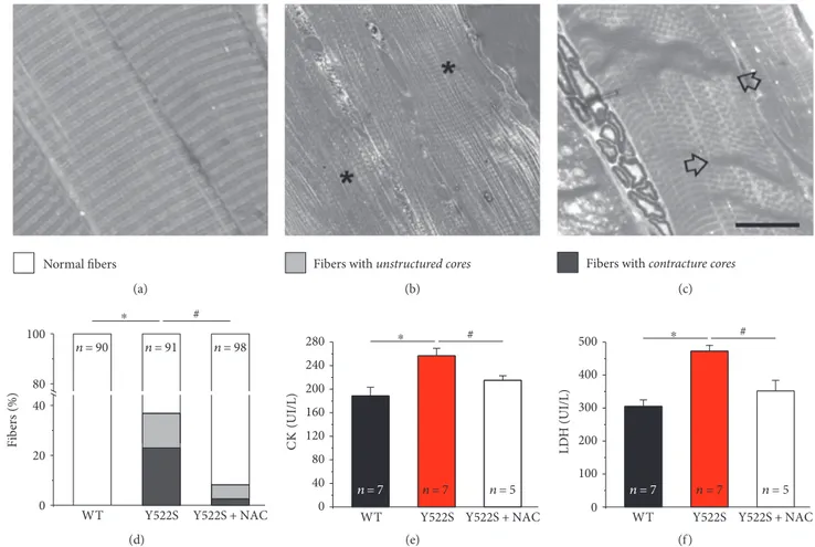
3.1. NAC Treatment Reduces Formation of Structural Cores, Fiber Damage, and Proteolytic Degradation in EDL Muscles
of RYR1Y522S/WT Mice. Skeletal fibers from RYR1Y522S/WT
mice develop unstructured and contracture cores [17]. To
evaluate the effect of NAC in reducing formation of cores,
using histological sections, we analyzed and classified
fibers in three different categories, as previously described
[17]: (a) normal fibers, presenting a well-preserved cross
striation pattern (Figure 1(a)); (b) fibers with unstructured
cores, presenting extensive areas lacking cross striation
(Figure 1(b), asterisks); and (c) fibers with contracture
cores, exhibiting areas of extreme sarcomere shortening (Figure 1(c), arrows). Quantitative analysis of the relative
percentage offibers, presenting the different features,
indi-cates that NAC treatment was effective in preventing the
formation of structural cores (Figure 1(d) and Table S1
available online at https://doi.org/10.1155/2017/6792694). Specifically, unstructured and contracture cores, which are
not present in WTfibers, were found, respectively, in 14%
and 23% of fibers in RYR1Y522S/WT EDL muscles
(Figure 1(d)). However, following NAC treatment, the
num-ber of RYR1Y522S/WTEDLfibers containing unstructured and
contracture cores was significantly reduced to 6% and 3%,
respectively (Figure 1(d)). The decrease in the number of fibers containing cores results in a parallel increase in the
percentage of normal fibers (from 63 to 92%). Data plotted
in Figure 1 are available in Table S1.
Asfiber damage results in changes in blood parameters,
we performed biochemical measurements of markers of muscle damage in blood samples. Specifically, we evaluated serum and plasma levels of the muscle-specific isoforms of creatine kinase (CK, in serum) and lactate dehydrogenase (LDH, in plasma) [28]. Consistent with the higher percentage
of fibers presenting structural alterations, in RYR1Y522S/WT
samples, both CK and LDH levels were about~30% higher
than those in aged-matched WT mice (Figures 1(e) and 1(f )). The protective effect of NAC was confirmed by these experiments: 2 months of NAC treatment was effective in
reducing the amount of both CK and LDH in blood samples
from RYR1Y522S/WT mice to values closer to that of WT
(Figures 1(e) and 1(f )), a result consistent with the significant
reduction in the incidence offibers presenting unstructured
and contracture cores (Figure 1(d)).
We also analyzed EDL muscles using a combination of immunofluorescence for CM and EM and assessed proteo-lytic degradation by measuring calpain activity and carbonyl
protein content (Figure 2). In adult EDL fibers from WT
mice, both CRUs, marked with an antibody that recognized RYR1, and mitochondria, marked with an antibody that labels translocase of outer mitochondrial membrane 20 homolog (TOM20), form double rows of cross striation
(Figure 2(a) and inset). Thisfluorescence pattern is
consis-tent with positioning of both CRUs and mitochondria at the I band, on either side of the Z-line (pointed by arrows in Figures 2(b) and 2(f ); see [20] for additional detail on the specific disposition of CRUs and mitochondria in adult
skeletal fibers). In RYR1Y522S/WT EDL fibers, this precise
cross striation pattern was often compromised (arrow in Figure 2(c) and inset). These areas lacking staining (pointed
by arrows in Figure 1(c)) reflect the presence of contracture
cores, visible in EM as regions with shortened sarcomeres
(arrow in Figure 2(d)). NAC treatment in RYR1Y522S/WT
mice restored the cross striation pattern in the large majority
of fibers (consistent with reduction of fibers with cores;
Figure 1(d)): both red and green staining were perfectly transversal (Figure 2(e)), while ultrastructure of myofibrils and sarcomeres at the EM examination was virtually
indis-tinguishable from that of WT fibers (compare Figures 2(b)
and 2(f )).
The RYR1-Y522S mutation causes leak of Ca2+from the
SR [16]. As excessive myoplasmic Ca2+concentration
acti-vates calpains that cleave a variety of substrates, including myofibrillar proteins [24, 29, 30], calpain-mediated degrada-tion could contribute to the ultrastructural alteradegrada-tions
observed in RYR1Y522S/WTmusclefibers. We measured the
total calpain activity, which was markedly elevated in
muscles from RYR1Y522S/WT compared to that from WT
(Figure 2(g)), but lowered to levels very similar to those from WT following NAC treatment (Figure 2(g)). In addition, we also evaluated the total carbonyl protein content in the same muscle homogenates, an important biomarkers
of oxidative modification of proteins [25, 26]. Also in this
Normal fibers
(a)
Fibers with unstructured cores
(b)
Fibers with contracture cores
(c)
0 20 40
WT Y522S Y522S+NAC 100 n=90 n=91 n=98 Fib er s (% ) 80 # ⁎ (d) 0 40 80 120 160 200 240 280 C K (UI/L)
WT Y522S Y522S+NAC
n = 7 n=7 n=5 # ⁎ (e) 0 100 200 300 400 500 LD H (UI/L)
WT Y522S Y522S + NAC
n = 7 n = 7 n = 5
# ⁎
(f )
Figure 1: Structural cores and blood markers of muscle damage (CK and LDH) at 4 months of age. (a–c) Representative histological images of
normalfibers (a), fibers with unstructured cores (b), and fibers with contracture cores (c). (d) Percentage of EDL fibers presenting the features
classified in (a–c) (white: normal fibers; grey: fibers with unstructured cores; and dark grey: fibers with contracture cores). See also Table S1.
(e and f) Serum levels of creatine kinase (CK) and lactate dehydrogenase (LDH). In (e) and (f ), data are given as mean± SEM;∗p < 0 05, WT
versus RYR1Y522S/WTmice;#p < 0 05, untreated RYR1Y522S/WTversus NAC-treated RYR1Y522S/WTmice. In (d), n = number offibers analyzed;
case, the total amount of carbonyl proteins, which was
abnormally elevated in RYR1Y522S/WT, was lowered of
about 20% by NAC treatment, although it remained still higher than that in WT (Figure 2(h)).
3.2. NAC Treatment Improves In Vivo Grip Strength and Ex
Vivo Muscle Contractile Function in RYR1Y522S/WTMice. As
NAC treatment was very effective in protecting muscle fibers
of RYR1Y522S/WT mice from structural damage, we
per-formed in vivo grip strength test and ex vivo contractile experiments on intact EDL muscles, to evaluate whether NAC was also able to improve muscle function of RYR1Y522S/WTknock-in mice. Grip strength was evaluated at 3 different time points between 2 (beginning of the short-WT RYR1 TOM20 (a) WT (b) Y522S RYR1 TOM20 (c) Y522S (d) RYR1 TOM20 Y522S + NAC (e) Y522S + NAC (f ) WT C al pa in ac tivi ty/ m g t issue ( ×10 3) n = 4 n = 4 n = 3
Y522S Y522S + NAC 25 20 15 10 5 0 ⁎ # (g) C arb on yl co nt en t/ mg tot al prote ins n = 4 n = 4 n = 3 0 6 5 4 3 2 1
WT Y522S Y522S + NAC
⁎ #
(h)
Figure 2: Fiber ultrastructure, calpain activity, and carbonyl protein content at 4 months of age. (a–f) Representative immunofluorescence
and EM images showingfluorescent cross striation (a, c, and e) and myofibrillar/sarcomeric organization (b, d, and f) in EDL fibers. In (b) and
(f ), small arrows point at Z-lines, while in (c and d) large arrows point to areas in which cross striation and sarcomeric structure are compromised. (g) Calpain activity expressed in total hind limb muscle homogenates. (h) Carbonyl protein content in EDL muscle
homogenates. In (g and h), data are given as mean± SEM;∗p < 0 05, WT versus RYR1Y522S/WTmice;#p < 0 05, untreated RYR1Y522S/WT
term NAC treatment) and 4 months of age (end of short-term NAC treatment). Results of these evaluations of force are plotted in Figures 3(a) and 3(b). While no significant differ-ence was found among the three groups of mice tested at 2 months of age, in the following 2 months, (a) WT mice showed a small, but progressive, rise in grip strength of about
10%; (b) untreated RYR1Y522S/WT mice exhibited a
pro-nounced decay, with a force reduction of about 20%; and
finally (c) NAC-treated RYR1Y522S/WT mice displayed an
ameliorated muscle function compared to untreated RYR1Y522S/WTmice, with a reduction in grasp force of only 4%. To verify if NAC treatment directly improved muscle function, we evaluated ex vivo force-frequency and 2-second tetanic force in isolated EDL muscles at 4 months of age using an in vitro setting (Figures 3(c) and 3(d)). The force-frequency relationship curves during high-frequency stimulation (from 80 Hz to 140 Hz) plotted in Figure 3(c)
showed that EDL muscles from RYR1Y522S/WT mice
0.11 0.10 0.09 0.08 2 3 4 WT (Months) Y522S Y522S + NAC G rip str en gt h (N/g) ⁎# ⁎# (a)
Change of grip strengt
h (from 2 to 4 mont hs of age) (%) ‒20 ‒10 20 10 0 WT
Y522S Y522S + NAC # ⁎ n = 15 n = 13 n = 12 (b) 300 250 200 150 100 50 0 WT Y522S Y522S + NAC 0 20 40 60 80 100 120 140 Frequency (Hz) Sp ecific f or ce (mN/mm 2) ⁎# ⁎# ⁎# ⁎# (c) n = 12 # ⁎ n = 11 n = 12 Tetanus: 120 Hz, 2 seconds 300 250 200 150 100 50 0 Sp ecific f or ce (mN/mm 2)
WT Y522S Y522S + NAC
(d)
Figure 3: In vivo grip strength and ex vivo specific force at 4 months of age. (a) Time-course of grip strength from 2 (beginning of short-term NAC treatment) to 4 months (end of short-term NAC treatment) of age expressed as force on body weight (N/g). (b) Change in grip strength from 2 to 4 months of age (shown as a percentage). (c and d) Force-frequency (1–140 Hz) relationship curves of specific force (c) and specific
force during a single 2 s, 120 Hz stimulation train (d) recorded for the same EDL muscles. Data are given as mean± SEM;∗p < 0 05, WT
versus RYR1Y522S/WTmice;#p < 0 05, untreated RYR1Y522S/WTversus NAC-treated RYR1Y522S/WTmice. In (b), n = number of mice; in (d),
developed a specific force (mN/mm2) significantly lower than
that from WT, force that was efficiently rescued by NAC
treatment. Interestingly, the percentage of relative force,
normalized to the maximum (F0), for the half-maximal
frequency (Hz60) was significantly higher in RYR1Y522S/WT
muscles (87.7± 2.8) than that in the WT (74.8 ± 7.4) and
NAC-treated RYR1Y522S/WT (77.0± 5.6) muscles
(Supple-mentary Figure S2 A). We also evaluated the maximal force developed by the same EDL muscles by applying a single train stimulus (120 Hz for 2 seconds) to generate a fused
tetanus (Figure 3(d)). EDL muscles from RYR1Y522S/WT
mice exhibited a maximal specific tetanic force significantly
lower to that developed by EDL muscles from WT mice
(227.2± 11.6 versus 264.0 ± 10.6 mN/mm2); while following
two months of NAC treatment, maximal specific tetanic
force expressed by EDL muscles from RYR1Y522S/WT mice
was rescued to values similar to those of WT muscles
(287.7± 8.8 mN/mm2) (Figure 3(d)).
3.3. NAC Treatment Reduces Mitochondrial Swelling and
Damage in EDL Muscle of RYR1Y522S/WTMice. As previously
reported, mitochondrial damage underlies formation of cores
in RYR1Y522S/WT mice [17]. Here, we confirmed previous
findings: (a) damaged mitochondria (such as those in Figures 4(b) and 4(c)) were significantly more frequent in
EDL fibers from RYR1Y522S/WT mice than in WT
(Figures 4(d) and 4(e)); (b) also, mitochondria that are apparently normal (dark appearance, such as that in Figure 4(a)) were larger in size, suggesting that the organelles are swollen (Figure 4(f )); (c) total mitochondrial volume
was increased in fibers from RYR1Y522S/WT (Figure 4(g)).
Short-term NAC treatment significantly rescued all these
Mit. (a) Mit. (b) Mit. (c) 25 ⁎ # 20 15 10 5 0 WT D amag ed mi to ch on dr ia (%) Y522S n = 546 n = 457 n = 551 Y522S + NAC (d) # ⁎ 5 4 2 3 1 0 A
verage no. of damaged mitoc
hondria/100
𝜇
m
WT Y522S Y522S + NAC
n = 30 n = 30 n = 30 (e) 120 Size o f a ppa re nt ly no rm al mi to ch on dr ia, nm 2 × 10 3 100 80 60 40 20 0 ⁎ #
WT Y522S Y522S+NAC
n = 571 n = 1552 n=832 (f) M it oc ho n dr ial v ol ume/ to ta l v ol ume (%) 1 0 2 3 4 5 6 7 ⁎ #
WT Y522S Y522S + NAC
n = 112
n = 80 n = 106
(g)
Figure 4: Mitochondrial damage, size, and volume at 4 months of age. (a–c) Representative EM images displaying apparently normal (a) and damaged mitochondria (b and c). (d) Percentage of damaged mitochondria. (e) Average number of damaged mitochondria/area of EM section. (f) Average size of apparently normal mitochondria (i.e., mitochondria not included in the quantitative analysis of (d and e)).
(g) Percentage of fiber volume occupied by mitochondria. See also Table S2. Data are given as mean ± SEM; ∗p < 0 05, WT versus
RYR1Y522S/WT mice; #p < 0 05, untreated RYR1Y522S/WT versus NAC-treated RYR1Y522S/WT mice. In (d and f ), n = number of
features: (a) the number of mitochondria presenting
struc-tural damage was decreased infibers from mice treated with
NAC (Figures 4(d) and 4(e)); (b) treatment of RYR1Y522S/WT
mice with NAC was also effective in reducing the size of
apparently normal mitochondria and the relative fiber
vol-ume occupied by these organelles (Figures 4(f ) and 4(g)). Data plotted in Figures 4(d), 4(e), 4(f ), and 4(g) are reported in Table S2.
As in adult skeletal muscle, mitochondria are usually closely associated with CRUs [20]; here, we verified whether NAC exert a beneficial effect on the CRU-mitochondrial interaction. We evaluated the frequency of mitochondria,
CRUs, and mitochondria-CRU pairs (Supplementary
Figure S1): (a) number/area of both mitochondria and CRUs
is decreased in RYR1Y522S/WT compared to that in WT
(Supplementary Figure S1 B and C), which in turn causes a great reduction in the number/area of mitochondria-CRU pairs (Supplementary Figure S1 D). Treatment with NAC was able to partially rescue the number/area of mitochondria (Supplementary Figure S1 B) and of mitochondria-CRU pairs (Supplementary Figure S1 D). However, NAC was not able to rescue the number/area of CRUs (Supplementary Figure S1 C). Data plotted in Supplementary Figure S1 B-C are reported in Table S3.
3.4. NAC Was Effective in Preventing Decay of Muscle
Structure/Function in RYR1Y522S/WTMice Also during
Long-Term Treatment. We have previously shown that fiber
damage in RYR1Y522S/WT mice becomes quite severe with
increasing age [17]. Here, we tested the long-term efficacy of NAC in reducing/preventing structural and functional decay
of RYR1Y522S/WTfibers by treating mice for 6 months with
NAC, starting at 4 months of age when structural damage was already at an advanced stage. Results of these
experi-ments are shown in Figure 5: (a) the percentage of fibers
presenting unstructured and contracture cores (resp., 18%
and 30% in RYR1Y522S/WT mice) was significantly reduced
(10% and 13%, resp.) by NAC administration (Figure 5(a)). Data plotted in Figure 5(a) are reported in Table S4. As done in samples from 4-month-old mice treated for 2 months, we also measured blood levels of CK and LDH at 10 months of age following 6 months of NAC treatment: both CK and
LDH were significantly elevated in RYR1Y522S/WT muscles
compared to WT, but significantly lowered by NAC
(Figures 5(b) and 5(c)).
Rescue of muscle damage by the long-term NAC treat-ment was also accompanied by functional improvetreat-ments (Figures 5(d), 5(e), 5(f ), and 5(g)). Indeed, although grip strength from 4 to 10 months of age show a slight decrease in all groups of animals (Figure 5(d)), the time-dependent decay of strength was significantly more
pro-nounced in RYR1Y522S/WTmice (~−30%), compared to both
WT (~−10%) and NAC-treated RYR1Y522S/WT (~−10%)
mice (Figure 5(e)). Finally, long-term NAC treatment was
also effective in restoring the kinetic-contractility properties
(i.e., force frequency and tetanic force) of isolated EDL
mus-cles from RYR1Y522S/WTmice, which were weaker than WT
when untreated (Figures 5(f ) and 5(g) and Supplementary Figure S2 B).
3.5. NAC Treatment Reduces 3-Nitrotyrosine (3-NT) and
SOD2 Levels in Skeletal Muscle from RYR1Y522S/WT Mice.
We measured in WT, and in untreated or NAC-treated RYR1Y522S/WTmice (both at 4 and 10 months of age), (i) levels of 3-Nitrotyrosine (3-NT), a product of nitration of tyrosine residues of proteins mediated by RNS such as perox-ynitrite anion and nitrogen dioxide, which are indicators of
oxidative protein damage and inflammation [31–34]; (ii)
expression levels of superoxide dismutase types 1 and 2 (SOD1 and SOD2), the two main intracellular enzymes, respectively, localized in the cytoplasm and within
mitochon-drial matrix, that catalyze the dismutation of anion (O2•−) into
O2and hydrogen peroxide (H2O2) [35–37], the first step in
the elimination of reactive species of oxygen (ROS). WB analyses (Figure 6(a and e)), performed on total hind limb muscle homogenates, revealed that the amount of 3-NT
was significantly higher in RYR1Y522S/WTcompared to WT
mice, with a ~1.5-fold increase (Figure 6(b and f)). NAC
treatment normalized 3-NT level to values similar to those of WT mice (Figure 6(a, b, e, and f )). In the same samples, we also measured SOD1 and SOD2 protein contents (Figure 6(a and e)): while there were no differences in SOD1 levels among the three groups of mice (Figure 6(a, c,
e, and g)), SOD2 in RYR1Y522S/WTmuscles was~1.5 times
higher than that in WT muscles (Figure 6(a, d, e, and h)); also, in this case, NAC treatment brought back SOD2 levels to values closer to those of WT (Figure 6(a, d, e, and h)).
4. Discussion
The substitution of a tyrosine with a serine in position 522
(Y522S) of RYR1 results in gain-of-function of the SR Ca2+
release channel linked, in humans, to MH with formation of cores [10]. The expression of this mutation in an animal model successfully reproduced the human phenotype, as
het-erozygous RYR1Y522S/WTknock-in mice are MH susceptible
[15] and develop structural abnormalities resembling human CCD [17]. The formation of unstructured and contracture
cores in RYR1Y522S/WT muscle fibers is initiated by
mito-chondrial damage, with abnormalities that then extend to sarcotubular system and contractile elements [17]. The mechanisms linking the RYR1 mutation to the
mitochon-drial damage in RYR1Y522S/WT muscle fibers are still not
fully understood: one possibility is that Ca2+-dependant
overproduction of ROS and RNS plays a direct role in the destruction of mitochondria [16, 17].
4.1. Main Findings of the Present Paper: NAC Ameliorates
Structure and Function of RYR1Y522S/WT Muscle by
Reducing Oxidative Stress. NAC was previously used to
reduce oxidative stress and normalize SR Ca2+ release in
RYR1Y522S/WTmice and in [16] mice lacking calsequestrin-1 (CASQcalsequestrin-1-null) [calsequestrin-19, 38]. NAC treatment also reduced the rate of mortality [19] and prevented mitochondrial damage
[39] in CASQ1-null mice that, similarly to RYR1Y522S/WT
mice, triggers lethal MH episodes when exposed to haloge-nated anesthetics and heat [40, 41]. The results of the present
paper demonstrate that NAC treatment in RYR1Y522S/WT
WT Y522S Y522S + NAC n = 78 n = 86 n = 90 Fib er s (% ) 0 20 40 100 ⁎ 80 # (a) 0 60 120 180 240 300 360 CK (UI/L) n = 5 n = 6 n = 5
WT Y522S Y522S + NAC
⁎ # (b) 0 100 200 300 400 500 600 700 LD H (UI/L) n = 5 n = 6 n = 5
WT Y522S Y522S + NAC
⁎ # (c) Grip strengt h (N/g) (Months) WT Y522S Y522S + NAC 4 6 8 10 0.06 0.08 0.10 0.12 # # # # ⁎ ⁎ ⁎ ⁎ (d) ‒30 ‒20 ‒10 0 C hange of g rip st re ng th (f ro m 4 t o 10 m on th s o f ag e) (%
) WT Y522S Y522S + NAC
n = 5 n = 7 n = 5 ⁎ # (e) WT Y522S Y522S + NAC Sp ecific f or ce (m N/mm 2) 0 50 100 150 200 250 300 Frequency (Hz) 0 20 40 60 80 100 120 140 # # # # ⁎ ⁎ ⁎ ⁎ (f ) 0 40 80 120 160 200 240 280 n = 7 n = 7 n = 8
WT Y522S Y522S + NAC
Sp ecific f or ce (mN/mm 2) Tetanus: 120 Hz, 2 seconds ⁎ # (g)
Figure 5: Effects of long-term NAC treatment at 10 months of age. (a) Quantitative analysis of EDL fibers presenting the features classified in
Figures 1(a), 1(b), and 1(c) shown as percentage offibers analyzed (white: normal fibers; grey: fibers with unstructured cores; and dark grey:
fibers with contracture cores). See also Table S4. (b and c) Serum levels of creatine kinase (CK) and lactate dehydrogenase (LDH). (d) Time course of grip strength from 4 (beginning of long-term NAC treatment) to 10 months (end of long-term NAC treatment) of age expressed as force on body weight (N/g). (e) Change in grip strength from 4 to 10 months of age (shown as a percentage). (f and g) Force-frequency
(1–140 Hz) relationship curves of specific force (f) and specific force during a single 2 s, 120 Hz stimulation train (g) recorded for the same
EDL muscles. In s (b–g), data are given as mean ± SEM;∗p < 0 05, WT versus RYR1Y522S/WTmice;#p < 0 05, untreated RYR1Y522S/WTversus
NAC-treated RYR1Y522S/WTmice. In (a), n = number of EDLfibers analyzed; in (b–e), n = number of mice; in (f and g), n = number of EDL
cores (Figures 1 and 2) and (ii) improves muscle function, both in vivo (grip strength) and ex vivo during electrical stim-ulation of isolated EDLs (Figure 3). The beneficial effect of
NAC treatment on structure/function of RYR1Y522S/WT
mus-cles is evident both when mice were treated starting at 2
months of age, that is, when mitochondrial andfiber damage
was still minimal, but also in mice treated for extended periods (6 months of NAC administration; Figure 5) starting at 4 months of age when structural damage was already at an advanced stage, as quantitative analysis in Figure 4 points to significant protection from mitochondrial damage. The
reduced formation of cores in NAC-treated RYR1Y522S/WT
mice could result from the beneficial effect that this treatment exerts on mitochondrial morphology (Figure 4), as we have previously shown that mitochondrial damage plays a pivotal role in the formation of structural cores in this mouse model [17].
The role that oxidative stress plays in the events leading to mitochondrial damage and formation of cores in CCD has been long discussed. Our present data shows that in
NAC-treated RYR1Y522S/WTmice, displaying improvedfiber
structure/function and reduced mitochondrial damage (Figures 1, 2, 3, 4, and 5), oxidative stress is significantly
lower than that in untreated RYR1Y522S/WTmice (Figure 6).
In line with these data, we have previously shown that treat-ment with antioxidants (NAC and Trolox) also lowered
oxidative stress in mice with a similar phenotype [19, 39]. Specifically, levels of 3-NT and SOD2, but not of SOD1,
which were significantly increased in RYR1Y522S/WTmuscles,
are reduced to levels more similar to WT following both short- and long-term NAC treatments (Figure 6). In general,
the augmented expression of SOD1 and SOD2 likely reflects
a compensatory response to excessive production of ROS
[42], a finding in agreement with the observation that
the expression of both isoforms increased under different physiopathological conditions in which oxidative stress is elevated [23, 43–46]. The fact that the SOD2 isoform, but not SOD1, is upregulated (Figure 6) suggests that (a)
the O2•− generated in the mitochondrial matrix plays a
central role in the elevated oxidative stress damaging
mito-chondria andfibers in RYR1Y522S/WTmuscles [18] and (b)
the effect of mitochondrial-targeted antioxidants should be
tested in future studies.
4.2. Possible Molecular Mechanisms Linking Excessive SR Ca2+
Leak to Oxidative Stress, Mitochondrial Damage, and
Formation of Cores. In RYR1Y522S/WT fibers, myoplasmic
Ca2+ and oxidative stress are both chronically elevated
[16, 18]. The Y522S gain-of-function mutation is directly
responsible of excessive SR Ca2+leak, whereas the elevated
oxidative stress could result from (a) excessive
mitochon-drial Ca2+uptake, which is known to stimulate the aerobic
GAPDH SOD1 SOD2 70 55 35 25 15 25 35 (a) (b) (c) (d) (e) (f) (g) (h) 70 55 35 25 15 25 35 KDa KDa 0.0 0.4 0.8 1.2 1.6 0.0 0.4 0.8 1.2 1.6 0.0 0.4 0.8 1.2 1.6 3-NT/GAPD H (A.U .) SO D1/GAPD H (A.U .) SO D2/GAPD H (A.U .) 0.0 0.4 0.8 1.2 1.6 0.0 0.4 0.8 1.2 1.6 0.0 0.4 0.8 1.2 1.6 3-NT/GA PD H (A.U .) SO D1/GA PD H (A.U .) SO D2/GA PD H (A.U .)
WT Y522S Y522S + NAC
WT Y522S Y522S + NAC WT Y522S Y522S + NAC
WT Y522S Y522S + NAC
WT Y522S Y522S + NAC WT Y522S Y522S + NAC
n = 5 n = 5 n = 5
n = 5 n = 5 n = 5
WT Y522S Y522S+ NAC WT Y522S Y522S+ NAC
3-NT GAPDH SOD1 SOD2 3-NT n = 5 n = 5 n = 5 n = 3 n = 3 n = 3 n = 3 n = 3 n = 3 n = 3 n = 3 n = 3
4 months (short-term NAC treatment) 10 months (long-term NAC treatment)
# # # # ⁎ ⁎ ⁎ ⁎
Figure 6: Oxidative stress markers at 4 and 10 months of age following either short- or long-term treatment. (a and e) Representative
immunoblots showing expression of 3-Nitrotyrosine (3-NT), SOD1, and SOD2 in total hind limb muscle homogenates. (b–d) and (f–h)
Relative band densities normalized to GAPDH levels. Data are given as mean± SEM;∗p < 0 05, WT versus RYR1Y522S/WTmice;#p < 0 05,
metabolism [47, 48] and/or (b) increased ATP demand due to the constant need of actively removing excessive myoplasmic
Ca2+(by ATP-dependent reuptake into the SR or extrusion
in the extracellular space). In turn, Ca2+leak and oxidative
stress are linked together in a loop where ROS/RNS results in
oxidative modifications of RYR1 channel that enhances the
opening probability of the channel and, thus, further Ca2+
release from the SR [16]. The accumulation of
myoplas-mic Ca2+ could also result from ROS/RNS-dependent
decrease in the activity of both sarcoplasmic/endoplasmic
reticulum Ca2+ ATPase (SERCA) [49] and plasma
membrane Ca2+-ATPase (PMCA). Although mitochondrial
Ca2+ uptake in RYR1Y522S/WT fibers has not been directly
measured, chronic elevation of myoplasmic Ca2+could likely
result in mitochondrial Ca2+overload, loss of mitochondrial
membrane potential, and swelling [50, 51]. The precise dis-position of mitochondria next to CRUs (which contain
RYR1) in skeletal musclefibers [20], and their cross
talk-based Ca2+ signaling [52–56], places these organelles in
the unfortunate location to be directly exposed to the
excessive Ca2+ leak through the mutated RYR1-Y522S
channel. In ongoing experiments, we are currently
investi-gating mitochondrial Ca2+ uptake in RYR1Y522S/WT fibers.
Chronic Ca2+ elevation and ROS/RNS overproduction
are likely key players also in proteolysis of contractile ele-ments and oxidation of proteins and lipids of intracellular organelles, such as CRUs and mitochondria. Increased
intracellular Ca2+ concentration has been linked to muscle
damage, mitochondrial swelling, and degeneration of myo-fibrils, [57–59] and activation of calpains, one of the most important nonlysosomal classes of proteases in skeletal
muscle fibers, is indeed activated by increased Ca2+ levels
[60–62] and by excessive oxidative stress [34, 63, 64]. Acti-vation of calpains has been shown to promote degradation
of specific sarcomeric proteins such as titin [24] and
disruption of myofibrils [34, 65, 66]. We recently reported
that RYR1Y522S/WT and CASQ1-null mice, both having
altered Ca2+handling and elevated oxidative stress, displayed
levels of calpain activity significantly higher than WT [67]. Consistent with these data, results of the present study indicate
that calpain activity is significantly elevated in RYR1Y522S/WT
muscles, suggesting that the activation of this proteolytic system could contribute to the disruption of membrane
organelles and myofibrillar architecture during formation of
cores. In support of this hypothesis, our data showed that NAC treatment reduces calpain levels, formation of structural cores, and mitochondrial damage (Figures 1, 2, and 4). 4.3. Conclusions. The mechanism linking mutations in RYR1 to mitochondrial damage (and consequent formation of structural cores) in human CCD has been long discussed and still far from being unraveled. Our work provides some additional insights in the pathogenic mechanisms that underlie mitochondrial damage/disappearance in cores of RYR1Y522S/WTmice, supporting the idea that SR Ca2+ leak and oxidative stress do play a central role in these events. In addition, our results suggest that NAC administration (or more generally treatment with antioxidants) could be taken into consideration as a long-term therapeutic intervention
to reduce the development of cores and improve muscle function in patients affected by CCD.
Abbreviations
CCD: Central core disease
CM: Confocal microscopy
CRU: Calcium release unit
EC-coupling: Excitation-contraction coupling
EDL: Extensor digitorum brevis
EM: Electron microscopy
NAC: N-acetylcysteine
ROS and RNS: Reactive oxygen and nitrogen species
RYR: Ryanodine receptor
SR: Sarcoplasmic reticulum
TT: Transverse tubules
WT: Wild type.
Disclosure
The abstract of the manuscript was presented in“XII annual
meeting of the interuniversity Institute of Myology.”
Conflicts of Interest
The authors declare that they have no conflicts of interest.
Authors
’ Contributions
Simona Boncompagni and Feliciano Protasi conceived and directed the study. Antonio Michelucci, Alessandro De Marco, Flavia A. Guarnier, and Simona Boncompagni per-formed the experimental work and data analysis. In detail, (a) Antonio Michelucci performed the spectrophotometric analysis of CK and LDH levels in blood samples and the in vivo grip strength and ex vivo force and contraction kinetics of intact EDL muscles; (b) Alessandro De Marco and Simona Boncompagni performed the qualitative and quantitative light and EM analysis; (c) Alessandro De Marco performed the western blot experiments; (d) Flavia A. Guarnier performed the measurements of the calpain activity and carbonyl protein content. Finally, Simona Boncompagni, Feliciano Protasi, and Antonio Michelucci wrote the manuscript. Antonio Michelucci and Alessandro
De Marco are the cofirst authors. Feliciano Protasi and
Simona Boncompagni are the cosenior authors.
Acknowledgments
The authors thank Dr. S. L. Hamilton (Baylor College of Medicine, Houston, TX, USA), who generated and kindly
provided the RYR1Y522S/WTmouse line for this study. This
study was supported by the following grants: (a) Italian Telethon ONLUS Foundation (Rome, Italy): GGP13213 to Feliciano Protasi; (b) Italian Ministry of Education, University and Research (Rome, Italy): RBFR13A20K to Simona Boncompagni; (c) Italian Ministry of Health, (Rome, Italy): GR-2011-02352681 to Simona Boncompagni; and (d) National Institute of Health (Bethesda, MD, U.S.A): AR053349-06 (subcontract to Feliciano Protasi).
References
[1] K. R. Magee and G. M. Shy, “A new congenital
non-progressive myopathy,” Brain, vol. 79, no. 4, pp. 610–621,
1956.
[2] H. Jungbluth,“Central core disease,” Orphanet Journal of Rare
Diseases, vol. 2, p. 25, 2007.
[3] V. Dubowitz and A. G. Pearse, “Oxidative enzymes and
phosphorylase in central-core disease of muscle,” Lancet,
vol. 2, no. 7140, pp. 23-24, 1960.
[4] K. Hayashi, R. G. Miller, and A. K. Brownell,“Central core
disease: ultrastructure of the sarcoplasmic reticulum and
T-tubules,” Muscle & Nerve, vol. 12, no. 2, pp. 95–102, 1989.
[5] D. H. Maclennan and E. Zvaritch, “Mechanistic models for
muscle diseases and disorders originating in the sarcoplasmic
reticulum,” Biochimica et Biophysica Acta, vol. 1813, no. 5,
pp. 948–964, 2011.
[6] C. Franzini-Armstrong and F. Protasi,“Ryanodine receptors
of striated muscles: a complex channel capable of multiple
interactions,” Physiological Reviews, vol. 77, no. 3, pp. 699–
729, 1997.
[7] M. F. Schneider, “Control of calcium release in functioning
skeletal musclefibers,” Annual Review of Physiology, vol. 56,
pp. 463–484, 1994.
[8] M. A. Denborough, K. C. Hopkinson, and D. G. Banney, “Firefighting and malignant hyperthermia,” British Medical Journal (Clinical Research Edition), vol. 296, no. 6634, pp. 1442-1443, 1988.
[9] D. H. MacLennan, K. Otsu, J. Fujii et al., “The role of the
skeletal muscle ryanodine receptor gene in malignant
hyper-thermia,” Symposia of the Society for Experimental Biology,
vol. 46, pp. 189–201, 1992.
[10] K. A. Quane, J. M. Healy, K. E. Keating et al.,“Mutations in
the ryanodine receptor gene in central core disease and
malignant hyperthermia,” Nature Genetics, vol. 5, no. 1,
pp. 51–55, 1993.
[11] M. A. Denborough, X. Dennett, and R. M. Anderson,
“Central-core disease and malignant hyperpyrexia,” British Medical Journal, vol. 1, no. 5848, pp. 272-273, 1973.
[12] G. D. Eng, B. S. Epstein, W. K. Engel, D. W. McKay, and
R. McKay,“Malignant hyperthermia and central core disease
in a child with congenital dislocating hips,” Archives of
Neu-rology, vol. 35, no. 4, pp. 189–197, 1978.
[13] G. B. Frank, “The current view of the source of trigger
calcium in excitation-contraction coupling in vertebrate skeletal muscle,” Biochemical Pharmacology, vol. 29, no. 18,
pp. 2399–2406, 1980.
[14] A. Shuaib, R. T. Paasuke, and K. W. Brownell,“Central core
disease. Clinical features in 13 patients,” Medicine (Baltimore),
vol. 66, no. 5, pp. 389–396, 1987.
[15] M. G. Chelu, S. A. Goonasekera, W. J. Durham et al., “Heat- and anesthesia-induced malignant hyperthermia in
an RyR1 knock-in mouse,” The FASEB Journal, vol. 20, no. 2,
pp. 329-330, 2006.
[16] W. J. Durham, P. Aracena-Parks, C. Long et al., “RyR1
S-nitrosylation underlies environmental heat stroke and sudden death in Y522S RyR1 knockin mice,” Cell, vol. 133, no. 1,
pp. 53–65, 2008.
[17] S. Boncompagni, A. E. Rossi, M. Micaroni et al.,
“Characteriza-tion and temporal development of cores in a mouse model of malignant hyperthermia,” Proceedings of the National Academy
of Sciences of the United States of America, vol. 106, no. 51, pp. 21996–22001, 2009.
[18] L. Wei, G. Salahura, S. Boncompagni et al., “Mitochondrial
superoxide flashes: metabolic biomarkers of skeletal muscle
activity and disease,” The FASEB Journal, vol. 25, no. 9,
pp. 3068–3078, 2011.
[19] A. Michelucci, C. Paolini, M. Canato et al., “Antioxidants
protect calsequestrin-1 knockout mice from halothane- and heat-induced sudden death,” Anesthesiology, vol. 123, no. 3,
pp. 603–617, 2015.
[20] S. Boncompagni, A. E. Rossi, M. Micaroni et al.,
“Mitochon-dria are linked to calcium stores in striated muscle by develop-mentally regulated tethering structures,” Molecular Biology of
the Cell, vol. 20, no. 3, pp. 1058–1067, 2009.
[21] B. A. Mobley and B. R. Eisenberg, “Sizes of components
in frog skeletal muscle measured by methods of stereology,”
The Journal of General Physiology, vol. 66, no. 1, pp. 31–45, 1975.
[22] A. V. Loud, “A method for the quantitative estimation of
cytoplasmic structures,” The Journal of Cell Biology, vol. 15,
no. 3, pp. 481–487, 1962.
[23] L. Pietrangelo, A. D'Incecco, A. Ainbinder et al.,
“Age-dependent uncoupling of mitochondria from Ca2+ release
units in skeletal muscle,” Oncotarget, vol. 6, no. 34, pp. 35358– 35371, 2015.
[24] D. E. Goll, V. F. Thompson, H. Li, W. Wei, and J. Cong, “The calpain system,” Physiological Reviews, vol. 83, no. 3, pp. 731–801, 2003.
[25] A. Z. Reznick and L. Packer,“Oxidative damage to proteins:
spectrophotometric method for carbonyl assay,” Methods in
Enzymology, vol. 233, pp. 357–363, 1994.
[26] D. Weber, M. J. Davies, and T. Grune,“Determination of
pro-tein carbonyls in plasma, cell extracts, tissue homogenates, iso-lated proteins: focus on sample preparation and derivatization conditions,” Redox Biology, vol. 5, pp. 367–380, 2015. [27] A. M. Connolly, R. M. Keeling, S. Mehta, A. Pestronk, and
J. R. Sanes, “Three mouse models of muscular dystrophy:
the natural history of strength and fatigue in dystrophin-, dystrophin/utrophin-, and laminin alpha2-deficient mice,”
Neuromuscular Disorders, vol. 11, no. 8, pp. 703–712, 2001.
[28] P. Brancaccio, G. Lippi, and N. Maffulli, “Biochemical markers
of muscular damage,” Clinical Chemistry and Laboratory
Medicine, vol. 48, no. 6, pp. 757–767, 2010.
[29] J. Huang and N. E. Forsberg, “Role of calpain in
skeletal-muscle protein degradation,” Proceedings of the National
Academy of Sciences of the United States of America, vol. 95,
no. 21, pp. 12100–12105, 1998.
[30] J. Huang and X. Zhu,“The molecular mechanisms of calpains
action on skeletal muscle atrophy,” Physiological Research,
vol. 65, no. 4, pp. 547–560, 2016.
[31] K. Ogino and D. H. Wang,“Biomarkers of
oxidative/nitrosa-tive stress: an approach to disease prevention,” Acta Medica
Okayama, vol. 61, no. 4, pp. 181–189, 2007.
[32] T. Grune, K. Merker, T. Jung, N. Sitte, and K. J. Davies, “Protein oxidation and degradation during postmitotic
senescence,” Free Radical Biology & Medicine, vol. 39, no. 9,
pp. 1208–1215, 2005.
[33] T. Jung, M. Engels, B. Kaiser, D. Poppek, and T. Grune,
“Intra-cellular distribution of oxidized proteins and proteasome in
HT22 cells during oxidative stress,” Free Radical Biology &
[34] S. K. Powers, J. Duarte, A. N. Kavazis, and E. E. Talbert, “Reactive oxygen species are signalling molecules for skeletal
muscle adaptation,” Experimental Physiology, vol. 95, no. 1,
pp. 1–9, 2010.
[35] J. M. McCord and I. Fridovich,“The utility of superoxide
dis-mutase in studying free radical reactions. I. Radicals generated
by the interaction of sulfite, dimethyl sulfoxide, and oxygen,”
The Journal of Biological Chemistry, vol. 244, no. 22,
pp. 6056–6063, 1969.
[36] S. Marklund and G. Marklund, “Involvement of the
super-oxide anion radical in the autoxidation of pyrogallol and a
convenient assay for superoxide dismutase,” European Journal
of Biochemistry, vol. 47, no. 3, pp. 469–474, 1974.
[37] I. Fridovich, “Superoxide anion radical (O2-), superoxide
dismutases, and related matters,” The Journal of Biological
Chemistry, vol. 272, no. 30, pp. 18515–18517, 1997.
[38] C. Paolini, M. Quarta, A. Nori et al.,“Reorganized stores and
impaired calcium handling in skeletal muscle of mice lacking
calsequestrin-1,” The Journal of Physiology, vol. 583, Part 2,
pp. 767–784, 2007.
[39] C. Paolini, M. Quarta, L. Wei-LaPierre et al., “Oxidative
stress, mitochondrial damage, and cores in muscle from
calsequestrin-1 knockout mice,” Skeletal Muscle, vol. 5,
p. 10, 2015.
[40] M. Dainese, M. Quarta, A. D. Lyfenko et al.,“Anesthetic- and
heat-induced sudden death in calsequestrin-1-knockout
mice,” The FASEB Journal, vol. 23, no. 6, pp. 1710–1720, 2009.
[41] F. Protasi, C. Paolini, and M. Dainese,“Calsequestrin-1: a new
candidate gene for malignant hyperthermia and exertional/
environmental heat stroke,” The Journal of Physiology,
vol. 587, Part 13, pp. 3095–3100, 2009.
[42] F. A. Guarnier, A. L. Cecchini, A. A. Suzukawa et al.,“Time
course of skeletal muscle loss and oxidative stress in rats with
Walker 256 solid tumor,” Muscle & Nerve, vol. 42, no. 6,
pp. 950–958, 2010.
[43] J. M. Gutteridge and B. Halliwell, Radicals in Biology and Med-icine, Oxford University Press, Oxford, Fourth edition, 2007.
[44] M. H. Disatnik, J. Dhawan, Y. Yu et al.,“Evidence of oxidative
stress in mdx mouse muscle: studies of the pre-necrotic state,”
Journal of the Neurological Sciences, vol. 161, no. 1, pp. 77–84,
1998.
[45] J. M. Lawler, W. Song, and S. R. Demaree,“Hindlimb
unload-ing increases oxidative stress and disrupts antioxidant capacity
in skeletal muscle,” Free Radical Biology & Medicine, vol. 35,
no. 1, pp. 9–16, 2003.
[46] P. M. Abruzzo, S. di Tullio, C. Marchionni et al.,“Oxidative
stress in the denervated muscle,” Free Radical Research,
vol. 44, no. 5, pp. 563–576, 2010.
[47] R. M. Denton and J. G. McCormack,“The calcium sensitive
dehydrogenases of vertebrate mitochondria,” Cell Calcium,
vol. 7, no. 5-6, pp. 377–386, 1986.
[48] P. R. Territo, S. A. French, and R. S. Balaban,“Simulation of
cardiac work transitions, in vitro: effects of simultaneous Ca2+
and ATPase additions on isolated porcine heart mitochondria,”
Cell Calcium, vol. 30, no. 1, pp. 19–27, 2001.
[49] S. K. Powers and M. J. Jackson,“Exercise-induced oxidative
stress: cellular mechanisms and impact on muscle force pro-duction,” Physiological Reviews, vol. 88, no. 4, pp. 1243– 1276, 2008.
[50] Q. Chen, Y. C. Chai, S. Mazumder et al.,“The late increase in
intracellular free radical oxygen species during apoptosis is
associated with cytochrome c release, caspase activation, and mitochondrial dysfunction,” Cell Death and Differentiation,
vol. 10, no. 3, pp. 323–334, 2003.
[51] W. Wang, H. Fang, L. Groom et al., “Superoxide flashes in
single mitochondria,” Cell, vol. 134, no. 2, pp. 279–290,
2008.
[52] R. Rudolf, M. Mongillo, P. J. Magalhaes, and T. Pozzan,
“In vivo monitoring of Ca2+ uptake into mitochondria
of mouse skeletal muscle during contraction,” The Journal
of Cell Biology, vol. 166, no. 4, pp. 527–536, 2004.
[53] C. Caputo and P. Bolanos,“Effect of mitochondria poisoning
by FCCP on Ca2+signaling in mouse skeletal musclefibers,”
Pflügers Archiv, vol. 455, no. 4, pp. 733–743, 2008.
[54] A. E. Rossi, S. Boncompagni, and R. T. Dirksen,“Sarcoplasmic
reticulum-mitochondrial symbiosis: bidirectional signaling in skeletal muscle,” Exercise and Sport Sciences Reviews, vol. 37,
no. 1, pp. 29–35, 2009.
[55] A. E. Rossi, S. Boncompagni, L. Wei, F. Protasi, and
R. T. Dirksen,“Differential impact of mitochondrial
position-ing on mitochondrial Ca2+uptake and Ca2+spark suppression
in skeletal muscle,” American Journal of Physiology. Cell
Physiology, vol. 301, no. 5, pp. C1128–C1139, 2011.
[56] C. Franzini-Armstrong and S. Boncompagni,“The evolution
of the mitochondria-to-calcium release units relationship in vertebrate skeletal muscles,” Journal of Biomedicine & Biotech-nology, vol. 2011, Article ID 830573, 9 pages, 2011.
[57] C. J. Duncan, “Role of intracellular calcium in promoting
muscle damage: a strategy for controlling the dystrophic condition,” Experientia, vol. 34, no. 12, pp. 1531–1535, 1978.
[58] D. G. Allen, N. P. Whitehead, and E. W. Yeung,“Mechanisms
of stretch-induced muscle damage in normal and dystrophic
muscle: role of ionic changes,” The Journal of Physiology,
vol. 567, no. 3, pp. 723–735, 2005.
[59] A. R. Burr and J. D. Molkentin,“Genetic evidence in the mouse
solidifies the calcium hypothesis of myofiber death in
muscu-lar dystrophy,” Cell Death and Differentiation, vol. 22, no. 9,
pp. 1402–1412, 2015.
[60] D. E. Croall and G. N. DeMartino, “Calcium-activated
neutral protease (calpain) system: structure, function, and
regulation,” Physiological Reviews, vol. 71, no. 3, pp. 813–
847, 1991.
[61] P. Costelli, P. Reffo, F. Penna, R. Autelli, G. Bonelli, and
F. M. Baccino, “Ca2+-dependent proteolysis in muscle
wast-ing,” The International Journal of Biochemistry & Cell Biology,
vol. 37, no. 10, pp. 2134–2146, 2005.
[62] H. Gissel,“The role of Ca2+in muscle cell damage,” Annals of
the New York Academy of Sciences, vol. 1066, pp. 166–180, 2005.
[63] A. J. Smuder, A. N. Kavazis, M. B. Hudson, W. B. Nelson,
and S. K. Powers, “Oxidation enhances myofibrillar protein
degradation via calpain and caspase-3,” Free Radical Biology
& Medicine, vol. 49, no. 7, pp. 1152–1160, 2010.
[64] E. Dargelos, C. Brule, P. Stuelsatz et al., “Up-regulation of
calcium-dependent proteolysis in human myoblasts under
acute oxidative stress,” Experimental Cell Research, vol. 316,
no. 1, pp. 115–125, 2010.
[65] T. Tiago, P. S. Palma, C. Gutierrez-Merino, and M. Aureliano, “Peroxynitrite-mediated oxidative modifications of myosin
and implications on structure and function,” Free Radical
[66] M. Fedorova, N. Kuleva, and R. Hoffmann, “Identification, quantification, and functional aspects of skeletal muscle
protein-carbonylation in vivo during acute oxidative stress,”
Journal of Proteome Research, vol. 9, no. 5, pp. 2516–2526,
2010.
[67] A. Michelucci, C. Paolini, S. Boncompagni, M. Canato,
C. Reggiani, and F. Protasi, “Strenuous exercise triggers a
life-threatening response in mice susceptible to malignant hyperthermia,” The FASEB Journal, 2017.
Submit your manuscripts at
https://www.hindawi.com
Stem Cells
International
Hindawi Publishing Corporation
http://www.hindawi.com Volume 2014
Hindawi Publishing Corporation
http://www.hindawi.com Volume 2014 INFLAMMATION
Hindawi Publishing Corporation
http://www.hindawi.com Volume 2014
Behavioural
Neurology
Endocrinology
International Journal of Hindawi Publishing Corporationhttp://www.hindawi.com Volume 2014 Hindawi Publishing Corporation
http://www.hindawi.com Volume 2014
Disease Markers
Hindawi Publishing Corporation
http://www.hindawi.com Volume 2014
BioMed
Research International
Oncology
Journal ofHindawi Publishing Corporation
http://www.hindawi.com Volume 2014
Hindawi Publishing Corporation
http://www.hindawi.com Volume 2014
Oxidative Medicine and Cellular Longevity
Hindawi Publishing Corporation
http://www.hindawi.com Volume 2014
PPAR Research
The Scientific
World Journal
Hindawi Publishing Corporationhttp://www.hindawi.com Volume 2014
Immunology Research
Hindawi Publishing Corporation
http://www.hindawi.com Volume 2014
Journal of
Obesity
Journal ofHindawi Publishing Corporation
http://www.hindawi.com Volume 2014
Hindawi Publishing Corporation
http://www.hindawi.com Volume 2014
Computational and Mathematical Methods in Medicine
Ophthalmology
Journal ofHindawi Publishing Corporation
http://www.hindawi.com Volume 2014
Diabetes Research
Journal ofHindawi Publishing Corporation
http://www.hindawi.com Volume 2014
Hindawi Publishing Corporation
http://www.hindawi.com Volume 2014
Research and Treatment
AIDS
Hindawi Publishing Corporationhttp://www.hindawi.com Volume 2014 Gastroenterology Research and Practice
Hindawi Publishing Corporation
http://www.hindawi.com Volume 2014