UNIVERSITÀ DELLA CALABRIA
Dipartimento di Farmacia e Scienze della Salute e della Nutrizione
Dottorato di Ricerca in
Biochimica Cellulare e Attività dei Farmaci in Oncologia
CICLO
XXVI
IN VITRO MECHANISM FOR DOWNREGULATION OF ERα
EXPRESSION BY EPIGALLOCATECHIN GALLATE IN
ER+/PR+ HUMAN BREAST CANCER CELLS
1
ABSTRACT
La prolungata esposizione agli estrogeni rappresenta un fattore di rischio per lo
sviluppo e la progressione del carcinoma mammario, pertanto la terapia
anti-estrogenica è considerata l’approccio terapeutico di elezione per il trattamento del
carcinoma mammario ormono-dipendente a tutti gli stadi. Purtroppo, spesso ci
può essere una ricomparsa del tumore che si manifesta come conseguenza dello
sviluppo di una resistenza ormonale secondaria. A tal proposito, sostanze in grado
di down-regolare il recettore estrogenico, in grado di esercitare una tossicità
minima e di agire su bersagli multipli, stanno suscitando un notevole interesse dal
punto di vista clinico.
Negli ultimi anni l’epigallocatechina-3-gallato (EGCG), un composto polifenolico
presente nel tè verde, ha suscitato una significativa attenzione per le sue proprietà
chemio-preventive e antitumorali.
In particolare, in questo studio si è cercato di delineare il meccanismo molecolare
attraverso il quale EGCG regola l’espressione di ERα in cellule di carcinoma
mammario ER+/ PR+.
I dati del presente lavoro hanno mostrato che EGCG down-regola ERα dal punto
di vista di proteina, di mRNA e di attività del promotore genico con una
concomitante riduzione del segnale genomico e non genomico di ERα. Noi
dimostriamo che questi eventi sono mediati dall’attivazione del signaling
p38MAPK/CK2 che porta al distacco dell’isoforma B di PR da hsp90 e di
conseguenza alla sua traslocazione nel nucleo.
I nostri saggi EMSA e ChIP hanno rivelato che, sotto trattamento con EGCG,
PR-B è reclutato all’emisito contenente elementi di risposta al progesterone (half-PRE
2
site) presente sul promotore di ERα. Questo avviene contemporaneamente alla
formazione del complesso con i corepressori N-Cor e HDAC1 che induce quelle
alterazioni nella struttura della cromatina, come la de acetilazione, che bloccano
l’accessibilità al promotore da parte dell’RNA polimerasi II che viene così
spiazzata.
I dati del presente lavoro permettono di delineare un nuovo modello secondo il
quale EGCG down-regola ERα con una conseguente azione inibitoria sulla
proliferazione delle cellule di carcinoma mammario ER+/PR+.
3
INDEX
Introduction _____________________________________________________ 5
Estrogens and estrogen receptor in breast cancer ______________________ 6
Progesterone and progesterone receptor in breast cancer ______________ 11
Role of hsp90 in the steroid hormone receptor action __________________ 15
Coregulators in steroid receptors transcriptional control_______________ 19
Hormonal therapy in breast cancer _________________________________ 22
Epigallocatechin-3-gallate and breast cancer _________________________ 24
Materials and Methods ___________________________________________ 29
Materials ___________________________________________________________ 29 Cell Culture _________________________________________________________ 29 Cell proliferation assays ______________________________________________ 30
MTT anchorage-dependent growth assay _______________________________________ 30 Anchorage-independent soft agar growth assays _________________________________ 30
Plasmids ___________________________________________________________ 31 Reverse transcription and real-time PCR ________________________________ 31 Western blotting (WB) and immunoprecipitation _________________________ 32 Immunofluorescence _________________________________________________ 33 Transfections and luciferase assays _____________________________________ 34 Site-directed mutagenesis _____________________________________________ 34 Lipid-mediated transfection of siRNA duplexes ___________________________ 35 Electrophoretic mobility shift assay (EMSA) _____________________________ 35 Chromatin immunoprecipitation (ChIP) assays ___________________________ 36 Statistical analysis ___________________________________________________ 36
Results ________________________________________________________ 37
EGCG treatment decreases ER+ PR+ cancer cell proliferation ______________ 37 EGCG downregulates ER-α expression and transcriptional activity __________ 39 EGCG downregulates ER-α mRNA via a region between −2769 and −1000 bp of its promoter_________________________________________________________ 45 EGCG inhibits Hsp90 client proteins and promotes PR-B translocation into the nucleus _____________________________________________________________ 49 Downregulation of ER-α expression by EGCG is dependent on PR-B _________ 55 The NCoR co-repressor is recruited with PR-B to the ER-α promoter upon EGCG treatment ____________________________________________________ 56
Discussion ______________________________________________________ 60
References _____________________________________________________ 66
4
Scientific Publication1. Cassano R., Trombino S., Ferrarelli T., Bergonzi M.C., Bilia A. R., De Amicis F.,
Russo A. and Picci N. Curcumin-based microspheres for azathioprine oral delivery:
development, antiproliferative and antioxidant activity evaluation. Journal of Bioactive and Compatible Polimers, 2011.
2. De Amicis F., Santoro M, Guido C, Russo A, Aquila S. Epigallocatechin gallate affects survival and metabolism of human sperm. Molecular Nutrition and Food Research, 2012.
3. De Amicis F., Russo A., Avena P., Santoro M., Vivacqua A., Bonofiglio D., Mauro L., Aquila S., Tramontano D., Fuqua S.A.W. and Andò S. In vitro mechanism for down-regulation of ERalpha expression by epigallocatechin gallate in ER+/PR+ human breast cancer cells. Molecular Nutrition and Food Research, 2013.
4. De Amicis F., Perri A., Vizza D., Russo A., Panno M.L., Bonofiglio D., Giordano C., Mauro L., Aquila S., Tramontano D., Andò S. Epigallocatechin gallate inhibits growth and Epithelial-to-Mesenchymal Transition in human thyroid carcinoma cell lines. Journal of Cellular Physiology, 2013.
5
Introduction
Breast cancer is the most commonly diagnosed cancer among women and it is second only to lung cancer in cancer-related deaths (Siegel et al., 2012; Aguas et al., 2005) (Fig.1). One in nine women in the UK and USA will develop the disease in their lifetimes. It is more common in Western countries, and many factors have been implicated in its etio-pathogenesis (Bonefeld-Jorgensen et al., 2011; Weinberg et al., 2005).
Figure 1: The ten most common causes of cancer death in females. Percentages of all cancer death, UK, 2010 (http://www.cancerresearchuk.org).
Breast cancer risk factors, such as early age at menarche (menstruation begins at 12 years), late age at menopause (menopause occurs after 55 years), postmenopausal hormone therapy, and high body mass index (BMI) are thought to affect breast
6
carcinogenesis by increasing the life-time exposure of the breast to estrogens and other sex hormones (Herynk and Fuqua, 2004).
Estrogens and estrogen receptor in breast cancer
The most potent and dominant estrogen in humans is 17β-estradiol (E2), but lower levels
of the estrogens estrone and estriol are also present (Zhu and Conney, 1998).
Estrogens particularly are mitogenic to breast epithelial cells, and since the effects of estrogens are mediated by both estrogen receptors (ERα and ERβ), the magnitude of their effects may be determined by the individual levels of ERs expressed in the breast.
Estrogen receptor (ER) belongs to a superfamily of nuclear receptors, including those of other steroid hormones, thyroid hormones, vitamin D, and retinoic acid (Weatherman et al.,1999). ERα and ERβ are produced by two distinct genes located on chromosome 6 and 14, respectively (Green et al., 1986; Kuiper et al., 1996). ERα and ERβ display a high relative tissue-specific expression, with overlapping distribution in some tissues. ERβ is more expressed in prostate, bone, ovaries (granulosa cells), lungs, and in various parts of the central and peripheral nervous system, while ERα is predominantly expressed in the pituitary gland, ovaries (thecal and interstitial cells), uterus, liver, kidneys, adrenals, and the mammary glands (Kuiper et al., 1997; Emmen et al., 2005; Harris, 2007).
Normally, when bound to their ligand, the ERs function as transcriptional factors of specific target genes (Osborne et al., 2001; Osborne and Shiff, 2005).
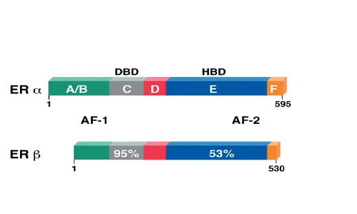
Like other steroid hormone receptors both the ER subtypes contain a well-defined domain organization, designated A/B through F (Kumar et al., 1987; Evans, 1988; Nilsson, 2000; Zilli et al., 2009) (Fig.2). The A/B domain, which is located at the
N-7
terminus, contains the activation function 1 (AF1) (Tora et al.,1989), a region responsible for the constitutive and ligand-independent transcriptional activity of ER. The C domain, referred to as DNA-binding domain (DBD), is responsible for specific DNA binding and receptor dimerization (Mader and Chambon, 1993). The D domain is a flexible hinge between the C and E domains and contains a nuclear localization signal (Picard et al., 1990). The E domain, referred to as ligand-binding domain (LBD), is a twelve-helix region involved in ligand binding and receptor dimerization. It harbors a second nuclear localization signal and the activation function 2 (AF2), responsible for the ligand-dependent activation of ER (Tora et al., 1989). The F domain, located at the extreme carboxy-terminus of the ERs, is a small region that although unnecessary for transcriptional activation exerts a complex modulatory role on both AF1 and AF2 activities of the receptor (Weatherman and Scanlan, 2001; Koide et al., 2007; Skafar and Zhao, 2008).
Fig. 2. Schematic representation of the structure of human ERα and ERβ nuclear receptors. Both ERα and ERβ are characterized by a well-defined domain organization: the A/B domain at the N-terminus contains the ligand-independent transcriptional activation function 1 (AF1), the C domain represents the DNA-binding domain (DBD) and contains a nuclear localization signal
8
(NLS), the D domain corresponds to the hinge region, the E domain harbors the ligand-binding domain and the ligand-dependent transcriptional activation function 2 (AF2) and the F domain at the C-terminus. Numbers outside each box refer to amino-acid number. Percentage of amino-acid homology for each domain is also shown. (Journal of Clinical Oncology, Vol 18, 2000: pp 3172-3186).
The classical mechanism of ER action involves estrogen binding to receptors in the nucleus, after which the receptors dimerize and bind to specific response elements known as estrogen response elements (EREs) located in the promoters of target genes (Nilsson et al., 2001). Hormone binding also induces a conformational change within the ligand binding domain of the receptors, and this conformational change allows coactivator proteins to be recruited (Rosenfeld and Glass, 2001). However, evidence for signaling pathways that deviate from this classical model has emerged, and it is now accepted that ERs can regulate gene expression by a number of distinct mechanisms. Around one third of the genes in humans that are regulated by ERs do not contain ERE-like sequences (O’Lone et al., 2004). The molecular mechanisms by which ERs regulate transcription at alternative response elements are not fully understood but are becoming increasingly clear. ERs can regulate gene expression without binding directly to DNA by modulating the function of other classes of transcription factors through protein-protein interactions in the nucleus (Gottlicher et al., 1998). The interaction of ERs with the activator protein 1 (AP-1) transcription factor complex is a typical example of such ERE-independent genomic actions (O’Lone et al., 2004).
Estrogens exert some of their effects through the action of ERs on gene expression, but a number of other effects of estrogens are so rapid that they cannot depend on the activation of RNA and protein synthesis. These actions are known as non-genomic
9
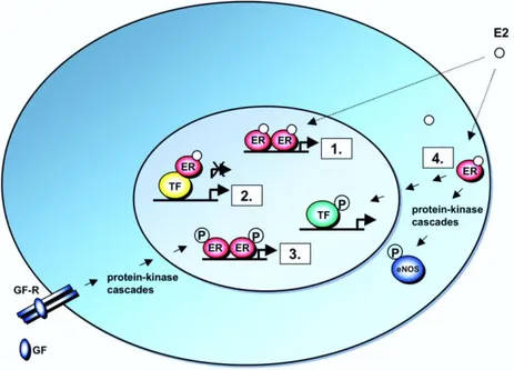
actions and are believed to be mediated through membrane-associated ERs. The actions are frequently associated with the activation of various protein-kinase cascades (Losel and Wehling, 2003). However, non-genomic actions of estrogens may indirectly influence gene expression, through the activation of signal transduction pathways that eventually act on target transcription factors. The functions of many transcription factors, including AP-1, are regulated through protein kinase-mediated phosphorylation, and these transcription factors may thus be targets of non-genomic actions of estrogens. This signalling pathway can be referred to as non genomic-to-genomic signaling, and it provides for a mechanism, distinct from protein-protein interactions in the nucleus, by which ERs can modulate the functions of transcription factors, and thus regulate the expression of genes that do not contain EREs (Fig.3).
Figure 3: Schematic Illustration of ER Signaling Mechanisms. 1. Classical mechanism of ER action. Nuclear E2-ERs bind directly to EREs in target gene promoters. 2. ERE-independent genomic actions. Nuclear E2-ER complexes are tethered through protein-protein interactions to a
10
transcription factor complex (TF) that contacts the target gene promoter. 3. Ligand-independent genomic actions. Growth factors (GF) activate protein-kinase cascades, leading to phosphorylation (P) and activation of nuclear ERs at EREs. 4. Nongenomic actions. Membrane E2-ER complexes activate protein-kinase cascades, leading to altered functions of proteins in the cytoplasm, e.g. activation of eNOS, or to regulation of gene expression through phosphorylation (P) and activation of a TF (Molecular Endocrinology 19:833-842).
A hypothesis is proposed that estrogens might play a dual role in affecting breast cancer risk. On one hand, there is evidence to indicate that estrogens might serve as preinitiators, initiators, and promoters of breast cancer. We generally associate estrogens with promotion of the growth of existing malignancies in the breast. However, these hormones and their metabolic products are also shown to induce direct and indirect free radical-mediated DNA damage, genetic instability, and mutations in cells in culture and in vivo (Liehr, 2000), suggesting a role for estrogens in cancer initiation. Furthermore, estrogens may serve as preinitiators. For example, elevated fetal estrogen levels can permanently alter the morphology of the mammary gland (Hilakivi-Clarke, 1997) and cause a persistent presence of epithelial structures (TEBs) that are known to be sites of malignant growth (Russo J. and Russo I.H., 1987). Data obtained in animal models and indirect evidence in humans indicate that high in utero estrogenicity increases breast cancer risk (Potischman and Troisi, 1999; Hilakivi-Clarke, 1997; Trichopoulos, 1990 ).
Up-regulated ERα expression in mice produces ductal hyperplasia, lobular hyperplasia, and ductal carcinoma in situ, demonstrating the consequences of unregulated ERα levels at all stages of breast cancer development (Frech et al., 2005); in contrast ERβ acts to regulate the degree of estrogen action by negatively modulating ERα, and the estrogen independent transcriptional activity of ERβ isoforms is inhibited by ERα (Poola et al., 2005). Further evidence shows that ERα levels in both hyperplastic lesions and
ER-11
positive tumors are greatly elevated as compared with adjacent normal tissue (van Agthoven et al., 1995) . Therefore it is not surprising that elevated levels of ERα in benign breast epithelium is, itself, considered a risk factor for progression to invasive breast disease (Khan et al., 1994).
Several studies demonstrated that ERβ expression is a favorable prognostic factor, correlating with low histological grading, longer disease-free survival and response to tamoxifen (Fuqua et al., 2003; Jarvinen et al.,2000).
Several studies have reported an increase in ERα/ERβ ratio in breast cancer as compared with benign tumors and normal tissues (Roger et al., 2001; Shaw et al., 2002; Park et al., 2003), suggesting that ERα is most closely associated with carcinogenesis, while ERβ can protect against the mitogenic activity of estrogens in mammary premalignant lesions. There is evidence that the estrogen-induced proliferation of ERα-positive breast cancer cells can be inhibited by ERβ overexpression (Strӧm et al., 2004; Paruthiyil et al., 2004; Williams et al., 2008). Routinely the evaluation of the ER expression in breast cancer, as determined by immunohistochemistry refers to ERα; consequently, the available information on the prognostic and predictive value of ER are related to this receptor form.
Progesterone and progesterone receptor in breast cancer
The steroid hormone, progesterone, is a key modulator of normal reproductive functions. These include ovulation, uterine and mammary gland development and the neurobehavioral expression associated with sexual responsiveness (Clarke and Sutherland, 1990; Lydon et al., 1995). The physiological effects of progesterone are mediated by interaction of the hormone with two specific intracellular progesterone
12
receptors (PRs) termed PR-A and PR-B that are transcribed from a single gene under the control of separate promoters (Kastner et al., 1990).
Binding of progesterone to PRs induces a significant conformational change on the receptor proteins (Allan et al., 1992 a,b) that results in dimerization of two ligand-receptor complexes (Tsai et al., 1988; O’ Malley et al., 1991), increased ligand-receptor phosphorylation (Weigel et al., 1995), binding of receptor dimers to specific hormone responsive DNA elements located in the promoter regions of target genes (Gronemeyer 1991, Tsai and O’Malley 1994), and interaction of the receptor complex with specific coactivator proteins and general transcription factors (Onate et al., 1995; Kamei et al., 1996) to form a productive transcription initiation complex on specific target gene promoters.
In human breast cancer the PR-A and B proteins, characterized in vitro (Lessey et al., 1983) and in vivo (Horwitz et al., 1983), are detected with molecular masses of approximately 81 kDa and 115 kDa, respectively.
Despite structural similarities, PR-A and PR-B regulate different subsets of genes and, although PR-B is transcriptionally more active, there are genes, known to be involved in breast cancer progression, that are uniquely regulated by the PR-A isoform (Kraus et al., 1993, Richer et al., 2002).
Approximately 75% of primary breast cancers express ERα, and more than half of these cancers coexpress PR.
Increased expression of ERα is an early event in breast carcinogenesis; in contrast, a decrease of PR levels is associated with breast cancer progression (Gross et al., 1984). PR is able to inhibit the growth of ER+ breast cancer cells in ovariectomized nude mice despite Progesterone (Pg) deficiency, addressing a specific inhibitory role of PR
13
independent of its natural ligand (Sartorius et al., 2003). It emerges from experimental models that ER+/PR+ breast cancers are well differentiated, presenting as low-risk, well-defined lesions whereas ER+/PR- metastatic tumors display a much more aggressive course after loss of PR compared with tumors retaining PR (Gross et al., 1984).
Importantly, in multivariate analyses including lymph node involvement, tumor size, and age, PR status was independently associated with disease-free and overall survival (Bardou et al., 2003).
In vivo the two PR isoforms are usually coexpressed at similar levels in normal cells, but their ratio varies dramatically in different tissues, in varying physiological states, and disease sites (Boyd-Leinen et al., 1982, Kato et al., 1993). With regard to the mammary gland, 3:1 overexpression of PR-A over PR-B in transgenic mice results in extensive epithelial cell hyperplasia, excessive ductual branching, and disorganized basement membrane, all features associated with neoplasia. In contrast, overexpression of PR-B leads to premature ductal growth arrest and inadequate lobulo-alveolar differentiation (Shymala et al., 2000, Shymala et al., 1998). Moreover the loss of coordinated PR-A and PR-B expression is thought to be an early event in carcinogenesis and is evident in premalignant breast lesions (Mote et al., 2002). A significant proportion of carcinomas express a predominance of the PR-A isoform, and elevated PR-A has been associated with poor clinical outcomes in endometrial cancer, indicating a direct association between PR-A isoform predominance and poor prognosis (Arnett-Mansfield et al., 2001).
Although ER and PR are members of different steroid hormone receptor subfamilies and recognize distinct hormone response elements, there is considerable biological evidence for cross-talk between their receptor-signaling pathways. For instance, progestins can suppress the stimulatory effects of estrogens in target cells; estrogen increases the expression of both c-fos and PR mRNA in uterine cells, and progestins block these
14
effects (Loose-Mitchell et al., 1988, Kirkland et al., 1992). This blockade appears to be mediated via the PRs, but it is unclear whether ER or some other component of the estrogen-ER signaling pathway is the target for repression. It is also known that liganded PRs can suppress E2-stimulated ER activity, with the magnitude of repression dependent
on the PR isoform, progestin ligand, promoter and cell type (Katzenellenbogen 2000, Weigel et al., 1993). The exact molecular mechanisms regulating ERα expression in breast tumors are unclear, but studies suggest that they are partly at the level of transcription (Martin et al., 1994). PR-B ablation studies using small interfering RNA further demonstrated that endogenous PR-B levels are determinant in regulating ERα.
Our previous studies have shown that PR-B overexpression repressed ERα levels in breast cancer cells in a ligand-independent manner as it emerges from data obtained on cells transiently overexpressing PR-B truncated in LBD. This inhibitory effect is mediated by the recruitment of NCoR bound to PR-B on ERα promoter (De Amicis et al., 2009). Furthermore we demonstrated previously that PR-B overexpression, but not PR-A, inhibited ERα transactivation together with the expression of estrogen-dependent genes such as pS2, cyclin D1, and IRS1. These effects correlate well with the inhibition of E2
-induced cell proliferation by Pg through endogenous PR-B isoform. Our findings corroborate previous clinical studies (Hopp et al., 2004; Graham et al. 1996) illustrating that high PR-A/PR-B ratios in breast tumors predict shorter disease-free survival. The protective action of PR-B in breast cancer is further reinforced by studies showing that PR is inversely associated with HER-2/neu, the signaling of which is known to drive estrogen-independent breast cancer cell growth (Huang et al., 2005, Bamberger et al., 2000). Also of note are the findings that the levels of Her-2 are significantly higher in ER+/PR-A+ xenografts, than in ER+/PR-B+ xenografts (Hopp et al., 2004). To clarify the molecular mechanisms through which PR-B may interfere with ERα gene
15
transcription, we analyzed previously the ERα promoter sequence and identified a PRE half-site located at -1757 bp to -1752 bp. Functional experiments using five deletion constructs of the ERα promoter showed that the down-regulatory effects induced by PR-B overexpression on ERα promoter activity were through the half-site and were not detected in the deletion constructs lacking the half-PRE site. Moreover site directed mutagenesis of the above region completely reversed down-regulation of ERα promoter activity. ChIP assay results further confirmed the specific recruitment of the PR-B isoform to the half-PRE site within the ERα gene promoter because recruitment was prevented in PR-B knockdown experiments. The inhibitory effects on the ERα promoter transcriptional machinery addressed the ability of PR-B to recruit corepressors interfering with ERα gene transcription (De Amicis et al., 2009).
Role of hsp90 in the steroid hormone receptor action
Steroid receptors form large oligomeric complexes in the absence of hormone (Toft and Gorski, 1966). Most of the receptor associated proteins belong to chaperoning proteins. In eukaryotes molecular chaperones are essential proteins that take part in then regulation of steroid receptors. Chaperones bind and stabilize unstable forms of protein and facilitate the folding of the protein, oligomeric assembly, transport in the cell compartment, switch between active and inactive conformations and assist in the regulation of signal transduction pathways. Their function is to ensure that the assembly of other polypeptide chains occurs correctly. They do not determine the tertiary structure of the folding protein and prevent incorrect interactions of protein folding intermediates. Chaperone-bound receptor is stabilized so that it has high affinity for hormone but cannot bind DNA. Many molecular chaperones are called heat shock proteins (hsps) (Hartl, 1996).
16
The heat shock response was first described in 1962 (Ritossa, 1962), and heat shock proteins (HSPs) are named for their increased synthesis after heat shock that is contrary to the reduced synthesis of most cellular proteins under these conditions. In addition to heat, these proteins are modulated by nutrient deprivation, and oxidative and other stresses where protein denaturation might otherwise occur (Zuehlke and Johnson, 2009). Many HSPs form multimolecular complexes that act as molecular chaperones binding other proteins, denoted as client proteins. These complexes play a regulatory role in the fate of proteins in several different ways including: folding of proteins in the cytosol, endoplasmic reticulum and mitochondria; intracellular transport of proteins; repair or degradation of proteins partially denatured by exposure to various environmental stresses; control of regulatory proteins; and refolding of misfolded proteins (Johnson and Brown, 2009). HSPs differ in their cellular localization and functions and mammalian HSPs have been classified into several families according to their molecular size: Hsp90, Hsp70, Hsp60 and Hsp40, and the small HSPs such as Hsp27 (Jackson, 2013).
Hsp90 defines a family of molecular chaperones that are highly conserved from prokaryotes to eukaryotes (Pearl et al., 2008; Wegele et al., 2004). Nonessential for normal growth in most bacteria, Hsp90 is abundantly expressed in higher eukaryotes where it has been shown to be necessary for viability (Zhao et al., 2005; McClellan et al., 2007). It functions as a homodimer that associates with co-chaperones to catalyze the maturation and/or activation of over 100 substrate proteins that are known to be involved in cell regulatory pathways (Theodoraki and Caplan, 2012). These ‘client proteins’ include protein kinases, nuclear hormone receptors, transcription factors, and an array of other essential proteins (Street et al., 2011).
17
It has been suggested that nuclear receptors as ER and PR are found in the cytoplasm in the form of heterocomplex with hsp90. Hormone binding stimulates hsp90 dissociation, and this leads to receptor dimerization, interaction with co-activators, DNA binding and target gene activation (Manninen et al., 2005).
Hsp90 forms a direct contact with the receptor and it acts on the ligand-binding domain of the steroid receptors (Pratt et al., 2004 a).
In humans, there are two hsp90 isoforms in the cytosol, hsp90α and hsp90β. These proteins are closely related. They are both induced by stress and no differences in their activities have been identified (Chen et al., 2005). Hsp90 is a phosphorylated homodimer containing two to three covalently bound phosphate molecules on each monomer. Hsp90 contains a highly conserved ATP binding domain near its N-terminus and the chaperoning activity of hsp90 requires both the binding and hydrolysis of ATP at this site (Mayer et al., 2009). A second nucleotide binding site appears to be present near the C-terminus, but this is less well characterized (Pratt and Toft, 2003). HSP90 functions as a part of a multichaperone complex, involving the dynamic association with various accessory cochaperones and client proteins. In an ATP-bound state, HSP90 adopts a closed conformation and becomes a mature complex that is essential for it to perform its function of client protein folding and stabilization.
18
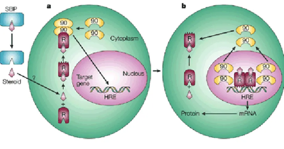
Figure 4: a The extracellular steroid released from its circulating steroid-binding protein (SBP) is transported into the cytoplasm of the target cell by passive diffusion or active transport. When bound to the non-ligand-bound receptor (R) it induces a conformational change that allows it to bind the Hsp90 dimer, which acts as its chaperone. The nuclear localization signal of the receptor allows the R–Hsp90 complex to translocate into the nucleus. b Once in the nucleus, the ligand– receptor complex dissociates from Hsp90 and itself dimerizes. The removal of Hsp90 unmasks the DNA-binding site of the receptor, which allows it to interact with the hormone response element (HRE) in the target gene promoter to activate transcription. This mechanism is only applicable to oestrogen receptor, progesterone receptor, glucocorticoid receptor and mineralocorticoid receptor, which do not heterodimerize with other nuclear receptors .
The association of hsp90 and breast cancer is of considerable interest, following studies showing the association of hsp90 and steroid receptors (Pratt, 1987; Shymala et al., 1989).
Inhibition of the Hsp90 chaperone cycle causes client proteins to undergo ubiquitination and subsequent degradation by the proteasome (Mimnaugh et al., 2004). Because many
19
of its clients include oncoproteins with important functions in the development and promotion of cancer, hsp90 is an important target in cancer therapy (Whitesell and Lindquist, 2005).
Levels of the heat shock proteins (hsp) molecular chaperones are elevated in many cancers, and hsp overexpression signals a poor prognosis in terms of survival and response to therapy in specific cancer types (van ’t Veer et al., 2002; Cornford et al., 2000; Ciocca et al., 1993). Elevated hsp expression in malignant cells plays a key role in protection from spontaneous apoptosis associated with malignancy as well as the apoptosis generated by therapy, mechanisms which may underlie the role of hsp in tumor progression and resistance to treatment (Gyrd-Hansen et al., 2004; Nylandsted et al., 2000; Volloch and Sherman, 1999).
In particular, the discovery of hsp90 as the target of anticancer activity of geldanamycin (GM) sparked much interest in the inhibition of hsp90 as a strategy for the treatment of cancer (Nickers and Workman, 2012).
Coregulators in steroid receptors transcriptional control
The activities of nuclear receptors are modulated by coregulators that are divided into coactivators and corepressors (Gao et al., 2002).
Coactivators are factors that can interact with nuclear receptors (NRs) in a ligand-dependent manner and enhance their transcriptional activity. Corepressors are factors that interact with NRs, either in the absence of hormone or in the presence of antihormone, and repress their transcriptional activity. Both types of coregulators are required for efficient modulation of target gene transcription by steroid hormones. Therefore, changes in the expression level and pattern of steroid receptor coactivators or
20
corepressors can affect the transcriptional activity of the steroid hormones and hence cause disorders of their target tissues.
The SRC (steroid receptor coactivator) family is composed of three distinct but structurally and functionally related members, which are named SRC-1 (NcoA-1), SRC-2 (TIF2/GRIP1/NcoA-2), and SRC-3 (p/CIP/RAC3/ACTR/AIB1/TRAM-1), respectively (McKenna et al.,1999). Histone acetyltransferase (HAT) activity was identified in the C-terminal region of SRC members. The acetylation of histones results in a less restrictive chromatin structure that is generally associated with transcriptional activation (Liu and Bagchi, 2004).
Although there are far fewer nuclear receptor corepressors, these molecules serve important roles in negatively regulating receptor-dependent gene expression (Smith and O’Malley, 2004).
Two corepressors, nuclear receptor corepressor (N-CoR) and silencing mediator for retinoid and thyroid hormone receptors (SMRT), were initially identified as a component of repression complexes associated with unliganded retinoic acid receptor (RAR) and thyroid-hormone receptor (TR) (Chen et al., 1996; Horlein et al.,1995).
It has been recently documented that NCoR and SMRT are also recruited by both ER and PR in the presence of ligand antagonist to repress their transcriptional activations (Agoulnik et al., 2003). The N-CoR and SMRT are thought to recruit histone deacetylase complex (HDAC) in the nuclear receptor complex to reduce the level of histone acetylation and repress transcription (Jones and Shi, 2003; Nagy et al., 1997).
For instance, our previous ChIP experiments showed that among potential corepressor molecules that are able to interact with PR, NCoR was the only one present on the PR-B/DNA complex regardless of its natural ligand. These results are consistent with
21
evidence reporting that corepressors have different preferences and determinants for interactions with nuclear receptors and transcription factors at specific genes (Hu and Lazar, 2001; Jepsen and Rosenfeld 2002). Moreover we show that formation of this bipartite complex leads to hypoacetylation of histone H4, which causes stabilization of nucleosome structure, limiting accessibility to the basal transcriptional machinery and thus repressing ERα gene expression. All these data support a model in which elevated expression levels of PR-B increase the interaction of the receptor with NCoR on the half-PRE site of the ERα promoter, an event incompatible with PR-coactivator interactions. The crucial role of NCoR emerges from our previous data showing that silencing of this corepressor was able to reverse the down-regulation of ERα expression induced by PR-B overexpression.
E2 is known to down-regulate the levels of ERα in breast cancer cell line through an
increased turnover of E2-bound receptor and via reduced transcription of its own gene
(Pink and Jordan, 1996). This down-regulation represents a classical feature of ERα transactivation. Our previous findings showed that PR-B is a determinant of these down-regulatory effects, because PR-B knockdown attenuated the feedback inhibition of ERα levels after estrogenic stimulus. Therefore we proposed that ERα modulation by E2 could
be due to early events independent from PR expression, and later transcriptional events leading to PR-B overexpression, which in turn can produce a decrease in ERα transcription, via recruitment of a corepressor complex containing NCoR and displacing RNA polymerase II (Pol II). Thus, in our previous study, we demonstrated that the E2
enhanced PR-B is crucial in determining the concomitant ERα down-regulation (De Amicis et al., 2009).
22
Hormonal therapy in breast cancer
Approximately 70% of breast cancer patients are positive for estrogen receptor (ER) or progesterone receptor (PR) expression at diagnosis. These patients are therefore suitable candidates for hormonal therapy, which aims to block estrogen stimulation of breast cancer cells (Normanno et al, 2005).
Although introduced more than 100 years ago, endocrine therapy is still an integral component in the systemic treatment of ER-positive breast cancer at any stage. Endocrine therapy is based on different approaches, including: (i) block of ER by the use of selective ER modulators (SERMs), such as tamoxifen; (ii) reduction of estrogen levels through aromatase inhibitors (AIs), such as anastrozole and letrozole; (iii) induction of ER degradation by selective estrogen receptor down-regulators (SERDs), such as fulvestrant.
Endocrine therapy is the most important component of adjuvant therapy for patients with early stage hormone receptor–positive breast cancer. Tamoxifen, a selective estrogen receptor modulator (SERM), binds to ERα and it blocks the estrogen action (Jordan and Morrow, 1999). Tamoxifen has been shown to improve relapse-free and overall survival in the adjuvant setting and to reduce the incidence of contralateral breast cancers (Moy and Goss, 2006). More recently, randomized data has shown that aromatase inhibitors (AI) as anastrozole, which deplete extragonadal peripheral estrogen synthesis, substantially improve disease-free survival in postmenopausal women with operable breast cancer in the adjuvant setting (Thurlimann et al., 2005; Coombes et al., 2004; Goss et al., 2003; Baum at al., 2002) (Fig. 5). In addition to these benefits in early stage disease, endocrine therapy is also of significant importance in the treatment of advanced metastatic disease (Moy and Goss, 2006). Recently fulvestrant has been reported to be as active as AIs in tamoxifen-resistant metastatic breast cancer patients (Howell et al.,
23
2005) and also shows a certain efficacy in patients progressing to AIs (Perey et al., 2007).
Figure 5: Both oestradiol and tamoxifen bind to the oestrogen receptor (ER) and lead to dimerization, conformational change in the activating function-2 (AF2) domain of ER and binding to oestrogen-response elements (EREs). The conformational change with tamoxifen is different from that with oestradiol and leads to persistent but less efficient transcription of most oestrogen-dependent genes. Oestrogen depletion leads to an absence of oestrogen-oestrogen-dependent transcription. Aromatase inhibitors block the enzyme aromatase and thus prevent the conversion of androgens into oestrogen. This decreases the level of oestrogen and the binding of oestrogen receptors. Finally, it stops the growth of cancer cells. (Nature Reviews Cancer 3, 821-831).
Despite these obvious benefits in a proportion of patients, acquired resistance to endocrine therapies frequently occurs; in fact, resistance to all forms of endocrine therapy remains a major problem. Furthermore, long-term use of tamoxifen is associated with several important side effects (increased incidence of endometrial cancer due to its
24
undesirable agonistic activity in the uterus, bone fractures and thrombo-embolism) (Coombes et al., 2004). Chemotherapy, in these cases, remains the treatment of choice, but is associated with severe adverse effects (Budzar, 2007).
Thus, looking for new antitumoral drugs with low toxicity to approach endocrine-related cancers treatment need to be considered as one of the most important priorities for research.
Epigallocatechin-3-gallate and breast cancer
The strong correlation between ERα expression, breast disease patho-physiology, and therapeutic response have justified the use of estrogen receptor down-regulators as attractive intervention, with significant clinical interest in their use. In this regard, many naturally occurring compounds, commonly present in the diet have gained considerable attention as well (De Amicis et al., 2011; Hong and Sporn, 1997). In recent years epigallocatechin gallate (EGCG), a polyphenolic compound found in green tea, has demonstrated chemo-preventative and antitumor properties (Mukhtar et al., 1992).
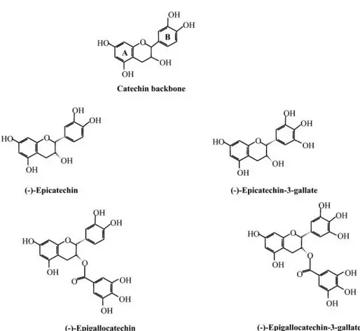
Green tea is a form of tea commonly consumed in Asia, derived from the leaf of the plant Camellia sinensis and its habitual consumption has long been associated with health benefits (Cabrera et al., 2006; Brown, 1999; Zloch, 1996). Most of the beneficial effects of green tea are attributed to its polyphenolic flavonoids, known as catechins, including epicatechin (EC), epigallocatechin (EGC), epicatechin-3- gallate (ECG) and the major flavonoid (−)-epigallocatechin-3-gallate (EGCG) (Fig.6) (Singh et al., 2011). These polyphenols account for up to 40% of the dry weight of green tea, and purified EGCG has been the focus of research in recent years (Kanwar et al., 2012).
25
Extensive research on green tea has taken place over the last decade, especially on the isolated catechin EGCG; however, most are based on in vitro and animal experiments. Green tea polyphenols are known antioxidants and it is proposed that these phytochemicals modulate biochemical and physiological processes leading to the initiation and propagation of carcinogenesis (Yuan et al., 2011).
The polyphenolic structure of EGCG consists of 4 rings, A, B, C and D (Fig.7). A and C rings constitute the benzopyran ring. This benzopyran ring has a phenyl group at C2 and gallate group at C3 positions. The B ring of EGCG has vicinal 3,4,5-trihydroxy groups, and the D ring galloyl moiety in EGCG is in the form of an ester at C3. The presence of this ester carbon makes EGCG highly susceptible to nucleophilic attack (Kanwar et al., 2012).
26
Figure 7: Chemical structure of EGCG (Int. J. Mol. Sci, 2011).
Epidemiological studies have suggested that green tea consumption is linked to a decrease in the incidence and recurrence of breast cancer (Nakachi et al., 1998).
Additionally, treatment with EGCG (50 mg/kg/day, 14 days) reduced the growth of MCF-7 implanted breast tumors in athymic nude mice by 40% (Liao et al., 1995), and it has been reported that catechin inhibited the proliferation of human breast cancer cells in vitro (Belguise et al., 2007). This is partly attributable to its effects on modulating the activity of mitogen-activated protein kinases (MAPKs), IGF/IGF-1 receptor, AKT, NF-κB, and CDKs (Cyclin-Dependent Kinases) (Fig.7) (Sah et al., 2004; Gupta et al., 2001). EGCG reduced angiogenesis and both local and distant invasion, and it could inhibit DNA methyltransferas and reactivate methylation-silenced genes in cancer cell lines (Fang et al., 2003).
EGCG also inhibits growth factor receptor extracellular signaling, the proteasome, mitochondrial depolarization, and fatty acid synthase (Siddiqui et al., 2011; Qanungo et al., 2005; Liang et al.,1997). However, the precise molecular targets and the exact anti-cancer mechanism of EGCG are not clearly defined.
27
Figure 8: Mechanism of actions of EGCG (Biochemical Pharmacology, Volume 82, Issue 12,
2011, 1807-1821).
Recent studies report that the anti-tumor activity of EGCG is mediated by targeting HSP70 and HSP90 in vitro and in vivo, through a mechanism that involves direct binding of EGCG to the C-terminal region of HSP90 (Tran et al., 2010), a chaperone protein that is constitutively expressed at high levels in many cancer cells. HSP90 is assembled into heterocomplexes with unliganded steroid receptors, such as GR and PR, thus influencing their cellular distribution and activity (Carson-Jurica et al., 1990).
Particularly recent studies on breast cancer models have implicated EGCG in the regulation of ERα. Since the catechin family is structurally similar to isoflavones, it has been shown that catechins, such as EGCG, were co-estrogenic for ERα at lower doses
28
(<5µM) but antiestrogenic at higher doses (100-150 µM) and eliciting a concomitant massive cell death at the higher levels (Belguise et al., 2007; Farabegoli et al., 2007). We considered that the interaction between EGCG and ERα might be potentially important and may have therapeutic implications.
In the present study we report that EGCG produces a significant inhibition of ER+ PR+ breast cancer cell proliferation and we define the molecular mechanisms associated with this growth effect. Concomitant with nuclear localization of PR, EGCG treatment causes a down-regulation of ERα protein, mRNA and gene promoter activity. We demonstrate that these effects are crucially mediated by PR-B via its recruitment to the ERα proximal promoter.
29
Materials and Methods
Materials
Epigallocatechin 3 gallate (EGCG), aprotinin, leupeptin, phenylmethylsulfonyl fluoride (PMFS), and sodium orthovanadate were from Sigma (Milan, Italy). PD 169316 and TBB from Calbiochem (Darmstadt, Germany). ICI 182,780 (ICI, ER antagonist), was purchased from Zeneca Pharmaceuticals (Cheshire, UK). Antibodies used in this study were GAPDH, laminin B, NCoR, RNA Pol II, HDAC1, total p38, phosphorylated p38, ERα, progesterone receptor B (PR-B), EGFR, Raf1, Her2, Hsp90, CK2 from Santa Cruz Biotechnology (Santa Cruz, CA); progesterone receptors (PR-B, PR-A), total MAPK, total AKT, phosphorylated p42/44 MAPK (Thr202/Tyr 204) and pAKT (Ser437), from Cell Signaling Technology (Beverly, MA).
Cell Culture
Cells utilized in the studies were obtained from American Type Culture Collection (Manassas, VA, USA). MCF-7 cells were maintained in DMEM/F-12 medium containing 5% fetal bovine serum (5% FBS), 1% L-glutamine, 1% Eagle’s nonessential amino acids, and 1 mg/ml penicillin/streptomycin in a 5% CO2 humidified atmosphere at 37 °C. T47D
cells were routinely maintained in RPMI 1640 supplemented with 10% FBS, 1 mg/ml insulin (Sigma), 1 mg/ml penicillin /streptomycin (Sigma), 1% L-glutamine (Sigma), 2.5 mg/ml D-glucose (Sigma), 1% Sodium Pyruvate (Sigma), 10 nM Hepes Buffer (Sigma). Ishikawa endometrial cancer cells (ISK) were maintained in DMEM without phenol red supplemented with 10% fetal bovine serum (FBS), 1 mg/ml penicillin /streptomycin (Sigma), 1% L-glutamine. SkBr3 cells were cultured in RPMI 1640 with 10% FBS, 1 mg/ml penicillin /streptomycin (Sigma), 1% L-glutamine. Treatments were performed,
30
after 48 hours of serum starvation, in 1% dextran charcoal stripped (CS) FBS to reduce steroid concentration (De Amicis et al., 2009).
Cell proliferation assays
MTT anchorage-dependent growth assay. Cells (3 × 10⁴ cells/mL) were plated in 24-well plates and serum-starved for 48 hours before the addition of treatment for 4 days. The MTT assay was performed as the following: 100 µL of MTT (3-(4,5-Dimethylthiazol-2-yl)-2,5-diphenyltetrazolium bromide) (2 mg/mL) (Sigma Aldrich, Milan, Italy) were added to each well, and the plates were incubated for 2 hours at 37 °C. Then, 500 µL of DMSO (Dimethyl sulfoxide) were added. The absorbance was measured with the Ultrospec 2100 Prospectrophotometer (Amersham-Biosciences, Milan, Italy) at a test wavelength of 570 nm.
Anchorage-independent soft agar growth assays. T47D cells (5000/well) were plated in
4 mL of 0.35% agarose with 5% CS FBS in phenol red free media, on a 0.7% agarose base in 6-well plates. Two days after plating, media containing control vehicle or hormonal treatments was added to the top layer, and the appropriate media was replaced every 2 days. After 14 days, 150 µL of MTT was added to each well and allowed to incubate at 37 °C for 4 hours. Plates were then placed in 4 °C overnight and colonies ≥50 µm diameter from triplicate assays were counted. Data are the mean colony number of three plates and representative of two independent experiments analyzed for statistical significance (p < 0.05) using a two-tailed student’s test, performed by Graph Pad Prism 5 (GraphPad Software, San Diego, CA, USA). SDs are shown.
31
Plasmids
XETL (Bunone et al., 1996) , the wild-type human ER-α (HEGO) (Tora et al., 1989 b), the full-length PR-B consisting of the full-length PR-B cDNA fused with the SV40 early promoter (a gift from Dr. D. Picard, University of Geneve, Switzerland) (De Amicis et al., 2009), the PR DNA-binding mutant C587A (mDBD PR) previously described by Faivre and Lange (2007) (gift from Dr. C. Lange, University of Minnesota Cancer Center, Minneapolis, MN, USA), the full-length PRA (Kastner et al., 1990), and the deletion fragments of the ER-α gene promoter (deGraffenried et al., 2002). The Renilla luciferase expression vector pRL-TK (Promega, Milan, Italy) was used as a transfection standard.
Reverse transcription and real-time PCR
Cells (6 × 10⁶) were treated as indicated and processed as described (Vivacqua et al., 2009). cDNA diluted were analyzed in triplicates by real-time PCR in an iCycler iQ Detection System (Bio-Rad, USA). The primers were:
(ER-α forward) 5’-AGAGGGCATGGTGGAGATCTT-3’; (ER-α reverse) 5’-CAAACTCCTCTCCCTGCAGATT-3’; (pS2 forward) 5’-TTCTATCCTAATACCATCGACG- 3’; (pS2 reverse) 5’-TTTGAGTAGTCAAAGTCAGAGC- 3’; (IRS1 forward) 5’- AGGATATTTAATTTGCCTCGG-3’; (IRS1 reverse) 5’- AAGCGTTTGTGCATGCTCTTG-3’; (CD1 forward) 5’-TCTAAGATGAAGGAGACCATC-3’; (CD1 reverse) 5’-GCGGTAGTAGGACAGGAAGTTGTT-3’; (18S forward) 5’- GGCGTCCCCCAACTTCTTA-3’;
32
Each sample was normalized on its GAPDH mRNA content. The relative gene expression levels were normalized to a calibrator that was chosen to be the basal, untreated sample. Final results were expressed as n-fold differences in gene expression relative to GAPDH mRNA and calibrator, calculated using the ΔCt method as follows:
n-fold=2- (ΔCtsample–ΔCtcalibrator)
where ΔCt values of the sample and calibrator were determined by subtracting the average Ct value of the GAPDH mRNA reference gene from the average Ct value of the gene analysed.
Western blotting (WB) and immunoprecipitation
Protein expression or complex formation were assessed as described (De Amicis et al., 2010) by Western blotting (WB) or immunoprecipitation (IP) followed by WB, using total protein lysates, cytoplasmic, or nuclear protein lysates, where appropriate. Cells (6 × 10⁶) were harvested to be analyzed using 500 µL of lysis buffer containing 50 mmol/L HEPES (pH 7.5), 150 mmol/L NaCl, 1% Triton X-100, 1.5 mmol/L MgCl2, 10 mmol/L
EGTA (pH 7.5), 10% glycerol, and inhibitors (0.1 mmol/L Na3VO4, 1% PMSF, and 2.0
mg/mL aprotinin) to obtain cytoplasmic proteins. After the collection using a scraper, incubation of 30’ on ice, we lysed the nuclei for 15’ at 4 °C using 250 µL of nuclear buffer containing 20 mmol/L HEPES (pH 8), 0.1 mmol/L EDTA, 5 mmol/L MgCl2, 0.5
mol/L NaCl, 20% glycerol, 1% NP-40, and inhibitors (1.7 mg/mL aprotinin, 1 mg/mL leupeptin 200 mmol/L PMSF, 200 mmol/L sodium orthovanadate, and 100 mmol/L sodium fluoride). Then lysates were collected and centrifuged at 10 000 × g for 10’ at 4 °C.
For total protein extracts, 500 µL RIPA buffer (50 mM Tris-HCl, pH 7.4, 150 mM NaCl, 1% NP-40, 0.25% Na deoxycholate, plus inhibitors 1.7 mg/mL aprotinin, 1 mg/mL
33
leupeptin 200 mmol/L PMSF, 200 mmol/L sodium orthovanadate, and 100 mmol/L sodium fluoride) was added to the 100 mL cell culture plate for 15’ at 4 °C. Then lysates were collected and centrifuged at 10 000 × g for 10’ at 4 °C. The protein content was determined using Bradford dye reagent (Bio-Rad). For WB, 50 µg of total, cytoplasmic or nuclear lysates were separated on an 11% polyacrylamide denaturing gel (SDS-PAGE) and transferred to nitrocellulose membranes. Proteins of interest were detected with specific Abs, recognized by peroxidase-coupled secondary Abs, and developed using the ECL PlusWestern Blotting detection system (Amersham Pharmacia Biotech, UK). For IP, 500 µg of protein of cytoplasmic or nuclear lysates were precleared for 1 hour with protein A/G-agarose (Santa Cruz), incubated with primary Abs at 4 °C for 18 hours in HNTG buffer (20 mmol/L HEPES, pH 7.5, 150 mmol/L NaCl, 0.1% Triton X-100, 10% glycerol, and 0.1 mmol/L Na3VO4), and then the antigen–Ab complexes were
precipitated with protein A/G agarose for 2 h in HNTG buffer. The immunoprecipitated proteins were washed three times with HNTG buffer, separated on SDS-PAGE, and processed by WB. The images were acquired by using an Epson Perfection scanner (Epson, Japan) using Photoshop software (Adobe). The optical densities of the spots were analyzed by using ImageJ software (NIH; http://rsb.info.nih.gov/IJ). Images are representative of three different experiments.
Immunofluorescence
T47D cells seeded on glass cover-lips were treated with 40 µM EGCG for 12 h, washed with PBS, and then fixed with 4% paraformaldehyde in PBS for 20’ at room temperature. Next, cells were permeabilized with 0.2% Triton X-100 in PBS for 5’, blocked with 5% BSA for 30’, and incubated with anti-PR-B antibody (1:50) in PBS overnight at 4 °C. The day after the cells were washed three times with PBS and incubated with the secondary antibody anti-rabbit IgG-FITC (1:200) for 1 hour at room temperature. To
34
check the specificity of immunolabeling, the primary antibody was replaced by normal rabbit serum (negative control). The blue fluorescent DAPI was used for nuclear stain. Immunofluorescence analysis was carried out on an OLYMPUS BX51 microscope using a ×40 objective. Images are representative of three different experiments.
Transfections and luciferase assays
Cells (1 x10⁵) were plated into 24-well dishes with 500µl of regular growth medium per well the day before transfection. The medium was replaced with that lacking serum on the day of transfection, which was done using Fugene 6 reagent as recommended by the manufacturer (Roche Diagnostics, Milan, Italy) with a mixture containing 0.5 µg of reporter plasmid, alone or in combination plasmids as indicated in the figure legends, and 5 ng of pRL-TK. Medium was renewed after which cells were treated for 24 hours. Luciferase activity was measured with the Dual Luciferase kit (Promega, Milan, Italy) according to the manufacturer’s recommendations.
TK Renilla luciferase plasmid was used to normalize the efficiency of the transfection. Firefly and Renilla luciferase activities were measured by Dual Luciferase kit. The firefly luciferase data for each sample were normalized based on the transfection efficiency measured by Renilla luciferase activity (De Amicis et al., 2009). Results represent mean of luciferase activities observed in three independent experiments done in triplicate.
Site-directed mutagenesis
Site-directed mutagenesis ia s method that is used to make specific and intentional changes to the DNA sequence of a gene and any gene products. Mutagenesis was performed on Fragment D of the ER-α promoter using the QuikChange mutagenesis kit (Stratagene, La Jolla, CA, USA) following the manufacturer’s instructions. The sequence for the sense primer was:
35
5’-AGCAGGGAGATGAGGATTGCTGAAGTCCATGGGGGTATGT-3’. The plasmids were then sequenced to confirm the mutation of the desired site.
Lipid-mediated transfection of siRNA duplexes
Custom-synthesized siRNA (Invitrogen) annealed duplexes (25-bp double-stranded RNA) were used for effective depletion of PR-B. A nonspecific siRNA (NS) (Invitrogen) that lacked identity with known gene targets was used as a control for nonsequence-specific effects. Cells were transfected using Lipofectamine 2000 reagent (Invitrogen, Paisley, UK) according to the manufacturer’s instructions and then treated as indicated.
Electrophoretic mobility shift assay (EMSA)
EMSA was carried out as previously described (deGraffenried et al., 2002), with a few modifications. Cells were treated for 6 hours before harvesting for the assay. The sequence of ER-α-half-PRE oligonucleotide used as probe or the unlabeled competitor was
5’-AGGATTGTGTTCTCCATGGG-3’,
mutated 5’- AGGATTGTTAAGTCCATGGG-3’.
The probe was generated by annealing single-stranded oligonucleotides, labelled with [γ32
P] ATP using T4 polynucleotide kinase, and purified using Sephadex G50 spin columns. To test specific binding, nuclear extracts were pre-incubated with rabbit polyclonal PR antibody or normal rabbit IgG. The reactions were separated on 6% polyacrylamide gel in 0.25× Tris borate–EDTA for 3 hours at 150 V. The gel was then dried for autoradiography. Images are representative of three different experiments.
36
Chromatin immunoprecipitation (ChIP) assays
Cells were treated for 12 hours before harvesting for the assay performed as described (Guido et al., 2012). DNA/protein complexes were cross-linked with 1% formaldehyde at 37 °C for 10’ and sonicated. Supernatants were immunocleared with salmon sperm DNA/protein A agarose for 1 hour at 4°C. The precleared chromatin was immunoprecipitated with specific antibodies. A normal mouse serum IgG was used as negative control. Pellets were washed, eluted with elution buffer (1% SDS, 0.1 M
NaHCO3) and digested with proteinase K. DNA was obtained by
phenol/chloroform/isoamyl alcohol extractions and precipitated with ethanol. A 5µl volume of each sample and input were used for PCR.
ER-α promoter primers used for PCR: forward, 5’-ACGTTCTTGATCCAGCAGGGTA-3’ and reverse, 5’-ACCTGCCAAATTATATGCAAATGGCAG- 5’-ACGTTCTTGATCCAGCAGGGTA-3’ containing the half-PRE site; and forward, 5’-GTGGCCATTGTTGACCTACAG-3’ and reverse, 5’CTGTAGGTCAACAATGGCCAC-3’ upstream the half-PRE site.
Images are representative of three different experiments.
Statistical analysis
Each datum point represents the mean ±SD of three different experiments. Data were analyzed by Student’s t-test using the GraphPad Prism 4 software program. p<0.05 was considered as statistically significant.
37
Results
EGCG treatment decreases ER+ PR+ cancer cell proliferation
We first investigated whether EGCG affects cancer cell proliferation following extended treatments with low doses of the catechin. We tested ER+ PR+ breast cancer cell lines,
including T47D cells, which are known to express ER-α and elevated levels of endogenous PRs (De Amicis et al., 2009), MCF-7 cells that express ER-α and low levels of PR, and a well-differentiated ER+ PR+ human endometrial adenocarcinoma cell line
(Ishikawa cells). Proliferating cells were exposed to different nontoxic concentrations of EGCG (10 µM; 20 µM; 40 µM) and then analyzed in MTT growth assays (Fig. 1A). After 4-day treatment, 10 µM EGCG inhibited basal cell proliferation of both T47D (50%), and MCF-7 (20%). With increasing doses of EGCG (20 and 40 µM), T47D cell number was further reduced by 52 to 67%, respectively. MCF-7 (25–50%) and ISK (55– 68%) cells growth was also significantly decreased. EGCG inhibitory action was still present after pre-treatment with 1 µM ICI 182 780, a potent and specific antagonist with excellent growth inhibitory effects in several cell and animal models of human breast cancer, which induce ER-α degradation through ubiquitin-mediated mechanism (Hu et al., 1993). This suggests that growth inhibition was not mediated by binding of EGCG to ER. Of note is that the antiproliferative effects produced by EGCG occur earlier in T47D cells with respect of those observed in MCF-7 and ISK. Consistent with MTT assay results, 40 µM EGCG treatment also markedly reduced basal and E2-induced colony formation of T47D cells growing in soft agar (Fig. 1B).
38
A
T47D T47DsiPRB 0,0 1,0 0,6 0,2 0,8 0,4 EGCG µM - 10 20 40 - 40 40 ICI F o ld C h a n g e o v er u n tr ea te d MCF-7 0,0 0,2 0,4 0,6 0,8 1,0 - 10 20 40 - 40 ICI EGCG µM F o ld C h a n g e o v er u n tr ea te d Ishikawa cells 0,0 0,2 0,4 0,6 0,8 1,0 - 10 20 40 EGCG µM F o ld C h a n g e o v er u n tr ea te d39
B
N u m b er of co lo n ie s 0 100 200 300 400 500 - E2 EGCG E2+ EGCG - E2 EGCG E2+ EGCGFigure 1. EGCG inhibits ER+ PR+ cancer cell proliferation (A) MTT assay. Cells, serum starved, were exposed to vehicle (−), or 1 µM ICI and/or different concentrations of EGCG in medium containing 1% dextran charcoal-stripped FBS for 4 days (treatments were renewed every 2 days). Results indicate mean of three independent experiments done in triplicate; bars SD; ◊ p < 0.05 compared with untreated cells. (B) EGCG blocks E2-induced anchorage independent
growth of T47D. Soft agar colony formation assay was performed in control conditions (−), or in the presence of 10 nM estrogen (E2) and/or 40 µM EGCG. Cells were allowed to grow for 14 days
and the number of colonies ≥50 µm were quantified. Bars SD; ◊ p < 0.05 compared with untreated cells; ◊◊ p < 0.05 compared with E2-treated cells. Pictures at the bottom show typical well for each
condition.
EGCG downregulates ER-α expression and transcriptional activity
It is well known that increased expression of ER-α is an early event in breast carcinogenesis, driving breast cancer cell proliferation. We thus hypothesized that
40
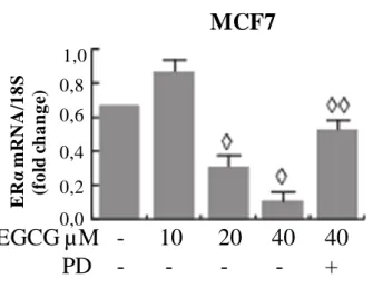
EGCG’s effects in ER-α-positive breast cancer cells could involve the regulation of ER-α itself. As shown in Fig. 2A, ER-α protein expression levels were decreased after 24 hourd of increasing EGCG concentrations, in MCF-7 and T47D. E2 treatment was used
as control for downregulation of ER-α.
To investigate the functional effects of downregulation of ER-α by EGCG, we next analyzed ER-α genomic activity after treatment. To this aim, a luciferase reporter plasmid containing a consensus estrogen-responsive element (XETL) was transiently transfected into MCF-7 cells. EGCG treatment caused a significant decrease of basal, and with more intensity of E2-induced luciferase activity (sevenfold) (Fig. 2B). To
confirm these results, we also evaluated mRNA levels of known estrogen-regulated genes, such as IRS1, pS2, and cyclin D1 (CD1). As assessed by real-time PCR, EGCG at 40 µM significantly decreased basal and E2-induced levels (Fig. 2C) of IRSI, pS2, and
cyclin D1 in T47D and MCF-7 cells.
Stemming from our evidence that EGCG inhibited ER-α levels, we hypothesized that it could also influence E2/ER-α nongenomic rapid effects within seconds to minutes
(Castoria et al., 2001). Therefore to demonstrate the effects of EGCG on E2-induced
nongenomic activity, we pretreated T47D cells with EGCG for 24 h causing ER-α levels to decrease, and subsequently we treated the same cells with short exposures to E2. As
shown in Fig. 2D, EGCG treatment blocked E2-induced phosphorylation of MAPK and
AKT. Five percent CS treatments were used as positive controls for activation of downstream signaling. The ERK/MAPK and PI3K/AKT signaling pathways that are rapidly activated by the ER-α–E2 complex, also have critical roles in estrogen action as
survival agents. In fact, these pathways enhance the expression of the anti-apoptotic protein Bcl-2 and block the activation of p38 kinase (Acconcia et al., 2005). Western blot analysis in MCF-7 cells revealed that p38MAPK phosphorylation was significantly upregulated in EGCG-exposed cells, effectively opposing estrogen’s effect (Fig. 2D).
41
These results suggest that EGCG-induced ER-α downregulation can affect nongenomic events in the E2/ER-α signaling cascade.