Edited by:
Cesare Mancuso, Catholic University of the Sacred Heart, Italy
Reviewed by:
Filippo Caraci, University of Catania, Italy Ai Na Ng, University of Bristol, United Kingdom
*Correspondence:
Robert Nisticò [email protected] †These authors have contributed
equally to this work.
Specialty section:
This article was submitted to Experimental Pharmacology and Drug Discovery, a section of the journal Frontiers in Pharmacology
Received: 09 May 2019 Accepted: 17 June 2019 Published: 16 July 2019 Citation:
Mango D, Saidi A, Cisale GY, Feligioni M, Corbo M and Nisticò R (2019) Targeting Synaptic Plasticity in Experimental Models of Alzheimer’s Disease. Front. Pharmacol. 10:778. doi: 10.3389/fphar.2019.00778
Targeting Synaptic Plasticity in
Experimental Models of Alzheimer’s
Disease
Dalila Mango
1†, Amira Saidi
1†, Giusy Ylenia Cisale
2, Marco Feligioni
1,3, Massimo Corbo
3and Robert Nisticò
1,4*
1 Laboratory of Neuropharmacology, EBRI Rita Levi-Montalcini Foundation, Rome, Italy, 2 Department of Physiology and Pharmacology, Sapienza University of Rome, Italy, 3 Department of Neurorehabilitation Sciences, Casa Cura Policlinico, Milan, Italy, 4 School of Pharmacy, Department of Biology, University of Rome Tor Vergata, Rome, Italy
Long-term potentiation (LTP) and long-term depression (LTD) of hippocampal synaptic
transmission represent the principal experimental models underlying learning and
memory. Alterations of synaptic plasticity are observed in several neurodegenerative
disorders, including Alzheimer’s disease (AD). Indeed, synaptic dysfunction is an early
event in AD, making it an attractive therapeutic target for pharmaceutical intervention.
To date, intensive investigations have characterized hippocampal synaptic transmission,
LTP, and LTD in in vitro and in murine models of AD. In this review, we describe the
synaptic alterations across the main AD models generated so far. We then examine the
clinical perspective of LTP/LTD studies and discuss the limitations of non-clinical models
and how to improve their predictive validity in the drug discovery process.
Keywords: long-term potentiation, long-term depression, synaptic plasticity, Alzheimer’s disease, predictive validity
INTRODUCTION
Long-term synaptic plasticity is considered the neural basis of learning and memory process (
Bliss
and Collingridge, 1993
). Long-term potentiation (LTP) and long-term depression (LTD) are the
major forms of durable synaptic strength changes in central nervous system abundantly studied in
the hippocampal region (
Malenka and Bear, 2004
). The magnitude of LTP and LTD is largely used in
many different experimental conditions and animal models as an indicator of cognitive function; on
the other hand, dysregulation of synaptic plasticity underlies a large number of neurodegenerative
disorders such as Alzheimer’s disease (AD) (
Selkoe, 2002
).
AD is a multifaceted neurodegenerative disorder typified by a progressive and irreversible
memory deficits and cognitive decline. To date, AD can only be diagnosed post-mortem, through
two characteristic neuropathological lesions in the brain: senile plaques, consisting of β-amyloid
protein oligomers aggregates (Aβo, residues 1–40/42), and intracellular neurofibrillary tangles
(NFT), constituted of abnormally hyperphosphorylated tau protein accumulation predominantly
Abbreviations: Aβ, amyloid β protein; Aβo, β-amyloid protein oligomers aggregates; AD, Alzheimer’s disease; AMPAR, L-α-amino-3-hydroxy-5-methyl-4-isoxazole propionate receptor; APP, amyloid precursor protein; BST, basal synaptic transmission; E-LTP, early-LTP; FAD, familiar Alzheimer’s disease; LFS, low frequency stimulation; LTD, long-term depression; LTP, long-term potentiation; L-LTP, late- LTP; mGlu, metabotropic glutamate receptor; NFT; neurofibrillary tangles; PPF, paired pulse facilitation; Tg, transgenic; WT PS1, wild-type human PS1; NA, not assessed; rTMS, repetitive transcranial magnetic stimulation; sAD, sporadic Alzheimer’s disease; tDCS, transcranial direct current stimulation.in hippocampal and cortical regions. The “amyloid cascade
hypothesis” is so far the prominent theory to describe the
time-course of AD neurodegeneration (
Hardy and Higgins, 1992
).
Impaired synaptic function of the hippocampus is an early event
leading to defective hippocampal-dependent memory appearing
long before the buildup of amyloid plaques and neuronal cell
death (
Selkoe, 2002
;
Tanzi, 2005
). Therefore, synaptic plasticity is
often used to evaluate part of the phenotype. Accordingly, many
electrophysiological studies on the different models have been
performed to delineate such changes.
Early impairments in synaptic transmission were highlighted
in different mouse models of AD and are caused, among other
factors, by Aβ which leads to impairment of LTP via tau protein
(
Shipton et al., 2011
). Notably, many studies investigated the
correlation between age and synaptic dysfunction in order to
describe the onset and development of pathology in a specific
mouse model. Discrepancy in the results obtained by the
different researchers across the various models of AD may reflect
the type of mutation studied, in addition to several other sources
of variations such as experimental design, age, or strain.
LTP AND LTD IN NORMAL CONDITIONS
Several studies indicate that the hippocampus plays a crucial
role in higher cognitive functions and in information-storage
(reviewed by
Neves et al., 2008
). LTP was first studied in
the hippocampus and has been widely characterized using
biochemical, electrophysiological, and molecular techniques
(reviewed by
Bliss et al., 2007
). LTP is characterized by a
short-term phase (early- or E-LTP) and a subsequent long-short-term phase of
potentiation (late- or L-LTP) (
Reymann et al., 1989
). Importantly,
distinct forms of N-methyl-D-aspartate (NMDA) receptor
LTP coexist at synapses (
Park et al., 2014
) and these can also be
distinguished based on their responsiveness to protein kinase A
(PKA) inhibitors (
Park et al., 2016
). E-LTP and L-LTP can be
induced in hippocampal slices by different induction protocols
and are sustained by distinct cellular and molecular pathways.
E-LTP (<1 h) is characterized by the recruitment of postsynaptic
2-amino-3-(3-hydroxy-5-methyl-isoxazol-4-yl)propionic acid
(AMPA) receptors, either from neighboring extra synaptic
receptors or from intracellular reserve pool by exocytosis (
Penn
et al., 2017
). On the other hand, L-LTP (>3 h) involves de novo
protein synthesis promoting structural and functional changes
(
Frey et al., 1988
). Among the key players facilitating the transition
from E-LTP to L-LTP, brain-derived neurotrophic factor (BDNF)
(
Panja and Bramham, 2014
) and transforming growth factor β1
(TGF-β1) (
Caraci et al., 2015
;
Caraci et al., 2018
) are noteworthy.
The classical form of LTD can be experimentally elicited using
specific electrical low-frequency stimulation (LFS) protocols
in slices (
Dudek and Friedlander, 1996
). Most of the LTD
forms studied imply activation of NMDA receptors (
Dudek
and Friedlander, 1996
) and/or metabotropic glutamate (mGlu)
receptors (
Fitzjohn et al., 1999
). Other chemical forms of LTD are
obtained either by exogenous application of NMDA or muscarinic
receptor agonists (reviewed by
Collingridge et al., 2010
), as well as
through activation of microglia (
Zhang et al., 2014
).
LTP AND LTD IN EXPERIMENTAL AD
In Vitro Models
The peptide amyloid beta is released endogenously during
physiologic neuronal activity and causes enhancement of
synaptic plasticity and memory formation when administered at
picomolar concentrations that likely resemble the physiological
level in the brain (
Puzzo et al., 2008
;
Morley et al., 2010
;
Puzzo
et al., 2011
;
Lawrence et al., 2014
). Conversely, a prolonged
exposure to the same amount is able to impair synaptic plasticity
by glutamate-induced excitotoxicity (
Koppensteiner et al., 2016
).
Specifically, it has been demonstrated that NR2B-containing
NMDA receptors and mGlu5 receptors mediate the synaptotoxic
effects of Aβo (
Rammes et al., 2017
).
The effects of acute application of exogenous Aβ oligomers
(Aβo) obtained from synthetic, secreted from AD transgenic cells,
or extracted from AD patients’ brain, on synaptic transmission,
have been widely studied in ex vivo hippocampal slices. All studies
suggest that treatment of hippocampal slices with Aβo 200–500
nM induces alteration in LTP and LTD, generally manifested as
loss of LTP and enhancement of LTD (
Lambert et al., 1998
;
Wang
et al., 2002
;
Shankar et al., 2008
;
Li et al., 2009
;
Jo et al., 2011
;
Cavallucci et al., 2013
;
Mango et al., 2016
). Additionally, another
study has shown that over-expression of Aβ in organotypic slices
reduces the number of surface
L-α-amino-3-hydroxy-5-methyl-4-isoxazole propionate receptor (AMPAR) similar to what occurs
in mGlu receptor-dependent LTD (
Kamenetz et al., 2003
).
Recently, we have demostrated that hippocampal mouse slices
treated with Aβo display an enhancement of mGlu
receptor-dependent LTD (
Mango and Nisticò, 2018
).
AD Mouse Models
Animal models of AD should fully model human features of
disease including gradual cognitive decline, synaptic dysregulation
and spine loss, plaque load and NFT accumulation, inflammation,
neurodegeneration, and atrophy of the central nervous sistem (CNS).
The breakthrough of amyloid precursor protein (APP) and PS
human mutations has led to the generation of transgenic (Tg)
animal models which strictly replicate the cardinal features of
AD. AD models are largely used to explore in a spatiotemporal
manner the pathogenic mechanisms of AD and the benefit of
therapeutic approaches.
Several lines of transgenic models of AD have been generated
so far; each recapitulates specific aspects of the disease. They
exemplify more integrated approaches to examine the complex
effects of Aβo on brain integrity and network function. Several
mouse models have been so far analyzed for hippocampal
synaptic function by means of electrophysiological techniques
(see Table 1). To simplify, we divide them in APP-derived,
PS1-derived, APP/PS1, 3xTg, and 5xTg models. Most of these models
are constructed on the overexpression of familial AD
(FAD)-linked mutated genes into the mouse genome, and magnitude of
LTP and LTD has been used to study synaptic plasticity alterations.
Several exhaustive reviews on this topic have been published in the
literature (
Morrissette et al., 2009
;
Ashe and Zahs, 2010
;
Elder et al.,
2010
;
Marchetti and Marie, 2011
;
Spires-Jones and Knafo, 2012
;
Peineau et al., 2018
or visit the Alzheimer forum at http://www.
alzforum.org/res/com/tra/default.asp).
APP-Derived Models
These mice models over-express the human APP, which is
mutated in one or more sites. The single mutations introduced
in the APP gene represent mutations associated with FAD, which
are termed the Swedish (swe, K670N & M671L,
Mullan et al.,
1992
), the Indiana (ind, V717F,
Murrell et al., 1991
;
Hsia et al.,
1999
), the London (Ld, V171I,
Goate et al., 1991
), and the Arctic
(E693G,
Nilsberth et al., 2001
) mutations. Other mouse models
show a double mutation, such as the Swe mutation together
with either the Indiana or Arctic mutation. These models
manifest progressive Aβ accumulation and plaques similar to
those found in humans. Aβ plaques observed in AD Tg mice
brain appear structurally comparable to those discovered in the
human brain; they start as diffuse plaques consisting mainly of
Aβ 42 with a dense Aβ 42 core that contains Aβ 40 with many
other non-Aβ components, among which are ubiquitin and
synuclein (
Yang et al., 2000
). Moreover, these mice models show
hyperphosphorylated tau and hippocampal-dependent memory
deficits similar to human AD pathology, but do not show NFTs,
cholinergic deficits, or neuronal death (
Morrissette et al., 2009
).
APP23 mice (
Sturchler-Pierrat et al., 1997
) display normal
LTP in the hippocampus and prefrontal cortex at all ages tested
(
Roder et al., 2003
). Tg2576 and the hAPPJ20 mouse models
present an age-dependent reduction in LTP in the CA1 area
(
Nalbantoglu et al., 1997
;
Chapman et al., 1999
;
Fitzjohn et al.,
2001
;
Balducci et al., 2011
;
D’Amelio et al., 2011
) and in the
dentate gyrus (
Palop et al., 2007
).
LTD has not been largely explored in APP-derived mouse
models; only few studies have reported LTD measurement
(
D’Amelio et al., 2011
;
Cavallucci et al., 2013
;
Lanté et al., 2015
).
Both groups performed field recordings in hippocampal slices
from Tg2576 mice and have shown enhanced NMDA
receptor-dependent LTD starting already from 3 to 4 months of age.
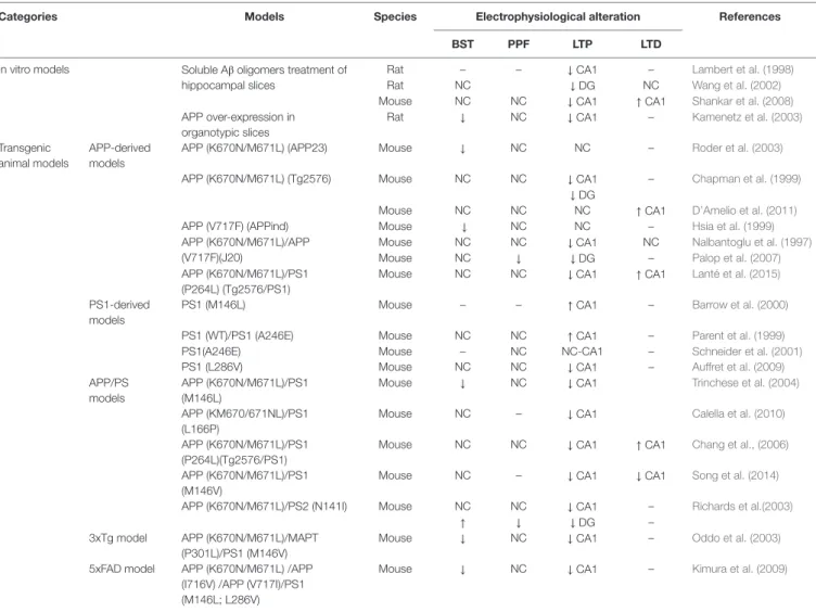
TABLE 1 | The table summarizes relevant data relative to experimental Alzheimer’s disease (AD) models for which hippocampal electrophysiological analyses were
performed.
Categories Models Species Electrophysiological alteration References BST PPF LTP LTD
In vitro models Soluble Aβ oligomers treatment of hippocampal slices
Rat – – ↓ CA1 – Lambert et al. (1998)
Rat NC ↓ DG NC Wang et al. (2002)
Mouse NC NC ↓ CA1 ↑ CA1 Shankar et al. (2008)
APP over-expression in organotypic slices
Rat ↓ NC ↓ CA1 – Kamenetz et al. (2003)
Transgenic animal models
APP-derived models
APP (K670N/M671L) (APP23) Mouse ↓ NC NC – Roder et al. (2003)
APP (K670N/M671L) (Tg2576) Mouse NC NC ↓ CA1 – Chapman et al. (1999)
↓ DG
Mouse NC NC NC ↑ CA1 D’Amelio et al. (2011)
APP (V717F) (APPind) Mouse ↓ NC NC – Hsia et al. (1999)
APP (K670N/M671L)/APP (V717F)(J20)
Mouse NC NC ↓ CA1 NC Nalbantoglu et al. (1997)
Mouse NC ↓ ↓ DG – Palop et al. (2007)
APP (K670N/M671L)/PS1 (P264L) (Tg2576/PS1)
Mouse NC NC ↓ CA1 ↑ CA1 Lanté et al. (2015)
PS1-derived models
PS1 (M146L) Mouse – – ↑ CA1 – Barrow et al. (2000)
PS1 (WT)/PS1 (A246E) Mouse NC NC ↑ CA1 – Parent et al. (1999)
PS1(A246E) Mouse – NC NC-CA1 – Schneider et al. (2001)
PS1 (L286V) Mouse NC NC ↓ CA1 – Auffret et al. (2009)
APP/PS models
APP (K670N/M671L)/PS1 (M146L)
Mouse ↓ NC ↓ CA1 Trinchese et al. (2004)
APP (KM670/671NL)/PS1 (L166P)
Mouse NC – ↓ CA1 Calella et al. (2010)
APP (K670N/M671L)/PS1 (P264L)(Tg2576/PS1)
Mouse NC NC ↓ CA1 ↑ CA1 Chang et al., (2006)
APP (K670N/M671L)/PS1 (M146V)
Mouse NC – ↓ CA1 ↓ CA1 Song et al. (2014)
APP (K670N/M671L)/PS2 (N141I) Mouse NC NC ↓ CA1 – Richards et al.(2003)
↑ ↓ ↓ DG –
3xTg model APP (K670N/M671L)/MAPT (P301L)/PS1 (M146V)
Mouse ↓ NC ↓ CA1 – Oddo et al. (2003)
5xFAD model APP (K670N/M671L) /APP (I716V) /APP (V717I)/PS1 (M146L; L286V)
Mouse ↓ NC ↓ CA1 – Kimura et al. (2009)
Models were grouped into in vitro models, APP-derived, PS1-derived, APP/PS1, 3xTg, and 5xTg models. Electrophysiological readouts include basal synaptic transmission (BST), paired pulse facilitation (PPF), long-term potentiation (LTP), and long-term depression (LTD). For each model, we also report the principal references to the electrophysiological studies.
In addition, a recent study described alteration of
mGlu-dependent LTD in 4-month-old Tg2576 mice, elicited by perfusion
of the group I mGlu agonist DHPG (
Mango and Nisticò, 2018
).
PS1-Derived Models
These models over-express the human presenilin gene (PS1)
encoding an FAD mutation. Being part of the secretase complex, this
gene is involved in the APP proteolysis. Presenilin variants do not
produce neuropathology, but potentiate plaque deposition in APP
transgenic mice. Electrophysiological studies have been performed
on M146L, A246E, and L286V mutants (
Parent et al., 1999
;
Barrow
et al., 2000
;
Schneider et al., 2001
;
Auffret et al., 2009
). A mouse
model was also generated harboring the PS1ÆE9 FAD mutation,
which results in a functional and non-cleavable modification of
PS1 (
Zaman et al., 2000
). One knock-in mouse was engineered
in which mouse PS1 was exchanged by its mutant M146V (
Sun
et al., 2005
). These presenilin FAD mutants consistently exhibit
an age-dependent increase of Aβ42 with minor effect on Aβ40;
however, they do show amyloid plaques, tau pathology, cholinergic
alterations, or neurodegeneration and present only a mild cognitive
deficit (
Games et al., 2006
). Most studies report that young adult
(up to 6 months) transgenic mice over-expressing PS1M146L,
PS1M146V, PS1A246E, PS1ΔE9, or PS1L286V display enhanced
CA1-LTP elicited using different conditioning protocols (
Schneider
et al., 2001
;
Oddo et al., 2003
;
Dewachter et al., 2008
;
Auffret et al.,
2009
). So far, no study described LTD in these mouse models.
APP/PS1 Models
To accelerate the brain Aβ accumulation and plaque aggregation,
researchers crossed APP- and PS1-derived animal models.
Most electrophysiological studies focused on double transgenic
models harboring the human APPswe transgene together with
the PS transgene.
APP/PS1 double mutant mice develop rapid and extensive Aβ
plaque accumulation, tau pathology, and cognitive defects, even
though they lack cholinergic deficits, neuronal loss, and NFTs
(
Morrissette et al., 2009
).
Most studies report a reduction of LTP in APPswe/PS1M146L
(
Gong et al., 2004
;
Trinchese et al., 2004
,
Trinchese et al., 2008
),
APPswe/PS1P246L (
Chang et al., 2006
), APPswe/PS1L166P
(
Calella et al., 2010
), and APPswe/PS2N141I (
Richards et al.,
2003
) mice. Using a standard LFS protocol,
Chang and coauthors
(2006)
report a linear decline in CA1-LTD expression between 9
and 20 months of age in the APPswe/PS1P246L model. Moreover,
loss of LTD was described in the APPswe/PS1M146V mouse
model (
Song et al., 2014
). Also, a recent study has shown mGlu
receptor-LTD impairment in the APPswe/PSEN1/ΔE9 (
Yang et
al., 2016
), whereas no alteration in the basal transmission was
found in this model (
Volianskis et al., 2010
).
3xTg Model
The 3xTg model over-expressing human APPswe and tau
MAPTP301L and encoding a knock-in of PS1M146V was
generated for the first time by
Oddo et al. (2003)
. These mice display
both Aβ aggregates and NFT and show hippocampal-dependent
memory decline during aging. Also, they show cholinergic
alterations and neuronal loss in the cortex (
Oddo et al., 2003
;
Perez et al., 2010
). This model is advantageous since it presents
a significant intracellular Aβ deposition before the occurrence of
extracellular plaques, which become evident around 12 months of
age, and notably, it develops NFTs comparable to humans.
Concerning the functional aspects, 6-month-old 3xTg mice
display impairment of LTP (
Oddo et al., 2003
), which correlates
with intracellular Aβ well before plaque and tangle pathology.
Recently, other triple transgenic mice have been generated
harboring APP, PS2, and tau mutations (
Rhein et al., 2009
;
Grueninger et al., 2010
) but electrophysiological investigations
have not been performed so far.
5xFAD Model
5xFAD mice (Tg6799 line) harbor three APP and PS2 (M146V
and L286V) mutations that are causally related to FAD (
Oakley
et al., 2006
). They exemplify one of the most early-onset mouse
models with robust amyloid pathology (
Oakley et al., 2006
;
Ohno et al., 2006
,
Ohno et al., 2007
). Indeed, 5xFAD mice
start developing amyloid deposition already from 2 months of
age and show early hippocampal dysfunction, as evidenced by
reduced basal synaptic transmission and LTP (
Kimura and
Ohno, 2009
;
Crouzin et al., 2013
). This mouse model exhibits
a strong pathology: at 1.5 months of age mice already express
intracellular Aβ42, which massively progresses at 2 months
of age with extracellular Aβ accumulation, senile plaques, and
lack of specific neuronal populations. Cognitive impairment is
reported at 4–6 months of age (
Oakley et al., 2006
). LTD has not
yet been investigated in this mouse model.
CLINICAL RELEVANCE OF LTP/LTD
Many of the mechanisms underlying LTP and LTD in the rodent
hippocampal slice preparation are shared also in hippocampal
tissue from patients undergoing surgery for intractable temporal
lobe epilepsy. Indeed, LTP is induced in the temporal lobe
and in the dentate gyrus in humans using similar protocols of
stimulation (
Chen et al., 1996
;
Beck et al., 2000
) and is modulated
by the different pharmacological approaches just as in
non-clinical models. These studies further support the notion that
also the human brain manifests LTP- and LTD-like events even
though linking synaptic plasticity to human learning and memory
remains a challenge (
Bliss and Cooke, 2011
). It also should be kept
in mind that the human tissue investigated in electrophysiological
studies is found in a pathological state, deriving from patients
with an epileptic focus in the temporal lobe.
Notably, LTP- and LTD-like events are nowadays exploited
in humans for therapeutic purposes. These plastic changes
can be induced through several noninvasive techniques such
as repetitive transcranial magnetic stimulation (rTMS) and
transcranial direct current stimulation (tDCS) and are used for the
treatment of a variety of neurological and psychiatric conditions,
such as epilepsy, drug addiction, depression, Parkinson disease,
neuropathic pain, tinnitus, and stroke (
Schulz et al., 2013
).
We can therefore hypothesize that stimulation of
neuroplasticity in the early stages of AD, through pharmacological
and noninvasive approaches, can attenuate disease progression.
Even though numerous therapeutic interventions reverse
synaptic alterations and improve behavior in the non-clinical
AD models, so far, there has been no successful translation into
disease-modifying compounds in humans (
Nisticò et al., 2012
).
PREDICTIVE VALUE OF AD MODELS
Considering the recent clinical trial failures in AD, there has
been considerable discussion as to whether results obtained from
non-clinical models are predictive or simply misleading. There
are numerous reasons why non-clinical studies may have failed
to predict clinical trial outcome. One of the main issues it that
Tg mice carry FAD mutations, accounting for only 5–10% of all
AD cases, while the vast majority of AD cases are sporadic (sAD).
As a consequence, these models have a low face and predictive
validity for the sporadic form of AD. Moreover, transgenic models
normally overexpress APP with consequent overproduction
several APP fragments. Of note, a knock-in mice with a phenotype
more similar to human was recently generated (
Saito et al., 2014
).
Another limitation is that each mouse model develops only
specific characteristics of AD (i.e. Aβ vs. tau pahology) and does
not recapitulate the complexity of the human disease. It has also
to be taken into account whether animal models display a similar
spatiotemporal profile of disease progression when compared
to AD patients. For example, cognitive deficits usually precede
plaque load in mice, whereas the opposite occurs in patients. An
important issue to be considered is that the majority of AD models
lack neuronal cell death, while a substantial neurodegeneration is
observed in the human AD brain.
It can be argued that the various non-clinical models typify
specific disease-related targets and pathways, a potential
advantage for testing candidate molecules on selected targets
involved in AD pathogenesis. Indeed, this target-driven approach
in non-clinical models has been translated over the years into
numerous clinical studies (
Nisticò et al., 2012
).
In addition to intrinsic limitations of animal models,
experimental bias is another crucial factor. For example,
gender- and litter-dependent differences, variability in transgene
expression, and the different genetic background among models
and even between active treatment and placebo groups should all
be considered in translation. Also, diversities in brain anatomy,
neuronal physiology, metabolism, and disease susceptibility play
a central role. Moreover, given the complex dynamics of
drug-target interactions, in vivo studies in non-clinical models should
include a complete pharmacokinetics/pharmacodynamics profile
in order to ensure that the dose range and timing are specific to
the target (e.g. based on receptor occupancy and/or correlative
biomarker data). On the other hand, the therapeutic window and
the off-target effects that may lead to adverse effects should also
be identified especially for behavioral experiments.
To improve clinical translation, non-clinical endpoints
need to be accurately selected and should employ a
combination of disease-relevant approaches such as integrated
neurophysiological/behavioral paradigms. Electrophysiological
techniques record neural activity from large neural populations
down to single cells and are ideal to measure synaptic plasticity,
as well as firing activity and neural oscillations. A limitation is
that this approach does not address spatiotemporal information
and is not suitable for noninvasively detecting activity from deep
brain regions. In contrast, functional neuroimaging provides
a noninvasive spatiotemporal readout of changes in brain
function, making it an invaluable tool for most clinical studies.
Unfortunately, the use functional imaging has been limited in
non-clinical setting due to the restricted applicability to animal
models and the relatively high cost.
CONCLUSIONS
We have previously hypothesized that alterations in normal
synaptic function are not only a key feature but also a leading cause
of disease (
Nisticò and Collingridge, 2012
). In this respect, LTP and
LTD can serve as synapse survival and death signals, respectively.
Thus, conditions that promote LTD, i.e. following excessive Aβ load
in the early-onset forms of disease, can lead to loss of synapses. On
the other hand, promoting LTP, which is known to inhibit LTD
(
Peineau et al., 2007
), can represent a protective mechanism to
preserve synaptic plasticity and brain connectivity.
It seems important to investigate the molecular mechanisms
that influence plasticity in the human brain and to determine
whether its vulnerability to aging and neurodegeneration can
be modified by pharmacological intervention. Considering that
AD is a complex disease affecting multiple signaling pathways,
therapeutic strategies should not be directed to a single target
rather to a combination of targets. To ensure a successful outcome,
therapy should start at an early stage of disease. In addition, highly
sensitive and specific biomarkers should identify susceptible
individuals at the onset of disease (
Hampel et al., 2014
).
Generally, the predictive value of non-clinical models in the
drug discovery process has been largely debated independently
of the therapeutic area (
McGonigle and Ruggeri, 2014
;
Mullane
and Williams, 2019
). Accordingly, several compounds showing
robust efficacy in experimental models of AD have failed so far in
clinical trials. Once a lead compound is selected, selection of
non-clinical endpoints through integrated approaches should reflect
the clinical endpoints in phase I studies. Correct design of
non-clinical studies can be a long, complex, and expensive process that
may slow down the course of drug development (
Mohs and Greig,
2017
); nonetheless, the probability of successful approval and
hence time saving and return on investment is certainly increased.
AUTHOR CONTRIBUTIONS
DM, AS and GC prepared the manuscript and reviewed the
drafts, MF and MC reviewed the drafts, RN conceived the idea,
prepared the manuscript and reviewed the drafts. All authors
contributed to the writing and final approval of the manuscript.
FUNDING
REFERENCES
Ashe, K. H., and Zahs, K. R. (2010). Probing the biology of Alzheimer’s disease in mice. Neuron 66, 631–645. doi: 10.1016/j.neuron.2010.04.031
Auffret, A., Gautheron, V., Repici, M., Kraftsik, R., Mount, H. T., Mariani, J., et al. (2009). Age-dependent impairment of spine morphology and synaptic plasticity in hippocampal CA1 neurons of a presenilin 1 transgenic mouse model of Alzheimer’ s disease. J. Neurosci. 29, 10144–10152. doi: 10.1523/ JNEUROSCI.1856-09.2009
Balducci, C., Mehdawy, B., Mare, L., Giuliani, A., Lorenzini, L., Sivilia, S., et al. (2011). The γ-secretase modulator CHF5074 restores memory and hippocampal synaptic plasticity in plaque-free Tg2576 mice. J. Alzheimers Dis. 24, 799–816. doi: 10.3233/JAD-2011-101839
Barrow, P. A., Empson, R. M., Gladwell, S. J., Anderson, C. M., Killick, R., Yu, X., et al. (2000). Functional phenotype in transgenic mice expressing mutant human presenilin- 1. Neurobiol. Dis. 7, 119–126. doi: 10.1006/nbdi.1999.0276 Beck, H., Goussakov, I. V., Lie, A., Helmstaedter, C., and Elger, C. E. (2000).
Synaptic plasticity in the human dentate gyrus. J. Neurosci. 20, 7080–7086. doi: 10.1523/JNEUROSCI.20-18-07080.2000
Bliss, T. V., and Collingridge, G. L. (1993). A synaptic model of memory: long-termpotentiation in the hippocampus. Nature 3616, 31–39. doi: 10.1038/ 361031a0
Bliss, T. V., and Cooke, S. F. (2011). Long-term potentiation and long-term depression: a clinical perspective. Clinics (Sao Paulo) 1, 3–17. doi: 10.1590/ S1807-59322011001300002
Bliss, T. V., Collingridge, G. L., and Morris, R. G. M., (2007). The Hippocampus
Book. Andersen P, Morris RGM, Amaral DG, Bliss TVP, O’Keefe J, editors. New
York: Oxford Univ. Press, 343–474.
Calella, A. M., Farinelli, M., Nuvolone, M., Mirante, O., Moos, R., Falsig, J., et al. (2010). Prion protein and Aâ-related synaptic toxicity impairment. EMBO Mol.
Med. 2, 306–314. doi: 10.1002/emmm.201000082
Caraci, F., Gulisano, W., Guida, C. A., Impellizzeri, A. A., Drago, F., Puzzo, D., et al. (2015). A key role for TGF-β1 in hippocampal synaptic plasticity and memory.
Sci. Rep. 5, 11252. doi: 10.1038/srep11252
Caraci, F., Spampinato, S. F., Morgese, M. G., Tascedda, F. 4., Salluzzo, M. G., Giambirtone, M. C., et al. (2018). Neurobiological links between depression and AD: the role of TGF-β1 signaling as a new pharmacological target.
Pharmacol. Res. 130, 374–384. doi: 10.1016/j.phrs.2018.02.007
Cavallucci, V., Berretta, N., Nobili, A., Nisticò, R., Mercuri, N. B., and D’Amelio, M. (2013). Calcineurin inhibition rescues early synaptic plasticity deficits in a mouse model of Alzheimer’s disease. Neuromolecular Med. 15, 541–548. doi: 10.1007/s12017-013-8241-2
Chang, E. H., Savage, M. J., Flood, D. G., Thomas, J. M., Levy, R. B., Mahadomrongkul, V., et al. (2006). AMPA receptor downscaling at the onset of Alzheimer’s disease pathology in double knockin mice. Proc. Natl. Acad. Sci.
U.S.A. 103, 3410–3415. doi: 10.1073/pnas.0507313103
Chapman, P. F., White, G. L., Jones, M. W., Cooper-Blacketer, D., Marshall, V. J., Irizarry, M., et al. (1999). Impaired synaptic plasticity and learning in aged amyloid precursor protein transgenic mice. Nat. Neurosci. 2, 271–276. doi: 10.1038/6374
Chen, W. R., Lee, S., Kato, K., Spencer, D. D., Shepherd, G. M., and Williamson, A. (1996). Long-term modifications of synaptic efficacy in the human inferior and middle temporal cortex. Proc. Natl. Acad. Sci. U.S.A. 93, 8011–8015. doi: 10.1073/pnas.93.15.8011
Collingridge, G. L., Peineau, S., Howland, J. G., and Wang, Y. T. (2010). Long-term depression in the CNS. Nat. Rev. Neurosci. 11, 459–473. doi: 10.1038/nrn2867 Crouzin, N., Baranger, K., Cavalier, M., Marchalant, Y., Cohen-Solal, C., Roman,
F. S., et al. (2013). Area-Specific alterations of synaptic plasticity in the 5XFAD mouse model of Alzheimer’s disease: dissociation between Somato sensory cortex and hippocampus. PLoS One 8, e74667. doi: 10.1371/journal. pone.0074667
D’Amelio, M., Cavallucci, V., Middei, S., Marchetti, C., Pacioni, S., Ferri, A., et al. (2011). Caspase-3 triggers early synaptic dysfunction in a mouse model of Alzheimer’s disease. Nat. Neurosci. 14, 69–76. doi: 10.1038/nn.2709
Dewachter, I., Ris, L., Croes, S., Borghgraef, P., Devijver, H., Voets, T., et al. (2008). Modulation of synaptic plasticity and Tau phosphorylation by wild-type and mutant presenilin 1. Neurobiol. Aging 29, 639–652. doi: 10.1016/j. neurobiolaging.2006.11.019
Dudek, S. M., and Friedlander, M. J. (1996). Developmental down-regulation of LTD in cortical layer IV and its independence of modulation by inhibition.
Neuron 16, 1097–1106. doi: 10.1016/S0896-6273(00)80136-1
Elder, G. A., Gama Sosa, M. A., and De Gasperi, R. (2010). Transgenic mouse models of Alzheimer’s disease. Mt. Sinai J. Med. 77, 69–81. doi: 10.1002/ msj.20159
Fitzjohn, S. M., Kingston, A. E., Lodge, D., and Collingridge, G. L. (1999). DHPG-induced LTD in area CA1 of juvenile rat hippocampus: characterisation and sensitivity to novel mGlu receptor antagonists. Neuropharmacology 38, 1577– 1583. doi: 10.1016/S0028-3908(99)00123-9
Fitzjohn, S. M., Morton, R. A., Kuenzi, F., Rosahl, T. W., Shearman, M., Lewis, H., et al. (2001). Age-related impairment of synaptic transmission but normal long-term potentiation in transgenic mice that overexpress the human APP695SWE mutant form of amyloid precursor protein. J. Neurosci. 21, 4691–4698. doi: 10.1523/JNEUROSCI.21-13-04691.2001
Frey, U., Krug, M., Reymann, K. G., and Matthies, H. (1988). Anisomycin, an inhibitor of protein synthesis, blocks late phases of LTP phenomena in the hippocampal CA1 region in vitro. Brain Res. 452, 57–65. doi: 10.1016/0006-8993(88) 90008-X
Games, D., Buttini, M., Kobayashi, D., Schenk, D., and Seubert, P. (2006). Mice as models: transgenic approaches and Alzheimer’s disease. J. Alzheimers Dis., 9, 133–149. doi: 10.3233/JAD-2006-9S316
Goate, A., Chartier-Harlan, M. C., Mullan, M., Brown, J., Crowford, F., Fidani, L., et al. (1991). Segregation of a missense mutation in the amyloid precursor protein gene with familial Alzheimer’s disease. Nature 349, 704–706. doi: 10.1038/349704a0
Gong, B., Vitolo, O. V., Trinchese, F., Liu, S., Shelanski, M., and Arancio, O. (2004). Persistent improvement in synaptic and cognitive functions in an Alzheimer mouse model after rolipram treatment. J. Clin. Invest. 114, 1624–1634. doi: 10.1172/JCI22831
Grueninger, F., Bohrmann, B., Czech, C., Ballard, T. M., Frey, J. R., Weidensteiner, C., et al. (2010). Phosphorylation of Tau at S422 is enhanced by Abeta in TauPS2APP triple transgenic mice. Neurobiol. Dis. 37, 294–306. doi: 10.1016/j. nbd.2009.09.004
Hampel, H., Lista, S., Teipel, S. J., Garaci, F., Nisticò, R., Blennow, K., et al. (2014). Perspective on future role of biological markers in clinical therapy trials of Alzheimer’s disease: a long-range point of view beyond 2020. Biochem.
Pharmacol. 88, 426–449. doi: 10.1016/j.bcp.2013.11.009
Hardy, J. A., and Higgins, G. A. (1992). Alzheimer’s disease: the amyloid cascade hypothesis. Science 256, 184–185. doi: 10.1126/science.1566067
Hsia, A. Y., Masliah, E., McConlogue, L., Yu, G. Q., Tatsuno, G., Hu, K., et al. (1999). Plaque-independent disruption of neural circuits in Alzheimer’s disease mouse models. Proc. Natl. Acad. Sci. U.S.A. 96, 3228–3233. doi: 10.1073/pnas.96.6.3228
Jo, J., Whitcomb, D. J., Olsen, K. M., Kerrigan, T. L., Lo, S. C., Bru-Mercier, G., et al. (2011). Abeta(1-42) inhibition of LTP is mediated by a signaling pathway involving caspase-3, Akt1 and GSK-3beta. Nat. Neurosci. 14, 545–547. doi: 10.1038/nn.2785
Kamenetz, F., Tomita, T., Hsieh, H., Seabrook, G., Borchelt, D., Iwatsubo, T., et al. (2003). APP processing and synaptic function. Neuron 37, 925–937. doi: 10.1016/S0896-6273(03)00124-7
Kimura, R., and Ohno, M. (2009). Impairments in remote memory stabilization precede hippocampal synapticand cognitive failuresin 5XFAD Alzheimer mouse model. Neurobiol. Dis. 33, 229–235. doi: 10.1016/j.nbd.2008.10.006 Koppensteiner, P., Trinchese, F., Fà, M., Puzzo, D., Gulisano, W., Yan, S., et al.
(2016). Time dependent reversal of synaptic plasticity induced by physiological concentrations of oligomeric Aβ42: an early index of Alzheimer’s disease. Sci.
Rep. 1, 6–32553. doi: 10.1038/srep32553
Lambert, M. P., Barlow, A. K., Chromy, B. A., Edwards, C., Freed, R., Liosatos, M., et al. (1998). Diffusible, nonfibrillar ligands derived from Abeta1-42 are potent central nervous system neurotoxins. Proc. Natl. Acad. Sci. U.S.A. 95, 6448– 6453. doi: 10.1073/pnas.95.11.6448
Lanté, F., Chafai, M., Raymond, E. F., Pereira, A. R., Mouska, X., Kootar, S., et al. (2015). Subchronic glucocorticoid receptor inhibition rescues early episodic memory and synaptic plasticity deficits in a mouse model of Alzheimer’s disease. Neuropsychopharmacology 40, 1772–1781. doi: 10.1038/npp.2015.25 Lawrence, J. L., Tong, M., Alfulaij, N., Sherrin, T., Contarino, M., White, M. M.,
fear conditioning by a N-terminal beta amyloid fragment. J. Neurosci. 34, 14210–14218. doi: 10.1523/JNEUROSCI.0326-14.2014
Li, S., Hong, S., Shepardson, N. E., Walsh, D. M., Shankar, G. M., and Selkoe, D. (2009). Soluble oligomers of amyloid Beta protein facilitate hippocampal long-term depression by disrupting neuronal glutamate uptake. Neuron 62, 788–801. doi: 10.1016/j.neuron.2009.05.012
Malenka, R. C., and Bear, M. F. (2004). LTP and LTD: an embarrassment of riches.
Neuron 44, 5–21. doi: 10.1016/j.neuron.2004.09.012
Mango, D., and Nisticò, R. (2018). Role of ASIC1a in Aβ-induced synaptic alterations in the hippocampus. Pharmacol. Res. 131, 61–65. doi: 10.1016/j. phrs.2018.03.016
Mango, D., Weisz, F., and Nisticò, R. (2016). Ginkgolic acid protects against Aβ-induced synaptic dysfunction in the hippocampus. Front. Pharmacol. 7, 401. doi: 10.3389/fphar.2016.00401
Marchetti, C., and Marie, H. (2011). Hippocampal synaptic plasticity in Alzheimer’s disease: what have we learned so far from transgenic models? Rev.
Neurosci. 22, 373–402. doi: 10.1515/rns.2011.035
McGonigle, P., and Ruggeri, B. (2014). Animal models of human disease: challenges in enabling translation. Biochem. Pharmacol. 87, 162–171. doi: 10.1016/j.bcp.2013.08.006
Mohs, R. C., and Greig, N. H. (2017). Drug discovery and development: role of basic biological research. Alzheimers Dement. (N Y) 3, 651–657. doi: 10.1016/j. trci.2017.10.005
Morley, J. E., Farr, S. A., Banks, W. A., Johnson, S. N., Yamada, K. A., and Xu, L. (2010). A physiological role for amyloid-beta protein: enhancement of learning and memory. J. Alzheimers Dis. 19, 441–449. doi: 10.3233/JAD-2010-1230 Morrissette, D. A., Parachikova, A., Green, K. N., and LaFerla, F. M. (2009).
Relevance of transgenic mouse models to human Alzheimer disease. J. Biol.
Chem. 284, 6033–6037. doi: 10.1074/jbc.R800030200
Mullan, M., Crawford, F., Axelman, K., Houlden, H., Lilius, L., Winblad, B., et al. (1992). A pathogenic mutation for probable Alzheimer’s disease in the APP gene at the N-terminus of beta-amyloid. Nat. Genet. 1, 345–347. doi: 10.1038/ ng0892-345
Mullane, K., and Williams, M. (2019). Preclinical Models of Alzheimer’s disease: Relevance and Translational Validity. Curr. Protoc. Pharmacol. 84 (1), e57. doi: 10.1002/cpph.57
Murrell, J., Farlow, M., Ghetti, B., and Benson, M. D. (1991). A mutation in the amyloid precursor protein associated with hereditary Alzheimer’s disease.
Science 254, 97–99. doi: 10.1126/science.1925564
Nalbantoglu, J., Tirado-Santiago, G., Lahsaïni, A., Poirier, J., Goncalves, O., Verge, G., et al. (1997). Impaired learning and LTP in mice expressing the carboxy terminus of the Alzheimer amyloid precursor protein. Nature 387, 500–505. doi: 10.1038/387500a0
Neves, G., Cooke, S. F., and Bliss, T. V. (2008). Synaptic plasticity, memory and the hippocampus: a neural network approach to causality. Nat. Rev. Neurosci. 9, 65–75. doi: 10.1038/nrn2303
Nilsberth, C., Westlind-Danielsson, A., Eckman, C. B., Condron, M. M., Axelman, K., Forsell, C., et al. (2001). The ‘Arctic’ APP mutation (E693G) causes Alzheimer’s disease by enhanced Abeta protofibril formation. Nat. Neurosci. 4, 887–893. doi: 10.1038/nn0901-887
Nisticò, R., and Collingridge, G. (2012). The synaptic basis of Alzheimer’s disease.
Eur. J. Neurodegener. Dis. 1, 21–33.
Nisticò, R., Pignatelli, M., Piccinin, S., Mercuri, N. B., and Collingridge, G. (2012). Targeting synaptic dysfunction in Alzheimer’s disease therapy. Mol. Neurobiol. 46, 572–587. doi: 10.1007/s12035-012-8324-3
Oakley, H., Cole, S. L., Logan, S., Maus, E., Shao, P., Craft, J., et al. (2006). Intraneuronal beta-amyloid aggregates, neurodegeneration, and neuron loss in transgenic micewith five familial Alzheimer’s disease mutations: potential factors in amyloid plaque formation. J. Neurosci. 26, 10129–10140. doi: 10.1523/JNEUROSCI.1202-06.2006
Oddo, S., Caccamo, A., Shepherd, J. D., Murphy, M. P., Golde, T. E., Kayed, R., et al. (2003). Triple-transgenic model of Alzheimer’s disease with plaques and tangles: intracellular A β and synaptic dysfunction. Neuron 39, 409–421. doi: 10.1016/S0896-6273(03)00434-3
Ohno, M., Chang, L., Tseng, W., Oakley, H., Citron, M., Klein, W. L., et al. (2006). Temporal memory deficits in Alzheimer’s mouse models: rescue by genetic deletion of BACE1. Eur. J. Neurosci. 23, 251–260. doi: 10.1111/j.1460-9568. 2005.04551.x
Ohno, M., Cole, S. L., Yasvoina, M., Zhao, J., Citron, M., Berry, R., et al. (2007). BACE1 gene deletion prevents neuron loss and memory deficits in 5XFAD APP/PS1 transgenic mice. Neurobiol. Dis. 26, 134–145. doi: 10.1016/j. nbd.2006.12.008
Palop, J. J., Chin, J., Roberson, E. D., Wang, J., Thwin, M. T., Bien-Ly, N., et al. (2007). Aberrant excitatory neuronal activity and compensatory remodeling of inhibitory hippocampal circuits in mouse models of Alzheimer’s disease.
Neuron 55, 697–711. doi: 10.1016/j.neuron.2007.07.025
Panja, D., and Bramham, C. R. (2014). BDNF mechanisms in late LTP formation: a synthesis and breakdown. Neuropharmacology 76, 664–676. doi: 10.1016/j. neuropharm.2013.06.024
Parent, A., Linden, D. J., Sisodia, S. S., and Borchelt, D. R. (1999). Synaptic transmission and hippocampal long-term potentiation in transgenic mice expressing FAD-linked presenilin 1. Neurobiol. Dis. 6, 56–62. doi: 10.1006/ nbdi.1998.0207
Park, P., Volianskis, A., Sanderson, T. M., Bortolotto, Z. A., Jane, D. E., Zhuo, M., et al. (2014). NMDA receptor-dependent long-term potentiation comprises a family of temporally overlapping forms of synaptic plasticity that are induced by different patterns of stimulation. Philos. Trans. R. Soc. Lond., B, Biol. Sci. 369, 20130131. doi: 10.1098/rstb.2013.0131
Park, P., Sanderson, T. M., Amici, M., Choi, S. L., Bortolotto, Z. A., Zhuo, M., et al. (2016). Calcium-permeable AMPA receptors mediate the induction of the protein kinase a-dependent component of long-term potentiation in the hippocampus. J. Neurosci. 36, 622–631. doi: 10.1523/JNEUROSCI.3625-15. 2016
Peineau, S., Rabiant, K., Pierrefiche, O., and Potier, B. (2018). Synaptic plasticity modulation by circulating peptides and metaplasticity: involvement in Alzheimer’s disease. Pharmacol. Res. 130, 385–401. doi: 10.1016/j.phrs.2018. 01.018
Peineau, S., Taghibiglou, C., Bradley, C., Wong, T. P., Liu, L., Lu, J., et al. (2007). LTP inhibits LTD in the hippocampus via regulation of GSK3beta. Neuron 53, 703–17.
Penn, A. C., Zhang, C. L., Georges, F., Royer, L., Breillat, C., Hosy, E., et al. (2017). Hippocampal LTP and contextual learning require surfacediffusion of AMPA receptors. Nature 549, 384–388. doi: 10.1038/nature23658
Perez, S. E., He, B., Muhammad, N., Oh, K. J., Fahnestock, M., Ikonomovic, M., et al. (2010). Cholinotrophic basal forebrain system alterations in 3xTg-AD transgenic mice. Neurobiol. Dis. 41, 338–352. doi: 10.1016/j.nbd.2010. 10.002
Puzzo, D., Privitera, L., Fa’, M., Staniszewski, A., Hashimoto, G., Aziz, F., et al. (2011). Endogenous amyloid-beta is necessary for hippocampal synaptic plasticity and memory. Ann. Neurol. 69, 819–830. doi: 10.1002/ana.22313 Puzzo, D., Privitera, L., Leznik, E., Fà, M., Staniszewski, A., Palmeri, A., et al. (2008).
Picomolar amyloid-beta positively modulates synaptic plasticity and memory in hippocampus. J. Neurosci. 28, 14537–14545. doi: 10.1523/JNEUROSCI.2692- 08.2008
Rammes, G., Mattusch, C., Wulff, M., Seeser, F., Kreuzer, M., Zhu, K., et al. (2017). Involvement of GluN2B subunit containing N-methyl-d-aspartate (NMDA) receptors in mediating the acute and chronic synaptotoxic effects of oligomeric amyloid-beta (Abeta) in murine models of Alzheimer’s disease (AD). Neuropharmacology 123, 100–115. doi: 10.1016/j.neuropharm. 2017.02.003
Reymann, K. G., Matthies, H. K., Schulzeck, K., and Matthies, H. (1989). N-methyl-D-aspartate receptor activation is required for the induction of both early and late phases of long-term potentiation in rat hippocampal slices. Neurosci. Lett. 96, 96–101. doi: 10.1016/0304-3940(89)90249-8
Rhein, V., Song, X., Wiesner, A., Ittner, L. M., Baysang, G., Meier, F., et al. (2009). Amyloid-beta and tau synergistically impair the oxidative phosphorylation system in triple transgenic Alzheimer’s disease mice. Proc. Natl. Acad. Sci.
U.S.A. 24;106, 20057–20062. doi: 10.1073/pnas.0905529106
Richards, J. G., Higgins, G. A., Ouagazzal, A. M., Ozmen, L., Kew, J. N., Bohrmann, B., et al. (2003). PS2APP transgenic mice, coexpressing hPS2mut and hAPPswe, show age-related cognitive deficits associated with discrete brain amyloid deposition and inflammation. J. Neurosci. 23, 8989–9003. doi: 10.1523/JNEUROSCI.23-26-08989.2003
Roder, S., Danober, L., Pozza, M. F., Lingenhoehl, K., Wiederhold, K. H., and Olpe, H. R. (2003). Electrophysiological studies on the hippocampus and prefrontal cortex assessing the effects of amyloidosis in amyloid precursor
protein 23 transgenic mice. Neuroscience 120, 705–720. doi: 10.1016/S0306-4522(03) 00381-6
Saito, T., Matsuba, Y., Mihira, N., Takano, J., Nilsson, P., Itohara, S., et al. (2014). Single app knock-in mouse models of Alzheimer’s disease. Nat. Neurosci. 17, 661–663. doi: 10.1038/nn.3697
Shipton, O. A., Leitz, J. R., Dworzak, J., Acton, C. E., Tunbridge, E. M., Denk, F., et al. (2011). Tau protein is required for amyloid {beta}-induced impairment of hippocampal long-term potentiation. J. Neurosci. 31, 1688–1692. doi: 10.1523/ JNEUROSCI.2610-10.2011
Schneider, I., Reverse, D., Dewachter, I., Ris, L., Caluwaerts, N., Kuiperi, C., et al. (2001). Mutant presenilins disturb neuronal calcium homeostasis in the brain of transgenic mice, decreasing the threshold for excitotoxicity and facilitating long-term potentiation. J. Biol. Chem. 276, 11539–11544. doi: 10.1074/jbc.M010977200 Schulz, R., Gerloff, C., and Hummel, F. C. (2013). Non- invasive brain stimulation
in neurological diseases. Neuropharmacology 64, 579–587. doi: 10.1016/j. neuropharm.2012.05.016
Selkoe, D. J. (2002). Alzheimer’s disease is a synaptic failure. Science 298, 789–791. doi: 10.1126/science.1074069
Shankar, G. M., Li, S., Mehta, T. H., Garcia-Munoz, A., Shepardson, N., Smith, I., et al. (2008). Soluble amyloid β-protein dimers isolated directly from alzheimer disease patients potently impair synaptic plasticity and memory. Nat. Med. 14, 837–842. doi: 10.1038/nm1782
Song, S., Wang, X., Sava, V., Weeber, E. J., and Sanchez-Ramos, J. (2014). In vivo administration of granulocyte colony-stimulating factor restores long-term depression in hippocampal slices prepared from transgenic APP/PS1 mice. J.
Neurosci. Res. 92, 975–980. doi: 10.1002/jnr.23378
Spires-Jones, T., and Knafo, S. (2012). Spines, plasticity, and cognition in Alzheimer’s model mice. Neural Plast., 28, 319836. doi: 10.1155/2012/319836 Sturchler-Pierrat, C., Abramowski, D., Duke, M., Wiederhold, K. H., Mistl, C.,
Rothacher, S., et al. (1997). Two amyloid precursor protein transgenic mouse models with Alzheimer disease-like pathology. Proc. Natl. Acad. Sci. U.S.A. 94, 13287–13292. doi: 10.1073/pnas.94.24.13287
Sun, X., Beglopoulos, V., Mattson, M. P., and Shen, J. (2005). Hippocampal spatial memory impairments caused by the familial Alzheimer’s disease-linked presenilin 1 M146V mutation. Neurodegener. Dis. 2, 6–15. doi: 10.1159/ 000086426
Tanzi, R. E. (2005). The synaptic Abeta hypothesis of Alzheimer disease. Nat.
Neurosci. 8, 977–979. doi: 10.1038/nn0805-977
Trinchese, F., Liu, S., Battaglia, F., Walter, S., Mathews, P. M., and Arancio, O. (2004). Progressive age-related development of Alzheimer-like pathology in APP/PS1 mice. Ann. Neurol. 55, 801–814. doi: 10.1002/ana.20101
Trinchese, F., Fa’, M., Liu, S., Zhang, H., Hidalgo, A., Schmidt, S. D., et al. (2008). Inhibition of calpains improves memory and synaptic transmission in a mouse model of Alzheimer disease. J. Clin. Invest. 118, 2796–2807. doi: 10.1172/ JCI34254
Volianskis, A., Kostner, R., Molgaard, M., Hass, S., and Jensen., M. S. (2010). Episodic memory deficits are not related to altered glutamatergic synaptic transmission and plasticity in the CA1 hippocampus of the APPswe/ PS1deltaE9-deleted transgenic mice model of ss-amyloidosis. Neurobiol. Aging 31, 1173–1187. doi: 10.1016/j.neurobiolaging.2008.08.005
Wang, H. W., Pasternak, J. F., Kuo, H., Ristic, H., Lambert, M. P., Chromy, B., et al. (2002). Soluble oligomers of beta amyloid (1-42) inhibit long-term potentiation but not long-term depression in rat dentate gyrus. Brain Res. 924, 133–140. doi: 10.1016/S0006-8993(01)03058-X
Yang, F., Uéda, K., Chen, P., Ashe, K. H., and Cole, G. M. (2000). Plaque-associated alpha-synuclein (NACP) pathology in aged transgenic mice expressing amyloid precursor protein. Brain Res. 853, 381–383. doi: 10.1016/S0006-8993 (99)02207-6
Yang, W., Zhou, X., Zimmermann, H. R., Cavener, D. R., Klann, E., and Ma, T. (2016). Repression of the eIF2α kinase PERK alleviates mGluR-LTD impairments in a mouse model of Alzheimer’s disease. Neurobiol. Aging 41, 19–24. doi: 10.1016/j. neurobiolaging.2016.02.005
Zaman, S. H., Parent, A., Laskey, A., Lee, M. K., Borchelt, D. R., Sisodia, S. S., et al. (2000). Enhanced synaptic potentiation in transgenic mice expressing presenilin 1 familial Alzheimer’s disease mutation is normalized with a benzodiazepine. Neurobiol. Dis. 7, 54–63. doi: 10.1006/nbdi.1999.0271 Zhang, J., Malik, A., Choi, H. B., Ko, R. W., Dissing-Olesen, L., and MacVicar, B. A.
(2014). Microglial CR3 activation triggers long-term synaptic depression in the hippocampus via NADPH oxidase. Neuron 82, 195–207. doi: 10.1016/j.neuron. 2014.01.043
Conflict of Interest Statement: The authors declare that the research was conducted in the absence of any commercial or financial relationships that could be construed as a potential conflict of interest.
Copyright © 2019 Mango, Saidi, Cisale, Feligioni, Corbo and Nisticò. This is an open-access article distributed under the terms of the Creative Commons Attribution License (CC BY). The use, distribution or reproduction in other forums is permitted, provided the original author(s) and the copyright owner(s) are credited and that the original publication in this journal is cited, in accordance with accepted academic practice. No use, distribution or reproduction is permitted which does not comply with these terms.