Analysis of the gene expression profile of mouse male meiotic germ cells
q
Pellegrino Rossi
a,*
,1, Susanna Dolci
a,1, Claudio Sette
a,1, Federica Capolunghi
a,
Manuela Pellegrini
a, Maria Loiarro
a, Silvia Di Agostino
a, Maria Paola Paronetto
a,
Paola Grimaldi
a, Daniele Merico
b, Enzo Martegani
b, Raffaele Geremia
a aDipartimento di Sanita’ Pubblica e Biologia Cellulare, Sezione di Anatomia, Universita’ di Roma Tor Vergata, Via Montpellier 1, 00133 Rome, Italy
bDipartimento di Biotecnologie e Bioscienze, Universita’ di Milano Bicocca, Piazza della Scienza 2, 20126 Milan, Italy
Received 31 October 2003; received in revised form 7 November 2003; accepted 18 November 2003
Abstract
Wide genome analysis of difference in gene expression between spermatogonial populations from 7-day-old mice and pachytene
spermatocytes from 18-day-old mice was performed using Affymetrix gene chips representing , 12,500 mouse known genes or EST
sequences, spanning approximately 1/3rd of the mouse genome. To delineate differences in the profile of gene expression between mitotic
and meiotic stages of male germ cell differentiation, expressed genes were grouped in functional clusters. The analysis confirmed the
previously described pre-meiotic or meiotic expression for several genes, in particular for those involved in the regulation of the mitotic and
meiotic cell cycle, and for those whose transcripts are accumulated during the meiotic stages to be translated later in post-meiotic stages.
Differential expression of several additional genes was discovered. In few cases (pro-apoptotic factors Bak, Bad and Bax), data were in
conflict with the previously published stage-dependent expression of genes already known to be expressed in male germ cells. Northern blot
analysis of selected genes confirmed the results obtained with the microarray chips. Six of these were novel genes specifically expressed in
pachytene spermatocytes: a chromatin remodeling factor (chrac1/YCL1), a homeobox gene (hmx1), a novel G-coupled receptor for an
unknown ligand (Gpr19), a glycoprotein of the intestinal epithelium (mucin 3), a novel RAS activator (Ranbp9), and the A630056B21Rik
gene (predicted to encode a novel zinc finger protein). These studies will help to delineate the global patterns of gene expression
characterizing male germ cell differentiation for a better understanding of regulation of spermatogenesis in mammals.
q
2003 Elsevier B.V. All rights reserved.
Keywords: DNA microarray; Transcriptome analysis; Spermatogenesis; Spermatocytes; Spermatogonia; Meiosis
1. Results and discussion
Spermatogenesis is characterized by a mitotic
(sperma-togonia), a meiotic (spermatocytes) and a differentiative
haploid (spermatids) phase. The dissection of the
mechan-isms that regulate the mitotic and meiotic cell cycles in
mammalian germ cells is useful for a better understanding
of the molecular requirements for spermatogenesis to
occur, and thus for the understanding of male sterility,
which is often based on lack of spermatogonial divisions or
meiotic blocks. Spermatogonia actively proliferate under
the control of growth factors released by somatic cells of
the seminiferous epithelium, such as Bone morphogenetic
protein 4 (Bmp4), which stimulates the Alk3 receptor
expressed in spermatogonial stem cells (
Pellegrini et al.,
2003
), and Kit Ligand (also called Stem Cell Factor), which
activates the c-kit receptor expressed in differentiating
spermatogonia (
Sorrentino et al., 1991; Yoshinaga et al.,
1991; Rossi et al., 1993; Schrans-Stassen et al., 1999; Dolci
et al., 2001
).
Few informations are available on the control of the
differentiation of spermatogonia into spermatocytes, i.e.
their entrance into the meiotic cell cycle, which is
characterized by two cell divisions and genetic exchange
(crossing-over) between homologous chromosomes, and
produce four haploid spermatids from each diploid
progenitor cell (
Roeder, 1997
). Insights into the molecular
mechanisms of the progression to the metaphase of the first
meiotic division have been obtained by treatment of
1567-133X/$ - see front matter q 2003 Elsevier B.V. All rights reserved. doi:10.1016/j.modgep.2003.11.003
www.elsevier.com/locate/modgep
q
Supplementary data associated with this article can be found, in the online version, at doi:10.1016/j.modgep.2003.11.003.
1 The first three authors contributed equally to this paper.
* Corresponding author. Tel.: þ39-672596272; fax: þ39-672596268. E-mail address: [email protected] (P. Rossi).
cultured spermatocytes with okadaic acid (OA), which
overcomes normal checkpoints that ensure in vivo the slow
progression of the meiotic prophase. OA triggers the
sequential activation of ERK1, p90Rsk2 and Nek2, thus
leading to chromosome condensation and progression to
metaphase with the concurrent activation of the cyclin
B/cdk1 complex (
Sette et al., 1999; Di Agostino et al.,
2002
). The spermatids that result at the end of meiosis will
then undergo spermiogenesis with the final production of
mature spermatozoa. The aim of our work was to obtain a
general profile of the expression pattern of genes
specifi-cally involved in the transition from the mitotic to the
meiotic cell cycle and in the progression through the meiotic
cell cycle.
DNA microarrays can be used to measure the expression
patterns of thousands of genes in parallel, allowing to
monitor changes in gene expression occurring during
developmental events (
Schena, 1996
). Analysis of the
results obtained with the microarray technique allows the
clustering of expressed genes in functional classes. We used
this approach in order to identify genes specifically involved
in the meiotic program and to group these genes in clusters
that should give more informations about the molecular
interactions required for this peculiar type of cell cycle in
mammals.
We prepared complementary RNAs from two germ cell
types at different developmental stages purified from testes
of pre-puberal mice, spermatogonia from 7-day-old mice
and spermatocytes from 18-day-old mice. The
spermato-gonial population obtained from 7-day-old mice (type A and
B spermatogonia) is contaminated by 10% of somatic cells,
whereas germ cells in the meiotic prophase are absent
(
Dolci et al., 2001; Pellegrini et al., 2003
; see also Section 2).
Spermatocytes obtained after elutriation from 18-day-old
mice are in the middle – late pachytene stage of the first
meiotic prophase (85%) and in the leptotene – zygotene
stage (10%). Contamination of spermatogonia and somatic
cells in the meiotic cell population is less than 5%, whereas
round spermatids, which are a common contaminant of
pachytene fractions when using adult testis (
Sette et al.,
1999; Di Agostino et al., 2002
), are absent, not being present
in the immature testis used. The cRNAs prepared from the
two different cell populations have been hybridized with
commercially available MG-U74Av2 GeneChip probe
arrays (Affymetrix Inc.), containing , 12,500 known
mouse genes or EST sequences, and thus spanning
approximately 1/3rd of the mouse genome. The analysis
was performed on duplicate chip arrays, using cRNAs from
two different cell preparations. The results of array data and
the comparative analysis were very similar in the duplicate
experiments
(
Fig.
1
)
and
are
available
in
the
supplemental data. The same data are also available online
at the addresses
http://www2.uniroma2.it/ricerca/ce/
absolutevaluestot.htm
and
http://www2.uniroma2.it/
ricerca/ce/comparativeanalysistot.htm
. The files named
‘Absolute Values’ contain filtered raw data of the absolute
analysis. Each of the five files contains data relative to
, 1/5th of the targets represented in the Affymetrix
MG-U74Av2 array. In the first column, the Affymetrix
identification number of the target oligonucleotide probe
pairs is indicated. Signal is a numeric value measuring the
abundance of a transcript revealed by the duplicate arrays
(C1 and C2: spermatocytes; G1 and G2: spermatogonia).
Detection indicates whether a transcript is present (P),
marginal (M), or absent (A) according to statistical analysis.
Detection P-value indicates the significance level of
detection call (P: P-value , 0.04; M: P-value between
0.04 and 0.06; A: P-value . 0.06). Descriptions contain the
Affymetrix informations about the target gene. More
informations about the target genes (especially those
corresponding to EST sequences) can be found with the
Interacting Query online facility at
www.affymetrix.com
.
The files named: ‘Comparative Analysis’ contain
compari-son data between paired samples of spermatocytes
(C2 – C1), spermatogonia (G2 – G1), and between the two
different cell populations in the duplicate arrays (G1 – C1
and G2 – C2). Signal log ratio indicates the change
expression level for a transcript between the compared
samples, and corresponds to the base 2 logarithm of the fold
difference (for instance, a signal log ratio of 3, or of 2 3,
indicates that the transcript corresponding to the target gene
is 8-fold more abundant in the first or the second,
respectively, of the two compared samples). Signal log
ratio low/high represent the lower and upper limit of signal
log ratio within a 95% of confidence interval. Change
indicates whether the target gene expression is increased (I),
marginally increased (MI), not changed (NC), marginally
decreased (MD) or decreased (D) in the first vs. the second
sample according to statistical analysis. Change in P-value
indicates
the
significance
level
of
change
call
(I: P-value , 0.0025; MI: P-value between 0.0025 and
0.003; NC: P-value between 0.003 and 0.997; MD: P-value
between 0.0997 and 0.9975; D: P-value . 0.9975).
A positive signal (detection parameter: P in both
samples) for , 3500 target sequences was obtained in
spermatocytes, vs. , 5500 in spermatogonia. Comparative
analysis identified , 2000 target sequences with a signal
significantly higher in spermatogonia (change parameter: I
in both samples), and , 700 target sequences with a signal
significantly higher in spermatocytes (change parameter: D
in both samples).
Between most of the targets that gave a higher signal in
spermatogonia, the selective expression in this cell
population was already known from published data. A
limited and representative list of such genes is shown in
Table 1
. We ordered these genes according to the average
Signal Log Ratio parameter, which was converted in
average fold difference of the signal in spermatogonia vs.
spermatocytes. We also considered whether the target,
besides giving a higher signal in spermatogonia, gave
a positive (detection parameter: P) or negative (detection
parameter: A) signal in spermatocytes. The calculation
of the fold difference of the signal between the two cell
populations does not take into account whether the target
gene is significantly expressed or not in spermatocytes. It
should be noticed that we found no X-linked genes whose
expression was higher in spermatocytes with respect to
spermatogonia, in agreement with the notion of the
inactivation of the X chromosome during the first meiotic
prophase (
Kelly, 1987
). The Ott (ovary – testis transcribed)
gene, a member of a mouse X-linked multigene family, was
found to be expressed at very high levels in spermatogonia,
whereas no significant expression was detected in
sperma-tocytes, even though, on the basis of the observation that
it was not expressed in the testes of adult sex-reversed mice
lacking germ cells, it was previously reported to be
expressed specifically during meiosis (
Kerr et al., 1996
).
Thus, it appears that this gene might play a role, if any, only
in the pre-meiotic stages of differentiation. On the other
hand, we confirmed the specific and strong pre-meiotic
expression of Stra8 (stimulated by retinoic acid gene 8)
(
Oulad-Abdelghani et al., 1996
), and of Atm (ataxia
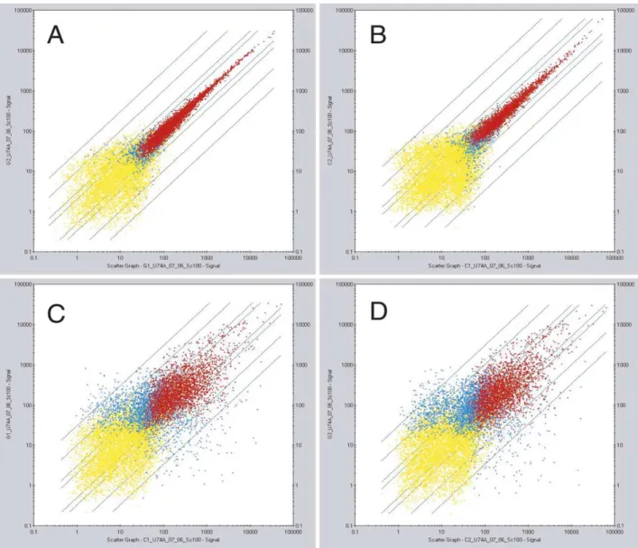
Fig. 1. Analysis of the homogeneity of microarray generated signals between duplicate samples of spermatogonia and spermatocyte cRNA probes, and comparative analysis of the divergence between spermatogonia and spermatocytes. Data represent scatter plots of (A) spermatogonia sample 1 intensities (G1) vs. spermatogonia sample 2 intensities (G2); (B) spermatocytes sample 1 (C1) vs. spermatocytes sample 2 (C2); (C) spermatogonia sample 1 (G1) vs. spermatocytes sample 1 (C1); (D) spermatogonia sample 2 (G2) vs. spermatocytes sample 2 (C2). In these scatter plots, each spot corresponds to the signal generated by a discrete Affymetrix target gene, each represented on the chip arrays by 16 specific 25mer oligonucleotide probes and by 16 one-mismatch probes. In A and in B the data fit a straight line with slope approximately equal to one and intercept near zero, demonstrating high reproducibility of the results. In C and D, the enlargement of spot distribution is very similar in both comparative analysis, and allows to define statistically significant difference in selective gene expression between the two cell populations. Red dots represent genes significantly expressed in both samples (P-value , 0.04). Blue dots genes expressed significantly only in one sample, and yellow dots genes not expressed (P-value . 0.06) in both samples. The P-values are calculated as described in the Statistical Algorithms Reference Guide by Affymetrix (see Section 2).
teleangectasia mutated homolog) (
Barlow et al., 1998
).
Between growth factor receptors, c-kit (encoding the KL
receptor) and Alk3 (encoding the Bmp4 receptor) were
confirmed to be expressed in pre-meiotic stages, and not
in spermatocytes (
Sorrentino et al., 1991; Yoshinaga et al.,
1991; Schrans-Stassen et al., 1999; Pellegrini et al., 2003
).
As expected, many of the genes selectively expressed in
spermatogonia and not in spermatocytes encode proteins
involved in the regulation of the mitotic cell cycle
(transcription factor E2f1,
dependent-kinase-inhibi-tors p57 and p21, cyclin D3, cyclin B1, cyclin A2,
cyclin-dependent-kinase 4), and replicative DNA synthesis (DNA
Table 1
Examples of genes selectively expressed in spermatogonia MG-U74Av2
target Affymetrix
Gene name Gene symbol Notes Fold difference
(spermatogonia vs. spermatocytes)
92306 Ovary – testis transcribed Ott Absent in spermatocytes. X-linked gene, previously defined as ‘meiosis specific’ (Kerr et al., 1996)
104
101194 Stimulated by retinoic acid gene 8 Stra8 Absent in spermatocytes. Pre-meiotic germ cell-specific cytoplasmic protein encoded by Stra8, a retinoic acid-responsive gene (Oulad-Abdelghani et al., 1996)
50
101180 Ataxia telangiectasia mutated homolog
Atm Absent in spermatocytes. Protein kinase involved in DNA repair and DNA damage response (induction of apoptosis by DNA damage). In knock-out mice gametogenesis is severely disrupted as early as leptonema of prophase I (Barlow et al., 1998)
15
93536 Bcl2-associated X protein Bax Absent in spermatocytes. Pro-apoptotic factor (Yan
et al., 2000)
9
99956 Stem cell factor receptor (KL receptor)
c-kit Absent in spermatocytes. Transmembrane tyrosine kinase receptor (Sorrentino et al., 1991; Yoshinaga et al.,
1991; Schrans-Stassen et al., 1999). Essential for
pre-meiotic spermatogenesis (Rossi et al., 1993; Kissel et al.,
2000; Blume-Jensen et al., 2000; Dolci et al., 2001)
7
102963 E2F transcription factor 1 E2f1 Absent in spermatocytes. Transcription factor crucial for mitotic cell cycle control (Dolci et al., 2001)
6
103207 DNA polymerase a 1, 180 kDa Pola1 Absent in spermatocytes. Dna replication (Orlando et al.,
1989)
6
94448 B-cell leukemia/lymphoma 10 Bcl10 Present in spermatocytes. Pro-apoptotic factor 6 160159 Cyclin B1 Ccnb1 Absent in spermatocytes. Subunit of cdc2/cdk1,
essential for G2/M transition
5
103057 DNA polymerase d 1, catalytic domain
Pold1 Absent in spermatocytes. Dna replication 4
92767 Bone morphogenetic protein receptor, type 1A
Bmpr1a, ALK3 Absent in spermatocytes. Bone morphogenetic receptor for Bmp2 and Bmp4. Involved in spermatogonial differentiation (Pellegrini et al., 2003)
4
96772 DNA primase, p49 subunit Prim1 Absent in spermatocytes. Dna replication (Orlando et al.,
1989)
4
95471 Cyclin-dependent kinase inhibitor 1C (P57)
p57 Absent in spermatocytes. Cdk2 inhibitor 4
98067 Cyclin-dependent kinase inhibitor 1A (P21)
p21 Absent in spermatocytes. Cdk2 inhibitor 3
103064 Checkpoint kinase 1 homolog Chk1, Chek1, rad27 Absent in spermatocytes. Protein kinase, which is required for the DNA damage checkpoint. In response to DNA damage, Chk1 phosphorylates and inhibits Cdc25C, thus preventing activation of the Cdc2 – cyclin B complex and mitotic entry
3
160538 D-type G1 cyclin catalytic subunit (PSK-J3/CDK4)
Cdk4 Present in spermatocytes. Cell cycle kinase activated and essential for G1/S transition (Dolci et al., 2001)
2
160545 Cyclin D3 Ccnd3 Absent in spermatocytes. Cell cycle control in G1/S Phase (Dolci et al., 2001)
2
104154 Transformation related protein 53 p53 Absent in spermatocytes (marginal in one sample). Apoptosis inducer and cell cycle control in G1/S Phase
2
92481 Checkpoint kinase 2 homolog Chk2, Chek2, rad53 Absent in spermatocytes (marginal in one sample). ATM-dependent. Function similar to that of Chk1
2
99186 Cyclin A2 Ccna2 Absent in spermatocytes. Mitotic cyclin, active in the S phase, cdk2 subunit (Dolci et al., 2001)
polymerase a, DNA polymerase d, DNA primase),
generally confirming previously published data (
Dolci
et al., 2001; Orlando et al., 1989
). Between genes encoding
proteins involved in pro-apoptotic programs, p53 and Bax
were found to be selectively expressed in spermatogonia.
The lack of Bax expression in spermatocytes, according to
the microarray statistical analysis, is in partial conflict with
previously published data (
Yan et al., 2000
, see below).
Bcl10 was found to be expressed also in spermatocytes,
even though at a much lower level.
As for the targets that gave a higher signal in
spermatocytes, we tried to classify them in a series of
functional clusters: apoptosis/cell-cycle,
chromatin/tran-scription, cytoskeleton/traffic, meiosis/spermatogenesis,
Table 2
Genes expressed in spermatocytes: apoptosis/cell cycle MG-U74Av2
target Affymetrix
Gene name Gene symbol Notes Fold difference
(spermatocytes vs. spermatogonia)
160644 BCL2-antagonist/killer 1 Bak1 Absent in spermatogonia. Involved in apoptosis; caspase activation via cytochrome c (Yan et al., 2000)
45
92911 Cyclin A1 Ccna1 Absent in spermatogonia. In knock-out mice, meiotic arrest during meiotic divisions (Liu et al., 1998)
23
103094 Small EDRK-rich factor 1 Serf1 Present in spermatogonia. (Survival of Motoneuron in SMA1) 18 99522 Germ cell-specific gene 2 Gsg2 Absent in spermatogonia (marginal in one sample). Atypical
serine – threonine kinase, named Haspin (for haploid germ cell-specific nuclear protein kinase)
17
160761 Upregulated during skeletal muscle growth 4
Usmg4 Absent in spermatogonia 11
100054 DNA segment Chr2 D2Wsu81e Present in spermatogonia. Endonuclease G: a mitochondrial protein released in apoptosis and involved in caspase-independent DNA degradation
10
92929 Cytochrome c, testis Cyct Present in spermatogonia. Null mice produce functional sperm but undergo early testicular atrophy (Narisawa et al., 2002)
10
94971 KAP1, Cdk inhibiting phosphatase
Cdkn3 Present in spermatogonia. cdk2-associated dual specificity phosphatase
9
94521 Cyclin-dependent kinase inhibitor 2D
Cdkn2d, Ink4d, p19 Absent in spermatogonia. Selective cdk4/6 inhibitor. Double p19 and p18 (Ink4c) knock-out provokes sterility due to a delayed exit of spermatogonia from the mitotic cell cycle
(Zindy et al., 2001)
9
101885 Growth arrest specific 5 Gas5 Absent in spermatogonia. Preferentially expressed in the growth phase arrest of the cell cycle
6
160638 Cyclin-dependent kinase inhibitor 2C
Cdkn2c, Ink4c, p18 Absent in spermatogonia. Selective cdk4/6 inhibitor. Double p19 (Ink4d) and p18 knock-out provokes sterility due to a delayed exit of spermatogonia from the mitotic cell cycle
(Zindy et al., 2001)
6
99670 Bcl-associated death promoter
Bad Absent in spermatogonia. Pro-apoptotic factor (Yan et al., 2000) 5
92902 Myeloblastosis oncogene-like 1
Mybl1, A-Myb Present in one sample in spermatogonia. Transcription factor. Knockout male mice are sterile due to arrest in pachytene
(Toscani et al., 1997)
5
98945 SH3-domain GRB2-like B1 (endophilin)
Sh3glb1 Present in spermatogonia. Synaptically enriched protein implicated in synaptic vesicle endocytosis. Might be involved in apoptotic programs since it interacts with Bax
4
92879 Protein phosphatase 1G, g isoform
Ppm1g, PP2C-g Present in spermatogonia. Formerly called protein phosphatase 2C. Magnesium-dependent serine – threonine phosphatase, known to be expressed in the testis and skeletal muscle
4
94294 Cyclin B2 Ccnb2 Present in spermatogonia. Interacts with cdc2 (cdk1) as a subunit. Component of MPF (Dolci et al., 2001)
4
104738 Zuotin related factor 2 Zrf2 Present in spermatogonia. A ribosome-associated DnaJ molecular chaperone. Also called MIDA-1, associates with Id HLH transcription factors
3
102734 Baculoviral IAP repeat-containing 3
Birc3, mIAP-2 Present in spermatogonia. Also called Apoptosis inhibitor 2. Caspase inhibitor
3
104476 Retinoblastoma-like 1 (p107)
Rbl1 Absent in spermatogonia (marginal in one sample). Homolog of pRb, involved in negative regulation of the cell cycle
2
101521 Baculoviral IAP repeat-containing 5
Birc5, TIAP Present in spermatogonia. Homologous to human survivin. Caspase inhibitor
membrane-bound-proteins/receptors, metabolism, RNA
binding
proteins,
signal-transduction/protein-kinases
(
Tables 2 – 9
). Also in this case, we ordered these genes
according to the average Signal Log Ratio parameter, which
was converted in average fold difference of the signal in
spermatocytes vs. spermatogonia. We also considered
whether the target, beside giving a higher signal in
spermatocytes, gave a positive (detection parameter: P) or
negative (detection parameter: A) signal in spermatogonia,
and the calculation of the fold difference of the signal
between the two cell populations does not take into account
whether the target gene is significantly expressed or not in
spermatogonia.
For the large majority of these targets, detection of a high
signal in spermatocytes by the microarray analysis
con-firmed data that are available in published literature or in
Table 3
Genes expressed in spermatocytes: chromatin/transcription MG-U74Av2
target Affymetrix
Gene name Gene symbol Notes Fold difference
(spermatocytes vs. spermatogonia)
160599 Testis-specific gene A2 Tsga2 Absent in spermatogonia. Male meiotic metaphase chromosome-associated acidic protein
56
102795 Mesoderm posterior 1 Mesp1 Absent in spermatogonia. HLH protein 42 161064 PHD finger protein 7 Phf7 Present in spermatogonia. Isolated from a mouse testis
cDNA library
42
102079 Mus musculus Aip1 Aip1, Aym1 Absent in spermatogonia. IME-1 functional homolog (Personal communication from Jeremy Don, Bar-Ilan University, Ramat Gan, Israel)
40
104622 Transcription elongation factor A (SII), 2
Tcea2 Absent in spermatogonia 34
97745 Homeo box A4 Hoxa4 Absent in spermatogonia 29
95755 Cold shock domain protein A Csda Present in spermatogonia 25 92190 Nuclear receptor subfamily 2, group
C, member 1
Nr2c1 Absent in spermatogonia 22
93182 Glial and testis-specific homeobox gene
Nkx6-2, Gtx Absent in spermatogonia. Murine homeobox-containing gene, expressed specifically in glial cells of the brain and germ cells of testis. Knock-out mice are viable and fertile (Cai et al., 2001)
17
102219 Regulatory factor X, 2 Rfx2 Absent in spermatogonia. Influences HLA class II expression
16
99987 RIKEN cDNA A630056B21 gene A630056B21Rik Absent in spermatogonia. Weakly similar to zinc finger protein 2 (Zfp2) (mKR2 protein)
15
98414 Zinc finger protein 42 Zfp42 Present in one sample in spermatogonia. Expressed also in embryonic stem cells
14
160204 RIKEN cDNA 3110013H01 gene 3110013H01Rik Present in spermatogonia. Nuclear protein p30, a protein of the nuclear pore complex
11
93221 RIKEN cDNA 4921540P06 gene (Homeo box D8)
4921540P06Rik Absent in spermatogonia. Other names: Hox-4.3 140, HOXD8, Hox5.4
11
100126 Chromatin accessibility complex 1 Chrac1 Present in spermatogonia. NF-YC-like protein. Also called YCL1 (Bolognese et al., 2000)
11
160068 Sin3 associated polypeptide, 30 kDa Sap30 Present in spermatogonia. Component of a histone deacetylase complex
10
97893 TATA box binding protein-like protein
Tlp Present in spermatogonia. Also named TLF, TRF2 or TBPL1. Knockout mice arrest at spermiogenesis
(Martianov et al., 2001)
9
92432 Zinc finger protein 93 Zfp93 Absent in spermatogonia 9 104604 Zinc finger protein 96 Zfp96 Present in one sample in spermatogonia 6 103629 Lymphoid enhancer binding factor 1 Lef1 Absent in spermatogonia 6 96144 Inhibitor of DNA binding 4 Idb4 Absent in spermatogonia. Id4, dominant negative
helix-loop-helix protein
6
92195 CCAAT/enhancer binding protein (C/EBP), g
Cebpg Present in spermatogonia 6
94406 Putative homeodomain transcription factor
Phtf Present in spermatogonia 5
98032 Zinc finger protein 35 Zfp35 Present in spermatogonia 5 160220 Zinc finger protein 110 Zfp110 Absent in spermatogonia 4 94102 H6 homeo box 1 Hmx1 Absent in spermatogonia (Yoshiura et al., 1998) 4
expression databases, indicating that our analysis faithfully
reflected the actual differences in the pattern of gene
expression between male mitotic and meiotic germ cells.
Many of these genes encode proteins specifically involved
in the control of the meiotic cell cycle, such as cyclin A1
(
Table 2
) (
Liu et al., 1998
), cdk4-inhibitors p18 and p19
(
Table 2
) (
Zindy et al., 2001
), A-myb (
Table 2
) (
Toscani
et al., 1997
), Nek2 (
Table 9
) (
Di Agostino et al., 2002
), but
in many cases their expression reflects meiotic
accumu-lation of transcripts destined to be translated later during
spermiogenesis, such as testis-specific lactate
dehydrogen-ase (
Table 7
) (
Li et al., 1998
), testis-specific poly(A)
polymerase b (
Table 8
)
Kashiwabara et al., 2002
), calmegin
(
Table 9
) (
Ikawa et al., 1997
), preproacrosin (
Table 5
)
(
Kremling et al., 1991
), fertilin b (
Table 5
) (
Cho et al.,
1998
), Trf2 (
Table 3
) (
Martianov et al., 2001
), MSJ-1 (
Table
5
) (
Berruti and Martegani, 2001
), Tpx1 (
Table 5
) (
Kasahara
et al., 1989
), Tekt1 (
Table 5
) (
Larsson et al., 2000
), Tesp1
(
Table 5
) (
Kohno et al., 1998
) and so on. It is noteworthy
that the spermatocyte-specific expression of a large number
of genes encoding enzymes is involved in glycolysis and
gluconeogenesis, beside that of Pgk2, encoding a well
known meiotic isoform of phosphoglycerate kinase (
Boer
et al., 1987
) (
Table 7
). Thus, metabolic pathways distinct
Table 4
Genes expressed in spermatocytes: cytoskeleton/traffic MG-U74Av2
target Affymetrix
Gene name Gene symbol Notes Fold difference
(spermatocytes vs.spermatogonia)
99995 Centrin 1 Cetn1 Absent in spermatogonia. Also called caltractin. Testis-specific centrosomal protein encoded by an intronless retroposon
588
101864 Actin-like 7b Actl7b Absent in spermatogonia. Testis-specific actin isoform, encoded by an intronless gene
76
161035 Kinesin family member 9 Kif9 Absent in spermatogonia. Microtubule motor associated protein abundantly expressed in the testis
58
160631 Sarcoglycan, a (50 kDa dystrophin-associated glycoprotein)
Sgca, adhalin Absent in spermatogonia. Integral plasma membrane protein considered specifically expressed in striated muscle
36
99531 Synaptogyrin 4 Syngr4 Absent in spermatogonia. Integral membrane protein present in synaptic vesicles
36
101195 Myosin light chain 2 Mylc2pl Absent in spermatogonia. Considered specifically expressed in precursor B and T lymphocytes
17
92496 Vesicle-associated membrane protein 5
Vamp5 Absent in spermatogonia. Also called synaptobrevin. Expressed during myogenesis in striated muscles
15
101520 RIKEN cDNA 1700062C23 gene 1700062C23Rik Absent in spermatogonia. Kinesin-related protein HASH. Rat homolog known to be expressed during spermatogenesis in meiotic cells
10
160487 Myosin light chain, alkali, cardiac atria
myla Absent in spermatogonia. Expressed during striated muscle development
9
103684 Tektin-2 Tekt2 Absent in spermatogonia. A sperm flagellar protein also called tektin-t and different from tektin-1
8
94321 Keratin complex 1, acidic, gene 10 Krt1-10 Absent in spermatogonia (marginal in one sample). Protein of intermediate filaments
4
95097 ARP10 actin-related protein 10 homolog
Actr10 Present in spermatogonia. Protein of the dynactin complex 4
93567 Profilin 2 Pfn2 Present in spermatogonia. Actin binding ubiquitous protein 4 102732 Talin tln Present in spermatogonia. Integrin and actin binding protein 4 93499 Capping protein a 1 cappa1 Present in spermatogonia. Actin binding protein 4 94248 Adaptor-related protein complex
AP-1, m subunit 1
Ap1m1 Present in spermatogonia. Adaptor protein of clathrin-coated vesicles involved in intracellular protein transport and endocytosis
4
104565 Adaptor-related protein complex AP-4, sigma 1
Ap4s1 Present in spermatogonia. Adaptor protein of
clathrin-coated vesicles involved in intracellular protein transport and endocytosis
3
92643 Neurofibromatosis 2 Nf2 Present in spermatogonia. Tumor suppressor protein involved in mediating interactions between the plasma membrane and the cytoskeleton
3
93333 Tubulin cofactor a Tbca Present in spermatogonia. Molecular chaperonin involved in tubulin folding
2
103878 Adaptor-related protein complex AP-3, b 1 subunit
Ap3b1 Present in spermatogonia. Adaptor protein of
clathrin-coated vesicles involved in intracellular protein transport and endocytosis
from those operating in mitotic germ cells and somatic cells
might drive carbohydrate utilization in meiotic and/or
post-meiotic germ cells, even though this hypothesis needs to be
substantiated by more specific studies. We also noticed
several targets whose relative gene expression in
sperma-togonia or in spermatocytes was either not known, or
controversial, or conflicting with data available in the
literature. The pattern of expression in spermatocytes vs.
Table 5
Genes expressed in spermatocytes: meiosis/spermatogenesis MG-U74Av2
target Affymetrix
Gene name Gene symbol Notes Fold difference
(spermatocytes vs. spermatogonia)
160219 Meiosis expressed gene 1 Meg1 Absent in spermatogonia (Don and Wolgemuth, 1992) 653 94927 Fatty acid binding protein 9 Fabp9, Perf 15 Absent in spermatogonia. Perforatorial protein in the
perinuclear theca of spermatozoa
388
92825 Testis-specific gene 1 Tpx1 Absent in spermatogonia (Kasahara et al., 1989) 349 100526 A disintegrin and metalloprotease
domain 3
Adam3 Absent in spermatogonia. Also known as cyritestin 326
92732 A disintegrin and metalloprotease domain 2
Adam2 Absent in spermatogonia. Also called Fertilin b. Knock-out mice are sterile for failure of sperm – egg or sperm – oviduct interactions (Cho et al., 1998)
256
103058 T-complex protein 10b Tcp10b Absent in spermatogonia 238 99545 Tektin 1 Tekt1 Absent in spermatogonia. Sperm axonemal protein (Larsson
et al., 2000)
187
100359 T-complex protein-10 Tcp10 Absent in spermatogonia 168 99134 T-complex-associated testis
expressed 3
Tcte3 Present in spermatogonia 163
93955 Zona-pellucida-binding protein Zpbp, sp38 Absent in spermatogonia 157 99474 A disintegrin and metalloprotease
domain 5
Adam5 Absent in spermatogonia 147
160506 A kinase anchoring protein-associated sperm protein
Akapasp Absent in spermatogonia 133
100358 T-complex protein 10a Tcp10a Absent in spermatogonia 128 97381 T-complex protein 11 Tcp11 Absent in spermatogonia 79 93207 Preproacrosin Acr Absent in spermatogonia (Kremling et al., 1991) 52 160122 RIKEN cDNA 2410004D18 gene 2410004D18Rik Absent in spermatogonia. Asparaginase-like sperm
autoantigen
50
97481 DnaJ (Hsp40) homolog, subfamily B, member 3
Dnajb3, MSJ-1 Absent in spermatogonia. Member of the DNAj co-chaperon family (Berruti and Martegani, 2001)
42
99456 Proacrosin binding protein Acrbp, sp32 Absent in spermatogonia 39 95299 Dynein, axonemal, heavy chain 8 Dnahc8 Absent in spermatogonia. Absent also in one sample of
spermatocytes
39
99816 Heat shock protein 2 Hspa2, Hsp70-2 Present in spermatogonia. Knock-out mice arrest in pachytene (Dix et al., 1996)
37
102244 Testicular serine protease 1 Tesp1 Absent in spermatogonia. Sperm acrosomal protein
(Kohno et al., 1998)
36
97785 DNA segment, human D6S2654E D0H6S2654E Present in one sample of spermatogonia. Also called X5L protein (XAP5 like protein, retroposon-encoded copy of an X-linked gene)
30
100626 Outer dense fiber 2 Odf2 Present in spermatogonia 16 103541 T-complex-associated testis
expressed 2
Tcte2 Absent in spermatogonia 16
103468 Meiosis-specific nuclear structural protein 1
Mns1 Present in spermatogonia 12
102747 T-complex-associated testis expressed 1
Tcte1 Absent in spermatogonia 11
103956 5-azacytidine induced gene 1 Azi1 Absent in spermatogonia. Pre-acrosomal protein 10 102818 Xlr-related, meiosis regulated Xmr Absent in spermatogonia 5 94891 Male enhanced antigen 1 Mea1 Present in spermatogonia 3 92888 Phosphoserine/threonine/tyrosine
interaction protein
Styx Present in spermatogonia. Complexes with a testicular phosphorylated RNA-binding protein and is essential for normal spermiogenesis (Wishart and Dixon, 2002)
2
spermatogonia for a selection of these genes is shown in
Fig. 2
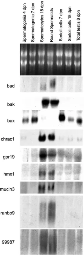
. Northern blot analysis confirmed both qualitatively
and quantitatively the data obtained by the microarray
experiments.
An expression pattern in partial conflict with previously
published data was particularly evident for several
pro-apoptotic members of the Bcl2 family. Bad was previously
reported to be expressed in spermatogonia and in Sertoli
cells, but not in spermatocytes, nor in spermatids (
Yan et al.,
2000
), while both microarray and Northern blot analysis
showed that Bad mRNA is expressed in spermatocytes, but
not in spermatogonia, nor in Sertoli cells (
Table 2
and
Fig. 2
). Moreover, its expression was very strong in
spermatids, in which a slower migrating transcript was
observed. Bak was reported to be expressed in Sertoli cells,
in spermatogonia and in spermatocytes, but not in
spermatids (
Yan et al., 2000
), but we found a very abundant
transcript only in meiotic and post-meiotic cells, and no
expression in spermatogonia, nor in Sertoli cells (
Table 2
and
Fig. 2
). Interestingly it has been recently reported that
apoptosis-like mechanisms are required for spermatid
differentiation in Drosophila (
Arama et al., 2003
). An
analogy between cytoplasmic apoptotic events and
the formation of residual bodies has been also noticed in
mammalian spermiogenesis (
Blanco-Rodriguez and
Marti-nez-Garcia, 1999
). On the other hand, Bax was reported to
be expressed, besides in spermatogonia and Sertoli cells,
also in spermatocytes (
Yan et al., 2000
), but we found an
abundant transcript in mitotic germ cells and in Sertoli cells,
with the highest level of expression at 7 dpn, whereas only a
very faint signal was detectable in spermatocytes (
Table 1
and
Fig. 2
).
Chrac1 (chromatin accessibility complex 1, also named
Ycl1) is a histone-fold protein that interacts with other
histone-fold proteins to bind DNA in a
sequence-indepen-dent manner. These histone-fold protein dimers combine
within larger enzymatic complexes for DNA transcription,
replication, and packaging (
Bolognese et al., 2000
). Chrac1
mRNA was found to be very abundant in spermatocytes
(
Table 3
and
Fig. 2
), suggesting that it might be involved in
chromatin remodeling during the first meiotic prophase.
This might help to regulate changes in gene expression
patterns that characterize specific developmental events
during spermatogenesis.
In the cluster of membrane-bound proteins and receptors,
microarray analysis revealed the unexpected expression of
Table 6
Genes expressed in spermatocytes: membrane-bound proteins/receptors MG-U74Av2
target Affymetrix
Gene name Gene symbol Notes Fold difference
(spermatocytes vs. spermatogonia)
101390 Mucin 3, intestinal Muc3 Absent in spermatogonia. Glycoprotein of the colon epithelium
62
92198 Decay accelerating factor 2 Daf2 Absent in spermatogonia. Integral membrane protein involved in complement activation
31
93390 Prominin 1 Prom1 Absent in spermatogonia. A microvilli-specific polytopic membrane protein of the apical surface of epithelial cells targeted to plasmalemmal protrusions of non-epithelial cells
11
103289 Low density lipoprotein receptor-related protein 4
Lrp4, corin Absent in spermatogonia. Atrial natriuteric peptide-converting enzyme (pro-ANP-peptide-converting enzyme). Serine protease of the trypsin family
10
103656 LanC (bacterial lantibiotic synthetase component C)-like
Lancl1, p40GPRT, p40/GPR69A
Present in spermatogonia. Originally proposed as a G-protein coupled receptor, was then characterized as a loosely membrane-associated protein related to the LanC family of bacterial proteins involved in the biosynthesis of
antimicrobial peptides
8
160876 B-cell receptor-associated protein 29
Bcap29 Present in spermatogonia. Associated with the membrane IgD and IgM receptors in B lymphocytes
8
100438 G protein coupled receptor 19 Gpr19 Absent in spermatogonia. G-protein coupled receptor for an unknown ligand (O’Dowd et al., 1996)
4
99160 and 99161 RIKEN cDNA 1110025J15 gene
1110025J15Rik Present in spermatogonia. Similar to membrane proteins related to a glutamate binding protein (NMDA receptor)
4
102343 Hypothetical protein 425O18-1 425O18-1 Present in spermatogonia. Contains a low density lipoprotein-receptor class A domain
4
103726 RIKEN cDNA 2610311I19 gene
2610311I19Rik Absent in spermatogonia. Similar to Golgi membrane protein SB140. I
3
161046 Cytokine receptor-like factor 1 Crlf1 Present in spermatogonia. Soluble cytokine receptor subunit or part of a cytokine responsive complex, possibly playing a regulatory role in the immune system and during fetal development
the transcript encoding mucin3, a protein known to be
specifically expressed in the colon epithelium (
Table 6
).
Northern blot analysis confirmed high levels of expression
of mucin3 mRNA in spermatocytes, and, at a lesser extent,
in spermatids (
Fig. 2
). Interestingly, another component of
the mucosal glycocalyx, contributing to anti-adhesive and
protective cell functions, mucin1, has been reported to be
expressed in maturing germ cells of the human testis
(
Franke et al., 2001
), and a mucin glycoprotein was found to
be an universal constituent of stable intercellular bridges in
the Drosophila melanogaster germ line (
Kramerova and
Kramerov, 1999
).
Table 7
Genes expressed in spermatocytes: metabolism MG-U74Av2
target Affymetrix
Gene name Gene symbol Notes Fold difference
(spermatocytes vs. spermatogonia)
93103 Lactate dehydrogenase 3, C chain, sperm specific
Ldh3 Absent in spermatogonia. Glycolysis and gluconeogenesis (Li et al., 1998)
3565
92599 Phosphoglycerate mutase 2 Pgam2 Absent in spermatogonia. Glycolysis and gluconeogenesis 1260 96918 Fructose bisphosphatase 1 Fbp1 Absent in spermatogonia. Glycolysis and gluconeogenesis 401 100931 Arylsulfatase A Arsa Absent in spermatogonia. Sulfuric ester hydrolase 194 92292 Solute carrier family 2
(facilitated glucose transporter), member 3
Slc2a3 Absent in spermatogonia 132
95060 Solute carrier family 16 (monocarboxylic acid transporters), member 7
Slc16a7 Absent in spermatogonia 68
93560 RIKEN cDNA 1110039O14 gene
1110039O14Rik Present in spermatogonia. Similar to human acylphosphatase 59
103982 Alcohol dehydrogenase 4 (class II), pi polypeptide
Adh4 Absent in spermatogonia. Glycolysis and gluconeogenesis 43
104328 Aquaporin 9 Aqp9 Absent in spermatogonia. Water transport 43 104372 RIKEN cDNA 0910001L24
gene
0910001L24Rik Absent in spermatogonia. Xenobiotic metabolism 39
99011 UDP-N-acetyl-a-D -galactosamine:polypeptide N-acetylgalactosaminyl-transferase 3
Galnt3 Absent in spermatogonia 39
103646 Carnitine acetyltransferase Crat Present in one sample in spermatogonia. Fatty acid metabolism 33 101388 Phosphoglycerate kinase 2 Pgk2 Absent in spermatogonia. Spermatocyte-specific PGK isoform
encoded by an intronless retroposon (Boer et al., 1987). Glycolysis and gluconeogenesis
33
99542 Pyruvate dehydrogenase E1 a 2 Pdha2 Present in spermatogonia. Glycolysis and gluconeogenesis 30 94540 RIKEN cDNA 1300006E06
gene
1300006E06Rik Absent in spermatogonia. Cytochrome C P-450-16a. Electron transport
22
103689 ATP-binding cassette, sub-family C (CFTR/MRP), member 3
Abcc3 Absent in spermatogonia. Similar to human multidrug resistance associated protein
19
161243 RIKEN cDNA 0910001L24 gene
0910001L24Rik Absent in spermatogonia. Xenobiotic metabolism 17
103531 RIKEN cDNA 1300013B24 gene
1300013B24Rik Absent in spermatogonia. Low similarity to endoplasmic oxidoreductase 1 b
16
92841 Chromogranin B Chgb Absent in spermatogonia 16
103068 Aldo-keto reductase family 1, member E1
Akr1e1 Absent in spermatogonia. Aldehyde reductase 15
99591 Retinol dehydrogenase 11 Rdh11 Absent in spermatogonia. Similar to human androgen-regulated prostate short-chain dehydrogenase/reductase 1
15
93557 Selenophosphate synthetase 2 Sps2 Absent in spermatogonia 13 97511 Monoglyceride lipase Mgll Absent in spermatogonia 13 97834 Phosphofructokinase-1 C Pfkp Absent in spermatogonia. Glycolysis and gluconeogenesis 12 160839 Solute carrier family 2
(facilitated glucose transporter), member 5
Slc2a5 Absent in spermatogonia 11
96072 Lactate dehydrogenase 1, A chain
Few receptors for potential growth factors were found to
be expressed in spermatocytes through the microarray
analysis. One of these was Gpr19 (
O’Dowd et al., 1996
), a
seven transmembrane G-coupled receptor for an unknown
ligand (
Table 6
). Northern blot analysis showed an high
abundance of the Gpr19 transcript in spermatocytes, a lower
level of expression in spermatids, while a faint band was
observed in spermatogonia from 7-day, but not 4-day-old
mice (
Fig. 2
). This receptor might thus play a role in the
regulation of meiotic entry and/or meiotic progression. The
gene encoding Ranbp9 (Ran binding protein 9), a protein
shown to be a positive regulator of Ras function (
Wang et al.,
2002
), was found to be highly expressed in spermatids, with
a complex migratory pattern, but the signals were evident
also in spermatocytes, implying its possible involvement in
the regulation of the Ras/MEK/ERK cascade during the
transition through the meiotic divisions and/or the
morpho-genetic events of spermiogenesis (
Table 9
and
Fig. 2
).
Finally, in the cluster of transcription factors (
Table 3
)
we confirmed through Northern blot analysis the selective
germ cell expression starting from the meiotic stage of
Hmx1, a homeodomain gene previously not known to be
expressed during spermatogenesis (
Yoshiura et al., 1998
),
and of a transcript corresponding to RIKEN cDNA
A630056B21Rik (Affymetrix target 99987_at in the
MG-U74Av2 array) predicted to encode a novel zinc finger
protein (
Fig. 2
). These transcription factors might play an
important role in driving the spermatogenic program. As in
the case of Ranbp9, the signal generated by the 99987 target
in Northern blots was rather complex: this might be due to
either the presence of multiple alternative transcripts, or to
cross-hybridization with closely related RNAs.
Even though, recently, an initial microarray screen of
spermatogenic cells at different developmental stages has
been reported (
Yu et al., 2003
), only 1176 mouse target
genes were represented in these arrays. We noticed partial
overlapping of our data with the ones published by
Yu et al.
(2003)
, but also some discrepancies were evident: for
instance, cyclin D3 was reported to be not expressed in
spermatogonia, but present in spermatocytes. One should
note that we used oligonucleotide based DNA chips, in
which each gene is represented by 16 couples of probes and
mismatch probes, whereas in the gene arrays used by Yu
et al. each target gene is represented by a single longer
Table 8
Genes expressed in spermatocytes: RNA binding proteins MG-U74Av2
target Affymetrix
Gene name Gene symbol Notes Fold difference
(spermatocytes vs. spermatogonia)
101938 Poly(A) binding protein, cytoplasmic 2
Pabpc2 Absent in spermatogonia. Encoded by an intronless retroposon during spermatogenesis
94
104440 Y box protein 2 Ybx2 Absent in spermatogonia. RNA-binding protein which might delay polysomal association of transcripts during spermiogenesis
90
161033 Poly(A) polymerase b (testis-specific)
Papolb Absent in spermatogonia. Responsible for cytoplasmic
polyadenylation of pre-existing mRNAs in male haploid germ cells. Knock-out results in the arrest of spermiogenesis (Kashiwabara et al., 2002)
20
97661 Testis nuclear RNA binding protein
Tenr Present in spermatogonia. Expressed in meiotic and haploid male germ cells
11
161041 and 92678 DEAD/H (Asp-Glu-Ala-Asp/His) box polypeptide 25
Ddx25 Present in spermatogonia. Gonadotropin regulated RNA helicase also expressed in Leydig cells
11
96850 Hypothetical protein 4833436O05
4833436O05 Present in spermatogonia. Similar to eukaryotic translation initiation factors
10
160429 NTF2-related export protein 1 Nxt1 Present in spermatogonia. RAN-binding protein involved in nuclear RNA export from the nucleus
7
100720 Poly(A) binding protein, cytoplasmic 1
Pabpc1 Present in spermatogonia 4
101579 Signal recognition particle 9 kDa
Srp9 Present in spermatogonia. Cytoplasmic ribonucleoprotein targeting nascent polypeptide chains to the endoplasmic reticulum
4
103101 TAR (HIV) RNA binding protein 2
Tarbp2, Prbp Present in spermatogonia. Interacts with the 30untranslated region of the Protamine-1 RNA
4
101519 Signal recognition particle 14 kDa (homologous Alu RNA binding protein)
Srp14 Present in spermatogonia. Cytoplasmic ribonucleoprotein targeting nascent polypeptide chains to the endoplasmic reticulum
3
94552 Poly(rC) binding protein 1 Pcbp1 Present in spermatogonia. Implicated in mRNA stabilization 2 103330 Spermatid perinuclear RNA
binding protein
Spnr Present in spermatogonia. Binds to the to the 30UTR of Protamine-1 mRNA. Microtubule-associated RNA-binding protein that localizes to the manchette in developing spermatids
oligonucleotide, making the possibility of
cross-hybridi-zation easier and hindering the statistical evaluation of
the generated signals (see also Section 2). In conclusion, our
results represent a first extensive attempt to delineate the
global patterns of gene expression characterizing male germ
cell differentiation, and should be extended to other germ
cell types, namely spermatogonial stem cells and
spermatids.
2. Experimental procedures
2.1. Cell preparations
Germ cell populations highly enriched in mitotic
spermatogonia were obtained as previously described
from testes of 4 – 7-day-old mice (
Rossi et al., 1993;
Pellegrini et al., 2003; Dolci et al., 2001
). Briefly, germ
Table 9
Genes expressed in spermatocytes: signal transduction/protein kinases MG-U74Av2
target Affymetrix
Gene name Gene symbol Notes Fold difference
(spermatocytes vs. spermatogonia)
104029 Calmegin Clgn Absent in spermatogonia. Knockout male mice are sterile for defective sperm function (Ikawa et al., 1997)
52
101850 Sperm autoantigenic protein 17 Spa17 Present in spermatogonia. A calmodulin-binding protein enriched in the sperm acrosome and interacting with the zona pellucida
31
161839 RAS-like, family 2, locus 9 Rasl2-9 Absent in spermatogonia. Strongly related to Ran GTPase, probably a testis-specific isoform
30
100972 Chemokine (C-C motif) ligand 27
Ccl27 Absent in spermatogonia. Also called ALP, CTAK, mILC, CTACK, PESKY, ESkine, skinkine
29
102948 hematopoietic cell transcript 1 Hemt1 Absent in spermatogonia. Contains a calcium-activated BK potassium channel a subunit signature
26
99869 Hepatoma-derived growth factor
HRP1, Pwwp1 Absent in spermatogonia. Also called PWWP domain containing 1. Present in the nucleus of spermatocytes and spermatids
23
98614 Nephrocystin Nphp1 Present in spermatogonia. SH3 containing protein which forms protein complexes with p130(Cas), proline-rich tyrosine kinase 2 (Pyk2), and tensin
23
103489 Socius Soc Absent in spermatogonia. A Rho-related GTPase-interacting protein involved in disassembly of actin stress fibers
22
93210 NIMA-related-kinase 4 Nek4 Absent in spermatogonia 18 93658 Protein tyrosine phosphatase,
non-receptor type 20
Ptpn20 Absent in spermatogonia 16
100287 Immunoglobulin (CD79A) binding protein 1b
Igbp1b Absent in spermatogonia. Binds to protein phosphatase 2A. Also called a4-b
16
160623 Cyclin-dependent kinase-like 2 Cdkl2 Absent in spermatogonia. CDC2-related kinase also called KKIAMRE
13
104166 Renal tumor antigen Rage, MOK Absent in spermatogonia. Protein kinase with homologies with members of the MAPK family
13
104135 ADP-ribosylation-like 3 Arl3 Present in spermatogonia. Small monomeric GTPase of the Ras superfamily
12
160948 Testis-specific calcineurin isoform
Ppp3cc Present in spermatogonia. Calmodulin-dependent protein phosphatase
11
92805 ADP-ribosylation-like 4 Arl4 Present in spermatogonia. Knock-out mice show a significant reduction of testis weight and sperm count (Schurmann et al., 2002)
10
100562 Ran. guanine nucleotide release factor
Rangnrf Present in one sample in spermatogonia. Also called MOG1 9
100291 Casitas B-lineage lymphoma Cbl Absent in spermatogonia. Adaptor protein 8 102033 Testis-specific protein kinase 1 Tesk1 Absent in spermatogonia 8 92639 Serine/threonine kinase 6 Stk6, Ayk1 Present in spermatogonia. Also called Aurora/IPL1-related kinase 1.
Specifically expressed in meiotic cells just before the first meiotic division
7
100885 NIMA-related kinase 2 Nek2 Present in spermatogonia. Involved in chromosome condensation during meiotic divisions (Di Agostino et al., 2002). Controls splitting of duplicated centrosomes
7
161575 Mitogen activated protein kinase 10
Mapk10, Absent in spermatogonia. Also called SAPK (b), JNK3, SERK2, p54bSAPK, p439F12
4
97812 RAN binding protein 9 Ranbp9 Present in spermatogonia. Stimulates Ras activation by recruiting Sos. Also called RanbpM (Wang et al., 2002)
cell suspensions were obtained by sequential collagenase –
hyaluronidase – trypsin digestions of freshly withdrawn
testes. A 3 h period of culture in E-MEM additioned with
10% FCS was performed to facilitate adhesion of
contami-nating somatic cells to the plastic dishes. At the end of this
pre-plating treatment, enriched mitotic germ cell
suspen-sions were rinsed from FCS. Purity of 7 dpn spermatogonia
was about 90% after the pre-plating treatment, whereas a
50% enrichment was obtained for 4 dpn spermatogonia. The
homogeneity of the cell populations was assessed through
both morphological criteria and by specific immunostaining
with antibodies directed against three specific markers of
mitotic germ cells, which are not expressed in testicular
somatic cells (Smad5, Alk3 and c-kit). Homogeneous
populations (purity . 90%) of spermatocytes and round
spermatids were obtained from testes of either 18-day-old or
36-day-old mice, respectively, by differential elutriation as
previously described (
Sette et al., 1999; Di Agostino et al.,
2002
). Spermatocyte populations from 18-day-old mice
(10% at the leptotene – zygotene and 85% at the middle – late
pachytene stage of the meiotic prophase) are devoid of
round spermatids, which contaminate elutriation fractions
from adult animals, and their purity was assessed through
morphological criteria (namely, cell size and the
character-istic aspect of partially condensed meiotic chromatin).
Sertoli cell monolayers from 7 to 17-day-old mice, devoid
of contaminating germ cells, were prepared as previously
described (
Grimaldi et al., 1993
).
2.2. RNA extraction, cDNA and cRNA preparation
RNA was purified by adding cold Trizol reagent
(Invitrogen) to freshly prepared cell samples and extracted
according to the manufacturer’s instructions.
Total cellular RNA (25 mg) was used to synthesize
cDNA using the cDNA Synthesis Kit (Life Technologies
BRL
11917-010)
and
T7-(dT)
24oligonucleotide
(5
0-GGCCAGTGAATTGTAATACGACTCACTATAGG-GAGGCGG-(dT)
24-3
0) according to manufacturer’s
instruc-tions. Second strand cDNA was synthesized by adding 10 U
of DNA ligase, 40 U of DNA polymerase and 2 U of
RNaseH and incubating at 16 8C for additional 2 h. At the
end of the incubation, 20 U of T4 DNA polymerase were
added to the reaction and incubated for 5 min at the same
temperature. Reactions were stopped by adding EDTA
(30 mM final concentration). Double stranded cDNA was
purified by phenol/chloroform extraction followed by
precipitation with 0.5 volumes of 7.5 M ammonium acetate
and 2.5 volumes of ethanol and its concentration measured
by optical densitometry.
Complementary RNA (cRNA) synthesis was performed
using the Essential ENZO kit (Bioarray High Yield TNA
transcription kit 900182) and following manufacturer’s
instructions. The resulting cRNA was purified using
QIAGEN Rneasy spin columns (74103) and the standard
procedure. RNA was then precipitated as described above
Fig. 2. Germ cell-type-specific expression of a selection of genes identified by Affymetrix microarray hybridizations was verified by Northern blot analysis, using 10 mg of total RNA for each indicated testicular cell type. The representative top panel shows ethidium bromide staining, indicating that RNA loading was qualitatively and quantitatively comparable for each sample. Specific labeled gene probes for hybridization were obtained by nick translation of RT-PCR amplified cDNAs, as indicated in Section 2. These Northern blots are representative of at least two experiments, which gave similar results.
for cDNA, resuspended in 15 ml of RNase-free H
2O and
quantified by optical densitometry. cRNA was then
fragmented in a Tris-acetate buffer (200 mM, pH 8.1)
containing 500 mM KOAc and 150 mM MgOAc by
incubation at 94 8C for 35 min. At the end of the
incubation, fragmented cRNA was stored at 2 80 8C
until hybridization.
2.3. DNA microarray analysis
cRNA samples from two independent cell preparations
were
used
for
hybridization
to
duplicate
mouse
MG-U74Av2 microarray sets from Affymetrix. This
array represents approximately 12,500 murine genes or
EST sequences. In each array, target genes are represented
by 16 pairs (exact match and single base mismatch) of
25-mer oligonucleotides for each gene. The signals of the
pairs are compared to assess specificity of hybridization,
thus, beside the intensity of the signal, its statistical
significance can be estimated. Biotinylated cRNA (15 mg)
was hybridized to the array and then processed following
the standard Affymetrix protocol. Phycoerythrin-coupled
avidin bound microarrays were scanned with a
Hewlett-Packard Gene Array Scanner (Hewlett-Hewlett-Packard Co., Palo
Alto, CA), and the results were analyzed using the
Affymetrix MAS5 statistical algorithm. For more
infor-mations about the statistical analysis, see the Affymetrix
Statistical Algorithms Reference Guide at
http://www.
affymetrix.com/support/technical/technotes/statistical_
reference_guide.pdf
.
Target genes represented in the MG-U74Av2 Affymetrix
chips were grouped in several functional clusters by using
specific keywords with the Interacting Query online facility
at
www.affymetrix.com
.
2.4. RT-PCR preparation of probes and Northern blot
analysis
cDNA probes for Northern blot hybridization of total
RNAs were prepared by RT-PCR amplification of selected
mRNAs, by using specific oligonucleotide primers
designed on the basis of the sequence of the corresponding
Affymetrix target genes. Specificity of the primers
was previously controlled through BLAST analysis (
http://
www.ncbi.nlm.nih.gov/blast/
). The couples of primers
used
were:
TAGCCCTTTTCGAGGACGCTCG
and
TGGAGCCTCCTTTGCCCAAGTT (for Bad,
amplifi-cation product 220 bp);
AGTTGGCTCTCAAGGAT-GGCTT and TCGTTGCACTGACAGAATCTTC (for
Bak1, 229 bp); ACCAAGAAGCTGAGCGAGTGT and
TCCAGCCCATGATGGTTCTGAT (for Bax, 253 bp);
ATCTGGAGAATAGGCACGGACG
and
CCGA-AATGCCCACATAGTTTCT
(for
gpr19,
378 bp);
TGCTCTACAGTGTACCGGACAG and
CAGCACTCTG-TACTGTCCCTTG (for hmx1, 291 bp);
GACTCTGT-GTACAACACCTTCC and GCCCTTGTAAAGACAGA
TGGTC
(for
mucin3,
524 bp);
CAAATTGG-GAGCTGTTCCGACC
and
CTACAACAGAAGT-CATCTGTAG (for Ranbp9, 267 bp); TGTGTAGCCG
GGAGTTTGGTA
and
TGAAAACGGACTCCG-CACTCCT (for A630056B21Rik, 357 bp). The cDNA
probe for Chrac1 was kindly provided by Prof. Roberto
Mantovani (University of Milan).
cDNAs were labeled by random priming with
a
32PdNTPs and hybridized using standard conditions to
blotted total RNA samples. After stringency washes, blots
were exposed overnight at 2 80 8C with intensifier screens
for autoradiography.
Acknowledgements
Due to space restrictions, we apologize for not being able
to cite all the relevant papers describing germ
cell-specific-expression of several genes that we have confirmed through
microarray analysis and included in our tables. We thank
Prof. Roberto Mantovani (University of Milan) for
supply-ing a Chrac1 cDNA probe. This work has been supported by
MIUR CoFin 2002, by a grant of ‘Centro di Eccellenza per
lo Studio del Rischio Genomico in Patologie Complesse
Multifattoriali’ and by Agenzia Spaziale Italiana.
References
Arama, E., Agapite, J., Steller, H., 2003. Caspase activity and a specific cytochrome C are required for sperm differentiation in Drosophila. Dev. Cell 4, 687 – 697.
Barlow, C., Liyanage, M., Moens, P.B., Tarsounas, M., Nagashima, K., Brown, K., et al., 1998. Atm deficiency results in severe meiotic disrup-tion as early as leptonema of prophase I. Development 125, 4007 – 4017. Berruti, G., Martegani, E., 2001. MSJ-1, a mouse testis-specific DnaJ protein, is highly expressed in haploid male germ cells and interacts with the testis-specific heat shock protein Hsp70-2. Biol. Reprod. 65, 488 – 495.
Blanco-Rodriguez, J., Martinez-Garcia, C., 1999. Apoptosis is physiologi-cally restricted to a specialized cytoplasmic compartment in rat spermatids. Biol. Reprod. 61, 1541 – 1547.
Blume-Jensen, P., Jiang, G., Hyman, R., Lee, K.F., O’Gorman, S., Hunter, T., 2000. Kit/stem cell factor receptor induced activation of phosphatidylinositol 30-kinase is essential for male fertility. Nat. Genet. 24, 157 – 162.
Boer, P.H., Adra, C.N., Lau, Y.F., McBurney, M.W., 1987. The testis-specific phosphoglycerate kinase gene pgk-2 is a recruited retroposon. Mol. Cell. Biol. 7, 3107 – 3112.
Bolognese, F., Imbriano, C., Caretti, G., Mantovani, R., 2000. Cloning and characterization of the histone-fold proteins YBL1 and YCL1. Nucleic Acids Res. 28, 3830 – 3838.
Cai, J., Qi, Y., Wu, R., Modderman, G., Fu, H., Liu, R., Qiu, M., 2001. Mice lacking the Nkx6.2 (Gtx) homeodomain transcription factor develop and reproduce normally. Mol. Cell. Biol. 21, 4399 – 4403.
Cho, C., Bunch, D.O., Faure, J.E., Goulding, E.H., Eddy, E.M., Primakoff, P., Myles, D.G., 1998. Fertilization defects in sperm from mice lacking fertilin beta. Science 281, 1857 – 1859.
Di Agostino, S., Rossi, P., Geremia, R., Sette, C., 2002. The MAPK pathway triggers activation of Nek2 during chromosome condensation in mouse spermatocytes. Development 129, 1715 – 1727.
Dix, D.J., Allen, J.W., Collins, B.W., Mori, C., Nakamura, N., Poorman-Allen, P., et al., 1996. Targeted gene disruption of Hsp70-2 results in failed meiosis, germ cell apoptosis, and male infertility. Proc. Natl Acad. Sci. USA 93, 3264 – 3268.
Dolci, S., Pellegrini, M., Di Agostino, S., Geremia, R., Rossi, P., 2001. Signaling through extracellular signal regulated kinase is required for spermatogonial proliferative response to stem cell factor. J. Biol. Chem. 276, 40225 – 40233.
Don, J., Wolgemuth, D.J., 1992. Identification and characterization of the regulated pattern of expression of a novel mouse gene, meg1, during the meiotic cell cycle. Cell Growth Differ. 3, 495 – 505.
Franke, F.E., Kraus, S., Eiermann, C., Pauls, K., Lalani, E.N., Bergmann, M., 2001. MUC1 in normal and impaired spermatogenesis. Mol. Hum. Reprod. 7, 505 – 512.
Grimaldi, P., Piscitelli, D., Albanesi, C., Blasi, F., Geremia, R., Rossi, P., 1993. Identification of 30,50-cyclic adenosine monophosphate-inducible nuclear factors binding to the human urokinase promoter in mouse Sertoli cells. Mol. Endocrinol. 7, 1217 – 1225.
Ikawa, M., Wada, I., Kominami, K., Watanabe, D., Toshimori, K., Nishimune, Y., Okabe, M., 1997. The putative chaperone calmegin is required for sperm fertility. Nature 387, 607 – 611.
Kasahara, M., Gutknecht, J., Brew, K., Spurr, N., Goodfellow, P.N., 1989. Cloning and mapping of a testis-specific gene with sequence similarity to a sperm-coating glycoprotein gene. Genomics 5, 527 – 534. Kashiwabara, S., Noguchi, J., Zhuang, T., Ohmura, K., Honda, A., Sugiura,
S., et al., 2002. Regulation of spermatogenesis by testis-specific, cytoplasmic poly(A) polymerase TPAP. Science 298, 1999 – 2002. Kelly, T.E., 1987. Inactivation of the mammalian X chromosome in
spermatogenesis. Am. J. Hum. Genet. 40, 288 – 289.
Kerr, S.M., Taggart, M.H., Lee, M., Cooke, H.J., 1996. Ott, a mouse X-linked multigene family expressed specifically during meiosis. Hum. Mol. Genet. 5, 1139 – 1148.
Kissel, H., Timokhina, I., Hardy, M.P., Rothschild, G., Tajima, Y., Soares, V., et al., 2000. Point mutation in kit receptor tyrosine kinase reveals essential roles for kit signaling in spermatogenesis and oogenesis without affecting other kit responses. Eur. Mol. Biol. Org. J. 19, 1312 – 1326.
Kohno, N., Yamagata, K., Yamada, S., Kashiwabara, S., Sakai, Y., Baba, T., 1998. Two novel testicular serine proteases, TESP1 and TESP2, are present in the mouse sperm acrosome. Biochem. Biophys. Res. Commun. 245, 658 – 665.
Kramerova, I.A., Kramerov, A.A., 1999. Mucinoprotein is a universal constituent of stable intercellular bridges in Drosophila melanogaster germ line and somatic cells. Dev. Dyn. 216, 349 – 360.
Kremling, H., Keime, S., Wilhelm, K., Adham, I.M., Hameister, H., Engel, W., 1991. Mouse proacrosin gene: nucleotide sequence, diploid expression, and chromosomal localization. Genomics 11, 828 – 834. Larsson, M., Norrander, J., Graslund, S., Brundell, E., Linck, R., Stahl, S.,
Hoog, C., 2000. The spatial and temporal expression of Tekt1, a mouse tektin C homologue, during spermatogenesis suggest that it is involved in the development of the sperm tail basal body and axoneme. Eur. J. Cell Biol. 79, 718 – 725.
Li, S., Zhou, W., Doglio, L., Goldberg, E., 1998. Transgenic mice demonstrate a testis-specific promoter for lactate dehydrogenase, LDHC. J. Biol. Chem. 273, 31191– 31194.
Liu, D., Matzuk, M.M., Sung, W.K., Guo, Q., Wang, P., Wolgemuth, D.J., 1998. Cyclin A1 is required for meiosis in the male mouse. Nat. Genet. 20, 377 – 380.
Martianov, I., Fimia, G.M., Dierich, A., Parvinen, M., Sassone-Corsi, P., Davidson, I., 2001. Late arrest of spermiogenesis and germ cell apoptosis in mice lacking the TBP-like TLF/TRF2 gene. Mol. Cell 7, 509 – 515.
Narisawa, S., Hecht, N.B., Goldberg, E., Boatright, K.M., Reed, J.C., Millan, J.L., 2002. Testis-specific cytochrome c-null mice produce functional sperm but undergo early testicular atrophy. Mol. Cell. Biol. 22, 5554 – 5562.
O’Dowd, B.F., Nguyen, T., Lynch, K.R., Kolakowski, L.F. Jr., Thompson, M., Cheng, R., et al., 1996. A novel gene codes for a putative G protein-coupled receptor with an abundant expression in brain. Fed. Eur. Biochem. Soc. Lett. 394, 325 – 329.
Orlando, P., Geremia, R., Frusciante, C., Grippo, P., 1989. Replicating premeiotic germ cells of the mouse contain a novel DNA primase stimulatory factor. Cell. Differ. Dev. 27, 129 – 136.
Oulad-Abdelghani, M., Bouillet, P., Decimo, D., Gansmuller, A., Heyberger, S., Dolle, P., et al., 1996. Characterization of a premeiotic germ cell-specific cytoplasmic protein encoded by Stra8, a novel retinoic acid-responsive gene. J. Cell Biol. 135, 469 – 477.
Pellegrini, M., Grimaldi, P., Rossi, P., Geremia, R., Dolci, S., 2003. Developmental expression of BMP4/ALK3/SMAD5 signaling pathway in the mouse testis: a potential role of BMP4 in spermatogonia differentiation. J. Cell Sci. 116, 3363 – 3372.
Roeder, G.S., 1997. Meiotic chromosomes: it takes two to tango. Genes Dev. 11, 2600 – 2621.
Rossi, P., Dolci, S., Albanesi, C., Grimaldi, P., Ricca, R., Geremia, R., 1993. FSH induction of steel factor (SLF) mRNA in mouse Sertolı´ cells and stimulation of DNA synthesis in spermatogonı´a by soluble SLF. Dev. Biol. 155, 68 – 74.
Schena, M., 1996. Genome analysis with gene expression microarrays. Bioessays 18, 427 – 431.
Schrans-Stassen, B.H., van de Kant, H.J., de Rooij, D.G., van Pelt, A.M., 1999. Differential expression of c-kit in mouse undifferentiated and differentiating type A spermatogonia. Endocrinology 140, 5894 – 5900. Schurmann, A., Koling, S., Jacobs, S., Saftig, P., Krauss, S., Wennemuth, G., et al., 2002. Reduced sperm count and normal fertility in male mice with targeted disruption of the ADP-ribosylation factor-like 4 (Arl4) gene. Mol. Cell. Biol. 22, 2761 – 2768.
Sette, C., Barchi, M., Bianchini, A., Conti, M., Rossi, P., Geremia, R., 1999. Activation of the mitogen-activated protein kinase Erk1 during meiotic progression of mouse pachytene spermatocytes. J. Biol. Chem. 274, 33571 – 33579.
Sorrentino, V., Giorgi, M., Geremia, R., Besmer, P., Rossi, P., 1991. Expression of the c-kit protooncogene in the murine male germ cells. Oncogene 6, 149 – 151.
Toscani, A., Mettus, R.V., Coupland, R., Simpkins, H., Litvin, J., Orth, J., et al., 1997. Arrest of spermatogenesis and defective breast develop-ment in mice lacking A-myb. Nature 386, 713 – 717.
Wang, D., Li, Z., Messing, E.M., Wu, G., 2002. Activation of Ras/Erk pathway by a novel MET-interacting protein RanBPM. J. Biol. Chem. 277, 36216– 36222.
Wishart, M.J., Dixon, J.E., 2002. The archetype STYX/dead-phosphatase complexes with a spermatid mRNA-binding protein and is essential for normal sperm production. Proc. Natl Acad. Sci. USA 99, 2112 – 2117. Yan, W., Samson, M., Jegou, B., Toppari, J., 2000. Bcl-w forms complexes with Bax and Bak, and elevated ratios of Bax/Bcl-w and Bak/Bcl-w correspond to spermatogonial and spermatocyte apoptosis in the testis. Mol. Endocrinol. 14, 682 – 699.
Yoshinaga, K., Nishikawa, S., Ogawa, M., Hayashi, S., Kunisada, T., Fujimoto, T., Nishikawa, S.-I., 1991. Role of c-kit in mouse spermatogenesis: identification of spermatogonia as a specific site of c-kit expression and function. Development 113, 689 – 699.
Yoshiura, K., Leysens, N.J., Reiter, R.S., Murray, J.C., 1998. Cloning, characterization, and mapping of the mouse homeobox gene Hmx1. Genomics 50, 61 – 68.
Yu, Z., Guo, R., Ge, Y., Ma, J., Guan, J., Li, S., et al., 2003. Gene expression profiles in different stages of mouse spermatogenic cells during spermatogenesis. Biol. Reprod. 69, 37 – 47.
Zindy, F., den Besten, W., Chen, B., Rehg, J.E., Latres, E., Barbacid, M., et al., 2001. Control of spermatogenesis in mice by the cyclin D-dependent kinase inhibitors p18(Ink4c) and p19(Ink4d). Mol. Cell. Biol. 21, 3244 – 3255.