UNIVERSITY OF CALABRIA
Department of Chemistry and Chemical Technologies
CHIM/03 General and Inorganic Chemistry
Doctorate School of Physical, Chemical, Materials Science and Technologies
PhD Thesis
Gold Nanoparticles: where Shape becomes Essence.
Synthesis and Characterization of an Outstanding
Nanomaterial
XXX Cycle
SUPERVISOR Prof. Massimo LA DEDA
PhD CANDIDATE Angela CANDREVA
First of all I would like to thank my supervisor Professor Massimo La Deda, I'm grateful for everything that he taught me. He supported me in any circumstance, respecting my opinion and leaving me free to synthesize and characterize “disproportionately” high amount of nanoparticles. Many thanks to Loredana and MATinLAB group: Professor Crispini, Professor Ghedini, Professor Aiello and Doctor Godbert, Franco, Andreea, Eugenia, Debora, Francesco, and in particular Francesca for sharing with me the office and the experience of writing down three years of work. It would have been harder alone. Each one of them helped me whenever I needed making me better professionally and personally.
Thanks to Pierluigi and Emilia for their kindness towards me, and Daniela for sharing my enthusiasm looking at TEM images.
Thanks to Professor Pagliusi who has been working with us on the project to organize the nanoparticles in the liquid crystalline medium.
My warm thanks to professor Luis M. Liz Marzán, Scientific Director of CIC biomaGUNE and leader of BioNanoplasmonics group, who allowed me to work in his group for six months. Thanks a lot to all “Plasmontiger” members for sharing with me their expertises, in particular Leo and Guille. I appreciated it very much guys! Thanks to Wiktor for supervising my experiments giving me great tips. Lastly I would like to thank my family and all people who supported me along this path.
A glance at the Thesis: scope and limitations
The present book collects the principal results obtained during the three-year PhD course at the Doctorate School of Physical, Chemical, Materials Science and Technologies (Cosenza, Italy). The research work, performed at the Inorganic Materials Laboratory (MAT_IN Lab) of the University of Calabria under the supervision of Prof. Massimo La Deda, and partially at the BioNanoPlasmonics Laboratory from the Centre for Cooperative Research in Biomaterials-CIC biomaGUNE (San Sebastian, Spain), in collaboration with Prof. Luis Liz-Marzán, concerns with
the synthesis and characterization of gold nanoparticles.
As well known, metallic nanoparticles with a size smaller than the wavelength of the incident radiation, show resonance phenomena with the magnetic field collectively called Localized Surface Plasmon Resonance (LSPR). LSPR properties are extremely interesting because they depends on, and consequently there are controlled by, metal, size, shape and environment. In this way, an accurate synthetic method capable of control size and shape, allow to control nanoparticles plasmonic properties. Ultimately, the plasmon energy can be determined
through the shape, which in turn is determined by synthetic procedure.
LSPR, moreover, can cooperate with other actors that interact with the electromagnetic field. In this way, plasmonic nanoparticles can “talk” with chromophores, increasing their absorption cross section, or, if they are emissive, interesting phenomena take place from the correlation between plasmonic field and de-excitation radiative paths.
More interestingly, LSPRs of two or more assembled nanoparticles can couple themselves. The way they interact depends on the nanoparticles shape and on the environment. Anisotropic
shaped nanoparticles, i.e. rod-like one, manifest various assembling modes, which determine
the properties of the plasmonic-coupled fields. On the other side, an ordered medium, acting as arrangement template, can pilot if and how plasmonic fields couple.
All this aspects, synthesis, morphological characterization, plasmonic optical study, assembly and coupling of various functionalized gold nanorods, have been performed, and the results are very encouraging to employs the prepared nanomaterials in various applications, from innovative energy collectors to phototherapeutical cancer-targeted agents.
The Thesis in a nutshell
Chapter 1 – Introduction. A brief review of the essential concepts necessary to introduce the reader
has been made. In particular, the synthetic methods to obtain gold nanorods have been illustrated, and the assembling process of these anisotropic nanoparticles has been described, including direct assembly (i.e. governed by interparticle forces) or indirect assembly (i.e. triggered by a template or an external field). The optical properties of individual and assembled nanorods have been illustrated, with particular emphasis regarding the plasmon coupling.
Chapter 2 – Gold Nanorods Preparation. Here we present synthesis, morphological and optical characterization of gold nanorods prepared via seed-mediated method. In addition, original surfactant coating agent has been substituted with other cover molecules, and solubility in various solvents was explored. In particular, we overcoated gold nanorods by a silica shell that deserve a deep study due to its tailorability. Finally, the control of the gold nanoparticles shape through synthetic parameters has been illustrated by preparing gold nanotriangles and gold nanospheres.
Chapter 3 – Silica shell on Gold Nanorods. Taking into account the high tailorability of the silica
shell that cover gold nanorods, we discuss the synthetic strategies to control shell thickness and its effects on the plasmonic bands.
Chapter 4 – Functionalization of the Silica shell. Another important feature of the gold nanorods
silica shell is the possibility to functionalize it. In particular, a high-luminescent Ir(III) complex has been embedded into the shell, and the exceptional “dialogue” between the radiative deactivation paths of the complex excited-states and the gold core plasmonic field, has been recorded.
Chapter 5 – Nanoparticles coupling. When two nanorods are placed in a close proximity, the
plasmon resonances carried by the two nanorods will interact to form different collective plasmon modes. Due to the geometrical anisotropy and the synthetically tailorable plasmon energy of nanorods, coupled nanorods exhibit extremely rich spectral responses. Here we have explored methods to assembly nanorods by using a functionalization of nanoparticles or a nanorods dispersion into organized medium, and the spectral response has been analyzed.
CONTENTS
Chapter I INTRODUCTION………... 1
I.1. Origin of surface plasmon resonance in noble metal nanoparticles... 2
I.2. Synthesis of anisotropic gold nanoparticles: seed-mediated growth approach 5
I.3. Self-assembly of nanoparticles ... 11
I.4. Optical properties of individual gold nanorods... 19
I.5. Plasmon coupling in assemblies of metal nanoparticles...… 29
References……….…………. 31
Chapter II. GOLD NANORODS SYNTHESIS………..
37II.1 Gold nanorods covered with CTAB: synthesis, morphological and optical characterization... 38
II.2 Playing with the gold nanorods surface: covering exchange and overcoating... 47
II.3 Shape control of gold nanoparticles: nanotriangles and nanospheres…………... 52
References………... 58
III. SILICA SHELL ON GOLD NANORODS………...
59III.1 Synthetic strategy and thickness control... 60
III.2 Obtained results... 63
III.3 Discussion... 74
III.4 Conclusions... 76
IV. FUNCTIONALIZATION OF THE SILICA SHELL………...
79IV.1 Silica-shell gold nanorods embedding a metal complex………... 80
IV.2. Results and discussion………... 84
IV.3 Conclusions………. 92
References………... 93
V. ASSEMBLED GOLD NANORODS AND THEIR PLASMONIC
BEHAVIOUR... 95
V.1 Gold nanorods direct assembly... ... 97
V.2 Gold nanorods indirect assembly... ... 111
References………... 116
CONCLUSIONS AND PERSPECTIVES... 119
1
I. INTRODUCTION
I.1. Origin of surface plasmon resonance in noble metal nanoparticles
I.2. Synthesis of anisotropic gold nanoparticles: seed-mediated growth approach
I.3. Self-assembly of nanoparticles
I.4. Optical properties of individual gold nanorods
I.1. Origin of surface plasmon resonance in noble metal nanoparticles
2
I.1. Origin of surface plasmon resonance in noble metal nanoparticles
The electrons in the metal (d electrons in silver and gold) are free to travel through the material. The mean free path in gold and silver is about 50 nm, therefore in particles smaller than this, no scattering is expected from the bulk. Thus, all interactions are expected to be with the surface. When the wavelength of incident light is much larger than the nanoparticle size it can set up standing resonance conditions, as represented in Fig. I.1. [1].Fig. I.1. Origin of surface plasmon resonance due to coherent interaction of the electrons in the conduction band with light [1]
Light in resonance causes the free-electrons in the metal to oscillate. As the wave front of the light passes, the electron density in the particle is polarized to one surface and oscillates in resonance with the light’s frequency causing a standing oscillation. The resonance condition is determined by absorption and scattering spectroscopy and is found to depend on the shape, size, and dielectric constants of both the metal and the surrounding material. This is referred to as the local surface plasmon resonance (LSPR), since it is located at the surface. As the shape or size of the nanoparticle changes, the surface geometry changes causing a shift in the electric field density on the surface. This causes a modification in the oscillation frequency of the electrons, generating different cross-sections for the optical properties including absorption and scattering.
The dielectric constant of the surrounding material will have an effect on the oscillation frequency due to the varying ability of the surface to accommodate electron charge density from the nanoparticles. Changing the capping agent and solvent will change the dielectric constant, determining the shift of the plasmon resonance due to the local nature of its effect on the surface of the nanoparticle. Chemically bonded molecules can be detected by the observed change they induce in the electron density on the surface, which results in a shift in the surface plasmon absorption maximum. This is the basis for the use of noble metal nanoparticles as sensitive sensors.
I.1. Origin of surface plasmon resonance in noble metal nanoparticles
3
Mie originally calculated the surface plasmon resonance by solving Maxwell’s equations for small spheres interacting with an electromagnetic field. Gan was able to extend this theory to apply to ellipsoidal geometries. Modern methods using the discrete dipole approximation allow one to calculate the surface plasmon resonance absorption for arbitrary geometries. Calculation of the longitudinal plasmon resonance for gold nanorods generates an increase in the intensity and wavelength maximum as the aspect ratio (length divided by width) increases. Thus, the plasmon resonance can be tuned across the visible region by changing the aspect ratio. The increase in the intensity of the surface plasmon resonance absorption leads to an enhancement of the electric field, exploited in many applications [2].
Many shapes of noble metal nanoparticles have been synthesized [3]. Nanorods [4] have attracted the most attention for the large number of synthetic methods available, the high monodispersity possible, and the rational control over the aspect ratio, which is primarily responsible for the change in their optical properties. Nanorods have been shown to have two plasmon resonances [5] one due to the transverse oscillation of the electrons, around 520 nm for gold, and the other due to the longitudinal plasmon resonance at longer wavelengths, as shown for various aspect ratios in Fig. I.2.
Fig. I.2. Gold nanoparticles: absorption of various sizes and shapes [5]
The transverse surface plasmon resonance does not depend on the aspect ratio and it is at the same wavelength as the plasmon resonance of spheres. The longitudinal surface plasmon resonance increases with larger aspect ratios. The anisotropy has been shown to generate large control over the
I.1. Origin of surface plasmon resonance in noble metal nanoparticles
4
optical absorbance for all shapes generated. Triangular nanoparticles have been generated by photochemical means and chemical growth. The edges and corners are very important with triangular nanoparticles. Snipping of the edges produces a visible blue shift in the plasmon resonance, which can be modelled theoretically. Disks also display a similar plasmon resonance absorption dependence on their aspect ratio.
I.2. Synthesis of anisotropic gold nanoparticles: seed-mediated growth approach
5
I.2. Synthesis of anisotropic gold nanoparticles: seed-mediated growth approach
The size and shape-controlled synthesis of metal nanoparticles is important in present day advanced materials, as almost every property within the nanometer regime are size and shape dependent [6]. Common methods for size control employ capping agents [7], such as surfactants, ligands, polymers, or dendrimers, to confine the growth in the nanometer regime. These methods commonly produce spherical particles due to the low surface energy associated with such particles. Structures other than spheres form as a result of specific interaction of the capping agents with different growing faces of the particles [8].A shape control of nanoparticles has been most successfully achieved using a template. Templates provide a constrained environment during the nanoparticle growth and thus shapes are tuned according to the template. Commonly used templates are porous alumina [9], polycarbonate membranes [10], carbon nanotubes [11] and micelles [12]. In practice, the presence of templates does not produce 100% shape monodispersity; rather, a significant fraction of thermodynamically favourable spheres is also formed. In addition, soft templates such as micelles may not be stable under the experimental conditions (e.g., boiling), and thus the template may not function [13]. A common reason for the failure of the template mechanism is the change in template microstructures by reactants.
Seed-mediated growth approach. Mechanism of conventional nanoparticle synthesis proceeds through successive nucleation and growth steps, both extremely sensitive to physical and chemical parameters [14]. In some of the solution-phase metal nanoparticle synthesis procedures, the control of nucleation and growth steps are done by changing the reducing agent or stabilizer concentration. In doing so, the size and shape of nanoparticles can be controlled. For solution-phase gold nanoparticle (AuNP) synthesis, it has been observed an initial slow nucleation followed by a nucleation burst associated with autocatalytic surface growth [15]. Through a physical separation of the nucleation process from the growth process, it was possible to control these steps to prepare different sized spherical particles; this method is known as seed-mediated growth approach. By using this growth strategy in the presence of a rod-shaped micelle, [16] a template-mediated shape control can be reached.
The basic principle of shape-controlled synthesis involves two steps: first, the preparation of small-size spherical AuNPs, and second, growth of the prepared spherical particle in rod-like micellar environment (Fig. I.3). This procedure, that take place in aqueous solution at or near room temperature, begins with the synthesis of AuNPs by chemical reduction of Au(III) salt such as
I.2. Synthesis of anisotropic gold nanoparticles: seed-mediated growth approach
6
tetrachloroauric acid (HAuCl4) with a strong reducing agent such as sodium borohydride (NaBH4)
in presence of a capping agent, to prevent particle coalescence. The gold spheres thus generated are 3±5 nm in diameter and serve as seeds on which to grow more anisotropic nanostructures. These seeds are then added to a solution containing more Au(III) salt, a weak reducing agent, such as ascorbic acid and a rodlike micellar template such as cetyltrimethyl ammonium bromide (CTAB). The seeds serve as nucleation sites for gold nanorods (AuNRs) growth; under these conditions, no metal salts are reduced to metal unless the seeds are present. Consequently, minimal additional nucleation occurs during particle growth. Typical product dimensions are 10-20 nm diameter short axis and aspect ratios up to 20 for AuNRs.
Fig. I.3. Schematic illustration of the seed-mediated method for the growth of Au nanorods [17]
The presence of additives, such as 5-bromosalicylic acid, in the reaction media can fine-tune the morphology of the resulting nanorods [18].
A small amount of AgNO3 is added for shape induction. The presence of silver salt is
essential for producing and controlling the aspect ratio of rods. In absence of Ag+, fewer rods (~10-20 % of the total particles) form and their aspect ratios are higher and have a wide distribution, ranging from ~5-15. Murphy et al. [19] proposed that the Ag+ forms AgBr (Br- from the CTAB), and because ascorbate is a weak reducing agent, it cannot reduce the silver ion. The mechanism by
I.2. Synthesis of anisotropic gold nanoparticles: seed-mediated growth approach
7
which Ag+ assists in controlling gold particle shape is not completely understood; however, it was hypothesized that Ag+ adsorbs at the Au particle surface in the form of AgBr and restricts growth. The adsorption of AgBr at the Au particle surface appears to stabilize the spheroids and rods. In absence of Ag+ the spheroids formed, but due to their instability, converted to spheres.
Size control. Surfactants are considered dynamic templates that in a precise concentration, called critical concentration, and temperature, form micelles. These are compartments of nanometer size able to confine the nanocrystals growth and control and limit physically their enlargement. A layer of surfactant covers the surface of the formed nanoparticles. In fact, acting like surface complexing agent it prevents the irreversibly aggregation of nanoparticles [20]. According to Scarabelli et al. [21] [18], the final size it is also dependent by the amount of seed solution added to the growth solution and by the pre-reduction time.
Shape control. The growth inside micellas not only confer a size control, but also a degree of shape control. It is studied that surfactants are able to adhere with different bonding strengths to the various facets of the nanocrystals and induce their preferential development along those crystallographic directions which grow fastest [20]. The surface covering plays a crucial role in the morphology of the product, each capping agent being selective for a specific shape [22]. Canbek et al. [23] demonstrated that the final shape of the nanoparticles depends also on the capping of the seeds. In particular, seeds with size around 3 nm and positive charge, coming from CTAB covering, are the best choice to synthesize nanorods with high yield. On the other hand seeds with size around 7 nm and negative charge which comes from sodium citrate have to be preferred if the spherical shape is the challenge.
Another important contribution on shape control is given by silver ions and halides in seed-mediated synthesis. Liz-Marzán and other groups [24] studied how they can be used to control particle shape and surface facet structure. It is clear that the halidates direct the anisotropic shape and that halide counterion plays a crucial role in the growth processes of anisotropic gold nanoparticles, but it is not yet clear which halide-metal interaction are primarily responsible for shape control [17]. Many study are trying to demostrate that halide ions may direct the anisotropic growth of metal nanocrystals modulating the redox potentials of the metal ions, acting as face-specific capping agents, and or controlling the extent of silver underpotential deposition at the nanocrystal surface. According to the literature [25] bromide is the right choice to obtain well-defined gold nanorods and iodide for nanoplates shapes. Iodide seems to prevent gold nanorods formation.
I.2. Synthesis of anisotropic gold nanoparticles: seed-mediated growth approach
8
CTAB: stabilizing and directing agent for rod-like nanoparticles. CTAB, composed by a hydrophilic cationic head group and a hydrophobic tail, is used as stabilizing and directing agent for the growth of anisotropic shape. CTAB forms complexes Au(I) and stabilizes metal nanoparticles
forming a bilayer around them (Fig. 1.4) [23].
Fig. I.4. CTAB bilayer on the surface of gold nanorods
CTAB is present on the whole nanorods surface as a bilayer through electrostatic interactions of the ammonium head group.
It is demonstrated that using CTAC (cetyltrimethyl ammonium chloride) instead of CTAB no nanorods are formed. This is because increasing the CTAB concentration (from 0.3 E-3 M to 0.1 M) spherical micelles transit towards elongated micelles, while CTAC forms always spherical micelles [23].
On this basis were performed various seeded growth method protocols to achieve different shapes. For example, Scarabelli et al. [21] proposed a successful synthesis and purification protocol to obtain well monodispersed gold nanotriangles covered with CTAC. Gold nanotriangles show interesting plasmonic properties with possible application in many fields. The synthetic protocol is based on the seed-mediated growth originally proposed by Mirkin and co-workers [26]. CTAC and iodide anions are requested to achieve the triangular shape. Adopted protocols involve three crucial steps: synthesis of CTAC-coated Au seeds, fast addition of the generated seeds to a final growth solution, and purification of the products from CTAC. Thanks this synthetic procedure the shape-yield increase up to 95%. This successful way to eleminate synthetic byproducts is based on the action of the depletion force. On this basis, Park et al. [27] demonstrated a robust and efficient procedure of shape and size selection of Gold nanoparticles (AuNPs) through the formation of reversible flocculates by surfactant micelle induced depletion interaction. AuNP flocculates form at a critical surfactant micelle molar concentration (Cm*), where the number of surfactant micelles is sufficient to induce an attractive potential energy between the AuNPs. Since the magnitude of this potential depends on the interparticle contact area of AuNPs, separation is achieved even for the
I.2. Synthesis of anisotropic gold nanoparticles: seed-mediated growth approach
9
NPs of the same mass with different shape by tuning the surfactant concentration and extracting flocculates from the sediment by centrifugation or gravitational sedimentation. The refined NPs are redispersed by subsequently decreasing the surfactant concentration to reduce the effective attractive potential. These concepts provide a robust method to improve the quality of large scale synthetic approaches of a diverse array of NPs, as well as fine-tune interparticle interactions for directed assembly, both crucial challenges to the continual realization of the broad technological potential of monodispersed NPs.
Bastus et al. [28] synthesized citrate-stabilized gold nanospheres following a kinetically controlled seeded growth strategy via the reduction of HAuCl4 by sodium citrate, improving the
Frens method and, by adjusting the reaction conditions: temperature, gold precursor, seed particle concentration and pH, it is guaranteed a homogeneous growth and inhibited any secondary nucleation.
Silver ions and anisotropic growth. Murphy [29] suggested three mechanism to interpret the role of silver in the seeded growth gold nanorods synthesis (Fig. I.5).
1. The complex CTA+−Br-−Ag+ could be able to avoid the growth along the longitudinal faces promoting, therefore, the growth at the AuNR caps.
2. CTAB micelles become cylindrical in the presence of silver ions, bromide ions, and the gold seed. Gold monomer then adds to the seed, while the CTAB micelle acts as a soft template controlling the shape.
3. A silver monolayer could deposit on the longitudinal faces of the AuNR, blocking the growth on these faces favouring the formation of structures like rod (silver under-potential deposition). Generally, the source of silver ions used in to the synthesis is silver nitrate. It promotes, during formation of gold nanorods, the anisotropy increasing the yield of the final product.
I.2. Synthesis of anisotropic gold nanoparticles: seed-mediated growth approach
10
Fig. I.5. Models of Ag+ action in seeded growth of AuNRs, according to literature [29].
Capping agents. Gold nanoparticles in general and gold nanorods in particular cannot stay “naked”; according to the surrounding environment, it is possible to cover them with different capping agents. It is possible alter the chemical behaviour, such as solubility, by changing the surface covering of nanoparticles. CTAB ensures the stability of nanoparticles in water [18], polyethylene glycol thiol (PEG-SH) in water and organic solvent [30]. Doping the layer of PEG-SH with dodecanethiol (DDT) [31], nanoparticles become selectively soluble in organic solvent like chloroform.
A winning move to confer stability and functionality to gold nanorods is improving the coating. On this perspective various research groups, such as Liz-Marzán (the first one), starting from Stober method [32], Tracy [33], tuned technique for coating gold nanorods with silica shells. Silica shell is a tuneable, protective and versatile covering. The thickness of the shell, which can controlled, influence the optical properties of nanorods.
I.3. Self assembly of nanoparticles
11
I.3. Self assembly of nanoparticles
Self-assembly refers to the process by which nanoparticles organize themselves by a direct specific interactions and or indirectly, through their environment. It is typically associated with thermodynamic equilibrium, the organized structures being characterized by a minimum in the system’s free energy, although this definition is too broad [34]. Self-assembly reflects features of individual components as shape, surface properties, charge, polarizability, magnetic dipole, mass, etc. responsible of the interactions between them. It is based on the evolution towards an equilibrium state with the consequent formation of ordered structures. In fact, essential in self-assembly is that the building blocks organize into ordered, macroscopic structures, either through direct interactions (e.g. by interparticle forces), or indirectly using a template (including the surface where NRs are deposited) or an external field. Generally, due to their anisotropic shape, gold nanorods can be interact themselves by an end-to-end or side-by-side arrangement, as reported in Fig. I.7.
a) b)
Fig.I.6. End-to-end (a), side-by-side (b) arrangement of AuNRs
Among the large number of stimuli easily accessible in nature or even in the chemistry lab, one can identify temperature, light, solvent polarity, or even ion concentration as suitable triggers for the assembly processes. The intermolecular forces involved, which can be modulated by the above listed stimuli, are related to hydrophobic interactions, hydrogen bonding, molecular dipole interactions, or π-π stacking.
There have been mainly four self-assembly strategies: solvent evaporation-induced assembly, assembly through small thiol molecules, assembly through biological molecules and polymers and assembly using templating method.
I.3. Self assembly of nanoparticles
12
Solvent evaporation-induced assembly
CTAB bilayer, formed around AuNRs, makes them positively charged. CTAB can induce electrostatic repulsion or interchain attraction, by van der Waals attraction, between different AuNRs when they approach each other. This apparent disagreement can be overcome if we consider that in aqueous environment an equilibrium between CTAB in free micelles and CTAB bilayered on NRs surfaces takes place. When an external stimulus such as purification depletes water solution from free CTAB, to restore the perturbed equilibrium, CTAB molecules leave AuNRs bilayer. In this way, CTAB aliphatic chain is exposed to the aqueous environment, which consequent interchain attraction. This balance between electrostatic repulsion and van der Waals attraction, results in an assembling of the AuNRs.
To trigger the assembly process, a small amount of the solution containing AuNRs is usually dropped on a flat substrate. As the solvent is progressively evaporated, AuNRs will be carried toward the edge of the droplet by the outward solvent flow due to the capillary force. The concentration of AuNRs at the edge therefore becomes higher and the spacing between different AuNRs is reduced. The counterbalances among the van der Waals attraction, electrostatic repulsion, and capillary forces thereafter lead to the formation of a mesophases-like organization of AuNRs, once the NR concentration at the edge increases up to a certain point [35].
El-Sayed et al. [17] and Murphy et al. [36] conducted the pioneering works using the solvent evaporation-induced assembly approach to fabricate ordered AuNRs superstructures.The obtained superstructures are similar to smectic-A liquid-crystalline phases with AuNRs aligned parallel with each other in a layer-by-layer manner. The mechanism governing the parallel alignment could be originated by the capillary force along the length of a nanorod which is larger than that along its width.
The concentration of the sample tremendously determines the nanoparticles organization [35]; in fact, an increasing of the sample concentration induces a high degree of organization. Fig.I.7 reports TEM images of drop-casted AuNR@CTAB solutions with different concentration: the organization degree increases from low (a) to high (c) concentration. Later studies have shown that the obtained AuNRs superstructures are strongly dependent not only on their concentration, but also on their aspect ratio. Increasing concentration, NRs with small (<7) or large (>7) aspect ratios show a transition from an isotropic arrangement towards a smectic-like or a nematic-like arrangement, respectively (Fig.I.8). In addition, the tuning of the aspect ratios, allows to obtain various superstructures, such as honeycomb structures, higher-order smectic two-dimensional structures, lower-order one-dimensional ribbon structures (Fig.I.9).
I.3. Self assembly of nanoparticles
13
a) b) c)
Fig.I.7. TEM images of drop-casted AuNR@CTAB derived from solutions with different concentration: the organization degree increases from low (a) to high (c) concentration [35].
a) b)
c) d)
Fig.I.8. TEM images of gold NRs assembly. a) Isotropic (b) and smectic-like assembly achieved increasing the concentration of NRs with aspect ratio 3. (c) Isotropic (d) and nematic-like assembly achieved increasing
the concentration of NRs with aspect ratio 14 [35].
I.3. Self assembly of nanoparticles
14
a) b)
c)
Fig.I.9. The cellular network structure formed by gold NRs with different aspect ratios: (a) aspect ratio of 4 and (b) aspect ratio of 6, (c) ribbon assembly of NRs [35]
The CTAB concentration is believed to affect the coverage of the CTAB molecules on AuNRs, which, as already observed, can perturb the assembly process via the van der Waals attraction, electrostatic repulsion, and molecular interchain attraction. So, the concentration of CTAB in solution has been shown to be an important parameter that governs the final NR superstructures [35].
Hamon et al.[37], proposed to exchange CTAB cover of AuNRs with a different surfactant, such as MUDOL (1-mercaptoundec-11-yl)hexa(ethylene glycol) to improuve the assembly. In fact, CTAB-coated AuNRs possess a positive surface charge (ζ = +40 mV), inducing a highly repulsive forces. Instead, the zeta potential of MUDOL-coated AuNRs was significantly lower (ζ = +10 mV) and self-assembly occurred at low AuNRs concentrations.
Shape and size homogeneity of the AuNRs is an important point in determining the assembling degree [38]. Increasing the homogeneity, the assembling degree improuves (Fig. I.10.a);
I.3. Self assembly of nanoparticles
15
this is due to the high affinity of nanoparticles with the same morphology, that as consequence, tend to assemble (Fig. I.10.b). This process is driven by the depletion force, that arises when large particles are placed in a solution of smaller ones, and sterically constrained to avoid them [39] [27].
a) b)
Fig. I.11 a) TEM image of AuNR@CTAB with a high shape yield; b) TEM image of of 3 different shapes of nanoparticles assembled separately [35] [38]
Assembly through small thiol molecules
In addition to the roles previously reported, functionalization of AuNRs is often needed for their assembly into a variety of superstructures and the inclusion of new functionalities. Small thiol molecules can be employed as linkers to assemble AuNRs into oligomers or long chains [40]. Molecules containing thiol groups are widely used to functionalize Au surfaces due to the formation of strong Au–S bonds. The end or side surfaces of Au nanorods can be selectively functionalized for end-to-end or side-by-side assembly with dithiol molecules or thiol-containing bifunctional molecules. Bifunctional molecules as 3-mercaptopropionic acid and 11-mercaptoundecanoic acid induce the assembly of AuNRs through intermolecular hydrogen bonding [41]. In this case, the assembly and disassembly can be controlled by changing the pH value of the solution: low pH is responsible of strong hydrogen-bond interactions while repulsive electrostatic interactions are observed at high pH.
I.3. Self assembly of nanoparticles
16
Assembly through polymers and biological molecules
Kumacheva et al. [42] have provided the details on polymer-induced assembly of AuNRs, realized by functionalising AuNRs with a polymeric ligands. When the affinity of the polymer for the solvent is low, an attractive interaction among AuNRs is produced. The assembly of polymer-tethered AuNRs can be manipulated by varying the position of the polymeric ligands on the NRs surfaces region, the volume ratio between the nanorod and the polymer blocks, and the solvent selectivity according to simulation results. The length of the polymer chain can also be varied to control the assembly of AuNRs. End-to-end or side-by-side assembly of AuNRs have been achieved by selectively attaching polymers to the side and end surfaces of the NRs, respectively, as showed in Fig.I.12.
a) b)
Fig.I.12. End-to-end (a) or side-by-side (b) assembly of AuNRs have been achieved by selectively attaching polymers (indicated with blue) to the side and end surfaces of the NRs, respectively [42].
Thiol-terminated biological molecules have been often utilized to assemble AuNRs. The assembly is achieved through biological ligands [42], such as avidin and biotin, antibody–antigen, aptamer–protein, oligonucleotides–metal ions, and DNA sequence recognition. DNA hybridization enables DNAs to act as templates to direct the assembly of AuNRs.
Assembly using templating method
In geometrical terms, templates can be considered as surface-modified substrates (in 1D, 2D, or 3D), containing active sites, which selectively induce nanoparticle deposition. The template is considered as any object serving as a scaffold onto which different particles can be arranged into a structure with a morphology that is complementary to that of the template. On this point of view a variety of elements, such as single molecules, microstructures (e.g. carbon nanotubes) or block copolymers, can become templates. Recently, an additional distinction between soft and hard templates has been proposed. [34] Soft templates possess a spatial distribution of specific reactive
I.3. Self assembly of nanoparticles
17
sites with affinity toward certain particles, resulting in a controlled periodicity of the assembled particles with a positional order, and eventual formation of hierarchical structures (e.g. DNA). On the contrary, a hard template, even if it define an order of the final structure, the control that it provides over periodicity is rather poor.
According to Murphy [43], Lacaze [44], Kumacheva [45[ and other, liquid crystals are successfully templates for nanoparticles assembly. Liquid crystal (LC)–nanoparticle (NPs) composites have drawn significant attention and are now one of the hot topics in LC research. The assembly of NPs in LC has been extensively studied particularly with the aim of forming ordered and predicable arrays that utilises the unique properties of NPs, as well as the collective properties of NP arrays.
The relationship between nanoscience and liquid crystals is so deep that a new term ‘‘liquid crystal nanoscience’’ has come into common use. Mobile at the molecular level, liquid crystalline phases are self-organized at the nanoscale into one-, two-, or three-dimensional nanostructures. In addition, because liquid crystals are composed of anisotropic molecules (rod-like, disk-like, bent-core), are very sensitive to external physical fields. Thus their spatial ordering as well as their physical properties can be tuned in a desirable way. LCs are very welcoming to nanoparticles (and in particular anisotropical shaped NPs) which are mixed with them, or are perfect to be embedded into other materials/confinements [46].
Various experts, like Lacaze, focused their attention on topological defects of liquid crystal matrices. They showed that NPs can be trapped and accumulated into defect cores, reducing the molecular disorder and the free energy of the LC phase [44]. Films of smectic LC deposited onto a solid substrate induce planar anchoring of the LC director, while a homeotropic (normal) anchoring was generated at the air interface. This produces a distorted texture, the so-called 'oily streaks', containing periodic curvature walls and disclination lines, perpendicular to the planar anchoring. Lacaze used films of smectic 8CB (4-n-octyl-4'-cyanobiphenyl) deposited onto a MoS2 substrate to
produces the 'oily streaks', in which were successfully trapped nanospheres (Fig.I.13) [47] and nanorods [48].
Fig.I.13. Schematic illustration of a linear array of straight parallel oily streaks in which only smectic layers are represented, with disclination lines (D) and curvature walls (W) [47].
I.3. Self assembly of nanoparticles
18
Cipparrone [49] reported a strategy to assemble and manipulate nanoparticles in defect lines arrays created in liquid crystal, exploiting periodic arrangements of twisted domains with opposite handedness. The defect lines self-organize in a planar cell guided by a polarization hologram recorded in one aligning substrate that provides planar periodic alignment. Depending on the relationship between the cell thickness and the hologram pitch Λ, the array period can be either Λ or 2 Λ.
Liquid crystals can also be considered as a model of cell membranes [46]. A number of biophysics experiments considering the interactions of gold nanoparticles with such model systems have been studied. Lyotropic liquid crystals have been found to be the most suitable systems for modelling of living objects. Recently, a lyotropic mesophase has been used as a selective filter for gold nanoparticles, giving new insight toward the understanding of complicated biological structures.
I.4. Optical properties of individual gold nanorods
19
I.4. Optical properties of individual gold nanorods
LSPRs, the surface electromagnetic modes associated with the confined collective oscillations of the conduction electrons, are the most exceptional properties of metal nanostructures. Under resonant excitation, noble metal nanostructures concentrate free-space electromagnetic waves within the near-field regions (<100 nm) close to their surfaces. This unique property enables noble metal nanostructures with various splendid effects, such as extremely large electric field enhancements, nanoantenna characteristics, huge light scattering and absorption, and striking photothermal conversion capabilities. In addition, the plasmonic properties of metal nanostructures are strongly affected by their geometries. For example, the plasmon wavelengths, where the strong light absorption or scattering occurs, of spherical metal nanocrystals are determined by their diameters, giving rise to the vivid colours of their colloidal solutions. Elongation of metal nanocrystals lowers their geometrical symmetry and thereby enriches their plasmonic properties. Compared to their spherical counterparts, Au nanorods exhibit anisotropic plasmon responses that are determined by the electron oscillation dynamics along different directions. In addition, such an anisotropic behaviour also enables various intriguing plasmon coupling properties, which depend strongly on the arrangements of AuNRs. Under the inspiration by all of these fascinating characteristics, tremendous research progress has been made on studying the plasmonic properties of AuNRs during the past few years [17].
Gold nanorods with cylindrical symmetry usually exhibit two plasmon modes, a longitudinal LSPR mode associated with the electron oscillations along the length direction and a transverse LSPR mode arising from the transverse electron oscillations. Due to the longer path of the electron movements, the longitudinal mode is located at the red side of the transverse one. Both modes are dipolar resonances and can be efficiently excited by external plane waves.
The plasmonic responses of metal nanostructures can be precisely simulated by various rigorous numerical algorithms, such as finite-difference time-domain (FDTD) methods, boundary element methods (BEMs), and discrete dipole approximation (DDA). However, these numerical procedures cost large amounts of computational resources without capturing the underlying physics. On the other hand, for metal nanostructures with sizes much smaller than the incident wavelength, quasistatic theories can be employed for describing the plasmonic properties associated with dipolar modes. Within the framework of quasistatic approximation, the electromagnetic field is treated to be the same throughout the entire particle. It is then solely determined by the scalar potential. Gans theory [50] is the most famous quasistatic theory for calculating the light scattering and absorption of nanocrystals with an ellipsoidal shape.
I.4. Optical properties of individual gold nanorods
20
From Gans theory, one can clearly see that the scattering or absorption cross-sections and plasmon wavelengths vary with the size and shape of Au nanorods. By progressively increasing the aspect ratio of the nanorod with a fixed diameter, the extinction cross-sections of both the longitudinal and transverse plasmon modes will be enlarged (Fig. I.14).
Fig. I.14. Extinction spectra of AuNRs with different aspect ratios, correlated with TEM images [18].
In addition, the plasmon wavelengths of these two modes will also be changed. For the transverse mode, its wavelength exhibits a small blue shift when the aspect ratio is increased [17]. On the contrary, the longitudinal mode of the nanorod exhibits a much more sensitive behavior. It red-shifts by up to several hundred nanometers for the same change in the aspect ratio. The much larger dependence of the longitudinal plasmon wavelength on the aspect ratio results from the higher polarizability of the nanorod at the longitudinal plasmon resonance. The polarizability usually determines how easily a nanocrystal can be polarized. Nanocrystals with higher polarizabilities can induce more polarization charges, leading to larger plasmon shifts as their
I.4. Optical properties of individual gold nanorods
21
geometry is changed. Moreover, the longitudinal plasmon wavelength is nearly linearly dependent on the aspect ratio of the nanorod (Fig. I.15). Such a linear behavior is expected from Gans theory by considering the nearly linear dependence of the real part of the Au dielectric function on the wavelength in the visible range [51].
Fig. I.15. Dependence of the longitudinal plasmon wavelength on the aspect ratio.
As predicted by the Gans theory, the scattering cross-section of the nanorod is a quadratic function of the particle volume while the absorption cross-section exhibits a linear dependence. Therefore, the scattering, absorption, and extinction cross-sections of AuNRs show nonlinear dependences on the diameter of the nanorod even if their aspect ratio is fixed (Fig. I.16).
Fig. I.16. Extinction (circles), scattering (triangles), and absorption (squares) cross-sections versus the diameter calculated according to Gans theory. The aspect ratio is 2.4.
I.4. Optical properties of individual gold nanorods
22
The electric field distribution around the nanorod can be computed from the gradient of the electric potential, whereby the electric field enhancement can be obtained. Regions with high curvatures, such as the two ends of the nanorod, can concentrate electrons at higher volume densities and therefore lead to higher electric field enhancements (Fig. I.17) [52].
Fig. I.17. Electric field enhancement contour of an AuNR with aspect ratio of 3
The electric field enhancement is also a function of the nanorod geometry. By extracting the maximum electric field enhancement around the nanorod at the longitudinal plasmon resonance, one can clearly see that the field enhancement grows monotonically with the aspect ratio of the nanorod (Fig. I.18) [53].
Fig. I.18. Change of the maximum electric field enhancement as a function of the aspect ratio at the longitudinal plasmon resonance
This dependence arises from the red shift of the longitudinal plasmon resonance into regions far away from the interband transition of gold, which reduces the damping of the plasmon
I.4. Optical properties of individual gold nanorods
23
resonance [54]. Rigorous numerical calculations have shown that the electric field enhancement can also be affected by the head curvature as well as the volume of the nanorod[55].
Once the plasmon resonance is excited, it will decay rapidly mainly by the generation of electron–hole pairs, thermalization with the lattice through Joule heating, and emission of photons (Fig. I.19).
Fig. I.19. Schematic showing the major plasmon damping pathways of a single Au nanorod. [49]
These three decay channels govern the plasmon damping dynamics and thereby the linewidth of the plasmon resonances. Feldmann et al. have shown that plasmon damping is drastically reduced in Au nanorods compared to that in Au nanospheres, which is due to the suppression of the electron– hole pair formation through interband transitions [53].
Besides the geometry, the electron density is also an important factor that affects the plasmon resonances of AuNRs, although not much attention has been paid to it. From Gans theory, one can derive the longitudinal plasmon wavelength of a nanorod as:
(1)
I.4. Optical properties of individual gold nanorods
24
In eqn (1), ∞ is the high-frequency dielectric constant of gold, L is the depolarization factor
along the length axis of the nanorods and m is the dielectric constant of the surrounding medium .
In eqn (2), c is the speed of light, m* is the electron effective mass in gold, e is the elemental charge, and N is the conduction electron density. The expressions above show clearly that the longitudinal plasmon wavelength of AuNRs can be strongly affected by the change in the electron density. An increase in the number of free electrons will result in a blue shift of the plasmon resonance due to the enhanced restoring force (Fig. I.20).
Fig. I.20. Schematic of charging of a single Au nanorod.
Mulvaney et al. have studied the effect of electron injection by adding reducing agents in Au nanorod solutions [56]. Addition of a reducing agent can introduce extra electrons to the nanorods, leading to blue shifts of the longitudinal plasmon resonance.
Refractive index-dependent plasmon resonance. The plasmon resonance of AuNRs is highly sensitive to the surrounding dielectric environment. This can be readily understood from eqn (1), which shows that the plasmon peak of AuNRs varies with (m)1/2. This dependence can be
understood from a physics point of view. Increase of the dielectric constant, or in other words, the refractive index, of the environment surrounding the nanorod can induce more polarization charges around the nanorod. Under external electromagnetic excitation, there will be an increased screening of the Coulombic restoring force, that acts on the free electrons of the nanorod. The reduced restoring force therefore results in a red shift in the plasmon resonance. If we consider nanorods dissolved in solution, the simplest way to modify the environmental refravtive index is to change the solvent. According to this, red-shift of plasmonic band the can be more clearly seen from the extinction spectra of AuNRs in solutions with different refractive indices (Fig. I.21) [56]
I.4. Optical properties of individual gold nanorods
25
Fig. I.14.Longitudinal nanorods plasmon band position tuned by the solvent [56]
The longitudinal plasmon modes red-shift with increasing refractive indices. The plasmon shift of the longitudinal mode can be up to 120 nm for an increment of 0.4 in the refractive index. Furthermore, the longitudinal plasmon shift follows a linear relationship with the refractive index (Fig. I.22). This linear relationship has been corroborated by a number of experimental measurements [56].
I.4. Optical properties of individual gold nanorods
26
The refractive index-induced plasmon shift has received increasing attention due to its potential for designing ultrasensitive biomedical sensors [57] [58] [59]. One important parameter that determines the sensing performance of metal nanocrystal-based plasmonic sensors is the refractive index sensitivity, which is defined as the plasmon shift per refractive index unit. The extent of the red shift of the plasmon resonance is governed by the amount of the polarization charges, which is determined by the local electromagnetic field distribution around the nanocrystal and the polarizability of the nanocrystal. These two factors are the functions of the material and geometry of the metal nanocrystal. The index sensitivities of AuNRs were found [49] to generally increase as their plasmon wavelengths become longer. This finding indicates that the index sensitivities are generally larger for AuNRs with longer plasmon wavelengths. Such a characteristic can be ascribed to the fact that the dispersion of the real part of the dielectric function of gold is larger in the longer-wavelength region. Therefore, the plasmon shift will be larger for nanorods with longer plasmon wavelengths.
Longitudinal plasmonic band is tuned by capping agent too. It causes the shift of the plasmon resonance due to the local nature of its effect on the surface of the nanoparticle [56]. In particular, the plasmonic band of nanorods, with the same solvent, doesn’t change position if covered with CTAB rather than PEG. Gold surface covered with molecules without a significant thickness is affected by the refractive index of the solvent. Different is the behaviour nanorods covered with SiO2 capable of shielding the gold surface from the external environment. The silica
thickness, that it is possible to control, influences the optical properties of nanorods.After coating with silica, the longitudinal LSPR peak of AuNR@SiO2 solution red-shifts compared to
AuNR@CTAB solution because refractive index of the mesoporous silica shell is higher (1.45) than refractive index of water (1.33). Increasing the thickness of the shell the longitudinal band shift towards low wavelength as shown in fig. I.23 [60].
I.4. Optical properties of individual gold nanorods
27
Fig. I.23. Extinction spectra of AuNR@SiO2 with increasing shell thickness [33]
Plasmonic photothermal conversion. Decay of the plasmon resonances in Au nanocrystals through thermalization with the lattice will generate heat, which can thereby cause temperature rises in the surrounding environment. This ability of converting light into heat through the excitation of plasmonic resonances has made Au nanocrystals excellent candidates for killing cancer cells, [61] controllable gene release [62] and delivery of drugs [63]. AuNRs offer additional merits as photothermal conversion agents. Their plasmon wavelengths can be synthetically tuned into the near-infrared region where organic tissues show small light absorption [64]. Furthermore, compared with other light-absorption species, such as organic dye molecules, semiconductor nanocrystals, and spherical Au nanoparticles, AuNRs exhibit much stronger light absorption at their plasmon resonances [65]. Therefore, in recent years, studies of the photothermal conversion properties of AuNRs and their related applications have gained much interest.
The photothermal conversion efficiency of plasmonic metal nanocrystals is the most critical factor for photothermal conversion-based applications. As the photothermal conversion of metal nanocrystals is governed by the competition between the radiative and non-radiative decays of the plasmon resonance, the photothermal conversion efficiency is straightforwardly expected to be strongly dependent on the geometries and plasmonic properties of metal nanocrystals. Photothermal conversion will be highest for AuNRs with longitudinal plasmon wavelengths very close to the incident laser wavelength [66]. Under this condition, the incident laser can efficiently excite the plasmon resonance of the nanorods, through which its energy can be effectively converted into heat. Both experimental measurements and theoretical simulations have shown that the photothermal
I.4. Optical properties of individual gold nanorods
28
conversion efficiency decreases as the particle volume of Au nanocrystals gets larger. Its origin is ascribed to the reduced radiative decay of the plasmon resonance in small nanocrystals, whereby the absorbed light energy is mainly converted into heat.
I.5. Plasmon coupling in assemblies of metal nanoparticles
29
I.5. Plasmon coupling in assemblies of metal nanoparticles
When metal nanoparticles are close to one another, their surface localized plasmonic resonances are coupled through electrostatic interaction. The plasmon coupling, which causes an enhancement of the plasmonic field, opens opportunities for tailoring the applications of plasmonic materials [17]. The plasmon coupling has been found to be strongly dependent on the inter-particle spacing, but it is necessary to take into account the contribution of the nanoparticle coating agent. The coverage, infact, can modulate the plasmon field in different ways: it can drive the nanoparticles’ assembling in a precise structural order [67] or avoid the plasmon interaction, increasing a lot the distance between nanoparticles. On this perspective, by using a tuneable coating agent like silica is possible to estimate the maximum distance between nanoparticles necessary for the plasmon interaction. [68]
Gold nanorods, for their anisotropic shape, can be assembled along two orientations: end-to-end or side-by-side, showed in Fig.I.24.
Fig.I.24. End-to-end (a) and side-by-side (b) nanorods arrangement
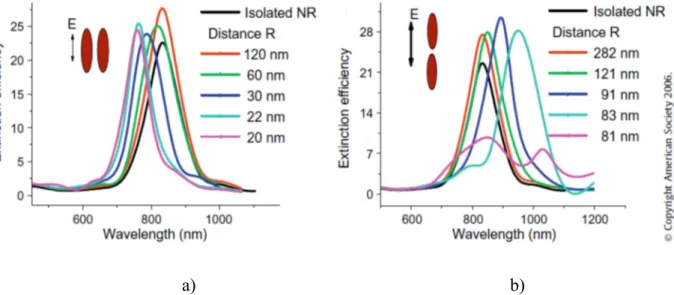
The plasmon resonances carried by the two nanorods will interact to form different collective plasmon modes. The extinction spectrum of gold nanorods changes according to the assembling arrangement. Precisely, the longitudinal plasmon band is found to shift to lower energies for end-to-end assembly, instead, a shift to higher energies is found for the side-by-side orientation. The effectivness of the plasmon coupling increases with decreasing internanorod distance and an increase in the number of the interacting nanorods, as showed in Fig.I.25. For both side-by-side and end-to-end assemblies, the strength of the longitudinal plasmon coupling increases with increasing nanorod aspect ratio as a result of the increasing dipole moment of the longitudinal plasmon [69],
a) Side-by-side arrangement
b) End-to-end arrangement
I.5. Plasmon coupling in assemblies of metal nanoparticles
30
a) b)
b) Fig.I.25. Extinction spectra at different internanorod center-to-center distance of side-by-side arranged AuNRs (a) and end-to-end arranged AuNRs (b) [69].
The plasmon coupling of metallic nanoparticles is clearly detected in assembled nanospheres; in this case, a simple linear arrangement of nanospheres leads to the formation of a rod-like shape. From a spectroscopic point of view this implies the appearance of a new band at lower energy than typical nanospheres plasmonic band [Fig. I.26] [70]. In the case of metallic nanorods, an assembly of a couple of nanoparticles, is comparable to a single nanorod with a different aspect ratio: a greater aspect ratio is obtained in the case of an end-to-end arrangement; a lower in the case of a side-by-side. Consequently, in the extinction spectra of an end-to-end assembled AuNRs, longitudinal plasmonic band is red-shifted respect to the isolated AuNRs, while in a side-by-side assembled AuNRs this band appears blue-shifted.
REFERENCES
31
REFERENCES
1. a) E. Hao, G. Schatz and J. Hupp, J. Fluoresc., 2004, 14, 331; b) K. L. Kelly, E. Coronado, L. L. Zhao and G. C. Schatz, J. Phys. Chem. B, 2003, 107, 668
2. S. Eustis and M. A. El-Sayed, Chem. Soc. Rev., 2006, 35, 209
3. a) C. Burda, X. Chen, R. Narayanan and M. A. El-Sayed, Chem. Rev., 2005, 105, 102; b) Y. Xia, P. Yang, Y. Sun, Y. Wu, B. Mayers, B. Gates, Y. Yin, F. Kim and H. Yan, Adv. Mater., 2003, 15, 353–389; c) M. P. Pileni, Nat. Mater., 2003, 2, 145; d) R. C. Jin, Y. W. Cao, C. A. Mirkin, K. L. Kelly, G. C. Schatz and J. G. Zheng, Science, 2001, 294
4. a) C. J. Murphy, T. K. Sau, A. M. Gole, C. J. G. Orendorff, J. L. Gou, S. E. Hunyadi and T. Li, J. Phys. Chem. B, 2005, 109; b) S. Link and M. A. El-Sayed, J. Phys. Chem. B, 1999, 103, 8410; c) S. Link and M. A. El-Sayed, Int. Rev. Phys. Chem., 2000, 19, 409; d) S. Link M. A. El-Sayed, Annu. Rev. Phys. Chem., 2003, 54, 331
5. a) M. A. El-Sayed, Acc. Chem. Res., 2001, 34, 257; b) S. Eustis, Mostafa A. El-Sayed, Chem. Soc. Rev., 2006, 35, 209
6. a) J. A. Creighton, D. G. Eadon, J. Chem. Soc., Faraday Trans. 1991, 87,3881; b) G. Schmid, Chem. Rev. 1992, 92; c) A. Henglein, J. Phys.Chem. 1993, 97, 5457; d) S. Nie, S. R. Emory, Science, 1997, 275, 1102; e) V.Russier, M. P. Pileni, Surf. Sci. 1999, 425, 313; f) N. R. Jana, T. K. Sau, T. Pal, J. Phys. Chem. B, 1999, 103, 115; g) S. Link, M. A. El-Sayed, J. Phys.Chem. B, 1999, 103, 8410
7. a) G. Frens, Nature, 1973, 241, 20; b) D. G. Duff, A. Baiker, P. P. Edwards, Langmuir, 1993, 9, 2301; c) M. T. Reetz, W. Helbig, J. Am. Chem. Soc.,1994, 116, 7401; d) D. V. Leff, P. C. Ohara, J. R. Heath, W. M. Gelbart, J. Phys. Chem., 1995, 99, 7036; e) J. H. Fendler, F. C. Meldrum, Adv. Mater., 1995, 7, 607; f) Y. Volokitin, J. Sinzig, L. J. de Jongh, G. Schmid, I. I. Moi-seev, Nature, 1996, 384, 621
8. a) T. S. Ahmadi, Z. L. Wang, T. C. Green, A. Henglein, M. A. El-Sayed, Science, 1996, 272, 1924; b) J. M Petroski, Z. L. Wang, T. C. Green, M. A.El-Sayed, J. Phys. Chem. B, 1998, 102, 3316; c) J. S. Bradley, B. Tesche, W. Busser, M. Maase, M. T. Reetz, J. Am. Chem. Soc., 2000, 122, 4631
9. a) B. R. Martin, D. J. Dermody, B. D. Reiss, M. M. Fang, L. A. Lyon, M. J. Natan, T. E. Mallouk, Adv. Mater., 1999, 11, 1021; b) B. M. I. van derZande, M. R. Bohmer, L. G. J. Fokkink, C. Schonenberger, Langmuir, 2000, 16, 451
10. a) C. R. Martin, Chem. Mater., 1996, 8, 1739; b) V. M. Cepak, C. R. Mar-tin, J. Phys. Chem. B, 1998, 102, 9985; c) C. Schonenberger, B. M. I. vander Zande, L. G. J. Fokkink,
REFERENCES
32
M. Henny, C. Schmid, M. Kruger, A. Bach-told, R. Huber, H. Birk, U. Staufer, J. Phys. Chem. B, 1997, 101, 5497
11. a) J. Sloan, D. M. Wright, H. G. Woo, S. Bailey, G. Brown, A. P. E.York, K. S. Coleman, J. L. Hutchison, M. L. H. Green, Chem. Commun., 1999,699; b) T. Kyotani, L. F. Tsai, A. Tomita, Chem. Commun., 1997, 701; c) A.Govindaraj, B. C. Satishkumar, M. Nath, C. N. R. Rao, Chem. Mater., 2000, 12, 202; d) B. K. Pradhan, T. Kyotani, A. Tomita, Chem. Commun., 1999, 1317
12. a) M. P. Pileni, T. Gulik-Krzywicki, J. Tanori, A. Filankembo, J. C. Dedieu, Langmuir, 1998, 14, 7359; b) M. P. Pileni, B. W. Ninham, T. Gulik-Krzywicki, J. Tanori, I. Lisiecki, A. Filankem, Adv. Mater., 1999, 11,1358; c) L. M. Qi, J. M. Ma, H. M. Cheng, Z. G. Zhao, J. Phys. Chem., B 1997, 101, 3460; d) M. Li, M. H. Schnablegger, S. Mann, Nature, 1999, 402,393; e) B. R. Heywood, S. Mann, Adv. Mater., 1994, 6,9
13. a) B. R. Heywood, S. Rajam, S. Mann, J. Chem. Soc., Faraday Trans., 1991,87, 735; b) D. Wash, S. Mann, Nature 1995, 377, 320; c) J. D. Hopwood, S. Mann, Chem. Mater. ,1997, 9, 1819; d) G. D. Rees, R. Evans-Gowing,S. J. Hammond, B. H. Robinson, Langmuir, 1999, 15, 1993; e) A. Filan-kembo, M. P. Pileni, J. Phys. Chem. B, 2000, 104, 5865
14. a) M. Q. Zhao, L. Sun, R. M. Crooks, J. Am.Chem. Soc., 1998, 120, 4877; b) M. J. Hostetler, J. E. Wingate, C. J. Zhong.J. E. Harris, R. W. Vachet, M. R. Clark, J. D. Londono, S. J. Green, J. J.Stokes, G. D. Wignall, G. L. Glish, M. D. Porter, N. D. Evans, R. W. Murray, Langmuir, 1998, 14, 17; c) G. W. Busser, J. G. van Ommen, J. A. Lerch-er, J. Phys. Chem. B, 1999, 103, 1651; d) T. Terani shi, M. Miyake, Chem.Mater., 1998, 10, 594; e) T. Teranishi, M. Hosoe, T. Tanaka, M. Miyake, J. Phys. Chem. B, 1999, 103, 3818
15. N. R. Jana, L. Gearheart, C. J. Murphy, Chem. Mater., 2001, 13, 2313 16. M. Törnblm, U. Henrikesson, J. Phys. Chem. B, 1997, 101, 6028 17. H. Chen, L. Shao, Q. Lia and J.Wang, Chem. Soc. Rev., 2013,42, 2679 18. L.Scarabelli, M. Grzelczak, L. M. Liz-Marzán, Chem. Mater., 2013, 25, 4232 19. N. R. Jana, L. Gearheart, C. J. Murphy, Adv. Mater., 2001, 13, 1389
20. P.D.Cozzoli, L.Manna, Synthetic Strategies to Size and Shape Controlled Nanocrystals and Nanocrystal Heterostructures in Bio-Application of Nanoparticles, Springer-Verlag New York, 2007, 620,1
21. L.Scarabelli, M. Coronado-Puchau, J. J. Giner-Casares, J. Langer, L. M. Liz-Marzán, ACS Nano, 2014, 8, 5833
REFERENCES
33
23. Zeliha Cansu Canbek. Crystal Engineering of Anisotropic Gold Nanoparticles through Modulation of Seed Size and Crystal Structure. Theoretical and/or physical chemistry. Université de Versailles-Saint Quentin en Yvelines, 2014.
24. S. E. Lohse, N. D. Burrows, L. Scarabelli, L. M. Liz-Marzán, C.J. Murphy, Chem. Mater., 2014, 26, 34
25. D. K. Smith, N. R. Miller, B. A. Korge, Langmuir, 2009, 16, 9518
26. M.R.Langille, M.L.Personick, J. Zhang, C.A. Mirkin., J. Am. Chem. Soc. 2012, 134, 14542 27. K. Park, H. Koerner, R. A. Vaia,Nano Lett., 2010, 10, 1433
28. N. G. Bastus, J. Comenge, V. Puntes, Langmuir, 2011, 27, 11098 29. S. E. Lohse and C. J. Murphy, Chem. Mater., 2013, 25, 1250
30. L. Jiang, H. Mundoor, Q. Liu, I. I. Smalyukh, ACS Nano, 2016, 10, 7064
31. A. B. Serrano-Montes, D. Jimenez de Aberasturi, J. Langer, J. J. Giner-Casares, L. Scarabelli, A.Herrero, L. M. Liz-Marzán, Langmuir, 2015, 31, 9205
32. I. Pastoriza-Santos, J. Perez-Juste, L. M. Liz-Marzán, Chem. Mater., 2006, 18, 2465 33. W. Wu and J. B. Tracy, Chem. Mater., 2015, 27, 2888
34. M. Grzelczak, J. Vermant, E. M. Furst, L. M. Liz-Marzán, ACS NANO, 2010,4, 3591 35. V. Sharma, K. Park, M.Srinivasarao, Materials Science and Engineering, 2009,65, 1
36. N. R. Jana, L.A. Gearheart, S. O. Obare, C. J. Johnson, K.J. Edler, S. Mann, C. J. Murphy, J. Mater. Chem., 2002, 12, 2909
37. C. Hamon, S. Novikov, L. Scarabelli, L. Basabe-Desmonts, L. M. Liz-Marzán, ACS NANO, 2014, 8, 10694
38. L. Scarabelli, A. Sánchez-Iglesias, J. Pérez-Juste, L. M. Liz-Marzán, J. Phys. Chem. Lett. 2015, 6, 4270
39. K. J. M. Bishop, C. E. Wilmer, S. Soh, B. A. Grzybowski,Small, 2009, 5, 1600 40. P. Pramod, K. G. Thomas, Adv. Mater., 2008, 20, 4300.
41. a) Z. Sun, W. Ni, Z.Yang, X. Kou, L. Li, J. Wang,Small, 2008, 4, 1287; b) W. Ni, R.A. Mosquera, J. Perez-Juste, L. M. Liz-Marzán,J. Phys. Chem. Lett., 2010, 1, 1181
42. Z. Nie, D.Fava, M. Rubinstein, E. Kumacheva, J. Am. Chem. Soc., 2008, 130, 3683
43. a)K. Caswell, J. N. Wilson, U. H. F. Bunz and C. J. Murphy, J. Am. Chem. Soc., 2003, 125, 13914; b) C. G. Wang, Y. Chen, T. T. Wang, Z. F. Ma and Z. M. Su, Chem. Mater., 2007, 19, 5809; c) S. J. Zhen, C. Z. Huang, J. Wang and Y. F. Li, J. Phys. Chem.C, 2009, 113, 21543; d) M. R. Jones, R. J. Macfarlane, B. Lee, J. Zhang, K. L. Young, A. J. Senesi, C. A. Mirkin, Nat. Mater., 2010, 9, 913
REFERENCES
34
44. Lacaze, E., Merchiers, O., Borensztein, Y. et al. Rend. Fis. Acc. Lincei, 2015, 26(Suppl 2): 183
45. Y. Lia, E. Princea, S. Choa, A. Salarib, Y.Mosaddeghian Golestanic, O. D. Lavrentovichc, Eugenia Kumachevaa, Proc Natl Acad Sci U S A. 2017, 114,2137
46. H. Qi, T. Hegmann,Liquid Crystals Today, 2011, 20, , 102
47. D. Coursault, J. Grand, B. Zappone, H. Ayeb, G. Lévi, N. Félidj, E. Lacaze,ACS Nano 24, 2012, 1461
48. B. Rožič, J.Fresnais, C. Molinaro, J. Calixte, S. Umadevi, S. Lau-Truong, N. Felidj, T.Kraus, F. Charra, V. Dupuis, T. Hegmann, C. Fiorini-Debuisschert, B. Gallas, E. Lacaze, ACS Nano, 2017, 11, 6728
49. a) D. Lysenko, P. Pagliusi,C. Provenzano,Y. Reznikov, K. Slyusarenko, G. Cipparrone, Appl. Phys. Lett., 2013, 103, 151913; b) D. Kasyanyuk, P. Pagliusi.A. Mazzulla, V. Reshetnyak, Y. Reznikov, C. Provenzano,M. Giocondo, M. Vasnetsov, O. Yaroshchuk, G. Cipparrone,Scientific Reports 2016, 17, 20742
50. H. Chen, L. Shao, Q. Lia and J.Wang, Chem. Soc. Rev., 2013,42, 2679
51. J. Perez-Juste, I. Pastoriza-Santos, L. M. Liz-Marzán, P. Mulvaney, Coord. Chem Rev, 2005, 249, 1870
52. P. B. Johnson and R. W. Christy, Phys. Rev. B: Solid State, 1972, 6, 4370 53. J. Zuloaga, E. Prodan and P. Nordlander, ACS Nano, 2010, 4, 5269
54. C. Sonnichsen, T. Franzl, T. Wilk, G. von Plessen and J. Feldmann, Phys. Rev. Lett., 2002, 88, 077402
55. a) X. S. Kou, W. H. Ni, C.-K. Tsung, K. Chan, H.-Q. Lin, G. D. Stucky and J. F. Wang, Small, 2007, 3, 2103. b) H. J. Chen, L. Shao, K. C. Woo, T. Ming, H.-Q. Lin and J. F. Wang, J. Phys. Chem. C, 2009, 113, 17691
56. a) C. Novo and P. Mulvaney, Nano Lett., 2007, 7, 520; J. Perez-Juste, I. Pastoriza-Santos, L. M. Liz-Marzán, P. Mulvaney, Coord. Chem. Rev., 2005, 249, 1870
57. a) C. X. Yu and J. Irudayaraj, Biophys. J., 2007, 93, 3684. b) C.-D. Chen, S.-F. Cheng, L.-K. Chau and C. R. C. Wang, Biosens. Bioelectron., 2007, 22, 926. c) J. Burgin, M. Z. Liu and P. Guyot-Sionnest, J. Phys. Chem. C, 2008, 112, 19279. d) Y. Khalavka, J. Becker and C. Sonnichsen, J. Am. Chem. Soc., 2009, 131, 1871. e) H. J. Chen, X. S. Kou, Z. Yang, W. H. Ni and J. F. Wang, Langmuir, 2008, 24, 5233
58. J. N. Anker, W. P. Hall, O. Lyandres, N. C. Shah, J. Zhao and R. P. van Duyne, Nature Mater., 2008, 7, 442.
![Fig. I.3. Schematic illustration of the seed-mediated method for the growth of Au nanorods [17]](https://thumb-eu.123doks.com/thumbv2/123dokorg/2867406.9112/13.892.104.793.423.806/fig-schematic-illustration-seed-mediated-method-growth-nanorods.webp)
![Fig. I.5. Models of Ag + action in seeded growth of AuNRs, according to literature [29]](https://thumb-eu.123doks.com/thumbv2/123dokorg/2867406.9112/17.892.256.665.106.656/fig-models-action-seeded-growth-aunrs-according-literature.webp)
![Fig. I.11 a) TEM image of AuNR@CTAB with a high shape yield; b) TEM image of of 3 different shapes of nanoparticles assembled separately [35] [38]](https://thumb-eu.123doks.com/thumbv2/123dokorg/2867406.9112/22.892.86.781.212.530/image-aunr-ctab-different-shapes-nanoparticles-assembled-separately.webp)
![Fig. I.14. Extinction spectra of AuNRs with different aspect ratios, correlated with TEM images [18].](https://thumb-eu.123doks.com/thumbv2/123dokorg/2867406.9112/27.892.203.756.281.784/extinction-spectra-aunrs-different-aspect-ratios-correlated-images.webp)
![Fig. I.14. Longitudinal nanorods plasmon band position tuned by the solvent [56]](https://thumb-eu.123doks.com/thumbv2/123dokorg/2867406.9112/32.892.255.634.139.441/fig-longitudinal-nanorods-plasmon-band-position-tuned-solvent.webp)
![Fig. I.23. Extinction spectra of AuNR@SiO 2 with increasing shell thickness [33]](https://thumb-eu.123doks.com/thumbv2/123dokorg/2867406.9112/34.892.210.648.114.420/fig-extinction-spectra-aunr-sio-increasing-shell-thickness.webp)