ARTICLE OPEN ACCESS
Anti-MOG encephalitis mimicking small vessel
CNS vasculitis
Kristina Patterson, MD, PhD, Estibaliz Iglesias, MD, PhD, Maclean Nasrallah, MD, PhD, Ver´onica Gonz´ alez-´
Alvarez, MD, Mariona Suñol, MD, Jordi Anton, MD, PhD, Albert Saiz, MD, PhD, Eric Lancaster, MD, PhD,
and Tha´ıs Armangue, MD, PhD
Neurol Neuroimmunol Neuroinflamm 2019;6:e538. doi:10.1212/NXI.0000000000000538
Correspondence Dr. Armangue [email protected]
Abstract
ObjectiveTo report 2 patients with anti–myelin oligodendrocyte glycoprotein (MOG)-associated en-cephalitis who were initially misdiagnosed with small vessel primary CNS vasculitis.
Methods
Review of symptoms, MRI and neuropathologic features, and response to treatment. MOG antibodies were determined in serum and CSF using a cell-based assay.
Results
Symptoms included fever, headache, and progressive mental status changes and focal neuro-logic deficits. CSF studies revealed lymphocytic pleocytosis, and both patients had abnormal brain MRIs. Brain biopsy samples showed prominent lymphocytic infiltration of the wall of small vessels; thesefindings initially suggested small vessel CNS vasculitis, and both patients were treated accordingly. Although 1 patient had a relapsing-remitting course not responsive to cyclophosphamide, the other one (also treated with cyclophosphamide) did not relapse. Retrospective assessment of serum and CSF demonstrated MOG antibodies in both cases, and review of biopsy specimens showed absence offibrinoid necrosis (a pathologic requirement for small vessel CNS vasculitis).
Conclusions
Anti–MOG-associated encephalitis can be mistaken for small vessel CNS vasculitis. This is important because the diagnosis of anti–MOG-associated encephalitis does not require brain biopsy and can be established with a serologic test.
RELATED ARTICLE
Editorial
Diagnostic challenges and pitfalls of myelin oligodendrocyte glycoprotein antibody–associated demyelination: Lessons from neuropathology Page e544
From the Neurology Department (K.P., E.L.), University of Pennsylvania, Philadelphia; Rheumatology Department, Sant Joan de Deu Children’s Hospital (E.I., J.A.), University of Barcelona, Spain; Department of Pathology (M.N.), University of Pennsylvania, Philadelphia; Pediatric Neuroimmunology Unit (V.G.-´A., T.A.), Sant Joan de D´eu Children’s Hospital, University of Barcelona, Spain; Department of Pathology (M.S.), Sant Joan de D´eu Children’s Hospital, University of Barcelona, Spain; and Neuroimmunology Program (A.S., T.A.), IDIBAPS-Hospital Clinic, University of Barcelona, Spain.
Funding information and disclosures are provided at the end of the article. Full disclosure form information provided by the authors is available with the full text of this article at Neurology.org/NN.
The Article Processing Charge was funded by the Clinic Foundation.
This is an open access article distributed under the terms of the Creative Commons Attribution-NonCommercial-NoDerivatives License 4.0 (CC BY-NC-ND), which permits downloading and sharing the work provided it is properly cited. The work cannot be changed in any way or used commercially without permission from the journal.
The diagnosis of small vessel primary CNS vasculitis is challenging because conventional and brain MRI angiog-raphy are negative, and brain biopsy remains as the only definite diagnostic test.1
However, brain biopsy is invasive and may be uninformative because of sampling error. Here, we describe 2 patients with myelin oligodendrocyte gly-coprotein (MOG) antibody–associated encephalitis2
who were initially misdiagnosed with small vessel CNS vascu-litis based on biopsyfindings. Physicians should be aware of this potential misdiagnosis because it has important clinical implications.
Case 1
A 5-year-old boy presented with 2 weeks of frontal headache and fever. His physical examination showed decreased alertness and bilateral papilledema (table). Brain CT and MRI (figure 1A) were normal, and the CSF showed pleo-cytosis. Meningoencephalitis was suspected, and he was started on steroids and acyclovir. During the following days, he developed visual hallucinations. There was gradual clin-ical improvement until complete recovery, and the patient was discharged on steroid taper 1 month later. In the en-suing 4 months, he was readmitted 3 times for relapsing symptoms while weaning from steroids. Repeat brain MRI showed T2 abnormalities in the basal ganglia, cerebellar peduncles, and supratentorial white matter (figure 1B-D), and CSF pleocytosis was identified in all episodes (table). All relapses substantially improved after treatment with steroids. At the last relapse, a conventional brain angiogra-phy was inconclusive. Brain biopsy showed infiltrates of lymphocytes involving the wall of small vessels and per-ivascular areas accompanied by perper-ivascular demyelination (figure 2A–D). The patient was diagnosed with primary CNS vasculitis, and he was started on monthly pulses of cyclophosphamide. After the 5th pulse, he developed acute right optic neuritis that was treated with steroids, resulting in little improvement. Extensive blood testing identified an elevation of lipoprotein A (also present in his asymptomatic father), and oral aspirin was added, together with myco-phenolate mofetil (MMF) and prednisone. He remained clinically and radiologically stable (figure 1E), with a right eye visual deficit for 2 years; at this time, immunosuppres-sion was weaned, and shortly after stopping the steroids (while on MMF and aspirin), he developed confusion and decreased level of consciousness. MRI showed extensive white matter abnormalities (figure 1F) and high serum titer of MOG antibodies (1:640). Retrospective assessment of stored serum and CSF obtained at onset of the disease were also positive for MOG antibodies (serum titer 1:20,480 and CSF 1:320, table). Review of the paraffin block containing
the brain biopsy showed that the inflammatory infiltrates were not confined to the vessel wall and also involved the white and gray matter. With thesefindings, the patient was diagnosed with anti-MOG encephalitis, and treatment with rituximab, azathioprine, and low-dose prednisone was initi-ated. No more relapses were observed; at the last follow-up, 3 years later, he remained clinically and radiologically stable on azathioprine and low-dose prednisone (eventually dis-continued), and the serum titer of MOG immunoglobulin G (IgG) antibodies had decreased (1:80) below the con-sensus limit of positivity (≥1:160).2,3
Case 2
A 39-year-old woman with a history of ulcerative colitis treated with mesalamine and budesonide developed severe, progressive left temporo-parietal headache associated with nausea and photophobia. Shortly after neurologic symptom onset, she developed intermittent fever. Peripheral leukocy-tosis with neutrophilic predominance and elevated C-reactive protein were identified (table). Brain MRI showed hyperin-tense gyriformfluid-attenuated inversion recovery signal over the left temporal, parietal, and occipital lobes (figure 1G–H). CSF analysis showed pleocytosis and elevated protein con-centration, and the patient was started on empiric antibiotics and acyclovir. Six days later, she developed expressive aphasia, confusion, and agitation, and bilateral VI cranial nerve palsies. Repeat brain MRI showed no changes, and CT angiography demonstrated no clear evidence of vasculitis. A biopsy of the left parietal lobe and dura showed interstitial and perivascular lymphocytic infiltrates with marked involvement of the vessel wall (figure 2E–H). There was no clear evidence of de-myelination (data not shown). Thesefindings led to suspect CNS vasculitis, and high-dose IV steroids and oral cyclophos-phamide were started. Four months later, cyclophoscyclophos-phamide was discontinued because of elevated transaminases. The dose of prednisone was tapered over the course of 2 years. During the follow-up, the brain MRI normalized at 4 months and the neurologic examination at 9 months; her only complaints were mild aphasia when tired and chronic daily headache. Retro-spective analysis of stored CSF obtained by the time of symptom onset showed MOG-IgG antibodies (1:8).
Discussion
We describe 2 patients initially diagnosed with small vessel CNS vasculitis but who in fact had anti–MOG-associated encephalitis. Thefirst patient received standard treatment for CNS vasculitis, including cyclophosphamide and steroid-sparing drugs. Although this treatment led to initial stabili-zation of symptoms, the patient developed new relapses until
Glossary
MMF = mycophenolate mofetil; MOG = myelin oligodendrocyte glycoprotein.
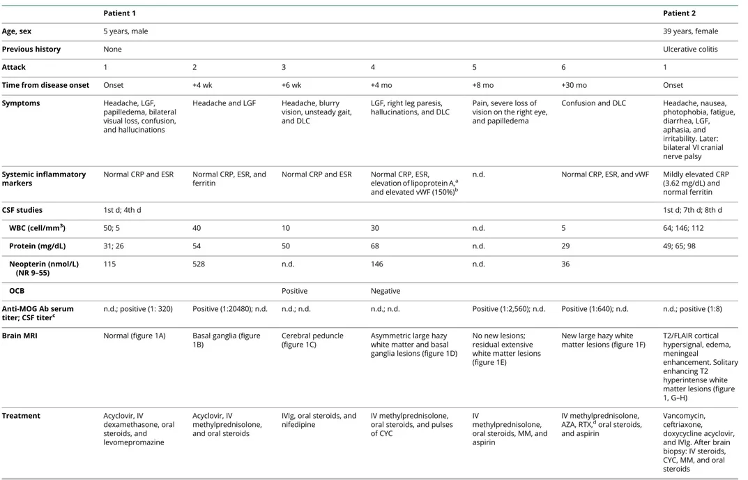
TableClinical and laboratory data of 2 patients with anti-MOG encephalitis initially misdiagnosed with small CNS vessel vasculitis
Patient 1 Patient 2
Age, sex 5 years, male 39 years, female
Previous history None Ulcerative colitis
Attack 1 2 3 4 5 6 1
Time from disease onset Onset +4 wk +6 wk +4 mo +8 mo +30 mo Onset Symptoms Headache, LGF,
papilledema, bilateral visual loss, confusion, and hallucinations
Headache and LGF Headache, blurry vision, unsteady gait, and DLC
LGF, right leg paresis, hallucinations, and DLC
Pain, severe loss of vision on the right eye, and papilledema
Confusion and DLC Headache, nausea, photophobia, fatigue, diarrhea, LGF, aphasia, and irritability. Later: bilateral VI cranial nerve palsy Systemic inflammatory markers
Normal CRP and ESR Normal CRP, ESR, and ferritin
Normal CRP and ESR Normal CRP, ESR, elevation of lipoprotein A,a
and elevated vWF (150%)b
n.d. Normal CRP, ESR, and vWF Mildly elevated CRP (3.62 mg/dL) and normal ferritin CSF studies 1st d; 4th d 1st d; 7th d; 8th d WBC (cell/mm3) 50; 5 40 10 30 n.d. 5 64; 146; 112 Protein (mg/dL) 31; 26 54 50 68 n.d. 29 49; 65; 98 Neopterin (nmol/L) (NR 9–55) 115 528 n.d. 146 n.d. 36
OCB Positive Negative
Anti-MOG Ab serum titer; CSF titerc
n.d.; positive (1: 320) Positive (1:20480); n.d. n.d.; n.d. n.d.; n.d. Positive (1:2,560); n.d. Positive (1:640); n.d. n.d.; positive (1:8) Brain MRI Normal (figure 1A) Basal ganglia (figure
1B)
Cerebral peduncle (figure 1C)
Asymmetric large hazy white matter and basal ganglia lesions (figure 1D)
No new lesions; residual extensive white matter lesions (figure 1E)
New large hazy white matter lesions (figure 1F)
T2/FLAIR cortical hypersignal, edema, meningeal enhancement. Solitary enhancing T2 hyperintense white matter lesions (figure 1, G–H) Treatment Acyclovir, IV dexamethasone, oral steroids, and levomepromazine Acyclovir, IV methylprednisolone, and oral steroids
IVIg, oral steroids, and nifedipine
IV methylprednisolone, oral steroids, and pulses of CYC
IV
methylprednisolone, oral steroids, MM, and aspirin
IV methylprednisolone, AZA, RTX,doral steroids,
and aspirin
Vancomycin, ceftriaxone, doxycycline acyclovir, and IVIg. After brain biopsy: IV steroids, CYC, MM, and oral steroids Continued Neurology. org/NN Neurology: Neuroimm unology & Neuroinflam mation | Volume 6, Number 2 | March 2019
he was transitioned to a B-cell depletion therapy with good clinical and serologic response. In the second patient, the finding of MOG antibodies did not change the treatment strategy because she was already clinically stable; however, an earlier detection of these antibodies could have prevented the brain biopsy.
In retrospect, none of these patients fulfilled the criteria for small vessel CNS vasculitis. Although the presence of in-flammatory cells in the vessel walls is sufficient to diagnose medium or large vessel vasculitis, signs of vessel damage such asfibrin deposition and/or necrosis (absent in our patients) must also be present to meet the criteria for small vessel vasculitis. Diapedesis of leukocytes takes place in small ves-sels, and the presence of inflammatory cells in the walls does not necessarily imply a pathologic process. Moreover, the pathology of CNS vasculitis is usually focal or segmental, and the brain biopsy specimen can be negative or not meet the full diagnostic criteria.4As our patients reveal, the pres-ence of lymphoid cells in vessel walls must be interpreted with caution, particularly when the differential diagnosis includes encephalitis with lymphocytic infiltrates. In this context, the presence of lymphocytes in the brain paren-chyma away from the vessel walls favors secondary in-volvement of the vessels rather than a primary vasculitic process. Whether other cases of anti-MOG encephalitis may have been misdiagnosed as CNS vasculitis is unknown. There are reports of patients diagnosed with CNS small vessel vasculitis by brain biopsy who later developed my-elitis or optic neuritis5 (symptoms suggestive of MOG encephalitis),2,3making us to postulate that this misdiagnosis may not be unusual.
Acknowledgment
The authors thank Professor Josep Dalmau for his valuable critical review of the manuscript.
Study funding
This study was supported in part by Fundaci´o Marat´o de TV3 (20141830).
Disclosure
K. Patterson received research support from the NIH/NINDS. E. Iglesias, M. Narallah, V. Gonzalez-Alverez, and M. Sunol report no disclosures. J. Anton served on the scientific ad-visory boards of Novartis, Sobi, and Gebro; received travel funding and/or speaker honoraria from AbbVie, Roche, Pfizer, Sobi, and Gebro; and served on the speakers’ bureau of AbbVie, Pfizer, Roche, and Gebro. A. Saiz received con-sulting compensation and speaker honoraria from Bayer-Schering, Merck Serono, Biogen Idec, Sanofi-Aventis, Teva, Novartis, and Roche. E. Lancaster served on the advisory boards of Grifols, Amgen, and Janssen; received speaker honoraria and travel funding from Grifols; consulted for Medimmune, Merck, and Novartis; received fees from the Federal Vaccine Injury Compensation Program; received research support from Grifols and the NINDS; and provided
Table Clinical and laboratory data of 2 patients with anti-MOG encephalitis initially misdiagnosed with small CNS vessel vasculitis (continued) Patient 1 Patient 2 Clinical response to immunotherapy Complete Complete Complete Complete; MRI residual lesions Right visual deficit, with optic atrophy Nearly complete; residual MRI lesions. Nearly complete; normalization o f b rain MRI Last follow-up 6 y , left eye visual loss and mild deficit of attention; extensive MRI white matter residual lesions. Serum anti-MOG titers c(1:80); current treatment: AZA and low-dose a spirin d 9 mo: mild aphasia when tired and chronic d aily headache Abbreviations: Ab = antibody; AZA = azathioprine; CRP = C-reactive protein; CYC = cyclophosphamid e ;DLC = decreased level of consciousness; ESR = ery throcyte sedimentation rate ;FLAIR = fluid-attenuated inversion recovery; IV = intravenous; IVIg = IV immunoglobulin; LGF = low-grade fever; MM = mycophenolate mofetil; MOG = myelin oligodendrocyte glycoprotein; n.d. = not d one; NR = normal range; OCB = oligoclonal band; RTX = rituximab, S = serum; vWF = von Willebrand factor; WBC = white blood cell. aAlso present in his a symptomatic father. bNR: 60% –120%. cThreshold for positive results, CSF (≥ 1:2), and serum (≥ 1:160). 2,3 dOnly 1 cycle of RTX was given (4 weekly 375 mg/m 2doses) due to CD19 counts <2% of lymphocytes since then.
medical legal consultation for Rogers Towers PA, Orlando Health, and Wilson, Elser, Moskowitz, Edelman & Dicker LLP. T. Armangue received research support from the Mutua
Madrileña Foundation. Full disclosure form information pro-vided by the authors is available with the full text of this article at Neurology.org/NN.
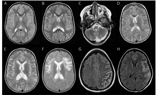
Figure 1MRI of 2 patients with anti-MOG encephalitis initially misdiagnosed with small vessel CNS vasculitis
Patient 1: (A) Axial T2 MRI sequence showing no abnormalities at disease onset; (B) bilateral involvement of the basal ganglia 4 weeks after disease onset while steroids were being decreased; (C) left cerebral peduncle abnormality at 6-week follow-up; (D) asymmetric large hazy white matter and basal ganglia lesions at 4 months; (E) residual white matter lesions and enlargement of ventricles due to brain atrophy; and (F) new asymmetric large hazy white matter lesions 30 months after disease onset when steroids were discontinued. Patient 2: (G and H) Axial FLAIR sequences showing gyriform hyperintensities with edema similar to abnormalities previously reported in cases of anti–MOG-associated cortical encephalitis.6
Figure 2Brain biopsy of 2 patients with anti-MOG encephalitis initially misdiagnosed with small vessel CNS vasculitis
In patient 1, biopsy of the right temporal lobe showed small vessel perivascular lymphocytic infiltration (A, hematoxylin-eosin staining; B, magnification of the vessel shown in panel A). Inflammatory infiltrates included T and B lymphocytes (not shown) in association with edema, perivascular demyelination, and reactive gliosis (C and D, luxol fast blue staining). In patient 2, biopsy of the left temporal lobe showed marked perivascular lymphocytic infiltrates involving the vessel wall (E, hematoxylin-eosin staining). The infiltrates were also composed of T lymphocytes (F, CD3 immunostaining), B lymphocytes (G, CD20 immu-nostaining), and macrophages (H, anti-CD68 immunostaining). Myelin staining did not show clear evidence of demyelination (not shown). No necrosis or fibrin deposition was identified (not shown). Scale bar 200μm in A and C, 500 μm in E, and 100 μm in B, D, and F–H.
Publication history
Received by Neurology: Neuroimmunology & Neuroinflammation August 16, 2018. Accepted infinal form October 19, 2018.
References
1. Benseler SM, deVeber G, Hawkins C, et al. Angiography-negative primary central nervous system vasculitis in children: a newly recognized inflammatory central ner-vous system disease. Arthritis Rheum 2005;52:2159–2167.
2. Jarius S, Paul F, Aktas O, et al. MOG encephalomyelitis: international recom-mendations on diagnosis and antibody testing. J Neuroinflammation 2018;15: 134.
3. H¨oftberger R, Sepulveda M, Armangue T, et al. Antibodies to MOG and AQP4 in adults with neuromyelitis optica and suspected limited forms of the disease. Mult Scler 2015;21:866–874.
4. Giannini C, Salvarani C, Hunder G, Brown RD. Primary central nervous system vasculitis: pathology and mechanisms. Acta Neuropathol 2012;123: 759–772.
5. Benseler S, Pohl D. Childhood central nervous system vasculitis. Handb Clin Neurol 2013;112:1065–1078.
6. Ogawa R, Nakashima I, Takahashi T, et al. MOG antibody-positive, benign, unilateral, cerebral cortical encephalitis with epilepsy. Neurol Neuroimmunol Neuroinflamm 2017;4:e322. doi: 10.1212/NXI.0000000000000322.
Appendix 1Author contributions
Name Location Role Contribution
Kristina Patterson, MD, PhD University of Pennsylvania, Philadelphia
Author Analyzed the data and drafted the manuscript
Estibaliz Iglesias, MD, PhD University of Barcelona, Barcelona
Author Analyzed the data and critically reviewed the manuscript Maclean Nasrallah, MD, PhD University of Pennsylvania, Philadelphia
Author Analyzed the data, critically reviewed the manuscript, and developed figure 2
Ver´onica Gonz´ alez-´ Alvarez, MD University of Barcelona, Barcelona
Author Analyzed the data and critically reviewed the manuscript Mariona Suñol, MD University of Barcelona, Barcelona
Author Analyzed the data, critically reviewed the manuscript, and developed figure 2
Jordi Anton, MD, PhD
University of Barcelona, Barcelona
Author Analyzed the data and critically reviewed the manuscript Albert Saiz, MD, PhD University of Barcelona, Barcelona
Author Analyzed the data and critically reviewed the manuscript Eric Lancaster, MD, PhD University of Pennsylvania, Philadelphia
Author Analyzed the data and critically reviewed the manuscript Tha´ıs Armangue, MD, PhD University of Barcelona, Barcelona
Author Designed and
conceptualized the study; analyzed the data; drafted the manuscript; and developed the figures
DOI 10.1212/NXI.0000000000000538
2019;6;
Neurol Neuroimmunol Neuroinflamm
Kristina Patterson, Estibaliz Iglesias, Maclean Nasrallah, et al.
Anti-MOG encephalitis mimicking small vessel CNS vasculitis
This information is current as of February 1, 2019
Services
Updated Information &
http://nn.neurology.org/content/6/2/e538.full.html
including high resolution figures, can be found at:
References
http://nn.neurology.org/content/6/2/e538.full.html##ref-list-1
This article cites 6 articles, 1 of which you can access for free at: Citations
http://nn.neurology.org/content/6/2/e538.full.html##otherarticles
This article has been cited by 3 HighWire-hosted articles: Subspecialty Collections http://nn.neurology.org//cgi/collection/vasculitis Vasculitis http://nn.neurology.org//cgi/collection/autoimmune_diseases Autoimmune diseases http://nn.neurology.org//cgi/collection/all_demyelinating_disease_cns All Demyelinating disease (CNS)
following collection(s):
This article, along with others on similar topics, appears in the
Permissions & Licensing
http://nn.neurology.org/misc/about.xhtml#permissions
its entirety can be found online at:
Information about reproducing this article in parts (figures,tables) or in
Reprints
http://nn.neurology.org/misc/addir.xhtml#reprintsus
Information about ordering reprints can be found online:
Academy of Neurology.. All rights reserved. Online ISSN: 2332-7812.
Copyright © 2019 The Author(s). Published by Wolters Kluwer Health, Inc. on behalf of the American Published since April 2014, it is an open-access, online-only, continuous publication journal. Copyright
is an official journal of the American Academy of Neurology.