Tethering of vesicles to the Golgi by GMAP210
controls LAT delivery to the immune synapse
Andres Ernesto Zucchetti
1
, Laurence Bataille
1
, Jean-Marie Carpier
1,2
, Stéphanie Dogniaux
1
,
Mabel San Roman-Jouve
1
, Mathieu Maurin
1
, Michael W. Stuck
3
, Rosa M. Rios
4
, Cosima T. Baldari
5
,
Gregory J. Pazour
3
& Claire Hivroz
1
The T cell immune synapse is a site of intense vesicular trafficking. Here we show that the
golgin GMAP210, known to capture vesicles and organize membrane traffic at the Golgi, is
involved in the vesicular transport of LAT to the immune synapse. Upon activation, more
GMAP210 interact with LAT-containing vesicles and go together with LAT to the immune
synapse. Regulating LAT recruitment and LAT-dependent signaling, GMAP210 controls T cell
activation. Using a rerouting and capture assay, we show that GMAP210 captures
VAMP7-decorated vesicles. Overexpressing different domains of GMAP210, we also show that
GMAP210 allows their specific delivery to the immune synapse by tethering LAT-vesicles to
the Golgi. Finally, in a model of ectopic expression of LAT in ciliated cells, we show that
GMAP210 tethering activity controls the delivery of LAT to the cilium. Hence, our results
reveal a function for the golgin GMAP210 conveying specific vesicles to the immune synapse.
https://doi.org/10.1038/s41467-019-10891-w
OPEN
1Institut Curie, PSL Research University, INSERM U932, Integrative analysis of T cell activation team, 26 rue d’Ulm, 75248 Paris Cedex 05, France.
2Immunobiology Department, Yale University School of Medicine, New Haven, CT 06520, USA.3Program in Molecular Medicine, University of
Massachusetts Medical School, Worcester, MA 01605, USA.4Cell Dynamics and Signaling Department, CABIMER-CSIC/US/UPO, 41092 Seville, Spain.
5Department of Life Sciences, University of Siena, 53100 Siena, Italy. Correspondence and requests for materials should be addressed to C.H. (email:claire.
123456789
C
ommunication of cells with their extracellular
environ-ment is a critical function for all eukaryotic cells. It is
particularly important for the cells of the immune system
that have to “sense” the danger in a full organism. This is
probably why T lymphocytes have evolved to form a structure,
specialized in cell–cell communication, which is generated upon
direct contact between T lymphocytes and antigen-presenting
cells and called the immune synapse
1,2.
The immune synapse is a place of intense vesicular endocytic
and exocytic traffic that control many aspects of T cell activities
and functions. Indeed, the polarized release, at the immune
synapse, of cytokines
3,4, extracellular vesicles
5–7, and receptors
and ligands such as CD40L
8,9, regulates their communication
with the interacting cells and environment. It also shapes the
adaptive immune response. This endocytic and exocytic traffic of
vesicular compartments has also been shown to regulate
TCR-induced signaling
10. Recent results including ours have shown
that molecules and enzymes, involved in signaling in T
lym-phocytes, are present both at the plasma membrane and in
intracellular vesicular pools
11–17. Remarkably different signaling
mediators are present in different vesicles associated to a unique
set of traffic regulators and effectors, such as Rab GTPase and
v-SNARE proteins
10,18. Hence different signaling molecules follow
different endocytic and exocytic pathways. This may control the
physical separation of signaling molecules in distinct modules
that can be assembled in response to specific triggering
19. These
results also raise the question of the nature of the different
vesicles, the control of their localization and of their polarized
delivery to the immune synapse. We previously showed that
trafficking to the immune synapse of the Linker for activation of
T cells (LAT), a transmembrane protein that plays a key role in T
lymphocyte signaling and function
20–23, is regulated by the
vesicular SNARE VAMP7
13. We also recently found that the
plasma membrane pool of LAT, after its TCR-induced
inter-nalization, is following the canonical retrograde Rab6/syntaxin 16
pathway to the Golgi before being transported back to the
immune synapse
24. This retrograde vesicular trafficking of LAT
controls the formation of signaling complexes
13,24,25, also known
as signalosomes and regulates some aspects of T cell activation.
Yet, the spatial organization of this traffic is not known. We
describe herein a new mechanism by which a golgin, specifically
conveys LAT-containing vesicles to the immune synapse.
Golgins are long coiled–coiled proteins attached to the Golgi
membrane via their C-terminal part, which can “capture” vesicles
at long distance through their N-terminal motifs increasing the
efficiency of trafficking
26,27. They have been proposed to ensure
the specific delivery of vesicles containing given cargoes to the
right membrane destination in the cells
26–29. GMAP210 is one of
these golgins. It anchors at the cis-Golgi via the interaction of its
C-terminal GRAB (GRIP-related Arf binding) domain with
Arf1
30–32. It captures vesicles through its N-terminal domain
31,
which contains a curvature-sensing amphipathic lipid-packing
sensor (ALPS) motif that binds liposomes of high membrane
curvature (radius < 50 nm)
33and of particular lipid composition
and packing
34. GMAP210 has been involved in trafficking of
Golgi resident membrane proteins and of ER to Golgi markers
28.
It has also been shown to bind the intraflagellar protein
IFT20
35,36and controls trafficking of some cargos to the primary
cilium and signaling by this structure
35,37,38.
Here we identify the golgin GMAP210 as a specific binder of
vesicles containing LAT/VAMP7. We show that it controls the
polarized recruitment of LAT at the immune synapse, the
for-mation of the LAT signalosome, and the TCR-induced activation
of T lymphocytes. Our results reveal a mechanism that controls
the correct localization of LAT to the immune synapse by
tethering the VAMP7/LAT-positive vesicles to the Golgi. Hence
our results reveal how the unique capacities of the golgin
GMAP210 to capture and tether membranes are being used by
T cells to selectively sort and deliver vesicles in the crowded
environment of the immune synapse.
Results
Presence of GMAP210 in LAT-containing membranes. We
have previously shown that LAT is present in vesicles that are
recruited to the immune synapse
13. To better characterize the
content of these vesicles as well as their mechanisms of transport,
we purified the vesicles containing LAT and performed a
pro-teomic analysis of their content using a method we set up in the
laboratory
39. After mechanical disruption of LAT-deficient Jurkat
T cells (JCAM-2.5)
40expressing the chimeric
LAT-twin-Strep-Tag (LAT-TST)
25, membranes were submitted to a floatation
gradient. Fractions were recovered from top (fraction 1) to
bot-tom (fraction 10). Fraction 3, which was enriched in both LAT
and the v-SNARE VAMP7 involved in LAT trafficking
13(Fig.
1
a), was submitted to pull down with Strep-Tactin
Sephar-ose. The proteomic analysis of the purified material revealed the
presence of the golgin GMAP210. Electron microscopy of fraction
3 confirmed the presence of GMAP210 and LAT on the same
membranes (Fig.
1
b). Western blot analysis performed on
membranes from fraction 3 purified with Strep-Tactin Sepharose,
confirmed the proteomic analysis. It showed an enrichment of
GMAP210 in membranes from JCAM-2.5 expressing LAT-TST
as compared to the control cells, whereas GMAP210 was present
at the same level in fraction 3 and lysates of both cell types
(Fig.
1
c). GM130, another cis-golgin expressed by the cells
(presence in the fraction 3 and whole lysates, Fig.
1
c) was less
present in the pull down (Fig.
1
c), showing the specificity of the
presence of GMAP210 together with LAT. Of note, VAMP7 was
also enriched in the membranes purified from JCAM-2.5 cells
expressing LAT-TST (Fig.
1
c) confirming the presence of this
vesicular SNARE on LAT-containing membranes
13. Thus,
GMAP210 is present together with LAT on intracellular
mem-branes. This was surprising since GMAP210 was known to be
present on the cis-Golgi, whereas the intracellular pool of LAT
was mainly present in recycling compartments
11,15. Electron
microscopy performed on T cells showed that at steady state
some LAT and GMAP210 were found together in small vesicles
with a diameter inferior to 100 nm, located in the vicinity of the
Golgi apparatus (Fig.
1
d, red arrows). LAT and GMAP210 were
also present together on the membrane of larger
electron-translucent vesicles that seemed to “cap” one of the centrosomes
(Fig.
1
d, blue arrows) resembling the primary ciliary vesicle
41.
Our confocal microscopy analysis confirmed that in T
lympho-cytes, like in other cell types
31,42, GMAP210 co-localized with
GM130 and CTR-433 two markers of the cis-medial-Golgi but
little with the Trans-Golgi network marker TGN-46
(supple-mentary Fig. 1).
GMAP210 is recruited together with LAT to the immune
synapse. Previous results were obtained at steady state. We asked
whether activation of T cells would regulate the presence of
GMAP210 on LAT-containing vesicles. Fractionation
experi-ments described above were performed on Jurkat T cells
acti-vated, for several times with anti-CD3 + anti-CD28. We first
noticed that T cell activation induced GMAP210 enrichment in
fraction 3 as compared to resting T cells (Fig.
2
a). This was
accompanied by a three-fold increase in the presence of
GMAP210 on the LAT-TST-containing vesicles purified with
streptactin (Fig.
2
a). These results show that T cell activation
induces the recruitment of GMAP210 on LAT-containing
vesi-cles. They correlated with confocal images realized on conjugates
1 2 3 4 5 6 7 8 9 10 GMAP210 GM130 LAT VAMP7 Top Bottom GMAP210 GM130 VAMP7 LAT-TST LAT-TST/JCAM2.5 LAT-TST/JCAM2.5 LAT-TST/JCAM2.5 Fractions LAT-TST/JCAM2.5 JCAM2.5 LAT-TST
Fraction 3 Strep tactin Lysate
LAT-TST
JCAM2.5 JCAM2.5 LAT-TST
0.99 1.30 0.97 1.48 1.12 1.08 118.84 31.07 11.30 1.12 1.54 0.97
a
b
c
GMAP210 LATd
GMAP210 LAT M.W. (kDa) 2 50 150 100 50 25 20 M.W. (kDa) 180 150 100 50 25 20 37 Nu m m m m m m m m G G G G CFig. 1GMAP210 is present in membranes purified from T lymphocytes and containing LAT. a JCAM2.5 LAT-deficient T-cells expressing a chimeric mouse
LAT coupled to two Strep-Tag (LAT-TST) were mechanically disrupted. The membrane fraction was then submitted to a floatation gradient on iodixanol. After ultracentrifugation, 10 fractions from top to bottom were collected and submitted to SDS–PAGE and Western blot analysis. b Transmission electron microscopy performed on membranes from fraction 3 showing an immunogold staining for LAT (6 nm gold particles) and GMAP210 (10 nm gold particles). Scale bar: 50 nm. c Fraction 3 prepared from JCAM2.5 (JCAM) or JCAM2.5 expressing LAT-TST (LAT-TST) were prepared as in a. They were mixed with Strep-Tactin Sepharose and submitted to SDS–PAGE and Western blot analysis. The presence of GMAP210, GM130, LAT-TST, and VAMP7 in: the fraction 3 before precipitation; the Strep-Tactin precipitates (StepTactin); and the total lysates obtained in the presence of detergent (Lysate), are shown. Ratios showing the relative expression of the different proteins in JCAM2.5 expressing LAT-TST as compared to the expression in JCAM2.5 are presented under each WB (LAT-TST/JCAM). d Transmission electron microscopy images of fixed Jurkat cells overexpressing LAT showing an immunogold staining for LAT (6 nm gold particles) and GMAP210 (10 nm gold particles); c centriole; g Golgi apparatus. Red arrows show small vesicles presenting both LAT and GMAP210 staining, blue arrows show a bigger vesicle “capping” a centriole. White Scale bar: 1 μm, gray scale bar: 500 nm, black scale bar: 200 nm. Data represent three independent experiments (a) and one experiment (b–d)
between Jurkat T cells and Raji B cells used as antigen-presenting
cells (APC). Indeed, at steady state, i.e. in the absence of SEE, the
intracellular pool of LAT showed only inconspicuous
colocali-zation with GMAP210. Activation with SEE induced the rapid
recruitment of LAT at the immune synapse, which was
accom-panied in the first 15 min by a polarization of GMAP210 toward
the synapse and an intertwined localization of both molecules
(Fig.
2
b and quantified in Fig.
2
c). At 30 min GMAP210 was
slightly behind the immune synapse. In these conditions, as
reported by others
43,44, we often observed the rims of the Golgi
stacks, labeled with GMAP210, in close proximity to the area of
the immune synapse where LAT was enriched (Fig.
2
b). To study
GMAP210 Merge
Raji B Cell LAT
GMAP210 GM130 SLP-76 LCK LAT VAMP7 CD3ζ Input +Anti-CD3/CD28 Time (min) PLC-γ
d
e
Live TIRF GMAP210-GFP/LAT-mCherry
g
f
LAT-GMAP210-GFP LAT-GM130 39 s 42 s 45 s 48 s 51 s 93 s 96 s 99 s 102 s 105 s Pearson R valueTIRF LAT/GMAP210-GFP
TIRF LAT/GM130 0.0 0.2 0.4 0.6 0.8 1.0 GMAP210 -GFP LAT LAT ns Number of puncta (per µ m 2) 0.0 0.2 0.4 0.6 0.8 1.0 Number of puncta (per µ m 2) GM130 LAT GM130 LAT ns Poly-Lys α-CD3 α-CD28 0.0 0.2 0.4 0.6 0.8 1.0 39 s 42 s 45 s 48 s 51 s 93 s 96 s 99 s 102 s 105 s 0 5 10 15 0 5 10 15 GMAP210 -GFP +SEE exposition
b
5 min 10 min 15 min 30 minc
0 min0 min 5 min 10 min 15 min 30 min 0.0 0.2 0.4 0.6 0.8 1.0 ns ns +SEE exposition Time (min)
a
GMAP210 LAT-TSTFraction 3 Strep tactin
0 5 15 30 0 5 15 30
1.0 3.3 2.9 3.4 1.0 3.0 3.0 3.5
1.0 1.0 1.2 1.2 1.0 1.1 1.6 1.8
Enrichement (fold increase)
Enrichement (fold increase)
Pearson R value M.W. (kDa) 250 50 37 M.W. (kDa) 180 150 75 100 65 50 30 25 20 20 15 130 100
more precisely if GMAP210 was recruited to the synaptic zone
together with LAT, we performed total internal reflection
fluor-escence microscopy (TIRFM) in T cells expressing
GMAP210-GFP. Cells were seeded on activating coverslips coated with
anti-CD3 and anti-CD28 mAbs, to mimic immune synapse formation,
or with poly-
L-Lysine as control (Fig.
2
d). Quantification of the
TIRFM images, performed 10 min after seeding, revealed
“
punctae” of GMAP210 that can be observed in activating
con-ditions (Fig.
2
d, upper panel and quantification on the right).
These “punctae” were revealed by TIRFM, showing that they were
present in a 200–300 nm of the contact zone. The density of
GMAP210 and LAT punctae were very similar (Fig.
2
d,
quanti-fication) and GMAP210 and LAT co-localized strongly (Fig.
2
e,
quantification). Of note, images were taken 10 min after
inter-action with the activating slide, at a time point shown by others to
correspond to a recruitment of abundant vesicles
45. Because
GMAP210 was never observed in the plasma membrane, our
results suggest that the punctae revealed by TIRFM correspond to
vesicular pool of LAT coming together with GMAP210 in the
synaptic zone (Fig.
2
d). The co-localization of LAT with
GMAP210 was not due to the mere recruitment of the Golgi
apparatus to the immune synapse, since neither the cis-golgin
GM130 (Fig.
2
d, lower panel, co-localization quantified in 2e),
nor the medial-Golgi marker CTR433
46(Supplementary Fig. 2a
and quantified in Ssupplementary Fig. 2b) co-localized with LAT
at the immune synapse. This was not an artifact of overexpression
of GMAP210-GFP, since colocalization of LAT with endogeneous
GMAP210 was also observed (Supplementary Fig. 2a, lower
panels and quantification of colocalization Supplementary
Fig. 2b). Live video microscopy of T cells expressing both
LAT-mCherry and GMAP210-GFP seeded on activating coverslips
showed that LAT and GMAP210 arrived at the same time in the
evanescent field. They also moved together suggesting that
GMAP210, which is not present at the plasma membrane, is
associated with the vesicular pool of LAT and recruited to the
inner face of the IS (Fig.
2
f, Supplementary Movie 1). We have
previously shown that the vesicular pool of LAT contributes to
the formation of a signalosome
47, which is assembled upon TCR
triggering
13. We reasoned that if GMAP210 is recruited together
with LAT at the immune synapse, it might be part of this
sig-nalosome. To test this hypothesis, we activated T cells with
magnetic beads coated with anti-CD3+CD28 mAb, retained the
bead-cell conjugates on a magnet and subjected them to cycles of
freezing and thawing. Western blot analysis of the
bead-associated complexes revealed the presence of LAT, as well as
different signaling molecules: the adaptor SLP76, the tyrosine
kinase p56lck and the phospholipase PLCγ1 (Fig.
2
f), which play
a role in T cell activation. It also revealed the progressive
recruitment of GMAP210 with the same kinetic as VAMP7
(Fig.
2
g). The absence of GM130 from this signalosome
demonstrated that the presence of GMAP210 was not due to a
mere contamination by material from the Golgi apparatus.
Altogether these results suggest that TCR activation induces
the rapid recruitment of GMAP210 on vesicles containing LAT
and suggest a role for GMAP210 in the delivery of the vesicular
pool of LAT.
GMAP210 controls the delivery of LAT to the immune
synapse. To test whether GMAP210 was involved in the
recruitment of the vesicular pool of LAT to the immune synapse,
we silenced GMAP210 in Jurkat T cells or human primary CD4
+-activated T cells using lentivirus encoding either of two different
short hairpin RNAs (shRNAs) targeting GMAP210 (Sh3 or Sh8).
In both Jurkat and primary T cells, GMAP210 protein expression
was decreased to 40% of the control cells (Supplementary Fig. 3a
for Jurkat and Supplementary Fig. 3c for primary T cells). We
controlled the expression at the plasma membrane of different
key markers of T cells, such as CD3, CD28, TCR, CD4, and
CD45, which was not affected by GMAP210 silencing
(Supple-mentary Fig. 3b for Jurkat and Supple(Supple-mentary Fig. 3d for primary
T cells) showing that GMAP210 silencing did not grossly affect
secretion at the plasma membrane in T cells. Besides,
GMAP210 silencing did not affect the expression of LAT in
T cells. Indeed, silenced T cells expressed the same amount of
LAT (Supplementary Fig. 3b for Jurkat and Supplementary Fig.
3d for primary T cells). Moreover, at steady state, expression of
LAT at the plasma membrane, as measured by FACS on T cells
expressing a chimeric LAT tagged with HA in its N-term
extra-cellular region (HA-LAT)
13,24was not affected either
(Supple-mentary Fig. 3b). We then quantified the recruitment of LAT to
the immune synapse. To do so we first quantified LAT
enrich-ment at the immune synapse in Jurkat T cells interacting with
Raji B cells in the absence of SEE (no synapse formation) or
presence of SEE (formation of the immune synapse) (average
density map representation in a “mean cell” Fig.
3
a and
Supple-mentary Fig. 4a, and quantification in Figs.
3
b and
4
b). We also
quantified on TIRFM images the number of LAT punctae in the
synaptic area in Jurkat T cells or CD4
+human primary T cell
blasts 10 min after seeding on activating slides (Fig.
3
e, f for
Jurkat and Supplementary Fig. 4c: quantification in primary
T cells). In both models, LAT recruitment was decreased when
GMAP210 was silenced (quantification Fig.
3
b, d and
Fig. 2GMAP210 is recruited together with LAT at the immune synapse. a JCAM2.5 LAT-deficient T-cells expressing LAT-TST were activated for different
time with anti-CD3ε+antiCD28, mechanically disrupted and membrane fractions were purified. Presence of GMAP210 and LAT-TST in fraction 3 and in precipitates (StepTactin), are shown. LAT-TST and GMAP210 intensities were quantified and expressed as fold increase of time 0. b Confocal images performed on Jurkat T-Raji conjugates (in blue) and pulsed with SEE for 0, 5, 10, 15 and 30 min, showing the relative localization of LAT and GMAP210. Images show the maximum intensity from z-projections of three–four z-stacks covering the GMAP210 staining. White scale bars: 5 μm, gray scale bars: 500 nm. c Quantification of GMAP210/LAT colocalization. Each dot = one cell; horizontal lines = median. *P < 0.05, ****P < 0.0001, ns = non-significant (one-way ANOVA). d TIRFM images of Jurkat cells incubated for 10 min on coverslips coated with anti-CD3ε+anti-CD28, before fixation and staining
for LAT, GMAP210-GFP, or GM130, scale bars: 5 μm. Dot plots show the quantification of the number of punctas/µm2formed by the different molecules
in the evanescent field (right). Poly-L-Lysine (Poly-Lys) alone (resting conditions) or anti-CD3/CD28 (α-CD3ε α-CD28) immune synapse formation.
eQuantification of the colocalization of LAT with GMAP210 or GM130. Each dot = one cell; horizontal lines = median. ****P < 0.0001, ns: non-significant
(one-way ANOVA). f Live TIRF imaging of the recruitment of LAT and GMAP210 at the immune synapse. Jurkat cells co-expressing GMAP210-GFP and LAT-mCherry were seeded on coverslips coated with anti-CD3ε+antiCD28. White squares indicate the magnified regions presented underneath that show the simultaneous appearance and displacement of LAT and GMAP210 in the evanescent field. White scale bars: 5 μm, gray scale bars: 2 μm. f Immunoblot of signalosomes prepared from Jurkat cells activated for 0, 5, 10 or 15 min with magnetic beads coated with mAb to CD3 and to CD28 (above blots). Proteins attached to the beads were purified by magnetic sorting after freezing and thawing of the cells. Presence of the different proteins in the corresponding cell lysates (with detergent) are shown in “input” lanes. Dashed line indicates a separate experiment. Data represent more than three experiments (g), two experiments (f), and one experiment (a–e)
Supplementary Fig. 4c). This defect in LAT recruitment at the
immune synapse led to a decreased phosphorylation of LAT
(Tyr-191) observed in conjugates (Fig.
3
c representative images,
quantification in Fig.
3
d) and by TIRFM (for Jurkat Fig.
3
e
quantified in Fig.
3
f; for primary T cells quantification in
Sup-plementary Fig. 4c). These results suggested that the pool of LAT
that is recruited in a GMAP210-dependent manner is
phos-phorylated. In contrast, recruitment of other signaling molecules,
such as CD3-ζ, the phosphorylated form of ZAP70 (Fig.
3
c–f for
Jurkat T cells, Supplementary Fig. 4c for primary T cells) and the
TCR (Supplementary Fig. 4a and 4b), which like LAT are
N = 21 N = 48 N = 37 N = 59 N = 62 N = 34 N = 70 N = 59 N = 64 N = 86 N = 60 N = 78a
C 3 8b
SEE ShC Sh3 Sh8 LAT – + +SEE –SEE ShC Sh3 Sh8 LAT P-LAT P-ZAP70 LAT P-LAT P-ZAP70 CD3ζ GFP α-CD3 α-CD28 CD3ζ-GFPf
e
CD3ζ-GFP – + LA T enr ichement (r atio IS/total) C D 3 ζ-GFP enr ichement (r atio IS/total) 0.0 0.2 0.4 0.6 0.0 0.2 0.4 0.6 ns ns ns ns ns ns C 3 8 C 3 8 C 3 8 +SEE –SEE shRNA shRNA LAT CD3ζ-GFP α-CD3 α-CD28 Poly-Lys ns ns ns ns C 3 8 0.0 0.2 0.4 0.6 0.8 1.0 0.0 0.2 0.4 0.6 0.8 1.0 0.0 0.2 0.4 0.6 0.8 1.0 0.0 0.2 0.4 0.6 0.8 1.0 SEE C 3 8 shRNA ns ns ns ns ns ns ns ns 0 1000 Nor maliz ed fluorescence 500 0 2 4 6 8 10 P-LAT enrichement(ratio IS/plasma membrane)
C 3 8 +SEE C 3 8 +SEE ns ns 0 2 4 6 8 10 P-ZAP70 enrichement
(ratio IS/plasma membrane)
P-LAT +SEE GMAP210 P-LAT Raji B-cell GMAP210 P-ZAP70 Raji B-cell +SEE P-ZAP70 Number of puncta (per µ m 2) Number of puncta (per µ m 2) Number of puncta (per µ m 2) Number of puncta (per µ m 2) P-LAT P-ZAP70
c
d
shRNA shRNA ShC Sh3 Sh8Fig. 3GMAP210 expression controls formation of the immune synapse. a, b Confocal images a and quantification b of the enrichment of LAT (left pannel) and CD3ζ-GFP (rigth pannel) at the immune synapse (depicted by the dotted white line) in Jurkat “mean cells” expressing non-targeting control ShRNA c or GMAP210-targeting ShRNA (3 and 8) and interacting for 30 min with Raji cells left unpulsed (−, unactivated state) or pulsed with SEE (+, immune synapse formation). N = number of cells constituting the mean image. Horizontal lines represent median. c and d Confocal images of conjugates of Jurkat cells expressing control (C) or GMAP210-specific shRNA (3 and 8) and SEE-pulsed Raji B cells (blue) labeled with anti-phospho LAT (P-LAT, showed in green, left pannel) or anti-phospho-ZAP70 (P-ZAP, showed in green, right pannel) and anti-GMAP210 (red) antibodies, assessed at 30 min c, and quantification d of the mean fluorescence intensity of P-LLAT and P-ZAP70, assessed in a fixed region of the immunimmune synapse and divided by the average of the mean intensities measured in three regions of the same size at the plasma membrane outside of the IS. Horizontal lines represent median.
eTIRF images of endogenous LAT, P-LAT, P-ZAP70, or CD3ζ-GFP in Jurkat cells expressing non-targeting control ShRNA c or GMAP210-specific ShRNA
(3, 8), incubated for 10 min on coverslips coated with poly-L-Lysine alone (resting conditions) or anti-CD3ε+antiCD28 Abs (α-CD3 α-CD28, activating
conditions) before fixation and staining. f Quantification, in the evanescent field, of the density of the number of punctas of different proteins and phospho-proteins in Jurkat cells. Each dot = one cell; horizontal lines = median. Scale bars = 5 μm, **P < 0.01, ****P < 0.0001, ns: non-significant (one-way ANOVA). Data are from two independent experiments in a, b, c, and d and three independent experiments in e and f
d
f
IFN-γ (ng/mL) 0.0 0.1 1.0 5.0 10.0 0.0 10.0 10.010.0 10.0 0.0 10.0 α-CD3 (µg/mL) α-CD28 (µg/mL)a
b
0.0 2.0 4.0 6.0 ShC Sh3 Sh8 ShRNA GMAP210 PLCγ SLP-76 LAT VAMP7 CD3ζ C 3 8 C 3 8 C 3 8 C 3 8 Time (min) 0 10 0 10 +Anti CD3/CD28 Inputc
[SEE ng/mL] IL-2 (ng/mL) 0.0 0.1 1.0 10.0 100.0 0.0 2.0 4.0 6.0 8.0 nsns IFN-γ (ng/mL) 0 50 100 ShRNA C 3 8 Primary CD4+ T cells IL-2 (2 Λ -dCT) 0 20 40 60 80 Jurkat T cells ShC Sh3 Sh8 Primary CD4+ T cells PMA/IONO ShC Sh3 Sh8 0.0 0.2 0.4 0.6 PLC γ recruitment (normalized to CD3 ζ A.U .) 0.0 0.1 0.2 0.3 0.4 SLP76 recruitment (normalized to CD3 ζ A.U.) ShC Sh3 Sh8 0.0 0.2 0.4 0.6 0.8 1.0 LAT recruitment (normalized to CD3 ζ A.U .) 1.2 ShC Sh3 Sh8 ShC Sh3 Sh8 VAMP7 recruitment (normalized to CD3 ζ A.U.) 0.0 0.2 0.4 0.6 0.8 1.0 1.2 1.4 [SEE ng/mL] 0.0 0.1 1.0 10.0 100.0e
Jurkat T cells ShC Sh3 Sh8 PLCγ (10 min) SLP-76 (10 min)LAT (10 min) VAMP7 (10 min)
1.6 M.W. (kDa) M.W. (kDa) 250 150 100 75 50 35 25 20 15 10 250 150 100 75 50 35 25 20 15 10
Fig. 4GMAP210 silencing inhibits activation of T lymphocytes. a Immunoblot analysis of signalosomes prepared from Jurkat cells expressing control (C) or
GMAP210 specific ShRNA (3, 8) activated for 0, or 10 min with magnetic beads coated with mAb to CD3ε and to CD28 (above blots). Proteins attached to the beads were purified by magnetic sorting after freezing and thawing of the cells without detergent (anti-CD3/CD28). Presence of the different proteins in the corresponding cell lysates are shown in “input” lanes. b Quantification of PLCγ, SLP-76, LAT, and VAMP7 band intensities at 10 min of activation and normalized on CD3ζ intensity band. c and e Enzyme-linked immunosorbent assay of IL-2 in supernatants of Jurkat cells (c) or IFN-γ in human primary CD4+ T cells (e) expressing control (circle) or GMAP210-specific ShRNA (triangle) and activated for 6 h by Raji B cells pulsed with various concentrations (horizontal axis) of SEE (c, Jurkat T cells) or anti-CD3 in the presence of 10 μg/mL of anti-CD28 (e, human primary T cells). d Quantitative PCR analysis of IL-2 in Jurkat cells activated as in c). f Enzyme-linked immunosorbent assay of IFN-γ in supernatants of human primary CD4+ T cells expressing control (ShC) or GMAP210-specific ShRNA (Sh3 and Sh8) and activated for 6 h with a combination of PMA+ionomycin that bypasses LAT-signaling. *P < 0.05, **P < 0.01, ***P < 0.001, ****P < 0.0001, ns: non-significant (paired parametric t-tests, one tail). Mean is represented by horizontal lines in b and each experiment is represented by one color. Data represent between three and five experiments in a, b, c, and e, and three experiments in d and f
enriched at the IS upon T-cell activation, was not affected by
silencing of GMAP210.
These results demonstrate that GMAP210 is involved in the
recruitment of the vesicular pool of LAT to the immune synapse
but that it does not participate in the recruitment of CD3-ζ.
GMAP210 controls signalosome formation and T-cell
func-tion. Others and us have shown that the recruitment of the
vesicular pool of LAT is involved in T-cell activation
13–16,24. We
reasoned that GMAP210 by altering LAT recruitment to the IS
should also control the activation of T lymphocytes. We activated
GMAP210-silenced T cells with activating magnetic beads and
purified the membranes associated with the beads as before
(Fig.
2
g) to study the composition of the signalosome. T cells
transfected with control shRNA showed enrichment, in the
sig-nalosome, of LAT, PLCγ1, and SLP-76, as well as the vesicular
SNARE VAMP7 (Fig.
4
a). Of note, Western blot analysis of
VAMP7 in the signalosome showed two bands. These two bands
were observed in the signalosome shown in Fig.
2
g and in the
LAT-containing purified vesicles (Fig.
1
c) but only one band was
observed in the total cell lysates (“lysate” Fig.
1
c, “input” Figs.
2
g
and
4
a). This might reflect the enrichment in LAT-containing
vesicles and in the signalosome of a pool of VAMP7 presenting
post-translational modifications that modify its apparent
mole-cular weight. In GMAP210-silenced cells the signaling complexes
were incomplete. They contained less LAT, and SLP76 and
almost no PLCγ1 (Fig.
4
a and quantified in Fig.
4
b)
demon-strating the role played by GMAP210 in the formation of this
signalosome. Moreover, they contain less VAMP7 suggesting a
defect in the recruitment of VAMP7-bearing vesicles. In contrast,
GMAP210 silencing did not alter the presence of CD3-ζ in the
signalosomes (Fig.
4
a), confirming the TIRF and confocal
microscopy results, which showed normal recruitment of CD3-ζ
to the synapse (Fig.
3
). Activation of helper T lymphocytes by the
TCR is characterized by the production of cytokines. To test if
GMAP210 is indeed involved in T cell activation, we activated
Jurkat T cells with APC or human CD4
+T lymphoblasts with
different concentrations of anti-CD3 Abs and measured the
production of cytokines (IL-2 for Jurkat and IFN-γ for primary
T cells). Silencing of GMAP210 decreased the production of
cytokines induced by TCR triggering at the protein (Fig.
4
c:
Jurkat and Fig.
4
e: primary T cells) and mRNA level (Fig.
4
d:
Jurkat T cells). In contrast, GMAP210 silencing did not affect
cytokine production induced by the PMA plus ionomycin (Fig.
4
f,
primary T cells) combination of pharmaceutical agents known to
bypass LAT signaling
40. These last results show that decrease of
TCR-induced cytokine secretion by GMAP210 silencing is not
due to a general effect on cytokine secretion but rather due to a
defect in early TCR signaling. Altogether, these results show that
GMAP210 is required for the formation of a functional
TCR-induced signalosome and for T-cell function.
GMAP210 captures vesicles carrying the VAMP7 vesicular
SNARE. We have previously shown that the vesicular SNARE
VAMP7 was required for the recruitment of LAT-containing
vesicles to TCR-activation sites
13. Moreover, results reported
herein showed that recruitment of VAMP7 to the signalosome
was decreased when GMAP210 was silenced (Fig.
4
a, quantified
in Fig.
4
b). These results suggested that GMAP210 might bind
VAMP7-bearing vesicles. We first observed that the distribution
of VAMP7 in the Golgi was altered by GMAP210 silencing
(Fig.
5
a). The expression of VAMP7 was not altered in these cells
(Supplementary Fig. 5a). Of note, the volume of the Golgi was not
significantly modified suggesting that GMAP210 silencing did not
grossly perturb the Golgi apparatus (Supplementary Fig. 5b). To
directly test our hypothesis we then used a strategy, already
described by others
28,48, which consists in attaching GMAP210 to
mitochondria and following the ectopic capture of different
car-goes on mitochondria. Jurkat cells were transfected with a
con-struct encoding a GFP chimeric GMAP210 molecule tagged with
the C-terminal hydrophobic anchor of ActA, which anchors
GMAP210 to mitochondria, or with a construct encoding GFP
tagged the same way as control (GFP-Mit)
48. Transfected Jurkat
cells were treated with nocodazole, because previous studies
showed that capture of vesicles by golgins on mitochondria was
more efficient when microtubules were depolymerized
28,49. The
ectopic localization of GMAP210 to mitochondria induced the
displacement of VAMP7 to the mitochondria (Fig.
5
b). This was
specific of VAMP7, since no displacement of VAMP3, another
vesicular SNARE that controls TCR
17but not LAT recruitment
13to the immune synapse (Fig.
5
b), was observed. Unfortunately, we
could not realize this test in activating conditions to see if TCR
activation increased the binding of GMAP210 to
VAMP7-decorated vesicles. Indeed, this assay requires depolymerization
of microtubules, which alters T-cell activation and LAT
trans-port
50. These results strongly support that GMAP210 specifically
binds VAMP7 “decorated” vesicles.
GMAP210 tethering activity controls vesicular traffic of LAT.
GMAP210 binds the intraflagellar protein IFT20
35,36and anchors
IFT20 to the Golgi complex
35. We previously showed that IFT20
regulates TCR recycling and LAT recruitment to the immune
synapse
51,52. We thus investigated if GMAP210 plays a role in the
localization of IFT20 in T lymphocytes. As already observed in
ciliated cells
35, absence of GMAP210 induced a dispersion of
IFT20 from the Golgi (Supplementary Fig. 6a and quantified in
Ssupplementary Fig. 6b). This dispersion of IFT20 was also
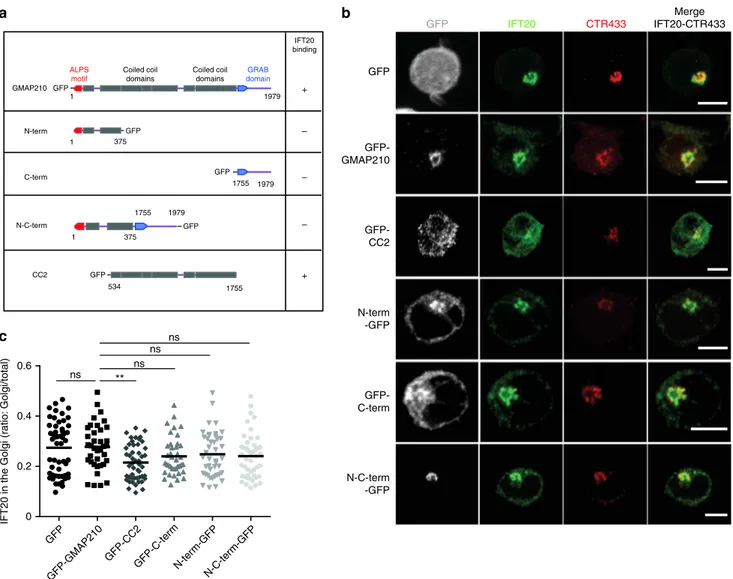
observed in cells overexpressing the IFT20-binding CC2 domain
(amino acids 534–1779) coupled to GFP
31(Fig.
6
), suggesting
that, in T lymphocytes, GMAP210 retains IFT20 close/at the
Golgi via its CC2 domain. Anchored to the Golgi membranes by
its C-terminal domain
30,32,53, GMAP210 which binds vesicles
through its N-terminus domain
33,34,49,54tethers them to the
Golgi. To investigate the role of this tethering activity, we
over-expressed the N-terminal domain encompassing amino acids
1–375, the C-terminal domain (amino acids 1778–1979) and a
shorter version of GMAP210 that contains both N-term and
C-term domains but lacks most of the coiled-coil domain (Fig.
6
a).
As described
31, all these constructs were localized to the Golgi
(Fig.
6
b). In contrast to the overexpression of CC2, none of them
displaced IFT20 from the Golgi (Fig.
6
b, c).
We then studied the effect of the overexpression of the
different GMAP210 constructs on LAT recruitment and
phos-phorylation at the immune synapse. Although CC2
overexpres-sion induced a disperoverexpres-sion of IFT20 from the Golgi (Fig.
6
), it did
neither alter LAT recruitment (Fig.
7
a, quantification in Fig.
7
b)
nor LAT phosphorylation (Supplementary Fig. 7a, quantified in
Supplementary Fig. 7b) to the immune synapse. These results
suggest that although GMAP210 is involved in the localization of
IFT20 to the Golgi apparatus, its binding activity is not required
for LAT trafficking.
In contrast, overexpression of the N-terminal and C-terminal
domains of GMAP210, as well as the short version of GMAP210
induced a significant decrease in LAT recruitment to the immune
synapse (Fig.
7
a, quantification in Fig.
7
b). This was accompanied
by less phosphorylated form of LAT at the immune synapse
(Supplementary Fig. 7a, quantified in Supplementary Fig. 7b). In
contrast overexpression of the short version of GMAP210, which
inhibits LAT recruitment and phosphorylation at the immune
synapse, did not affect the phosphorylation of ZAP70 at the
a
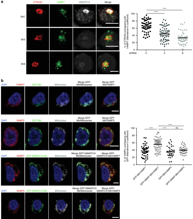
CTR433 VAMP7 GMAP210 Merge ShC Sh3 Sh8 C 3 8 shRNA 100 80 60 40 20 % of CTR433 overlapping withVAMP7 (Manderson’s coefficent) 0
0 20 40 60 80 100
% of VAMPs overlapping with GFP (Manderson’s coefficent)
b
GFP-Mit/VAMP7 GFP-GMAP-Mit/VAMP3 GFP-GMAP-Mit/VAMP7 GFP-Mit/VAMP3 nsVAMP7 GFP-GMAP210-Mit Mitotracker
Merge GFP-GMAP210-Mit/Mitotracker DAPI
Merge GFP-GMAP210-Mit/VAMP7
VAMP3 GFP-GMAP210-Mit Mitotracker DAPI
Merge GFP-GMAP210 -Mit/Mitotracker
Merge GFP-GMAP210-Mit/VAMP3 VAMP7 GFP-Mit Mitotracker
Merge GFP-Mit/Mitotracker DAPI
Merge GFP -Mit/VAMP7
VAMP3 GFP-Mit Mitotracker
Merge GFP-Mit/Mitotracker DAPI
Merge GFP -Mit/VAMP3
Fig. 5GMAP210 captures vesicles carrying VAMP7. a Confocal images showing the relative localization of VAMP7 (green) and CTR433 (red) in Jurkat
cells expressing control (ShC) or GMAP210 specific shRNA (Sh3, Sh8) (GMAP210, nucleus in gray). Dot plot on the right side of the panel show the quantification of the percentage of CTR433 (Golgi marker) overlapping with VAMP7 (Manderson coefficient). Scale bar 5 μm. Each dot = one cell; horizontal lines = median. ****P < 0.0001 (one-way ANOVA). b Confocal images showing the localization of VAMP7 or VAMP3 (red) in Jurkat cells expressing a GFP-GMAP210-ActA chimera (GFP-GMAP210-Mit) or a GFP-ActA chimera (GFP-Mit), treated for 4 h with nocodazol (5 μg/mL) (nucleus in blue and mitochondria in Gray). Dot plot on the right side of the panel shows the quantification of the percentage of VAMPs (VAMP7 or VAMP3) overlapping with the GFP staining (GFP-mit or GFP-GMAP210-mit, Manderson coefficient). Scale bar 5 μm. Each dot = one cell; horizontal lines = median. ****P < 0.0001, ns: non-significant (one-way ANOVA). Data and Images represent two independent experiments in a and b
immune synapse (Supplementary Fig. 7c and quantified in
Supplementary Fig. 7d). Altogether, these results suggest that
the tethering activity of GMAP210 is involved in the delivery of
vesicular LAT to the immune synapse.
Traffic of ectopically expressed LAT to the primary cilium.
GMAP210 was also shown to control trafficking of specific cargos
to the primary cilium
35,37,38. We reasoned that LAT, which is not
expressed in ciliated cells, might follow a transport pathway,
which is used by cargoes specifically going to the cilium. To test
this hypothesis, we expressed LAT in the mIMCD-3-ciliated cells.
LAT was transported to the cilium where it co-localized with
ARL13B, a marker of the cilium
55, demonstrating that the
intraciliary trafficking machinery could take care of the vesicular
transport of LAT (Fig.
8
a). This was rather specific since once
introduced in ciliated cells, the transmembrane protein CD3-ζ,
whose recruitment to the immune synapse does not depend on
GMAP210 (Fig.
3
) and VAMP7
13, was not transported to the
cilium (Fig.
8
a). Overexpression of the GMAP210-encoding
constructs described earlier also showed that the tethering activity
of GMAP210 was involved in transport of LAT to the cilium
(Fig.
8
b). As observed for the synapse, overexpression of the CC2
domain that binds IFT20 did not block LAT trafficking to the
cilium.
These results show that, when ectopically expressed in ciliated
cells, LAT traffics to the primary cilium. Moreover, they show
that the tethering activity of GMAP210 to the Golgi controls LAT
delivery to the primary cilium highlighting the similarities
between transport to the immune synapse and to the cilium.
Discussion
We show herein that GMAP210 by tethering vesicles containing
LAT to the Golgi helps their correct delivery to the immune
synapse. By doing so it organizes the formation of
LAT-containing signalosomes involved in T lymphocyte activation
revealing a new molecular player in the formation of the immune
synapse.
Several studies have investigated the cellular function of
GMAP210. In some cells, depletion of GMAP210 has been
reported to cause Golgi fragmentation without defect in secretory
trafficking
53,56. This is not the case in human T cells, in which no
defect in the Golgi morphology and volume are observed
(Sup-plementary Fig. 5b). GMAP210 is a long coiled-coil molecule that
binds to the cis-Golgi via its C-terminal domain
30–32and
GFP-GMAP210 N-term -GFP GFP-C-term N-C-term -GFPb
c
GFP-CC2 GFP ns ns ns ns GFP-GMAP210 N-ter m-GFP GFP-C-ter m N-C-ter m-GFP GFP-CC2 GFP 0.6 0.4 0.2 0IFT20 in the Golgi (r
atio: Golgi/total)
a
1 1 GMAP210 ALPS motif GRAB domain Coiled coil domains Coiled coil domains 1979 IFT20 binding GFP – 375 1755 GFP 534 + – 1979 1755 GFP 1 375 1979 1755 GFP – N-term C-term N-C-term CC2 GFP + IFT20 CTR433 Merge IFT20-CTR433 GFPFig. 6IFT20 localization in the Golgi in Jurkat cells expressing different GMAP210-GFP domains. a Schematic representation of GMAP210 and fusion
constructs with different IFT20-binding capacity. b Confocal images showing the relative localization of IFT20 (green) and CTR433 (red) in Jurkat cells expressing GFP, GFP-GMAP210, or different GMAP210 domains coupled to GFP. c Quantification of the ratio of IFT20 fluorescence present in the Golgi versus total fluorescence in the different conditions. Scale bar 5 μm. Each dot = one cell; horizontal lines = median. **P < 0.01, ns: non-significant (one-way ANOVA). Data and images represent two independent experiments in b and c
captures vesicles through its N-terminal domain
34,49,54. Using the
mitochondrial relocation assay, it was shown to capture vesicles
containing Golgi, as well as endoplasmic resident proteins
28. We
show herein that it also captures vesicles bearing VAMP7
(Fig.
5
b), which is expressed on Golgi membrane in T cells
(Fig.
5
a). Concerning the specificity of vesicular binding of
GMAP210, GMAP210 contains an ALPS motif at its N-terminal
end. This motif does not present a sequence-specific interaction
site, but rather senses the curvature of the vesicles, preferentially
binding highly curved liposomes (radius < 50 nm) with
mono-unsaturated lipids
34. The N-terminal domain of GMAP210 also
contains a sequence-specific interaction, suggesting that this
golgin can bind two different types of vesicles
54. It is worth noting
that the sizes of the vesicles containing both LAT and GMAP210,
found in our study (Fig.
1
b, d), are compatible with the size
preference of the GMAP210 ALPS domain. This gives us precious
insights and hypothesis to be tested on the potential lipid
com-position of the LAT-containing vesicles, the cargoes they may
contain and the consequences this composition may have on the
formation of the immune synapse.
Humans with mutations in GMAP210 and
GMAP210-knockout mice die early on from a severe skeletal dysplasia
57.
This is associated with a defective trafficking of some cargo
proteins in the early secretory pathway of chondrocytes
57,58.
The role of GMAP210 in the early secretory pathway, i.e. both
anterograde and retrograde trafficking has been confirmed in
other cell types
59,60. We recently showed that the endocytic
LAT is following a retrograde pathway back to the Golgi
apparatus. This transport pathway which exists at steady state is
increased upon TCR activation and is crucial for LAT transport
to the immune synapse
24. These results associated to the data
reported herein suggest the following model (Fig.
9
): Upon TCR
activation, LAT is endocytosed and transported through the
retrograde transport pathway to the Golgi. From there,
vesicles-containing LAT and VAMP7 are budding and are captured by
GMAP210 to be delivered back to the immune synapse. This
happens 10–15 min after activation and corresponds to an
active phase of vesicle recruitment
45. The Golgi apparatus is
polarized close to the immune synapse, as reported early on
61and showed by electron microscopy
43,44,62. Yet, it does not
dock to the immune synapse, as shown by the absence of
GM130, a marker of the Golgi, in the evanescent field of the
TIRFM. Thus the long coiled-coil domain of GMAP210,
200–300 nm
31, could allow the proximity and docking at the
immune synapse of the LAT/VAMP7-vesicles bound to the
N-terminal domain of GMAP210. This pool of vesicular LAT
delivered in a GMAP210-dependent manner participates to the
formation of LAT signalosomes (Fig.
4
a, b).
GMAP210 has also been shown to bind IFT20 in several cell
types including T lymphocytes
35,36and to anchor this protein to
the Golgi
35. This intraflagellar transport (IFT) protein, which
regulates the assembly of the primary cilium, also regulates
traf-ficking of the TCR
51and LAT
52to the immune synapse. Yet, our
results suggest that the IFT20-binding activity of GMAP210 is not
involved in the vesicular transport of LAT (Fig.
7
). Moreover,
whereas IFT20 controls TCR/CD3-ζ recruitment to the immune
synapse
51, GMAP210 does not (shown here Fig.
3
and
Supple-mentary Fig. 3 and in ref.
36). Thus, at least some effects of IFT20
0.0 0.2 0.4 0.6a
b
GFP N-term-GFP GFP-C-term N-C-term-GFP – + SEE LAT GFP N-ter m-GFP GFP-C-ter m N-C-ter m-GFP G FP N-ter m-GFP GFP-C-ter m N-C-ter m-GFP LA T enr ichement (r atio: IS/total) LAT –SEE +SEE GFP-GMAP GFP-CC2 ns ns GFP-GMAPGFP-CC2 GFP-GMAPGFP-CC2 ns ns ns ns ns N = 126 N = 99 N = 46 N = 59 N = 67 N = 73 N = 71 N = 68 N = 77 N = 82 N = 80 N = 81 0 1000 Nor maliz ed fluorescence 500Fig. 7GMAP210 tethering activity controls LAT recruitment at the immune synapse. a, b Confocal images (left panels) and quantification (right panels) of
the enrichment of LAT (a, b) at the immune synapse (depicted by the dotted white line) in Jurkat “mean cells” expressing GFP alone, GMAP210-GFP, or different GMAP210 domains coupled to GFP and interacting for 30 min with Raji cells left unpulsed (−, unactivated state) or pulsed with SEE (+, immune synapse formation). N = number of cells constituting the mean image. Horizontal line represents median. **P < 0.01, ***P < 0.001, ****P < 0.0001, ns: non-significant (one-way ANOVA). Data are representative from two independent experiments
a
DAPI LAT-mCherry i ii i ii CD3ζ-GFP i ii i ii ARL13b i ii i ii Merge i ii i iib
GFP-CC2 GFP-C-term N-term -GFP N-C-term -GFP GFP Ac-tubulin Merge DAPI LAT-mCherry GFP LAT-mCherry Ac-tubulinFig. 8Specific recruitment of LAT to the primary cilium depends on GMAP210 tethering activity. a Confocal images showing the localization of ARL13b
(gray) and LAT-mCherry (red) or CD3ζ-GFP (green) in ciliated mIMCD-3 epithelial cells. Staining of the nucleus by DAPI in blue. White squares indicate the magnified regions presented underneath that shows the primary cilia. White scale bars: 5 μm, gray scale bars: 1.5 μm. b Confocal images showing the relative localization of LAT-mCherry (red) and acetylated-tubulin (gray), in mIMCD-3 cells co-expressing GFP alone, GMAP210-GFP, or different domains coupled to GFP. White squares indicate the magnified regions presented on the right that shows the primary cilia. White scale bars: 5 μm, gray scale bars: 1.5 μm. Images representative of two independent preparations in a and one preparation in b
on the formation of the immune synapse formation do not rely
on its binding to GMAP210.
One interesting aspects of our study is that, as reported for
transport to the cilium
37, GMAP210 does show some specificities
for vesicles and cargoes delivered to the immune synapse. Indeed,
GMAP210 silencing does not affect CD3-ζ recruitment to the
immune synapse (Fig.
3
), this might be due to the difference in
the “nature” of the vesicles containing CD3-ζ.
We now have to characterize if GMAP210 controls the delivery
of other molecules to the immune synapse but also if other
gol-gins are involved in immune synapse formation.
Although, T lymphocytes are non-ciliated cells, morphological
similarities have been shown between the immune synapse and
the primary cilium
63,64. In particular, the central role of
con-trolled vesicular trafficking in signaling by these structures has
been demonstrated
65–67. Our results further extend this analogy.
Indeed, we show that LAT, an emblematic signaling molecule of
the immune synapse, is transported to the primary cilium
(Fig.
8
a) when ectopically expressed in ciliated cells. Like for the
immune synapse, LAT trafficking to the cilium depends on the
tethering activity of GMAP210 (Fig.
8
b). Hence, studying LAT
trafficking to the immune synapse can bring information on
vesicular trafficking to the cilium.
Together, our results reveal a new player and a new mechanism
in transport to-and formation of-the immune synapse that may
also be a key player of cilium generation. These results open a
new field of research that may have valuable implications in the
study of ciliopathies and immunodeficiencies.
Methods
Cells. Jurkat T cells (validated by SSTR method present 88% of homology with DSMZ Leibniz ACC 282), JCAM2.5 cells stably expressing the mouse LAT-Streptag
construct25, Jurkat expressing the CD3-ζ-GFP chimera described elsewhere68, and
Raji B (ATCC, CCL-86) cells were cultured at 37 °C 5% CO2in RPMI 1640
Glutamax (Gibco, 61870-010) supplemented with 10% fetal calf serum (FCS,
Lonza, DE14-801F, lot no. 0SB017) and were passed every 2–3 days at ~0.5 × 10⁶
cells/mL.
Inner medullar collecting duct cells (IMCD3, ATCC CRL-2123), a kind gift from Alexandre Benmerah (Laboratory of Hereditary Kidney Diseases, Imagine Institute, Paris, France), were grown in Dulbecco’s modified Eagle’s medium DMEM-F12 1:1 (GIBCO, 31331-028) supplemented with 10% FCS for basic cell culture conditions.
Mononuclear cells were isolated from peripheral blood of healthy donors on a ficoll density gradient. Buffy coats from healthy donors (both male and female donors) were obtained from Etablissement Français du Sang (Paris, France) in accordance with INSERM ethical guidelines. Human total CD4+ isolation kit (Miltenyi Biotech, 130-096-533) was used for the purification of T cells. To obtain
lymphoblastoid effector T cells13, six-well plastic plates were coated overnight
at 4 °C withαCD3 (OKT3 clone, eBioscience, 16-0037-85, 2.5 μg/mL final
concentration in 1.3 mL). Wells were washed and 5.4 × 10⁶purified primary
human total CD4+ T cells were then plated per well in the presence of soluble anti-CD28 (LEAF Purified anti-human anti-CD28 from anti-CD28.2 clone, Biolegend,
BLE302923) at 2.5μg/mL final concentration and recombinant human IL-2
(20 U/mL, Novartis, Basel, Switzerland) in RPMI culture medium supplemented with 10% FCS, penicillin/streptomycin (100 U/mL, Gibco, 15-140-122), 10 mM
HEPES (Gibco, 15630-080) and 0.05 mMβ-mercaptoethanol, (Gibco, 31350-010)
at 37 °C, 5% CO2.
Reagents and antibodies. Recombinant Staphyloccocus Enterotoxin type E (SEE, Cellgenetech, MBS1112600), Ionomycin (407950; Calbiochem), PMA
(Sigma-Aldrich, 79346) and Poly-L-lysine (Poly-Lys, Sigma-Aldrich, P8920) were used.
For detailed information on dilutions, companies, and reference numbers see Supplementary Table 1. APC Extracellular Centrosome Endosomes Recycling endosomes GMAP210 Arf1 VAMP7 LAT LAT+/VAMP7+ Vesicles LAT+endocytic Vesicles Trans Golgi Cis Golgi ER Nucleus CD4 CD3 TCR CD3 MHC-II Cytoplasm Cytoplasm
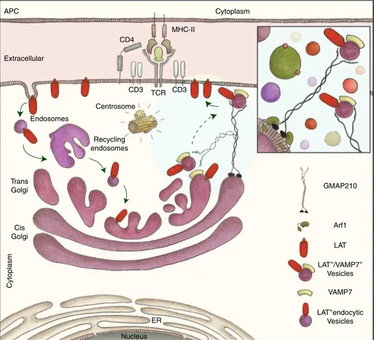
Fig. 9Graphical abstract: GMAP210 facilitates the delivery of vesicles containing LAT to the immune synapse. Upon TCR triggering, LAT is internalized in
recycling endosomes. This endocytic pool of LAT is “retrotransported” to the Golgi apparatus24, where it meets the vesicular SNARE VAMP7 that is
involved in LAT trafficking13. GMAP210, which binds the Golgi through Arf1, sorts and captures the LAT/VAMP7 vesicles via its N-terminal domain (inset).
Production of lentiviruses and infection of CD4+T cells. Non-replicative VSV-g pseudotyped lentiviral particles were produced by transfecting HEK-293T cells with Gag, Pol, rev, encoding plasmid (pPAX2), envelop encoding plasmid (pMD2.G) and
either the HA-Tev-LAT construct13encoded in a pWXLD vector, or short hairpin
RNA (shRNA) sequences encoded in pLKO.1 plasmid: Non-targeting control shRNA (shC, Sigma-Aldrich, Mission shRNA SHC002), GMAP-210-specific shRNA, sh3 (Sigma-Aldrich, Mission shRNA, sequence GCAAAGGAACAA-GAACTCAAT), and sh8 (Sigma-Aldrich, Mission shRNA, sequence GCAGAA-GATAGAGAGGCTAAACT). Lentivirus were recovered in supernatant after 2 days
and concentrated. 5 × 10⁶Jurkat T cells were infected for 24 h, cells infected with
shRNA encoding virus were selected in puromycine (2 µg/mL, InvivoGen, ant-pr) and used 5 days post infection.
Primary human CD4+ T cells were activated in six-well plates coated with
anti-CD3 (2.5μg/mL) in the presence of soluble anti-CD28 (2.5 μg/mL) and
recombinant IL-2 (20 U/mL). Concentrated virus was added 36–48 h later. Cells were washed and then put in fresh medium with IL-2 (20 U/mL) and puromycin
(2.5μg/mL) 24 h later and used 72 h later.
Plasmids. The plasmid encoding LAT-mCherry and CD3ζ-GFP were reported
previously13,68. Plasmids encoding chimeric molecules GFP-GMAP210,
N-term-GFP, GFP-C-term, N-C-term-N-term-GFP, GFP-CC2, GFP-GMAP-Mit, GFP-Mit are
described elsewhere31,48.
Transfection. Jurkat T cells were transfected using the Amaxa Cell Line
Nucleoefector Kit V (Lonza, VCA-1003). To do so, 5 × 10⁶cells were washed,
resuspended in 100 µL of nucleoefector solution and combined with 5–10 µg of DNA. Cells were passed into the electroporation cuvettes and then electroporated (Amaxa program X-005). Cells were then incubated at room temperature for 10 min, recovered and diluted in warmed RPMI supplemented with 10% FCS and
cultured for 24 h at 37 °C, 5% CO2.
To transfect IMCD3, cells were grown to confluence on 12 mm coverslips in 24-wells plate. Medium was removed and 500 µL of Optimem (GIBCO, 31985-047) was added. In a final volume of 50 µL of Optimem, 2 µL of Lipofectamine 2000 (Invitrogen, 11668019), and 2 µg of DNA were mixed and incubated for 10 min at room temperature. The mix Lipofectamine/DNA was added and cells were incubated for 24 h to induce ciliogenesis.
Preparation of lysates from Jurkat or Human CD4+ T lymphoblasts. 1 × 10⁶
cells/mL of Jurkat T-cells or human lymphoblasts were washed three times with cold PBS and incubated on ice for 20 min in 30 µL ice-cold lysis buffer (50 mM Tris
pH 8, 150 mM NaCl, 1,5 mM MgCl2, 1% Glycerol, 1% TritonX100, 0.5 mM EDTA
pH 8, 5 mM NaF) supplemented with a protease inhibitor cocktail (Sigma-Aldrich, 11873580001). Post-nuclear lysates were obtained by centrifugation at maximum velocity for 15 min at 4 °C. Laemmli Sample Buffer (BIORAD, 161-0747) and reducing agent (Thermo Fisher Scientific, NP0009) were added and samples were heated at 95 °C for 5 min and kept at −20 °C before immunoblot analysis. Preparation of LAT-containing membranes. JCAM2.5 LAT-deficient Jurkat cells
(150 × 106) expressing the mouse LAT-StrepTag (LAT-TST) protein25or
non-transfected JCAM.2.5 were washed, resuspended in RPMI at 100 × 106/mL and
activated with soluble CD3 (125 ng/mL) and CD28 (250 ng/mL) anti-bodies for different times (0, 5, 15, and 30 min). The activation was stopped by adding cold PBS and the cells were centrifuged at 1800 × g at 4 °C for 5 min. The cell pellet was then suspended in homogenization buffer (0.25 M sucrose, 10 mM Tris–HCl pH 7.4, 1 mM EDTA) supplemented with a EDTA-free protease inhi-bitor cocktail (Roche, 1123000) and a phosphatase inhiinhi-bitor cocktail (Thermo Scientific, 78420). Cell breakage was induced on ice by 25 successive stokes of a Dounce homogenizer. The cell suspension was then passed 15 times through a 25GA needle to achieve cell disruption and centrifuged for 5 min at 900 × g at 4 °C to remove nuclei and unbroken cells. The supernatant was transferred into Ultra-clear centrifugation tubes (Beckman Coulter) and centrifuged at 65,000 × g for 1 H at 4 °C in a SW55Ti rotor (Beckman Coulter). The pellet was suspended in 1.2 mL of homogenization buffer supplemented as before and passed several times through a 25GA needle to ensure complete resuspension of the membranes. This suspen-sion was transferred into a new tube and mixed with 1.2 mL of a 60% solution of Optiprep/iodixanol (Axis-shield) to reach a 30% iodixanol suspension. The Opti-prep solution was diluted extemporaneously into 0.25 M sucrose, 60 mM Tris–HCl pH 7.4, 6 mM EDTA to prepare 1.3 mL of a 20% solution and 1.2 mL of a 10% solution. The 20% and the 10% iodixanol solutions were layered successively on top of the 30% suspension and centrifuged at 350,000 × g for 3 h at 4 °C in a SW55Ti rotor without brake when stopping. Ten fractions of 490 µl were collected from the top of the tube. To purify LAT-TST-associated membranes, the third fraction (fraction 3) was incubated for 90 min at 4 °C on a rotating wheel with pre-washed Strep-Tactin Sepharose resin in presence of protease and phosphatase inhibitors. Resin was washed in StrepTag washing buffers (Buffer W: Tris–HCl 100 mM, NaCl 150 mM, EDTA 1 mM, pH 8.0) and suspended in RIPA lysis buffer before being submitted to SDS–PAGE and immunoblot analysis.
Purification of LAT-signalosome. Jurkat cells (5 × 10⁶) were resuspended in
200 µl of RPMI medium, and magnetic beads (1 × 10⁷) coated with anti-CD3 and
anti-CD28 (Gibco, 11132D) were added in a volume of 100 µl. Beads were incu-bated with T cells for the appropriate time at 37 °C. Activation was stopped with the addition of 500 µl cold PBS, and 80 µl (1/10) of samples were collected as ‘input’
and lysed as described above (‘'Preparation of lysates from Jurkat or human CD4+
T lymphoblasts' section). Bead-cell conjugates were then magnetically restrained, resuspended in 500 µl of ‘freeze–thaw’ buffer (600 mM KCl, 20 mM Tris, pH 7.4, and 20% glycerol) supplemented with, EDTA-free Protease Inhibitor Cocktail Tablet (Roche, 1123000). Samples were submitted to seven cycles of freezing and thawing. After the final cycle, 5 µl benzonase (Novagen, 2733353) was added, followed by incubation for 20 min at room temperature. Samples were magnetically restrained to purify the bead-attached proteins and then were washed five times in the supplemented ‘freeze–thaw’ buffer described above. Bead-associated proteins were resuspended in lysis buffer and separated by SDS–PAGE and analyzed by immunoblot.
Immunoblot analysis. Samples were resolved on NuPage 4–12% Bis–Tris gel (Thermo Fisher Scientific, NP0323BOX) and liquid transferred (Thermo Fisher Scientific, NP00061) on PVDF membranes (Bio-Rad, 162-0177). After blocking with TBS 0.05% Tween20 5% BSA for 1 h 30 min on rocking platform shaker, membranes were incubated overnight at 4 °C with primary antibodies. Membranes were washed three times with TBS 0.05% Tween and incubated for 40 min in TBS 0.05% Tween on rocking platform shaker with the secondary antibody. Bound
antibodies were revealed using the ClarityTMWestern ECL substrate (Bio-Rad,
#170-5061), according to the manufacturers’ directions. The intensity of the bands was quantified by densitometry using Image Lab 5.2.1 software (Bio-Rad Labora-tories) and was expressed as arbitrary units. All original gel images are included in the Source Data file.
Coverslips and dishes preparation for immunofluorescence assay. 12mm ø coverslips (VWR, 631-0666) for fixed cells or fluorodishes (World Precision
Instrument Inc., FD35-100) for live imaging were pre-coated with 0.02% of poly-L
-Lysine for 20 min at room temperature and were washed three times in water before being dried and kept for maximum 2 days.
Preparation of Jurkat T cells and Raji B cells conjugates. Raji B cells were
washed, resuspended at a concentration of 1 × 10⁶cells/mL in RPMI without FCS
and labeled with CellTracker™ Blue CMAC dye (10 µM, Thermo Fisher, C2110) for 20 min at 37 °C. Labeling was stopped with RPMI 10% FCS and cells were washed
once and resuspended at 1 × 10⁶cells/mL. Cells were pulsed with SEE (100 ng/mL)
or left untreated for 30 min at 37 °C before being washed once and resuspended at a
concentration of 1 × 10⁶cells/mL. 100,000 Raji cells were incubated on coverslips
for 30 min, washed once with warmed PBS and 150,000 Jurkat cells resuspended in RPMI 10% FCS were added for 30 min. Coverslips were washed once with cold PBS before fixation.
Mitochondrial capture assay in cells expressing GFP-GMAP-Mit. Jurkat cells
were washed, resuspended at 1 × 10⁶cells/mL and incubated 4 h with nocodazol
(5μg/mL) in RPMI containing 10% of FCS at 37 °C. 150,000 Jurkat cells were then
incubated on coverslip for 30 min, washed once with cold PBS and fixed.
Fixed and live TIRF microscopy. Poly-L-Lysine-coated coverslips were left
untreated or coated overnight at 4 °C withαCD3ε αCD28, washed three times and
pre-warmed at 37 °C for 10–15 min. 150,000 Jurkat or primary CD4+ T cells were incubated on coated coverslips for 15 min at 37 °C before being washed once with cold PBS and fixed. For live imaging, fluorodishes were coated following this same protocol. 200,000 Jurkat T cells were plated and images for 491 and 561 channels were acquired every 3 s.
Fixation. Cells were fixed with 4% paraformaldehyde (Life technologies, FB002) for 15 min at room temperature, washed once in PBS and excess of paraformaldehyde was quenched for 10 min with PBS 10 mM Glycine (Thermo Fisher Scientific, G8898). Coverslips were kept at 4 °C in PBS until permeabilization and staining. Staining and mounting. Cells were permeabilized for 30 min at room temperature with PBS 0.2% Bovine Serum Albumin (BSA, Euromedex, 04-100-812) 0.05% Saponin (Sigma-Aldrich, S4521). Cells were then incubated for 1 h at room tem-perature with primary antibody, followed by washing three times with PBS 0.2% BSA 0.05% Saponin and incubated protected from light for 20 min in the same buffer with spinned secondary antibodies. After washing once with PBS BSA Saponin, and once with PBS, coverslips were soaked three times in PBS, three times in water, and mounted on slides.
For regular confocal microscopy, coverslips were mounted with 4–6 µL of Fluoromount G (SouthernBiotech, 0100-01) on slides (KNITTEL Starfrost) and dried overnight protected from light before microscope acquisition.